Embed Size (px)
Citation preview

Ch 27: Female Reproductive Ch 27: Female Reproductive SystemSystem
All organs are internal and closely associated– Primary reproductive organs: ?
– Secondary reproductive organs: ?
Female repro system must produce gametes AND maintain developing embryo
Fig 27-11

OvariesOvaries
Suspended by ovarian ligament & suspensory ligament
Functions: 1. Ova production 2. Hormone production

Oogenesis Oogenesis ((= ovum production)= ovum production)
takes place inside ovarian follicles in ovaries as part of ovarian cycle
Oogonia (= stem cells) complete mitotic divisions before birthAt birth: ~ 2 mio primary oocytes
At puberty: ~ 400,000 primary oocytes
40 years later: 0 (even though only ~ 500 used) Atresia

OogensisOogensisOvarian cycles start at puberty under influence of ___
Primordial follicle
Primary follicle
Secondary follicle
Tertiary (Graafian follicle)
Each month some proceed
Few proceed
Few proceed
Fig 27-12

(simple squamous layer)
Primordial Follicle or Egg Nests
in cortex
Present at birth

Primary Follicle
OocytesFollicle cells
Follicles enlarge in response to FSH and produce estrogens

Few relative to number of primary follicles
Produce follicular fluid
Rapid enlargement
= Clear glycoprotein layer
Secondary Follicle

Tertiary or Graafian Follicle
Spans entire width of cortex
First meiotic division being completed: 1oocyte divides into one 2 oocyte and one polar body

Ovulation
Happens in tertiaryfollicle
OogenesisOogenesisSuspended in prophase I
Stops in Metaphase II

Oocyte and follicular cells shed into abdominal cavity
then1. Empty follicle forms corpus luteum which produces
progesterone
2. Corpus luteum degenerates and becomes corpus albicans
3. GnRH increases under low estrogen and progesterone levels
Ovulation

Uterine Tube= Fallopian tube = oviduct
= salpinx
Two muscular tubes– infundibulum with fimbriae– Ampulla (place of fertilization)– Isthmus– intramural portion
Tubal ligation
Fig 27-14

Uterine Tube HistologyCiliated and non-ciliated
simple columnar epithelium
Ciliary movement and periodic peristaltic contractions move ova
Secretion of nutrient substances

The Uterus
Uterine wall ~ 1.5 cm
made up of
1. Endometrium,
2. Myometrium,
3. Incomplete perimetrium
Blood supply– Uterine arteries from internal iliac– Ovarian arteries from abdominal
aorta (inferior to renal arteries)
Fig 27-16

Histology of Endometrium
Functional zone – deciduum, sheds during menses – menstruation - flow sheds functionalis layer of
endometrium– proliferative phase - under influence of estrogen
basal cells proliferate– secretory phase - progesterone maintains
functionalis
Basilar zone – permanent layer, deep to functionalis
Fig 27-16
Functions of UterusFunctions of Uterus
Protection of embryo/fetus
Nutritional support
Waste removal
Ejection of fetus at birth

Cervix and VaginaCervix attaches to vagina at ~ 90° angle
Fornix – pocket surrounding uterine cervix (surgical access to pelvic cavity; location of birth control device)
Vagina – fibro-muscular organ serving as– receptacle for intercourse– passageway for menstrual products– birth canal
Fig 27-20b

The Mammary GlandModified sweat gland
Overlaying the ____________ muscle
15-20 separate lobes separated by suspensory ligaments; each lobe contains several secretory lobules
Lactiferous ducts leaving lobules; converge into 15-20 lactiferous sinuses
Milk stored in lactiferous sinus until released at tip of nipple
Fig 27-21
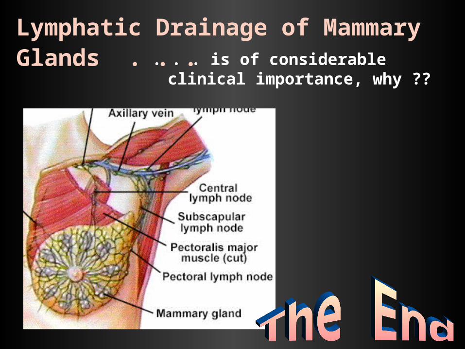
Lymphatic Drainage of Mammary Glands . . . . . . is of considerable clinical
importance, why ??