Embed Size (px)
Citation preview
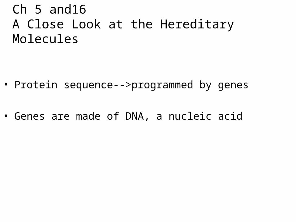
Ch 5 and16 A Close Look at the Hereditary Molecules
• Protein sequence-->programmed by genes
• Genes are made of DNA, a nucleic acid
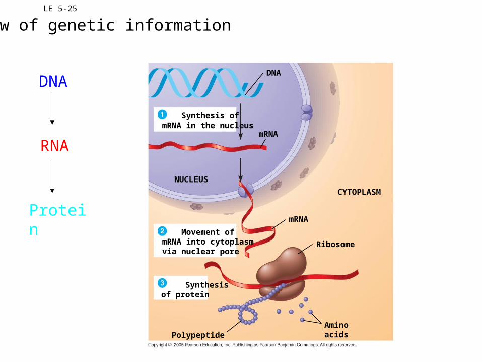
LE 5-25
NUCLEUS
DNA
CYTOPLASM
mRNA
mRNA
Ribosome
Aminoacids
Synthesis ofmRNA in the nucleus
Movement ofmRNA into cytoplasmvia nuclear pore
Synthesis of protein
Polypeptide
DNA
RNA
Protein
Flow of genetic information
The Roles of Nucleic Acids
• Two types:– Deoxyribonucleic acid (DNA)– Ribonucleic acid (RNA)
• DNA provides directions for its own replication.
• DNA directs synthesis of messenger RNA (mRNA)
• mRNA controls protein synthesis.
• Protein synthesis occurs on ribosomes.
LE 5-26a5 end
3 end
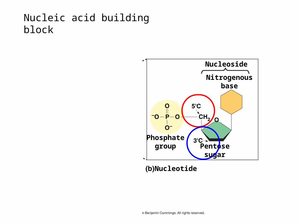
Nucleoside
Nitrogenousbase
Phosphategroup
Nucleotide
Polynucleotide, ornucleic acid
Pentosesugar
Nucleic acid building block
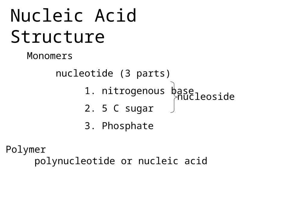
Nucleic Acid Structure
Monomers
nucleotide (3 parts)
1. nitrogenous base
2. 5 C sugar
3. Phosphate
Polymerpolynucleotide or nucleic acid
nucleoside

LE 5-26b
Nitrogenous bases
Pyrimidines
Purines
Pentose sugars
CytosineC
Thymine (in DNA)T
Uracil (in RNA)U
AdenineA
GuanineG
Deoxyribose (in DNA)
Nucleoside components
Ribose (in RNA)
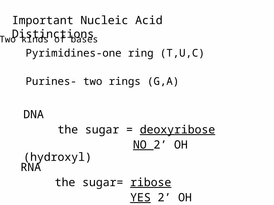
Important Nucleic Acid Distinctions
Pyrimidines-one ring (T,U,C)
Purines- two rings (G,A)
DNA the sugar = deoxyribose
NO 2’ OH (hydroxyl)
Two kinds of bases
RNA the sugar= ribose
YES 2’ OH
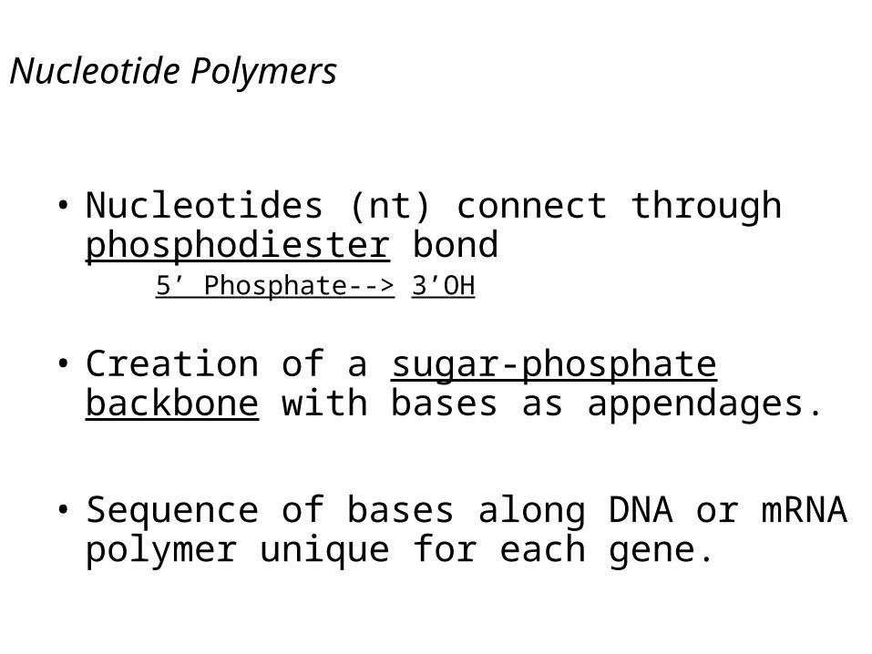
Nucleotide Polymers
• Nucleotides (nt) connect through phosphodiester bond
5’ Phosphate--> 3’OH
• Creation of a sugar-phosphate backbone with bases as appendages.
• Sequence of bases along DNA or mRNA polymer unique for each gene.
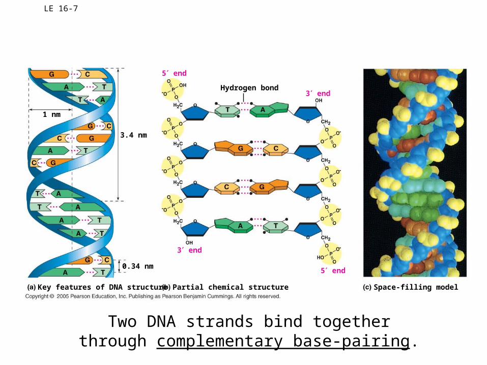
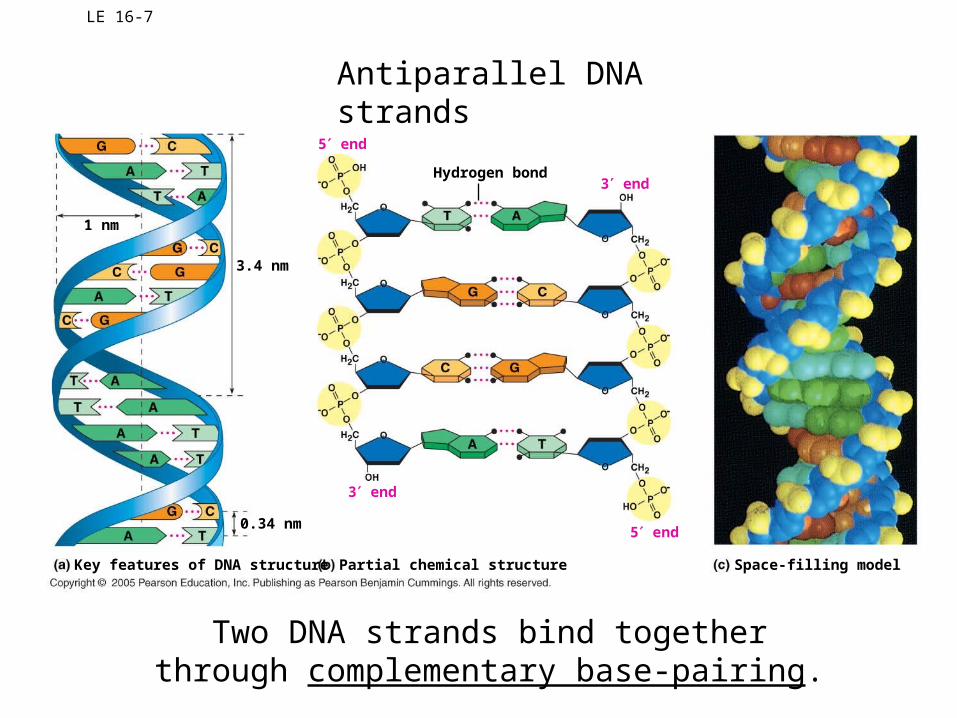
LE 16-7
5 end
3 end
5 end
3 end
Space-filling modelPartial chemical structure
Hydrogen bond
Key features of DNA structure
0.34 nm
3.4 nm
1 nm
Two DNA strands bind togetherthrough complementary base-pairing.
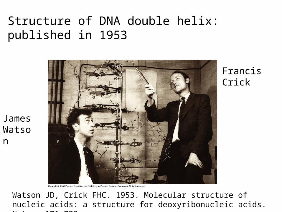
FrancisCrick
JamesWatson
Structure of DNA double helix: published in 1953
Watson JD, Crick FHC. 1953. Molecular structure of nucleic acids: a structure for deoxyribonucleic acids. Nature 171:738.
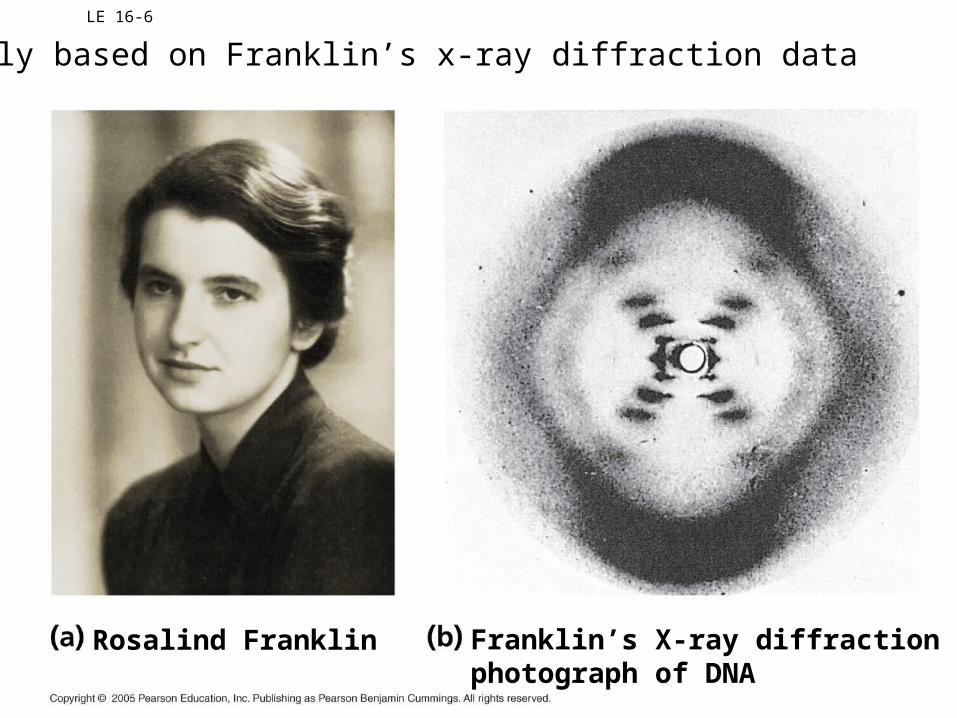
LE 16-6
Franklin’s X-ray diffractionphotograph of DNA
Rosalind Franklin
Partly based on Franklin’s x-ray diffraction data
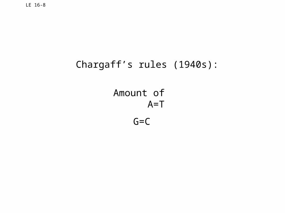
LE 16-8
Chargaff’s rules (1940s):
Amount of A=T
G=C
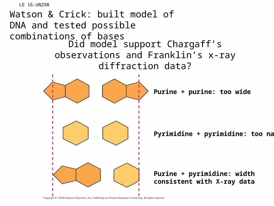
LE 16-UN298
Purine + purine: too wide
Pyrimidine + pyrimidine: too narrow
Purine + pyrimidine: widthconsistent with X-ray data
Watson & Crick: built model of DNA and tested possible combinations of bases
Did model support Chargaff’s observations and Franklin’s x-ray diffraction data?
LE 16-7
5 end
3 end
5 end
3 end
Space-filling modelPartial chemical structure
Hydrogen bond
Key features of DNA structure
0.34 nm
3.4 nm
1 nm
Antiparallel DNA strands
Two DNA strands bind togetherthrough complementary base-pairing.
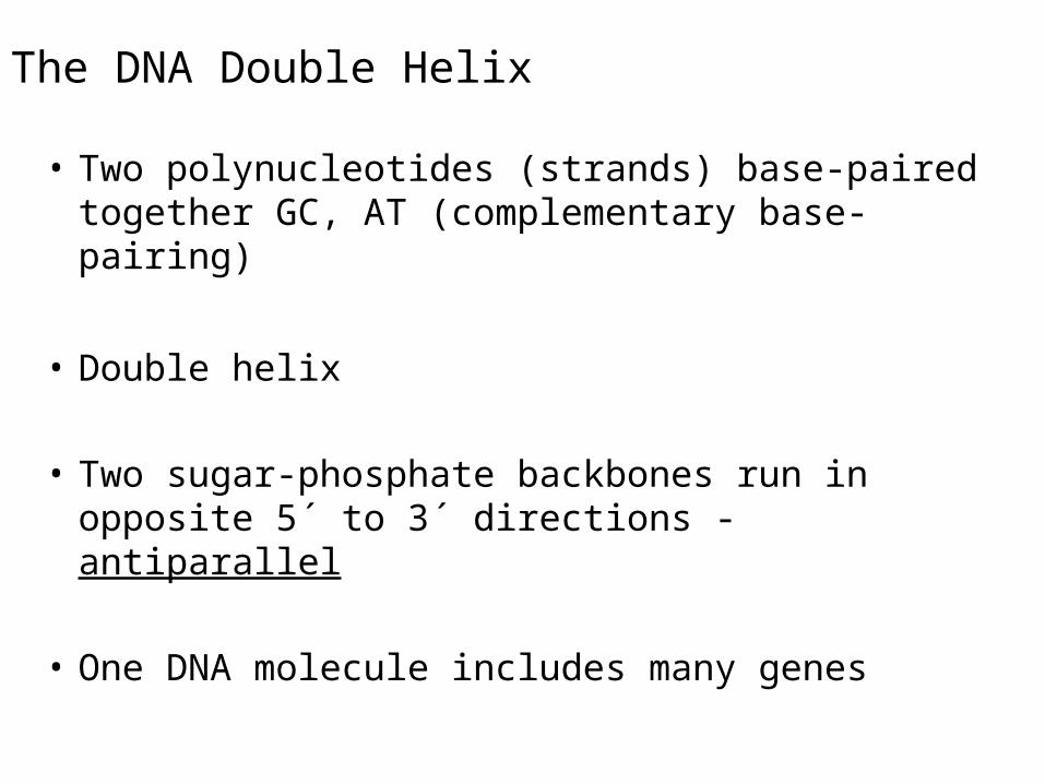
The DNA Double Helix
• Two polynucleotides (strands) base-paired together GC, AT (complementary base-pairing)
• Double helix
• Two sugar-phosphate backbones run in opposite 5´ to 3´ directions - antiparallel
• One DNA molecule includes many genes
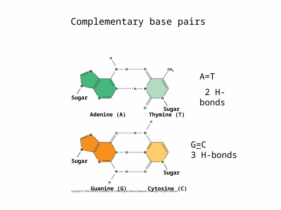
Adenine (A) Thymine (T)
Guanine (G) Cytosine (C)
Sugar
Sugar
Sugar
Sugar
Complementary base pairs
G=C3 H-bonds
A=T
2 H-bonds
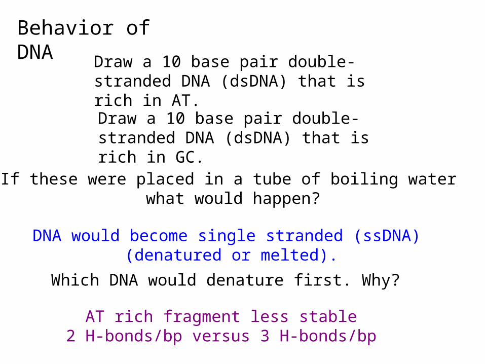
Behavior of DNA
Draw a 10 base pair double-stranded DNA (dsDNA) that is rich in AT.
Draw a 10 base pair double-stranded DNA (dsDNA) that is rich in GC.
If these were placed in a tube of boiling water what would happen?
DNA would become single stranded (ssDNA) (denatured or melted).
Which DNA would denature first. Why?
AT rich fragment less stable2 H-bonds/bp versus 3 H-bonds/bp
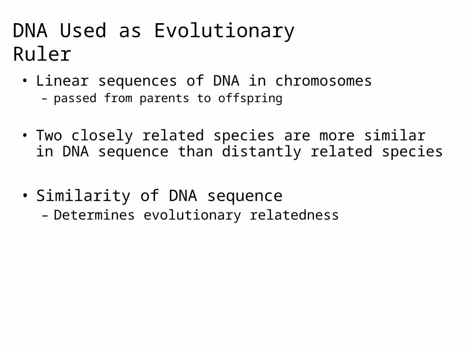
DNA Used as Evolutionary Ruler
• Linear sequences of DNA in chromosomes – passed from parents to offspring
• Two closely related species are more similar in DNA sequence than distantly related species
• Similarity of DNA sequence– Determines evolutionary relatedness
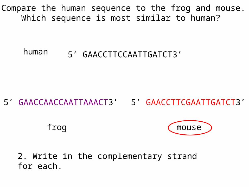
5’ GAACCTTCCAATTGATCT3’
5’ GAACCAACCAATTAAACT3’ 5’ GAACCTTCGAATTGATCT3’
1. Compare the human sequence to the frog and mouse. Which sequence is most similar to human?
human
mousefrog
2. Write in the complementary strand for each.
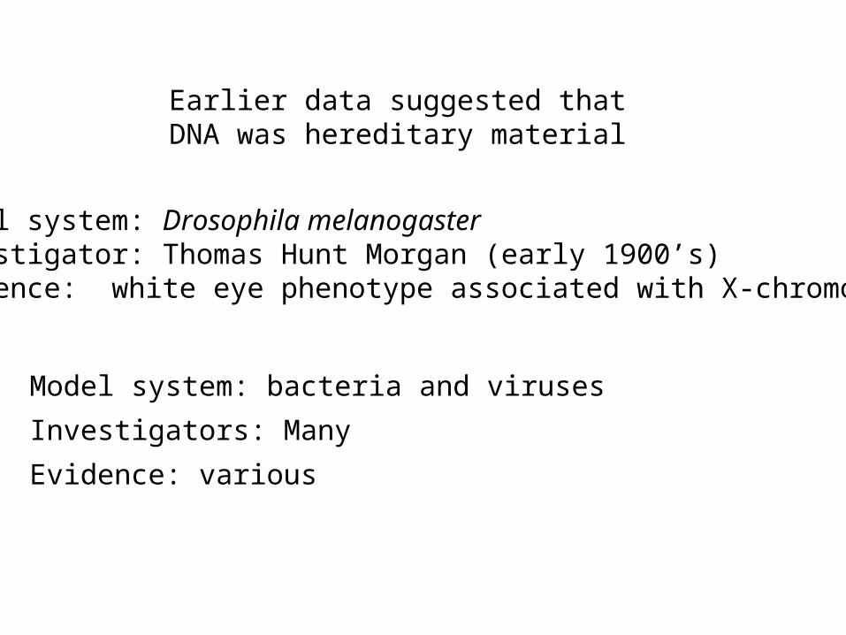
Earlier data suggested that DNA was hereditary material
Model system: Drosophila melanogasterInvestigator: Thomas Hunt Morgan (early 1900’s)Evidence: white eye phenotype associated with X-chromosome
Model system: bacteria and viruses
Investigators: Many
Evidence: various
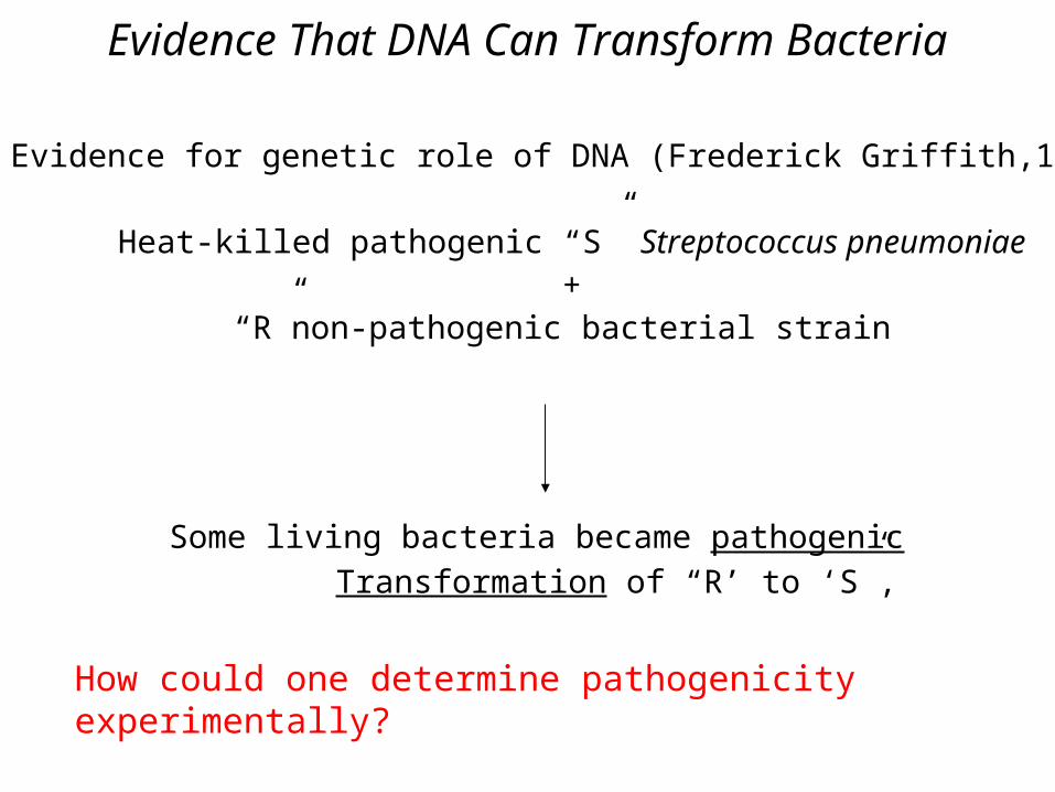
Evidence That DNA Can Transform Bacteria
Evidence for genetic role of DNA (Frederick Griffith,1928)
Heat-killed pathogenic “S” Streptococcus pneumoniae+
“R”non-pathogenic bacterial strain
Some living bacteria became pathogenicTransformation of “R’ to ‘S”,
How could one determine pathogenicity experimentally?
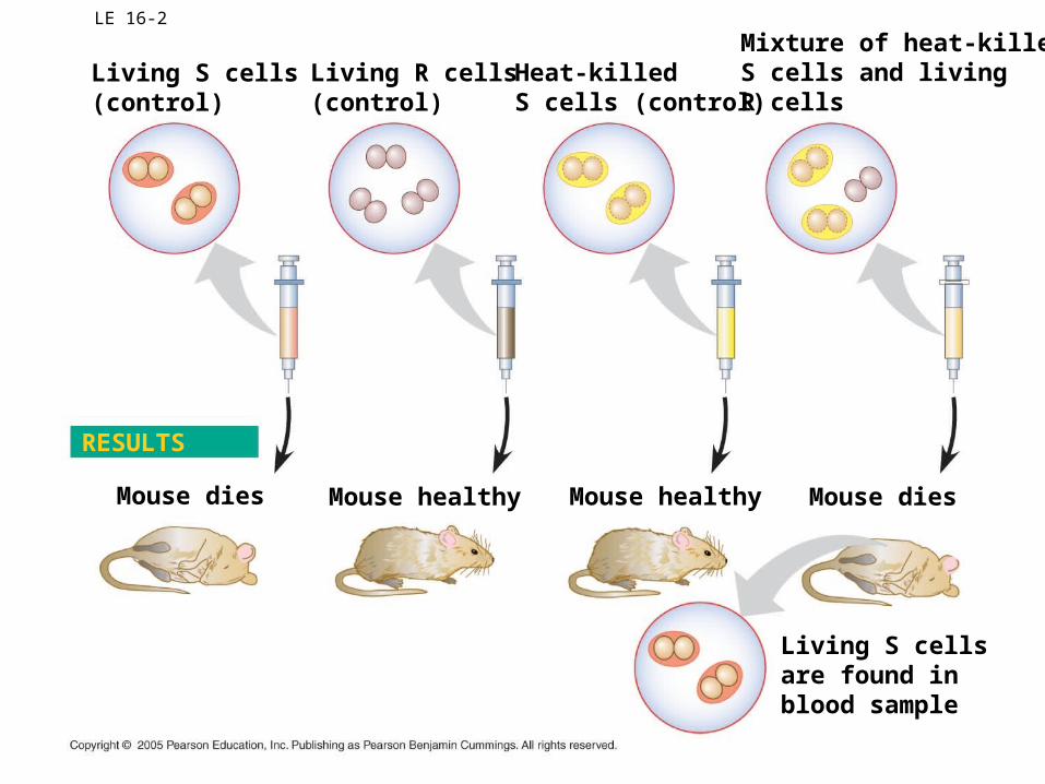
LE 16-2
Living S cells(control)
Living R cells(control)
Heat-killedS cells (control)
Mixture of heat-killedS cells and livingR cells
Mouse dies
Living S cellsare found in blood sample
Mouse healthy Mouse healthy Mouse dies
RESULTS
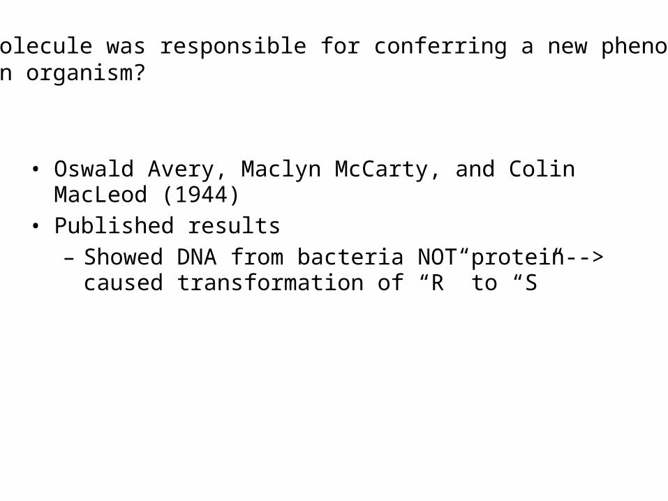
• Oswald Avery, Maclyn McCarty, and Colin MacLeod (1944)• Published results
– Showed DNA from bacteria NOT protein--> caused transformation of “R” to “S”
What molecule was responsible for conferring a new phenotypeinto an organism?

• Alfred Hershey and Martha Chase (1952)
– Used bacterial virus (bacteriophage) (T2) to ask whether DNA or protein was hereditary material
Independent confirmation
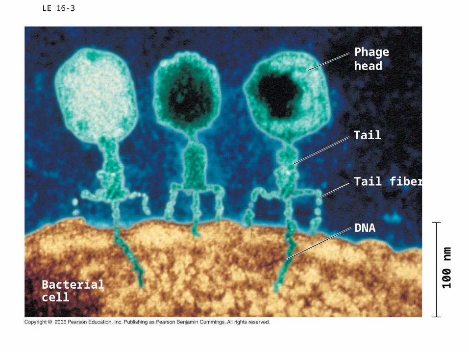
LE 16-3
Bacterialcell
Phagehead
Tail
Tail fiber
DNA
100
nm
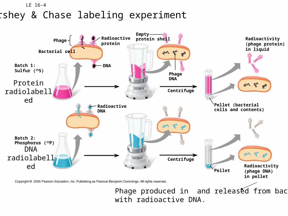
LE 16-4
Bacterial cell
Phage
DNA
Radioactiveprotein
Emptyprotein shell
PhageDNA
Radioactivity(phage protein)in liquid
Batch 1:Sulfur (35S)
RadioactiveDNA
Centrifuge
Pellet (bacterialcells and contents)
PelletRadioactivity(phage DNA)in pellet
Centrifuge
Batch 2:Phosphorus (32P)
Hershey & Chase labeling experiment
Protein radiolabelled
DNA radiolabelled
Phage produced in and released from bacteriawith radioactive DNA.
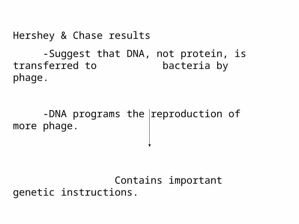
Hershey & Chase results
-Suggest that DNA, not protein, is transferred to bacteria by phage.
-DNA programs the reproduction of more phage.
Contains important genetic instructions.
I’m a pretty cool molecule butI’ll still answer yourquestions.