Embed Size (px)
Citation preview

Glyco-
proteinCarbohydrate
Glycolipid
Microfilaments
of cytoskeleton
EXTRACELLULAR
SIDE OF
MEMBRANE
CYTOPLASMIC SIDE
OF MEMBRANE
Integral
protein
Peripheral
proteins
Cholesterol
Fibers of extra-
cellular matrix (ECM)
Ch 5. Cell Membrane• Phospholipid bilayer• Cholesterol• Protein• Carbohydrates (on lipid or protein)
• Cell boundary• 8 ƞm thick• Selective permeable• Hydrophobic interaction• 1972 Singer & Nicholson
• Fluid mosaic model

Membrane Structure
•
Which of the following best describes the structure of a biological
membrane?
a) two layers of phospholipids with proteins embedded between the two layers
b) a mixture of covalently linked phospholipids and proteins that determines which solutes can cross the membrane and which cannot
c) two layers of phospholipids with proteins either crossing the layers or on the surface of the layers
d) a fluid structure in which phospholipids and proteins move freely between sides of the membrane
e) two layers of phospholipids (with opposite orientations of the phospholipids in each layer) with each layer covered on the outside with proteins

Figure 7.4
Knife
Plasma membrane Cytoplasmic layer
Proteins
Extracellular
layer
Inside of extracellular layer Inside of cytoplasmic layer
TECHNIQUE
RESULTS
Freeze Fracturing
• 1972 Singer & Nicholson Fluid mosaic model confirmation
• 1935 Davson & Danielli

Figure 7.3
Phospholipid
bilayer
Hydrophobic regions
of proteinHydrophilic
regions of protein
1972 Fluid Mosaic Model• Amphipatic phospholipids & proteins

Figure 7.2Hydrophilic
head
Hydrophobic
tail
WATER
WATER
Fluid Mosaic Model1) Phospholipids
• Amphipatic
• hydrophilic & hydrophobic

Figure 7.6
Lateral movement occurs
107 times per second.
Flip-flopping across the membrane
is rare ( once per month).
Fluid Mosaic Model
1) Phospholipids
• Amphipatic
• Movement
• Lateral
• Flip-flopping
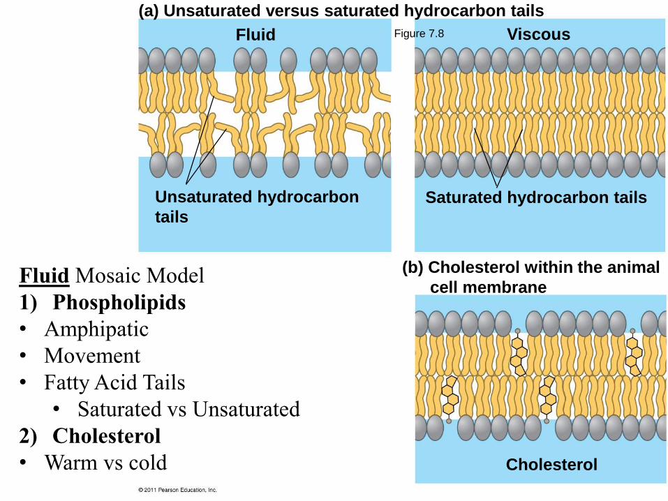
Figure 7.8Fluid
Unsaturated hydrocarbon
tails
Viscous
Saturated hydrocarbon tails
(a) Unsaturated versus saturated hydrocarbon tails
(b) Cholesterol within the animal
cell membrane
Cholesterol
Fluid Mosaic Model
1) Phospholipids
• Amphipatic
• Movement
• Fatty Acid Tails
• Saturated vs Unsaturated
2) Cholesterol
• Warm vs cold

Membrane Lipids and Fluidity
An animal cell membrane will be
more fluid at room temperature if it
contains
a) more cholesterol.
b) longer chain fatty acids.
c) more cis-unsaturated and polyunsaturated fatty acids.
d) more trans-unsaturated fatty acids.
e) any of the above

Glyco-
proteinCarbohydrate
Glycolipid
Microfilaments
of cytoskeleton
EXTRACELLULAR
SIDE OF
MEMBRANE
CYTOPLASMIC SIDE
OF MEMBRANE
Integral
protein
Peripheral
proteins
Cholesterol
Fibers of extra-
cellular matrix (ECM)
Cell Membrane
Fluid Mosaic Model• Phospholipid bilayer
• Cholesterol
Fluid Mosaic Model• Protein (integral & peripheral)
• Carbohydrates (on lipid or protein)

Figure 7.9
N-terminus
helix
C-terminus
EXTRACELLULAR
SIDE
CYTOPLASMIC
SIDE
Fluid Mosaic Model
3. Proteins
• Intergral proteins
• amphipatic

Figure 7.7
Membrane proteins
Mouse cellHuman cell
Hybrid cell
Mixed proteins
after 1 hour
RESULTS
Membrane Proteins

Membrane Protein Functions
1) Transport• Uniport: 1 in 1 direction
• Symport: 2 in same direction
• Antiport: 2 in opposite directions
• Carrier proteins• Pumps
• Facilitated diffusion
• Ion channels• Na+, K+, Ca++, Cl-.

Membrane Protein Functions
2) Receptor site
• For chemical messengers
• Cellular recognition
3) Signal transduction
Glyco-
protein

Membrane Protein Functions
4) Enzymatic• Chemical rxn
• Energy production
• Signal transduction

Figure 7.11
Receptor(CD4)
Co-receptor(CCR5)
HIV
Receptor (CD4)but no CCR5 Plasma
membrane
HIV can infect a cell thathas CCR5 on its surface,as in most people.
HIV cannot infect a cell lackingCCR5 on its surface, as in resistant individuals.
Receptor protein & binding HIV+ & Leukemia

Membrane Protein Functions
5) Cytoskeleton attachment• Cell shape
• Coordinate extracellular & intracellular changes

Membrane Protein Functions
6) Cell adhesion

Figure 7.10
Enzymes
Signaling molecule
Receptor
Signal transduction
Glyco-
protein
ATP
(a) Transport (b) Enzymatic activity (c) Signal transduction
(d) Cell-cell recognition (e) Intercellular joining (f) Attachment to
the cytoskeleton
and extracellular
matrix (ECM)

Fluid Mosaic Model
4) Carbohydrates
Glycoproteins
Glycolipids
• Function:
Cell-cell recognition
Organ donor/blood type
Glyco-
protein

Transmembrane Domains
Which of the following amino acids
would most likely be present in the
transmembrane domain of an integral
membrane protein?
a) a charged amino acid like lysine
b) a polar amino acid like serine
c) a special amino acid like glycine or proline
d) a hydrophobic amino acid like valine
e) any of the above, with no preference

Figure 7.12
Transmembraneglycoproteins
ER
ER lumen
Glycolipid
Plasma membrane:
Cytoplasmic face
Extracellular face
Secretoryprotein
Golgiapparatus
Vesicle
Transmembraneglycoprotein Secreted
protein
Membraneglycolipid
Endomembrane System & sidedness

Membrane Synthesis and Sidedness
An oligosaccharide covalently linked to a
lipid in the lumen of the rough ER may
end up on the
a) cytoplasmic face of the plasma membrane.
b) extracellular face of the plasma membrane.
c) cytoplasmic face of a secretory vesicle.
d) any of the above

Membrane Permeability & Transport• Size:
– Small molecules
• Charge:
– Uncharged easy
– Charged: can not
• Lipid solubility
– Hydrophobic molecules
• Carrier proteins
– Assist small charged
– Assist non-soluble
Glyco-
proteinCarbohydrate
Glycolipid
Microfilaments
of cytoskeleton
EXTRACELLULAR
SIDE OF
MEMBRANE
CYTOPLASMIC SIDE
OF MEMBRANE
Integral
protein
Peripheral
proteins
Cholesterol
Fibers of extra-
cellular matrix (ECM)

Molecules of dyeMembrane (cross section)
WATER
(a) Diffusion of one solute
Net diffusion Net diffusion Equilibrium
Membrane Transport
Simple Diffusion (single molecule):• [High] [Low]
• Down its [gradient]
• Passive no energy

Figure 5.8 – Diffusion through membranes

(b) Diffusion of two solutes
Net diffusion Net diffusion
Net diffusion Net diffusion
Equilibrium
Equilibrium
Membrane Transport
Simple Diffusion (two molecules):• [High] [Low]
• Down its [gradient]
• Passive no energy
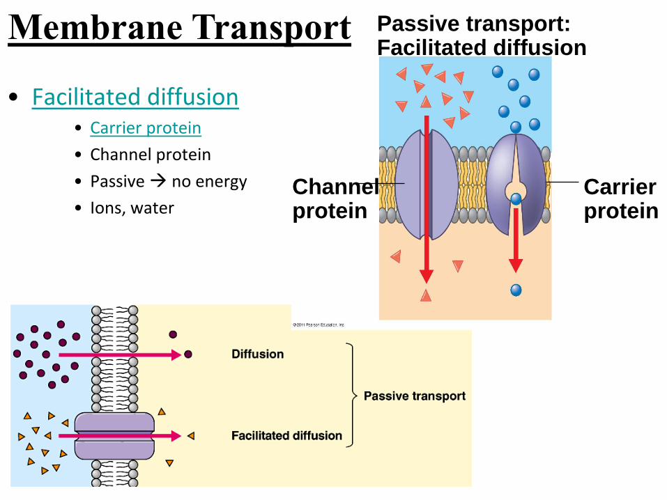
• Facilitated diffusion• Carrier protein
• Channel protein
• Passive no energy
• Ions, water
Passive transport:Facilitated diffusion
Channelprotein
Carrierprotein
Membrane Transport

Figure 7.17
EXTRACELLULARFLUID
CYTOPLASM
Channel proteinSolute
SoluteCarrier protein
(a) A channelprotein
(b) A carrier protein
Membrane Transport

• Osmosis• Diffusion of water
• Across a selectively permeable
membrane
• Passive
• Hypotonic• Lower [solute] & High [H2O]
• Isotonic• Equal/same [solute] &
equal/same [H2O]
• Hypertonic• Higher [solute] & Low [H2O]
Hypotonic hypertonic
Membrane Transport

Figure 7.14
Lowerconcentrationof solute (sugar)
Higher concentrationof solute
Sugarmolecule
H2O
Same concentrationof solute
Selectivelypermeablemembrane
Osmosis
MembraneTransportFigure 5.11

• Isotonic solution
Equal/same [solute]
Inside cell: 3% NaCl, 97% H2O
Beaker: 3% NaCl, 97% H2O
Animal cells = Normal
Plant cells = Normal
3% NaCl
97% H2O
Membrane Transport - Osmosis

• Hypotonic solution
Lower [solute] Inside cell: 3% NaCl, 97% H2O
Beaker: 3% NaCl, 97% H2O
H2O: more IN than out
Plant cells turgid
Animal cells burst/lyse
Why? 1% NaCl
99% H2O
Membrane Transport - Osmosis

• Hypertonic solution
Higher [solute]
Inside cell: 3% NaCl, 97% H2O
Beaker: 5% NaCl, 95% H2O
H2O: more OUT than in
Animal cells shrinks
Plant cells plasmolysis 5% NaCl
95% H2O
Membrane Transport - Osmosis

Osmosis – hypertonic solutions
• Animal cells
Shrinks/shrivels
• Plant cells
Plasmolysis
• Cell membrane separates from
cell wall
5% NaCl
95% H2O

Figure 7.15
Hypotonicsolution
Osmosis
Isotonicsolution
Hypertonicsolution
(a) Animal cell
(b) Plant cell
H2O H2O H2O H2O
H2O H2O H2O H2OCell wall
Lysed Normal Shriveled
Turgid (normal) Flaccid Plasmolyzed
Membrane Transport - Osmosis

Figure 5.12

Figure 5.13

Osmosis
If a marine algal cell is suddenly
transferred from seawater to freshwater,
the algal cell will initially
a) lose water and decrease in volume.
b) stay the same: neither absorb nor lose water.
c) absorb water and increase in volume.

Membrane Transport• Active Transport
• [lower] [higher]
• Against [gradient]
• Carrier protein
• REQUIRES energy

Figure 7.18-6
EXTRACELLULAR
FLUID[Na] high
[K] low
[Na] low
[K] highCYTOPLASM
Na
Na
Na
1 2 3
456
Na
Na
Na
Na
Na
Na
K
K
K
K
K
K
PP
PP i
ATP
ADP
Active transport – Na+/K+ pump

Figure 7.19
3 types of Passive transport Active transport
Diffusion Facilitated diffusion ATP

Figure 5.18 – Active Transport
• Primary active transport moves ions across a membrane, creating an electrochemical
gradient (electrogenic transport). (credit: modification of work by Mariana Ruiz Villareal)

Resting Membrane Potential
• Unequal distribution of
ions (Na+/K+/Cl-/A-)
• Separation of ions
• Movement of ions due
to leak channels• Concentration gradient
• Electrical charge
• Electrochemical gradient
• Excitable cells (nerve &
muscle)

Figure 5.16 – Electrochemical gradient
Active transport & electrochemical gradient

Ion distribution across the membrane

Figure 48.7KeyNa
K
Sodium-potassiumpump
Potassiumchannel
Sodiumchannel
OUTSIDEOF CELL
INSIDEOF CELL
Resting Membrane Potential

For a nerve cell at its resting
potential, the forces acting on
potassium ions (K+) area) none: K+ ions do not move at the resting potential.
b) an electrical gradient, pulling K+ inward, and a
chemical gradient, pushing K+ outward.
c) an electrical gradient, pushing K+ outward, and a
chemical gradient, pulling K+ inward.
d) an electrical gradient, pulling K+ inward, and a
chemical gradient, pushing K+ outward.
e) an electrical gradient, pushing K+ outward, and a
chemical gradient, pushing K+ outward.

Figure 7.20
CYTOPLASM
ATP EXTRACELLULAR
FLUID
Proton pumpH
H
H
H
H
H
Type of transporter?
What’s a “pump?”

Figure 7.21
ATP
H
H
HH
H
H
H
H
Proton pump
Sucrose-H
cotransporter
SucroseSucrose
Diffusion of H
Co-transportType of transporters?

Solutes
Pseudopodium
“Food” or
other particle
Food
vacuole
CYTOPLASM
Plasma
membrane
Vesicle
Receptor
Ligand
Coat proteins
Coated
pit
Coated
vesicle
EXTRACELLULAR
FLUID
Phagocytosis Pinocytosis Receptor-Mediated Endocytosis
3 types of Bulk Transport for large molecules (Endocytosis)
Figure 5.22

Figure 5.22
Pseudopodium
Solutes
“Food”
or other
particle
Food
vacuole
CYTOPLASM
EXTRACELLULAR
FLUID
Pseudopodium
of amoeba
Bacterium
Food vacuole
An amoeba engulfing a bacterium
via phagocytosis (TEM).
1) Phagocytosis
1
m
Phagocytosis1
Phagocytosis2 = cell eating

Pinocytosis vesicles forming
in a cell lining a small blood
vessel (TEM).
Plasma
membrane
Vesicle
0.5
m
2) Pinocytosis
Pinocytosis = cell drinking
Figure 5.22

Top: A coated pit. Bottom: Acoated vesicle forming duringreceptor-mediated endocytosis(TEMs).
Receptor
0.2
5
m
3) Receptor-Mediated Endocytosis
Ligand
Coat proteins
Coated
pit
Coated
vesicle
Coat
proteins
Plasma
membrane
Receptor mediatedSubstance triggers receptor protein
Figure 5.22

• Large molecules• Endocytosis = in take
1. Phagocytosis = cell eating
2. Pinocytosis = cell drinking
3. Receptor mediatedSubstance triggers receptor protein
Membrane Transport

• Bulk Transport Large molecules out
• Exocytosis – Figure 5.23 Getting rid of substances
Transport vessicle fuses w/ cell membrane
Contractile
vacuole
Membrane Transport

Figure 7.16
Contractile vacuole50 m
Membrane Transport – Contractile Vacuole

c) Photomicrograph showingvarious stages of endocytosis.
a) Endocytosis. In endocytosis, material is surrounded bythe cell membrane and brought into the cell.
Extracellular environment
Plasma membrane
Cytoplasm
Vesicle
b) Exocytosis. In exocytosis, a membranous vesicle fuseswith the plasma membrane, expelling its contents outsidethe cell.
Comparison of endocytosis & exocytosis

Insulin results in the uptake
and lowering of blood sugar
would be an example of?
a) Endocytosis
b) Receptor mediated endocytosis
c) Phagocytosis
d) Pinocytosis
e) exocytosis

The resting membrane potential
results from:
a) An unequal distribution of ions across the cell membrane
b) Separation of ions
c) Osmosis
d) Diffusion
e) Aren’t there two correct answers?