Embed Size (px)
DESCRIPTION
Ch. 5 The Structure and Function of Macromolecules. The 4 Macromolecules. Carboyhydrates Lipids Proteins Nucleic acids may consist of thousands of covalently bonded atoms. Similarities:. chainlike molecules ( polymers ) - PowerPoint PPT Presentation
Citation preview
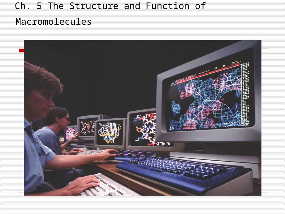
Ch. 5 The Structure and Function of Macromolecules
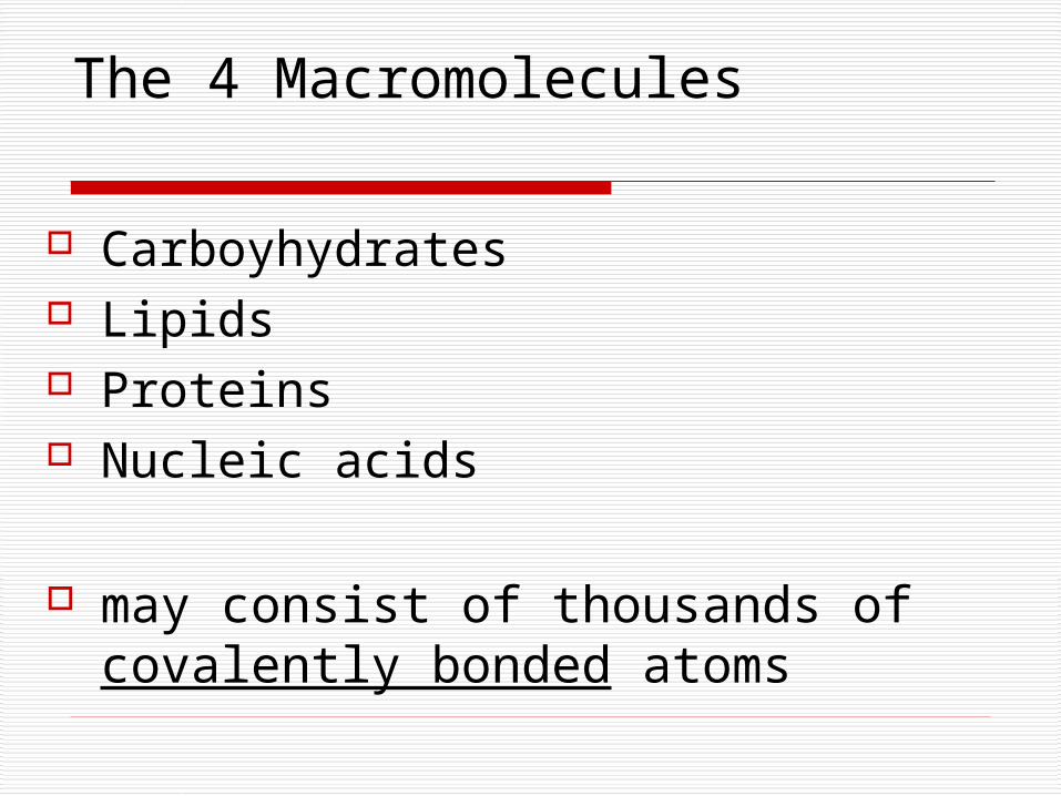
The 4 Macromolecules
Carboyhydrates Lipids Proteins Nucleic acids
may consist of thousands of covalently bonded atoms
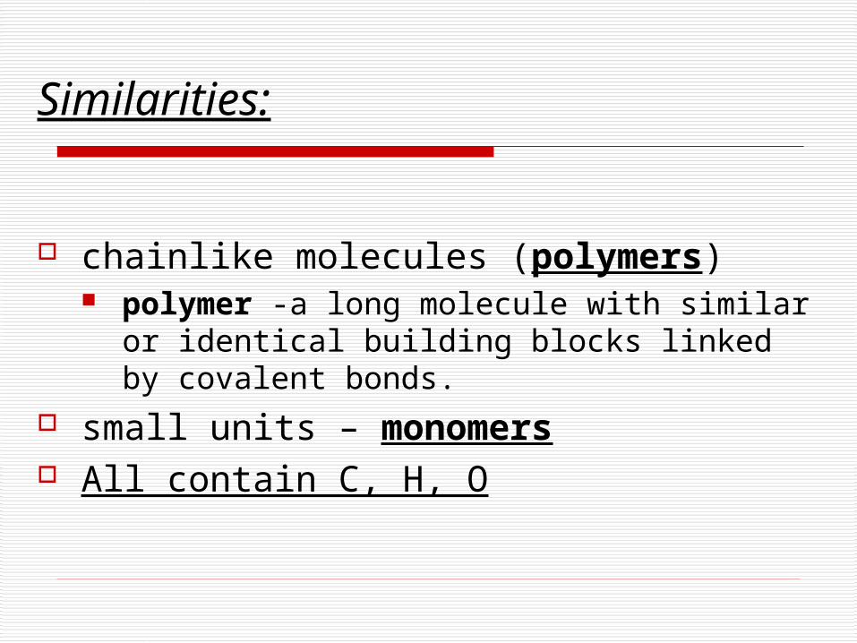
Similarities:
chainlike molecules (polymers) polymer -a long molecule with similar or
identical building blocks linked by covalent bonds.
small units – monomers All contain C, H, O

How are polymers made and broken down?
hydrolysis
dehydration synthesis reaction
Both involve water
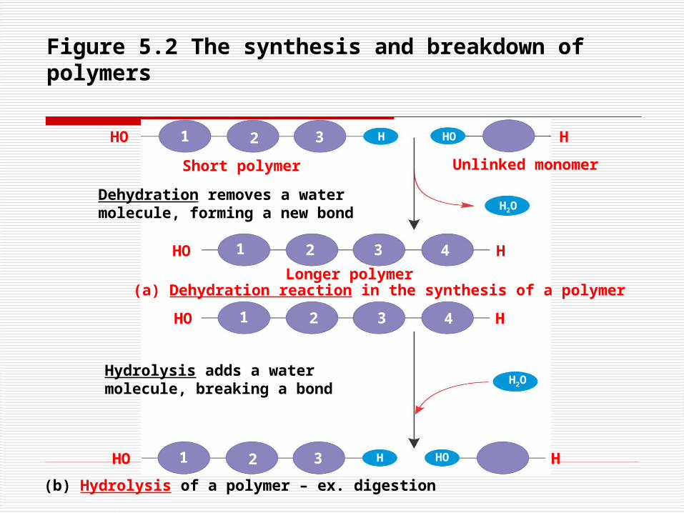
Figure 5.2 The synthesis and breakdown of polymers
(a) Dehydration reaction in the synthesis of a polymer
(b) Hydrolysis of a polymer – ex. digestion
HO H1 2 3 HO
HO H1 2 3 4
H
H2O
HO 1 2 3 H
HO H1 2 3 4
H2O
HHO
Short polymer Unlinked monomer
Longer polymer
Hydrolysis adds a watermolecule, breaking a bond
Dehydration removes a water molecule, forming a new bond
Carbohydrates
Sugars Cellulose
Chitin
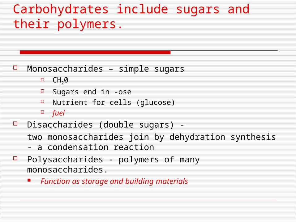
Carbohydrates include sugars and their polymers.
Monosaccharides – simple sugars CH20 Sugars end in -ose Nutrient for cells (glucose) fuel
Disaccharides (double sugars) - two monosaccharides join by dehydration
synthesis - a condensation reaction Polysaccharides - polymers of many monosaccharides.
Function as storage and building materials
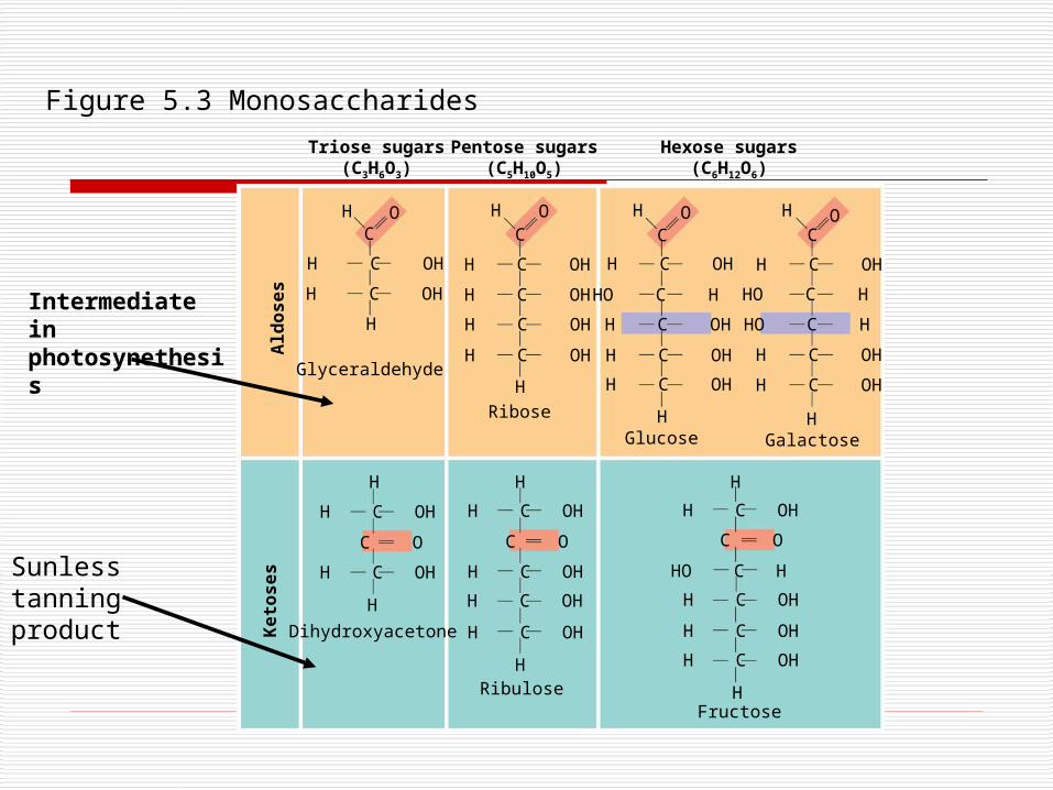
Triose sugars(C3H6O3)
Pentose sugars(C5H10O5)
Hexose sugars(C6H12O6)
H C OH
H C OH
H C OH
H C OH
H C OH
H C OH
HO C H
H C OH
H C OH
H C OH
H C OH
HO C H
HO C H
H C OH
H C OH
H C OH
H C OH
H C OH
H C OH
H C OH
H C OH
H C OH
C OC O
H C OH
H C OH
H C OH
HO C H
H C OH
C O
H
H
H
H H H
H
H H H H
H
H H
C C C COOOO
Ald
os
es
Ke
tos
es
Glyceraldehyde
Ribose
Glucose Galactose
Dihydroxyacetone
RibuloseFructose
Figure 5.3 Monosaccharides
Sunless tanning product
Intermediate in photosynethesis
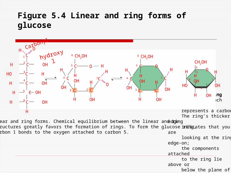
Figure 5.4 Linear and ring forms of glucose
(b) Abbreviated ring structure. Each corner represents a carbon. The ring’s thicker edge indicates that you are looking at the ring edge-on; the components attached to the ring lie above or below the plane of the ring.
H
H C OH
HO C H
H C OH
H C OH
H C
O
C
H
1
2
3
4
5
6
H
OH
4 C
6 CH2OH 6 CH2OH
5 C
HOH
C
H OH
H
2 C
1C
H
O
H
OH
4 C
5 C
3 C
H
HOH
OH
H
2C
1 C
OH
H
CH2OH
H
H
OHHO
H
OH
OH
H5
3 2
4
(a) Linear and ring forms. Chemical equilibrium between the linear and ring structures greatly favors the formation of rings. To form the glucose ring, carbon 1 bonds to the oxygen attached to carbon 5.
OH3
O H OO
6
1
Carbonyl
hydroxyl
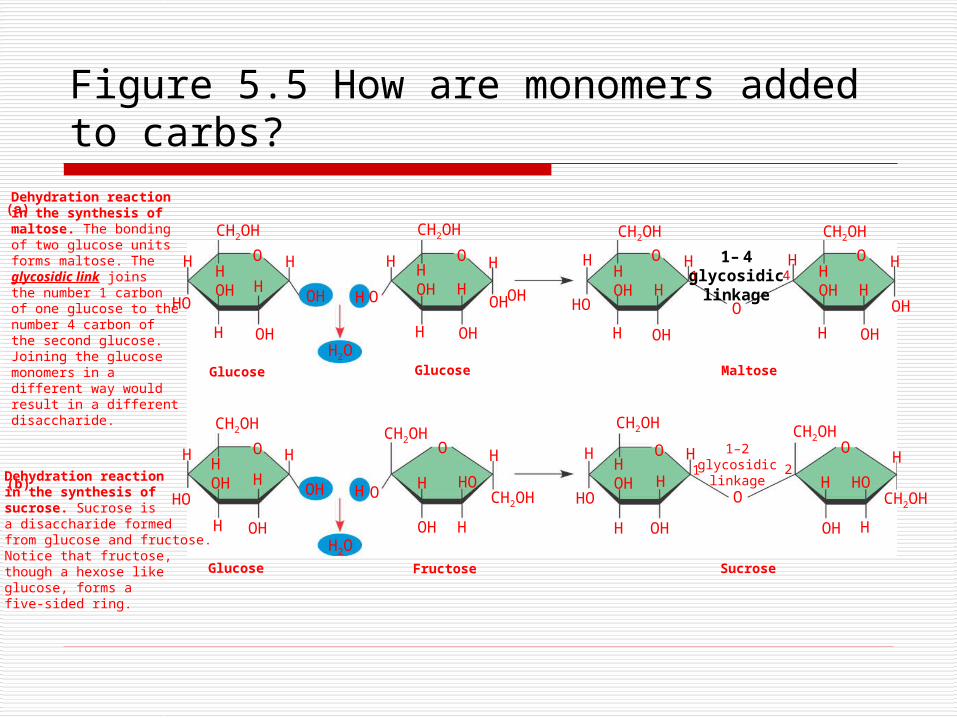
Figure 5.5 How are monomers added to carbs?
Dehydration reaction in the synthesis of maltose. The bonding of two glucose units forms maltose. The glycosidic link joins the number 1 carbon of one glucose to the number 4 carbon of the second glucose. Joining the glucose monomers in a different way would result in a different disaccharide.
Dehydration reaction in the synthesis of sucrose. Sucrose is a disaccharide formed from glucose and fructose.Notice that fructose,though a hexose like glucose, forms a five-sided ring.
(a)
(b)
H
HO
H
HOH H
OH
O H
OH
CH2OH
H
HO
H
HOH H
OH
O H
OH
CH2OH
H
O
H
HOH H
OH
O H
OH
CH2OH
H
H2O
H2O
H
H
O
H
HOH
OH
O HCH2OH
CH2OH HO
OHH
CH2OH
HOH H
H
HO
OHH
CH2OH
HOH H
O
O H
OHH
CH2OH
HOH H
O
HOH
CH2OH
H HO
O
CH2OH
H
H
OH
O
O
1 2
1 41– 4
glycosidiclinkage
1–2glycosidic
linkage
Glucose
Glucose Glucose
Fructose
Maltose
Sucrose
OH
H
H
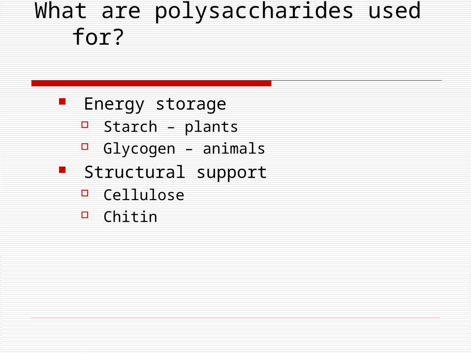
What are polysaccharides used for?
Energy storage Starch – plants Glycogen – animals
Structural support Cellulose Chitin
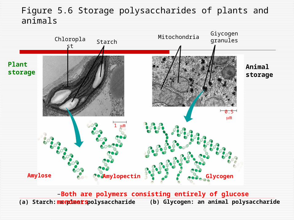
Figure 5.6 Storage polysaccharides of plants and animals
Mitochondria Giycogen granulesChloroplast Starch
Amylose Amylopectin
1 m
0.5 m
(a) Starch: a plant polysaccharide (b) Glycogen: an animal polysaccharide
Glycogen
–Both are polymers consisting entirely of glucose monomers
Plant storage
Animal storage
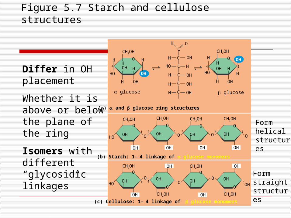
Figure 5.7 Starch and cellulose structures
(c) Cellulose: 1– 4 linkage of glucose monomers
H O
O
CH2OH
HOH H
H
OH
OHH
H
HO
4
C
C
C
C
C
C
H
H
H
HO
OH
H
OH
OH
OH
H
O
CH2OH
HH
H
OH
OHH
H
HO
4 OH
CH2OH
O
OH
OH
HO41
O
CH2OH
O
OH
OH
O
CH2OH
O
OH
OH
CH2OH
O
OH
OH
O O
CH2OH
O
OH
OH
HO4
O1
OH
O
OH OHO
CH2OH
O
OH
O OH
O
OH
OH
(a) and glucose ring structures
(b) Starch: 1– 4 linkage of glucose monomers
1
glucose glucose
CH2OH CH2OH
1 4 41 1
Differ in OH placement
Whether it is above or below the plane of the ring
Isomers with different “glycosidic linkages”
Form helical structures
Form straight structures
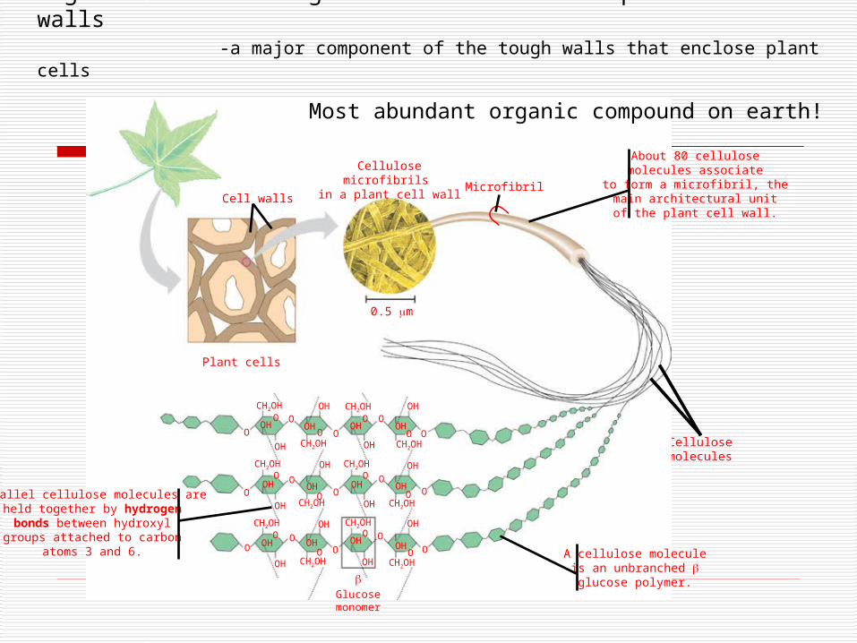
Figure 5.8 The arrangement of cellulose in plant cell walls -a major component of the tough walls that enclose plant cells
Cellulosemolecules
Plant cells
0.5 m
Cell walls
Cellulose microfibrils in a plant cell wall
Microfibril
CH2OH
CH2OH
OH
OH
O
OOH
OCH2OH
O
OOH
OCH2OH OH
OH OHO
O
CH2OH
OO
OH
CH2OH
OO
OH
O
O
CH2OHOH
CH2OHOH
OOH OH OH OH
O
OH OH
CH2OH
CH2OH
OHO
OH CH2OH
O
O
OH CH2OH
OH
Glucose monomer
O
O
O
O
O
O
Parallel cellulose molecules areheld together by hydrogenbonds between hydroxylgroups attached to carbon
atoms 3 and 6.
About 80 cellulosemolecules associate
to form a microfibril, themain architectural unitof the plant cell wall.
A cellulose moleculeis an unbranched glucose polymer.
OH
OH
O
OOH
Most abundant organic compound on earth!
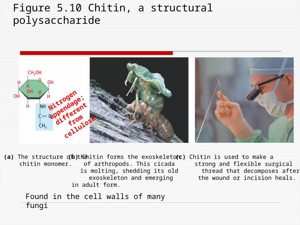
Figure 5.10 Chitin, a structural polysaccharide
(a) The structure of the chitin monomer.
O
CH2OH
OHH
H OH
H
NH
C
CH3
O
H
H
(b) Chitin forms the exoskeleton of arthropods. This cicada is molting, shedding its old exoskeleton and emergingin adult form.
(c) Chitin is used to make a strong and flexible surgical
thread that decomposes after the wound or incision heals.
OH
Nitrogen
appendage;
different fr
om
cellulose
Found in the cell walls of many fungi
Review Questions
The building blocks of carbohydrates are? Function in?
A glycosidic linkage is between what? What is the polysaccharide of plants called?
Of animals? How does a cellulose molecule differ from a
starch? Differ from a chitin?
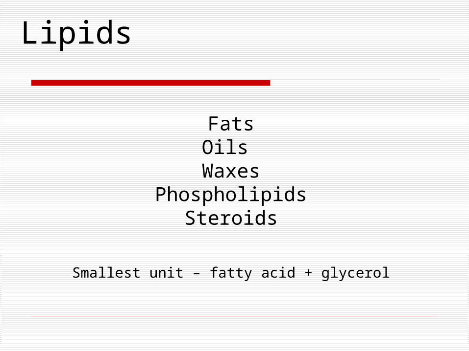
Lipids
FatsOils
WaxesPhospholipids
Steroids
Smallest unit – fatty acid + glycerol
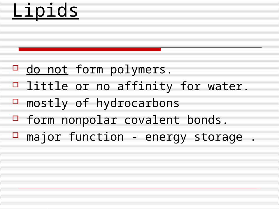
Lipids
do not form polymers. little or no affinity for water. mostly of hydrocarbons form nonpolar covalent bonds. major function - energy storage .
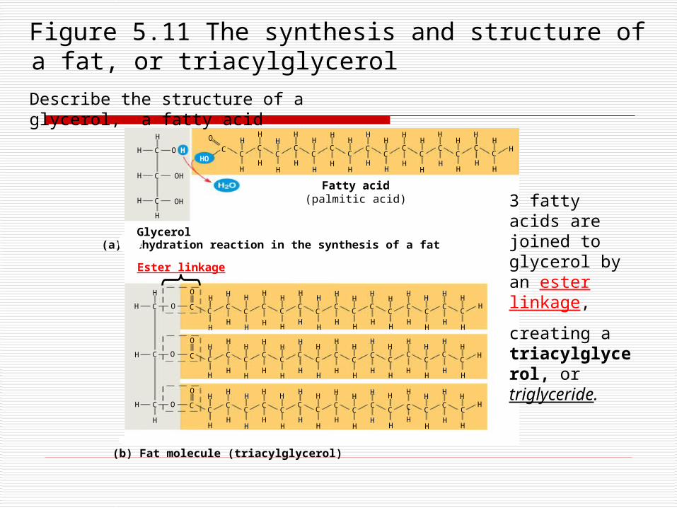
(b) Fat molecule (triacylglycerol)
H
H O HC
C
C
H
H OH
OH
H
HH H
HH
HH
H
HHH
H
HH
H
HH
HH
HH
H
HH
HH
H
HH
H
HC
CCC
CC
CC
CC
CC
CC
CC
Glycerol
Fatty acid(palmitic acid)
H
H
H
H
HH
HH
HH
HH
HH
HH
HH
HH
HH
HH
HHHH
HHH
HH
HH
H
H
HH
HH
HH
HH
HH
HH
HH
HH
HH
HH
HH
HH
HH
HH
HH
HH H
HH
HH
HH
HH
HH
HH
H
HH
HH
HH
HH
HH
HHH
HH
HO
O
O
O
O
OC
C
C C CC
CC
CC
CC
CC
CC
CC
C
C
CC
CCCC
CC
CC
CC
CC
CC
C CC
CC
CC
CC
CC
CC
CC
O
O
(a) Dehydration reaction in the synthesis of a fat
Ester linkage
Figure 5.11 The synthesis and structure of a fat, or triacylglycerol
3 fatty acids are joined to glycerol by an ester linkage,
creating a triacylglycerol, or triglyceride.
Describe the structure of a glycerol, a fatty acid
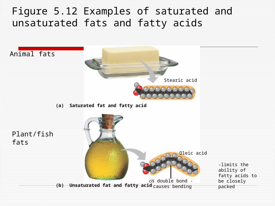
Figure 5.12 Examples of saturated and unsaturated fats and fatty acids
(a) Saturated fat and fatty acid
Stearic acid
(b) Unsaturated fat and fatty acidcis double bond - causes bending
Oleic acid
Animal fats
Plant/fish fats
-limits the ability of fatty acids to be closely packed
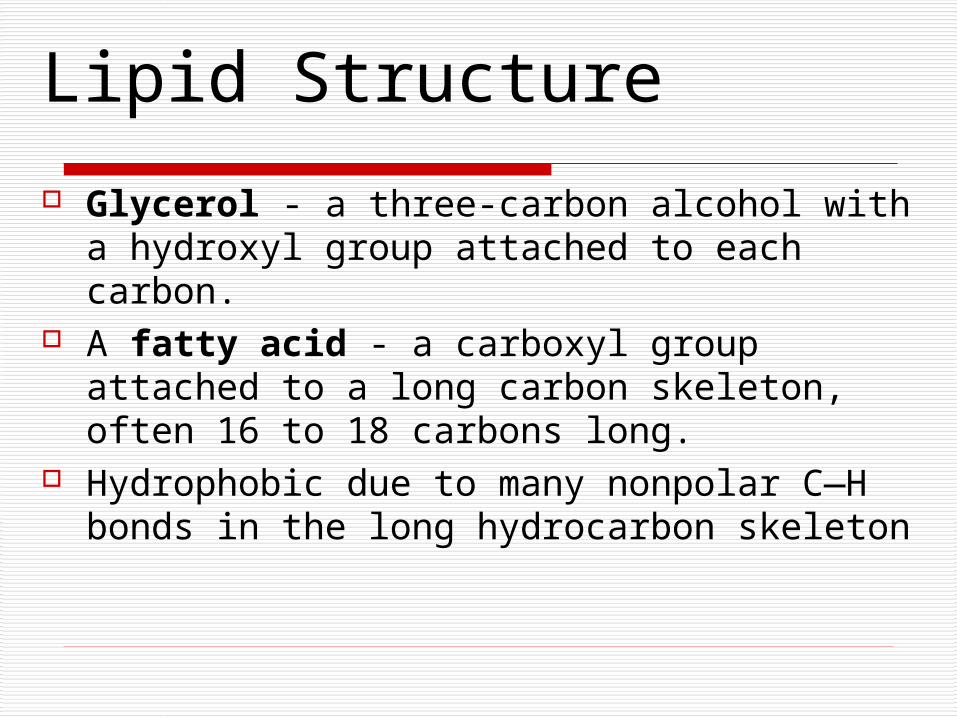
Lipid Structure
Glycerol - a three-carbon alcohol with a hydroxyl group attached to each carbon.
A fatty acid - a carboxyl group attached to a long carbon skeleton, often 16 to 18 carbons long.
Hydrophobic due to many nonpolar C—H bonds in the long hydrocarbon skeleton
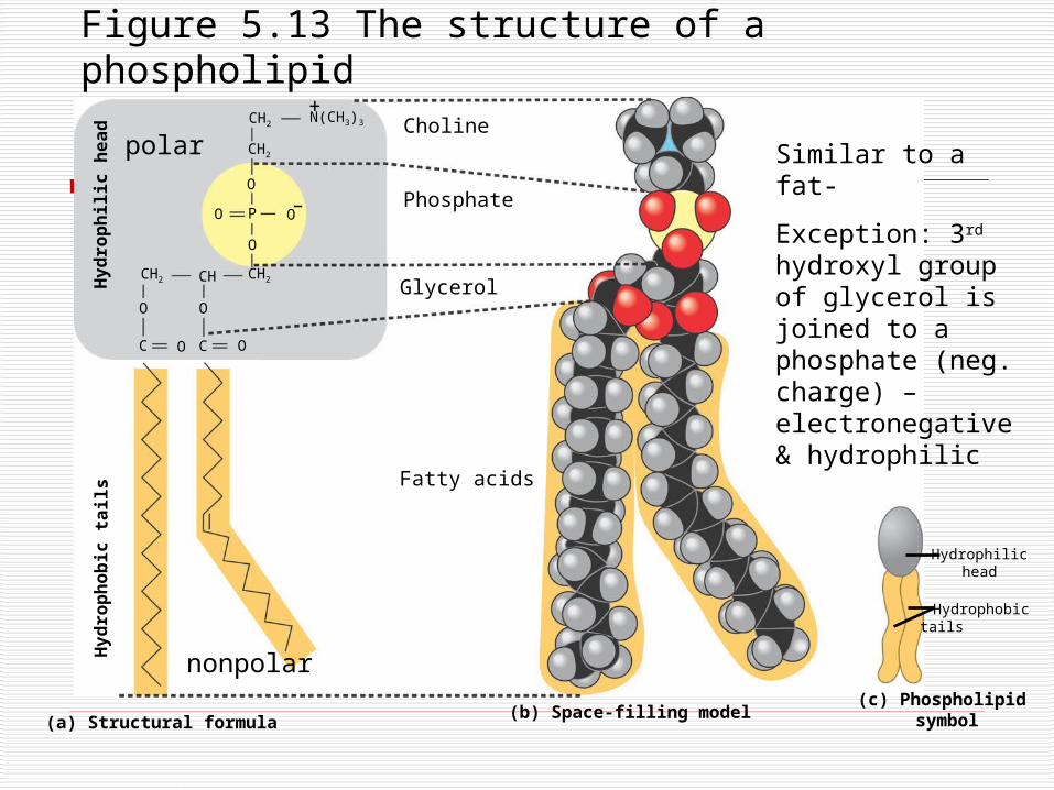
Figure 5.13 The structure of a phospholipidH
yd
rop
hil
ic h
ea
dCH2
N(CH3)3
CH2
O
PO O
O
CH2CHCH2
OO
C O C O
Choline
Phosphate
Glycerol
(a) Structural formula(b) Space-filling model
Fatty acids
(c) Phospholipid symbol
Hy
dro
ph
ob
ic t
ail
s
Hydrophilichead
Hydrophobictails
+
–
polar
nonpolar
Similar to a fat-
Exception: 3rd hydroxyl group of glycerol is joined to a phosphate (neg. charge) – electronegative & hydrophilic
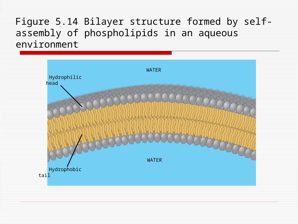
Hydrophilichead
WATER
WATER
Hydrophobictail
Figure 5.14 Bilayer structure formed by self-assembly of phospholipids in an aqueous environment
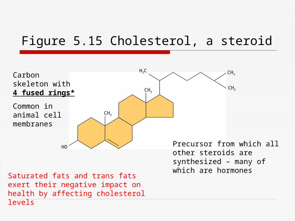
Figure 5.15 Cholesterol, a steroid
HO
CH3
CH3
H3C CH3
CH3
Carbon skeleton with 4 fused rings*
Common in animal cell membranes
Precursor from which all other steroids are synthesized – many of which are hormones
Saturated fats and trans fats exert their negative impact on health by affecting cholesterol levels
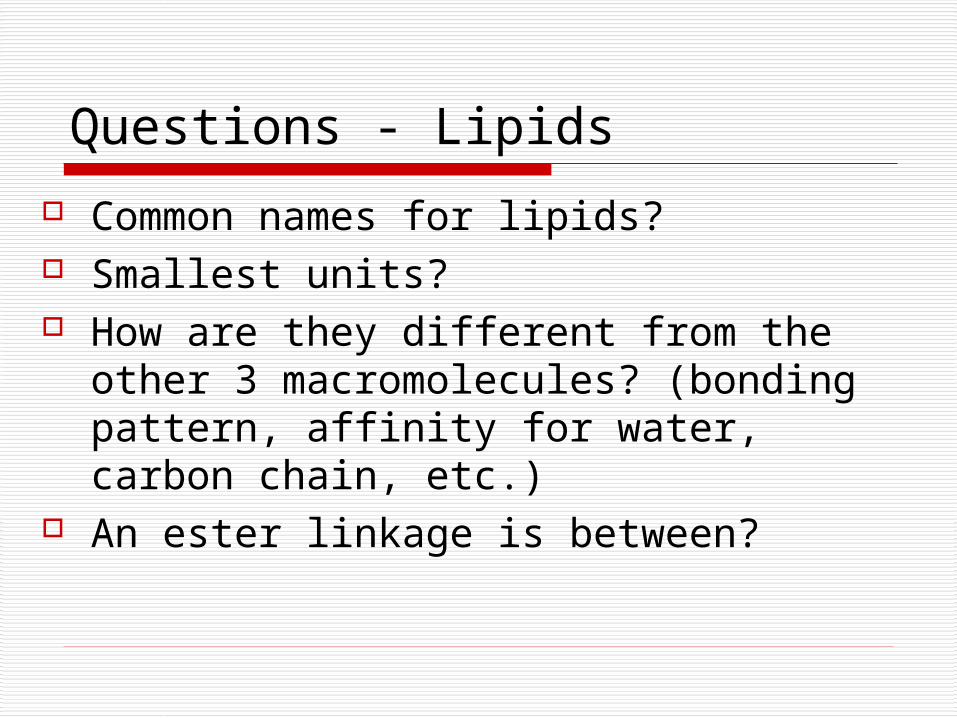
Questions - Lipids
Common names for lipids? Smallest units? How are they different from the other 3
macromolecules? (bonding pattern, affinity for water, carbon chain, etc.)
An ester linkage is between?
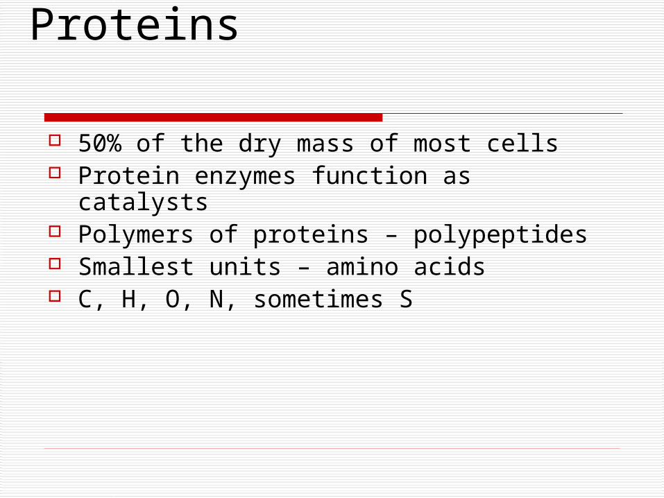
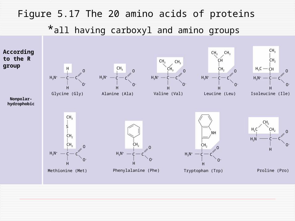
Proteins
50% of the dry mass of most cells Protein enzymes function as catalysts Polymers of proteins – polypeptides Smallest units – amino acids C, H, O, N, sometimes S
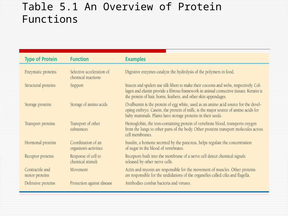
Table 5.1 An Overview of Protein Functions
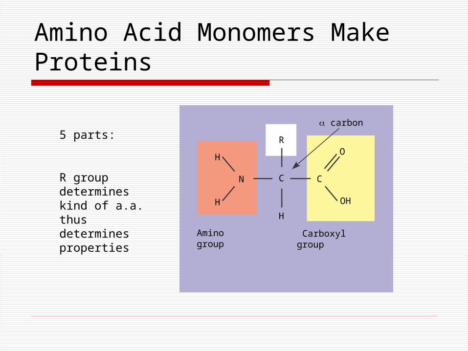
Amino Acid Monomers Make Proteins
H
H
N C
R
H
C
O
OH
Aminogroup
Carboxylgroup
carbon
5 parts:
R group determines kind of a.a. thus determines properties
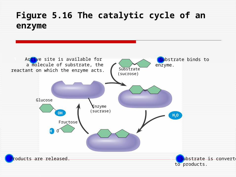
Figure 5.16 The catalytic cycle of an enzyme
Substrate(sucrose)
Enzyme (sucrase)
Glucose
OH
H O
H2O
Fructose
1 Active site is available for a molecule of substrate, the
reactant on which the enzyme acts.
2 Substrate binds toenzyme.
4 Products are released. 3 Substrate is convertedto products.
S
Figure 5.17 The 20 amino acids of proteins
*all having carboxyl and amino groups
O
O–
O
O–
H
H3N+ C C
O
O–
H
CH3
H3N+ C
H
C
O
O–
CH3 CH3
CH3
C C
O
O–
H
H3N+
CH
CH3
CH2
C
H
H3N+
CH3
CH3
CH2
CH
C
H
H3N+ C
CH3
CH2
CH2
CH3N+
H
C
O
O–
CH2
CH3N+
H
C
O
O–
CH2
NH
H
C
O
O–
H3N+ C
CH2
H2C
H2N C
CH2
H
C
Nonpolar-hydrophobic
Glycine (Gly) Alanine (Ala) Valine (Val) Leucine (Leu) Isoleucine (Ile)
Methionine (Met) Phenylalanine (Phe)
C
O
O–
Tryptophan (Trp) Proline (Pro)
H3C
According to the R group
O–
OH
CH2
C C
H
H3N+
O
O–
H3N+
OH CH3
CH
C C
HO–
O
SH
CH2
C
H
H3N+ C
O
O–
H3N+ C C
CH2
OH
H H H
H3N+
NH2
CH2
OC
C C
O
O–
NH2 O
C
CH2
CH2
C CH3N+
O
O–
O
Polar-hydrophilic
ElectricallyCharged-Ionized
Refers only to R groups
–O O
C
CH2
C CH3N+
H
O
O–
O– O
C
CH2
C CH3N+
H
O
O–
CH2
CH2
CH2
CH2
NH3+
CH2
C CH3N+
H
O
O–
NH2
C NH2+
CH2
CH2
CH2
C CH3N+
H
O
O–
CH2
NH+
NHCH2
C CH3N+
H
O
O–
Serine (Ser) Threonine (Thr)Cysteine
(Cys)Tyrosine
(Tyr)Asparagine
(Asn)Glutamine
(Gln)
Acidic – negative charge Basic – positive charge
Aspartic acid (Asp)
Glutamic acid (Glu)
Lysine (Lys) Arginine (Arg) Histidine (His)
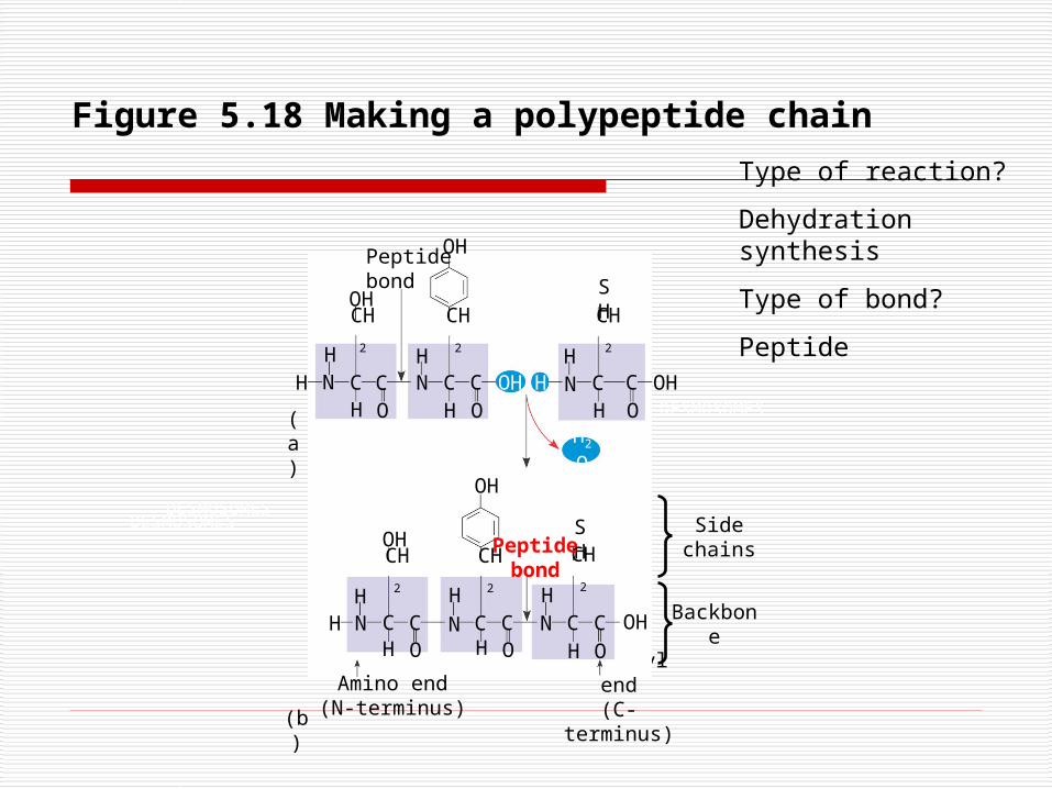
Figure 5.18 Making a polypeptide chain
Carboxyl end
(C-terminus)
DESMOSOMES
OH
DESMOSOMESDESMOSOMES
OHCH
2
CN
HC
H OH OH OH
Peptidebond
OH
OH
OH
H H
HHH
HH
H
H
H H
HN
N N
N N
SH
Side chains
SH
OO
O O O
H2
O
CH
2
CH
2
CH
2
CH
2
CH
2
C C C C C C
C CC C
Peptidebond
Amino end(N-terminus)
Backbone
(a)
(b)
Type of reaction?
Dehydration synthesis
Type of bond?
Peptide
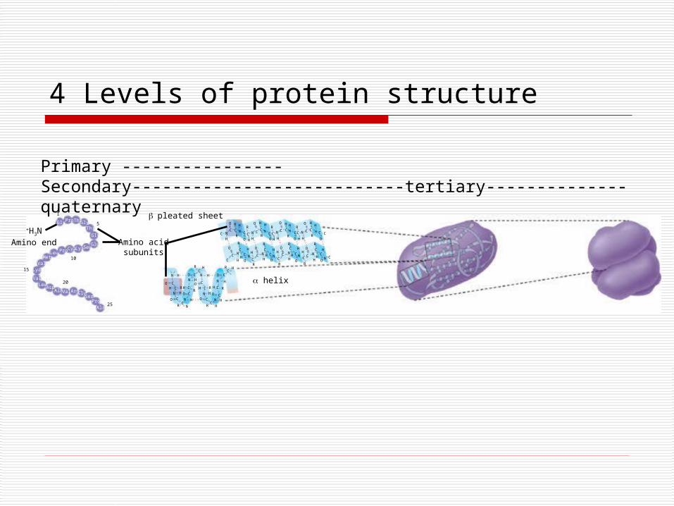
4 Levels of protein structure
NC
H
C
O
CH
O
H
NR
C
R
C
H
N
H
O
C
H
C
R
H
NCO
R
C
H
N
H
O
C
H
C
RH
H
C
O
R
C
H
N
H
O
C
H
C
R
H
H C
O
O
CC
N
H
R
C
HC
O
H
NH
CR
O
C N
H
R
C
H C
O
H
NH
C
R
O
C N
H
RC
H C
O
H
NH
C
R
O
C N
H
R
C
HC
O
H
N C
N H O C N H O C
RC
H RC
H
O CN H
CON H
H C RH CR
CN H O
O C N HC
R N
H C R H C R
N H O CO C N H
CR H
Gly GlyThr
Gly
GluSeuLyaCyaProLeu
MetVal
Lya
ValLeu
AspAla Val ArgGly
SerPro
Ala
Pro Thr
Amino acidsubunits
pleated sheet
helix
+H3NAmino end
1
5
10
15
20
25
C
Primary ----------------Secondary---------------------------tertiary--------------quaternary
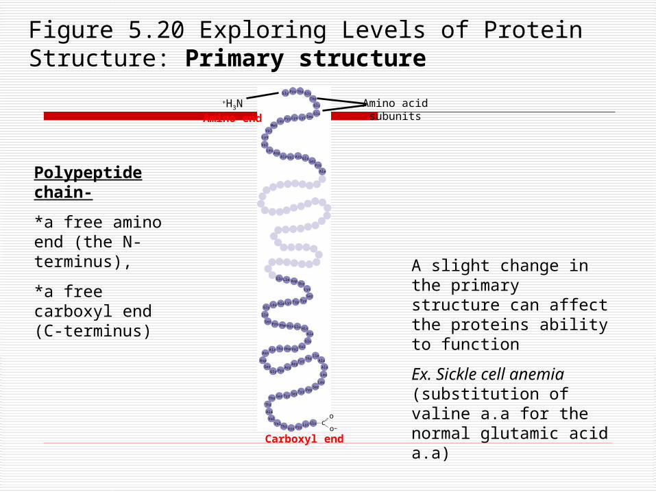
Figure 5.20 Exploring Levels of Protein Structure: Primary structure
–
Amino acid subunits
+H3NAmino end
o
Carboxyl end
oc
Gly Pro Thr Gly
Thr
Gly
GluSeuLysCysPro
LeuMet
Val
Lys
Val
LeuAsp
Ala Val Arg GlySer
Pro
Ala
Gly
lle
SerPro Phe His Glu His
Ala
Glu
ValValPheThrAla
Asn
Asp
SerGly Pro
ArgArg
TyrThr
lleAla
Ala
Leu
LeuSer
ProTyrSer
TyrSerThr
Thr
Ala
ValVal
ThrAsn Pro
Lys Glu
Thr
Lys
SerTyrTrpLysAlaLeu
Glu Lle Asp
Polypeptide chain-
*a free amino end (the N-terminus),
*a free carboxyl end (C-terminus) A slight change in the
primary structure can affect the proteins ability to function
Ex. Sickle cell anemia (substitution of valine a.a for the normal glutamic acid a.a)
O C 2. Helix (coiled)
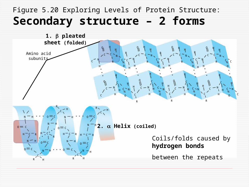
1. pleated sheet (folded)
Amino acidsubunits N
C
H
C
O
CN
H
C
OH
R
C N
H
C
O H
C
R
N
HH
RC
O
R
C
H
N
H
C
OH
N
C
O
R
C
H
N
H
H
C
R
C
O H
C
R
N
H
C
O
C
C
O
C
N
H
H
R
C
C
O
N
HH
C
R
C
O
N
H
R
C
H C
O
N
HH
C
R
C
O
N
H
R
C
H C
O
N
HH
C
R
C
O
N
H
R
C
H C
O
N
H
C
N H
H C R
N HO
O CN
C
RC
H
H O
CHR
N H
O C
R
CH
N H
O C
H C R
N H
C
C
N
R H
H
O C
H C R
N H
O C
R
CH
Figure 5.20 Exploring Levels of Protein Structure:
Secondary structure – 2 forms
Coils/folds caused by hydrogen bonds
between the repeats
Figure 5.20 Exploring Levels of Protein Structure: Tertiary structure
CH2
OH
O
COH
CH2
CH2 NH3+ C-O CH2
O
CH2SSCH2
CH
CH
CH3
CH3
H3C
H3C
Hydrophobic interactions and van der Waalsinteractions
Polypeptidebackbone
Hydrogenbond
Ionic bond
Disulfide bridge
Determined by interactions among various R groups
Among hydrophobic R groups
Between polar and/or charged areas
Between charged R groups
Strong covalent bonds between sulfydryl groups of 2 cysteine
monomers
Bonds:
Hydrogen
Disulfide covalent
Ionic
Van der Waals interactions
Peptide
Polypeptidechain
Collagen
Chains
ChainsHemoglobin
IronHeme
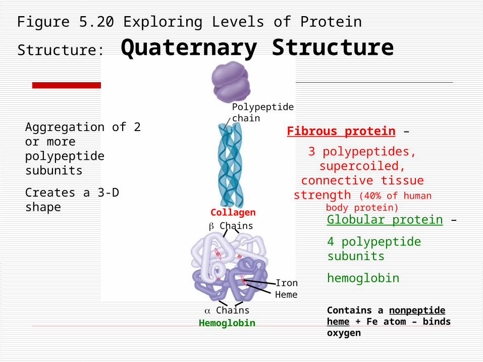
Figure 5.20 Exploring Levels of Protein Structure: Quaternary Structure
Aggregation of 2 or more polypeptide subunits
Creates a 3-D shape
Fibrous protein –
3 polypeptides, supercoiled, connective tissue strength
(40% of human body protein)
Globular protein –
4 polypeptide subunits
hemoglobin
Contains a nonpeptide heme + Fe atom – binds oxygen
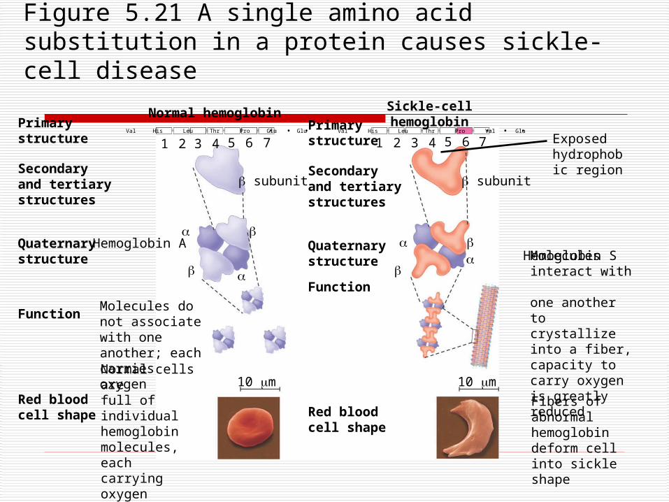
Exposed hydrophobic region
Figure 5.21 A single amino acid substitution in a protein causes sickle-cell disease
Primary structure
Secondaryand tertiarystructures
Quaternary structure
Function
Red bloodcell shape
Hemoglobin A
Molecules donot associatewith oneanother; eachcarries oxygen
Normal cells arefull of individualhemoglobinmolecules, eachcarrying oxygen
10 m 10 m
Primary structure
Secondaryand tertiarystructures
Quaternary structure
Function
Red bloodcell shape
Hemoglobin S
Molecules interact with one another tocrystallize into a fiber, capacity to carry oxygen is greatly reduced
Fibers of abnormalhemoglobin deform cell into sickle shape
subunit subunit
1 2 3 4 5 6 7 3 4 5 6 721
Normal hemoglobin Sickle-cell hemoglobin. . .. . .Val His Leu Thr Pro Glu Glu Val His Leu Thr Pro Val Glu
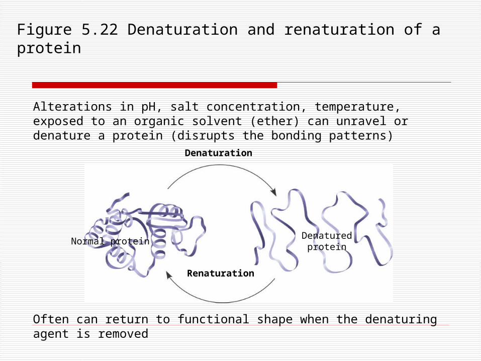
Figure 5.22 Denaturation and renaturation of a protein
Denaturation
Renaturation
Denatured proteinNormal protein
Alterations in pH, salt concentration, temperature, exposed to an organic solvent (ether) can unravel or denature a protein (disrupts the bonding patterns)
Often can return to functional shape when the denaturing agent is removed
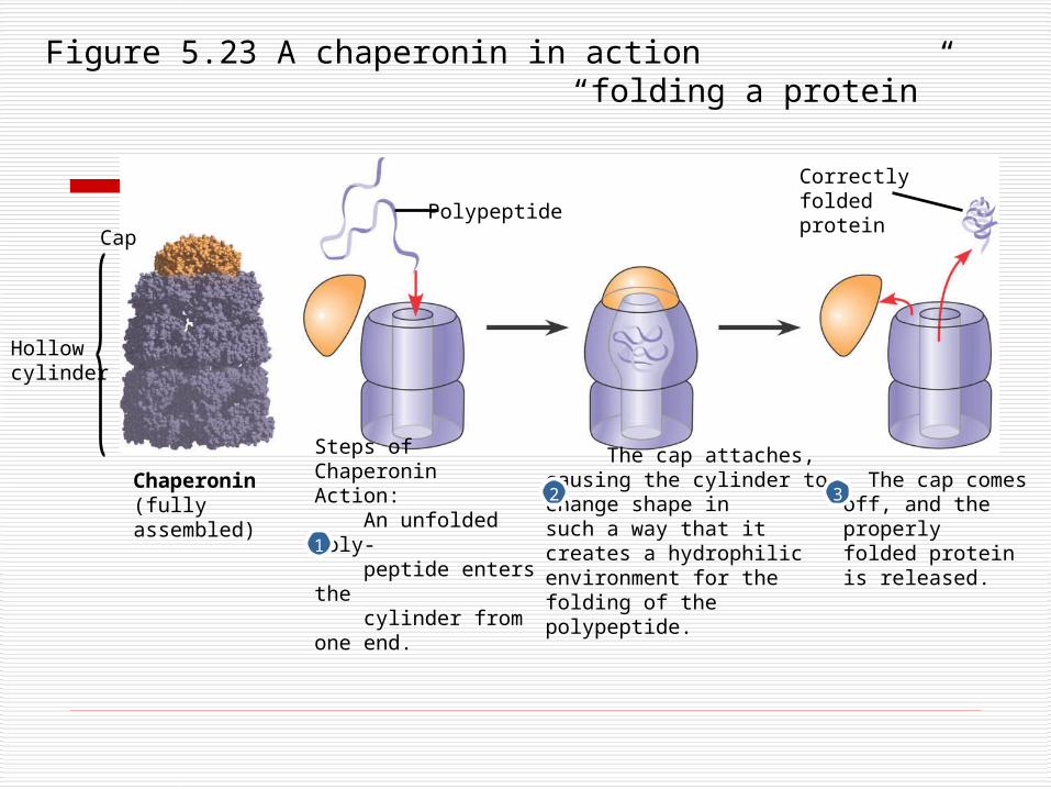
Figure 5.23 A chaperonin in action“folding a protein”
Hollowcylinder
Cap
Chaperonin(fully assembled)
Steps of ChaperoninAction: An unfolded poly- peptide enters the cylinder from one end.
The cap attaches, causing the cylinder to change shape insuch a way that it creates a hydrophilic environment for the folding of the polypeptide.
The cap comesoff, and the properlyfolded protein is released.
Correctlyfoldedprotein
Polypeptide
2
1
3
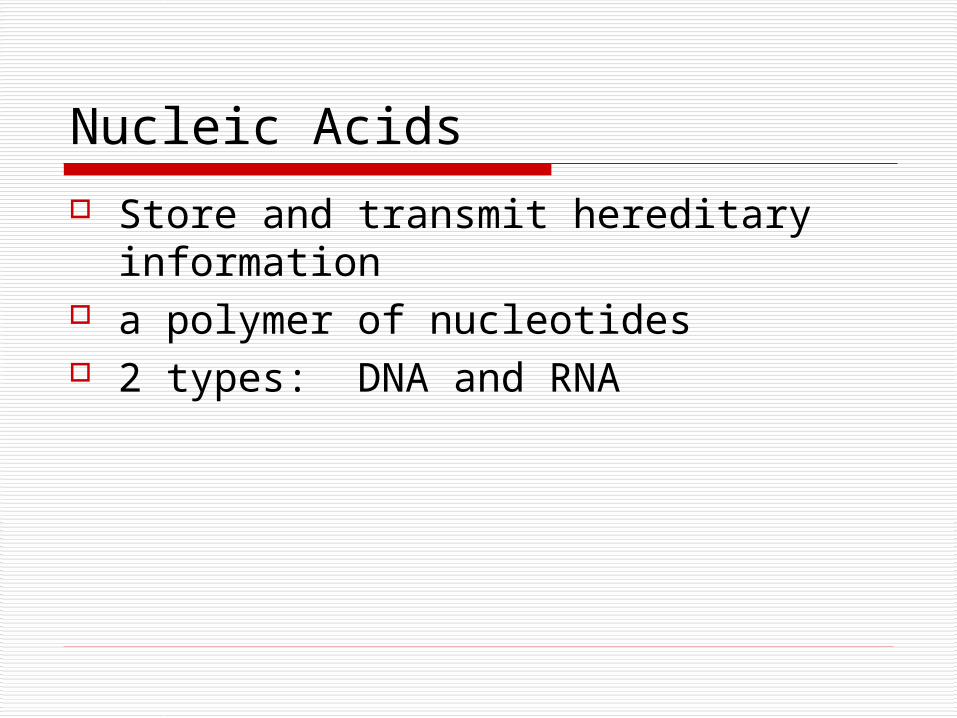
Nucleic Acids
Store and transmit hereditary information
a polymer of nucleotides 2 types: DNA and RNA
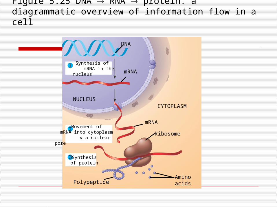
Figure 5.25 DNA RNA protein: a diagrammatic overview of information flow in a cell
1
2
3
Synthesis of mRNA in the nucleus
Movement of mRNA into cytoplasm
via nuclear pore
Synthesisof protein
NUCLEUSCYTOPLASM
DNA
mRNA
Ribosome
AminoacidsPolypeptide
mRNA
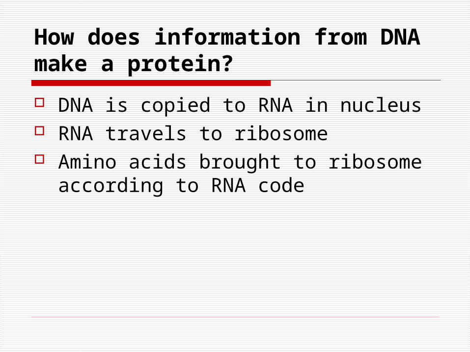
How does information from DNA make a protein?
DNA is copied to RNA in nucleus RNA travels to ribosome Amino acids brought to ribosome
according to RNA code
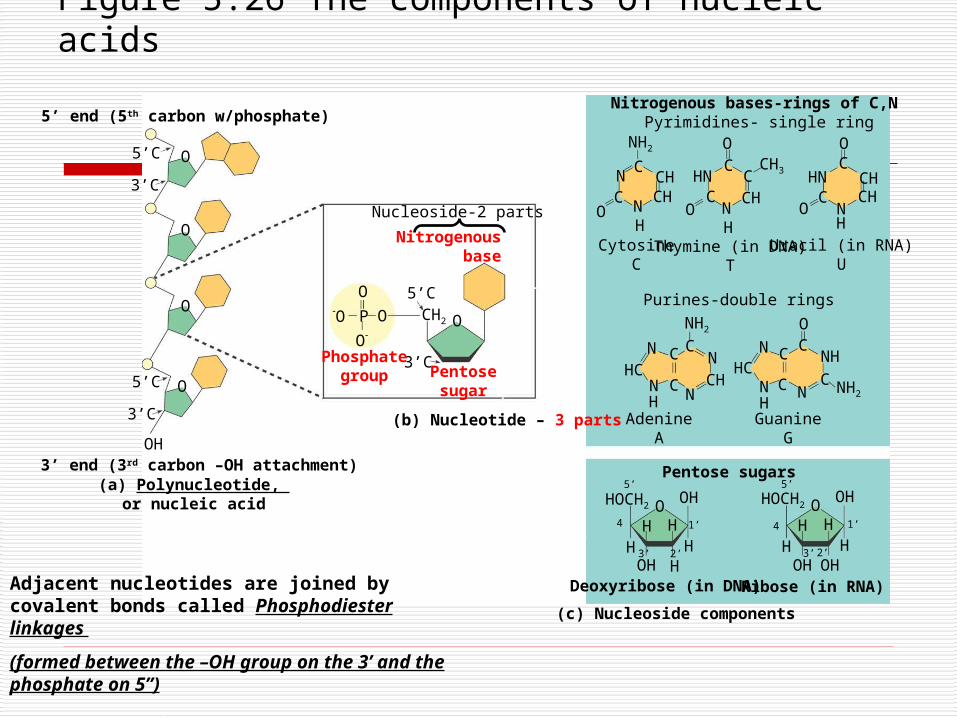
Figure 5.26 The components of nucleic acids
3’C
CHCH
Uracil (in RNA)U
5’ end (5th carbon w/phosphate)
5’C
3’C
5’C
O
O
O
O
3’ end (3rd carbon –OH attachment)OH
Nitrogenousbase
Nucleoside-2 parts
O
O
O
O P CH2 O
5’C
3’CPhosphategroup Pentose
sugar
(b) Nucleotide – 3 parts
CN
NC
OH
NH2
CHCH
OC
NH
CHHN
CO
CCH3
N
HNC
C
HO
O
CytosineC
Thymine (in DNA)T
NHC
N C
CN
C
CHN
NH2 O
NHC
NHH
C
C
C
N
NH
C NH2
AdenineA
GuanineG
Purines-double rings
OHOCH2
HH H
OH
HOHOH
OHOCH2
HH H
OH
HOH H
5’
4
3’ 2’
1’
3’ 2’
1’4
5’Pentose sugars
Deoxyribose (in DNA) Ribose (in RNA)
Nitrogenous bases-rings of C,N Pyrimidines- single ring
(c) Nucleoside components
(a) Polynucleotide, or nucleic acid
Adjacent nucleotides are joined by covalent bonds called Phosphodiester linkages
(formed between the –OH group on the 3’ and the phosphate on 5”)
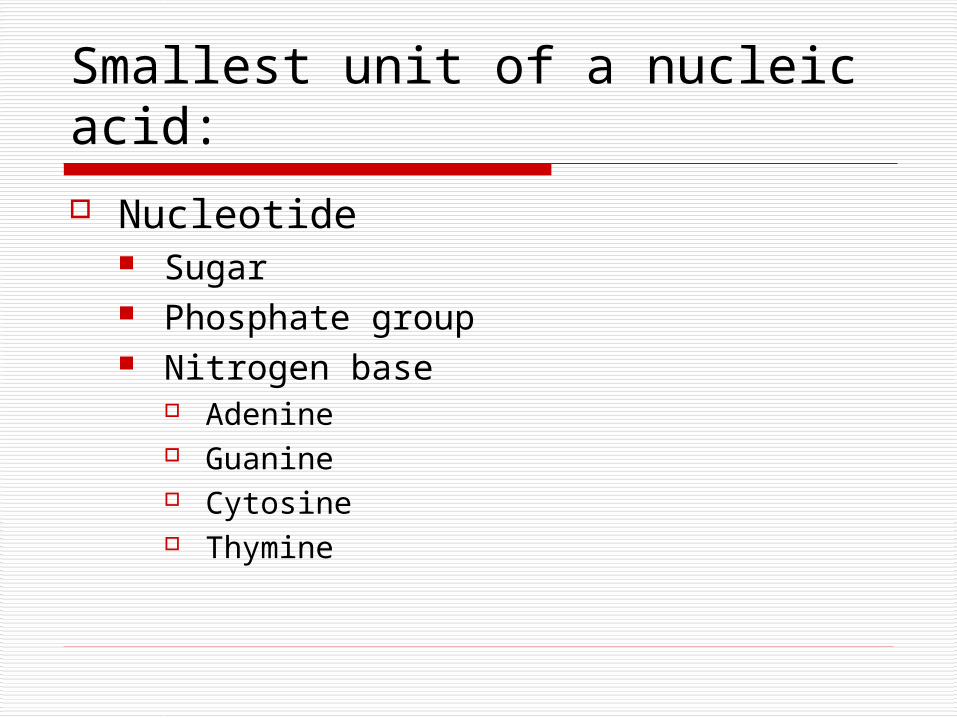
Smallest unit of a nucleic acid:
Nucleotide Sugar Phosphate group Nitrogen base
Adenine Guanine Cytosine Thymine
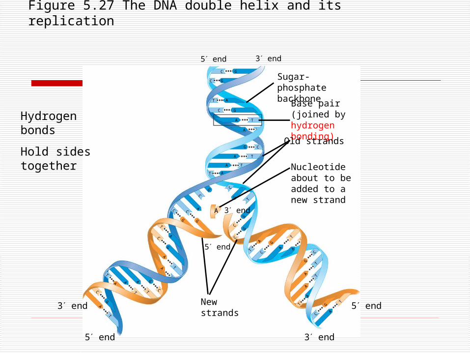
Figure 5.27 The DNA double helix and its replication
3 end
Sugar-phosphatebackbone
Base pair (joined by hydrogen bonding)Old strands
Nucleotideabout to be added to a new strand
A
3 end
3 end
5 end
Newstrands
3 end
5 end
5 end
C G
C G
AT
C G
A T
A T
G C
A T
A T
T A
G
AC
C
C
G G
T
A
A
T
C
G
A
T
G
C
A
T
A
T
T
A
C
GA
T
A
T
G
C
T
AA
TT
A
C
G
A
T
T
A
C
G
T
A
C
GG
C
T
CG
5 end
Hydrogen bonds
Hold sides together