Embed Size (px)
Citation preview
CH11 Cell Communication
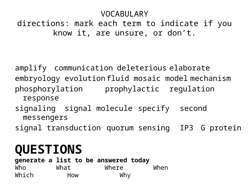
VOCABULARYdirections: mark each term to indicate if you know it, are unsure,
or don’t.
amplify communication deleterious elaborateembryology evolution fluid mosaic model mechanismphosphorylation prophylactic regulation responsesignaling signal molecule specify second
messengerssignal transduction quorum sensing IP3 G protein
QUESTIONS generate a list to be answered todayWho What Where When Which How Why
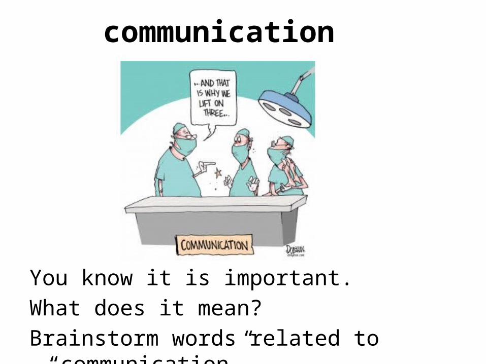
communication
You know it is important.What does it mean?Brainstorm words related to “communication”.
Where on the cell does the “communication” occur?
• “speaking” occurs through the production and release of LIGANDS hormones or other chemical messengers using the cells machinery, vesicles, and exocytosis.
• “Listening” occurs by receptor proteins embedded in the cell membrane and by way of signal transduction pathways (domino effect of sorts) .
• “Reacting” occurs when specific enzymes become activated (because of the signal transduction pathway) and the cell performs a specific function.
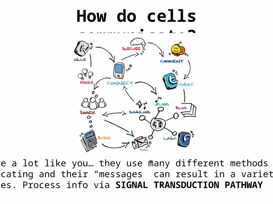
How do cells communicate?
They are a lot like you… they use many different methods for communicating and their “messages” can result in a variety of responses. Process info via SIGNAL TRANSDUCTION PATHWAY
COMMUNICATIONTalking, listening, & responding.
Cells communicate by generating, transmitting and receiving chemical signals.
Big Idea 3: Living systems store, retrieve, transmit and respond to information essential to life processes.
Big Idea 1: The process of evolution drives the diversity and unity of life.
The expression of genetic material controls cell products, and these products determine the metabolism and nature of the cell.
1) Gene expression is regulated by both – 1)environmental signals and – 2)developmental cascades or stages.
2) Cell signaling mechanisms can also modulate and control gene expression.
3) Structure and Function in biology involve two interacting aspects: – 1) the presence of necessary genetic information and – 2) the correct and timely expression of this information.
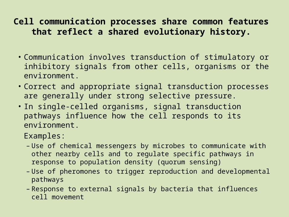
Cell communication processes share common features that reflect a shared evolutionary history.
• Communication involves transduction of stimulatory or inhibitory signals from other cells, organisms or the environment.
• Correct and appropriate signal transduction processes are generally under strong selective pressure.
• In single-celled organisms, signal transduction pathways influence how the cell responds to its environment.Examples:– Use of chemical messengers by microbes to communicate with other
nearby cells and to regulate specific pathways in response to population density (quorum sensing)
– Use of pheromones to trigger reproduction and developmental pathways
– Response to external signals by bacteria that influences cell movement
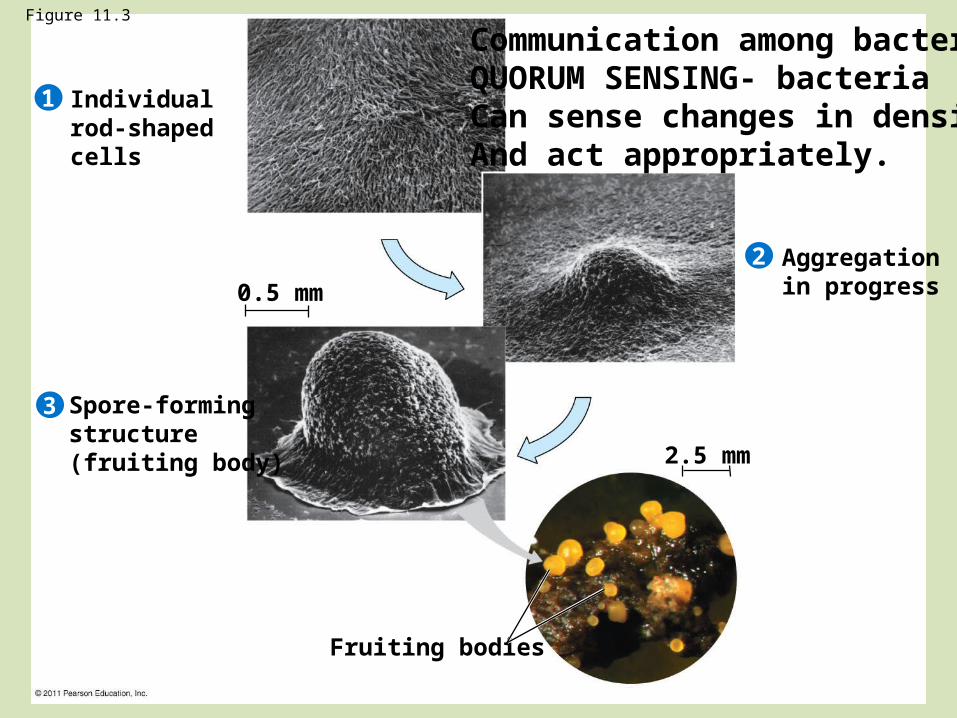
Figure 11.3
Individualrod-shapedcells
Spore-formingstructure(fruiting body)
Aggregation in progress
Fruiting bodies
1
2
3
0.5 mm
2.5 mm
Communication among bacteriaQUORUM SENSING- bacteriaCan sense changes in densityAnd act appropriately.
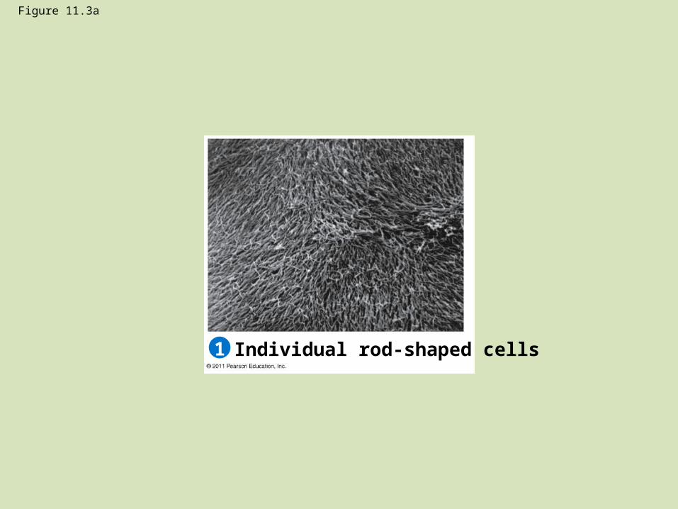
Figure 11.3a
Individual rod-shaped cells1
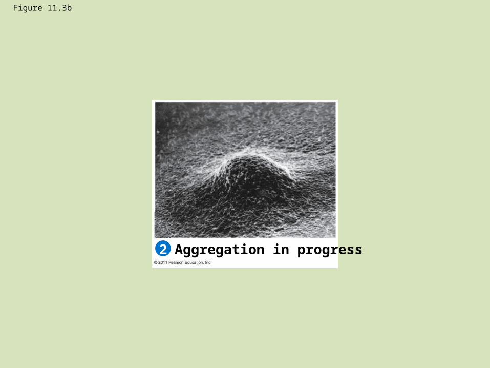
Figure 11.3b
Aggregation in progress2

Figure 11.3c
Spore-forming structure(fruiting body)
0.5 mm
3
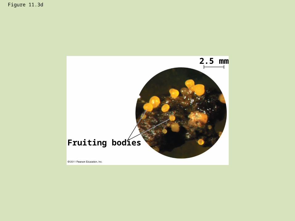
Figure 11.3d
Fruiting bodies
2.5 mm
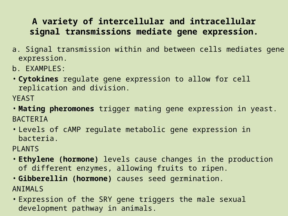
A variety of intercellular and intracellular signal transmissions mediate gene expression.
a. Signal transmission within and between cells mediates gene expression.b. EXAMPLES: • Cytokines regulate gene expression to allow for cell replication and
division.YEAST• Mating pheromones trigger mating gene expression in yeast.BACTERIA• Levels of cAMP regulate metabolic gene expression in bacteria.PLANTS• Ethylene (hormone) levels cause changes in the production of different
enzymes, allowing fruits to ripen.• Gibberellin (hormone) causes seed germination.ANIMALS• Expression of the SRY gene triggers the male sexual development pathway
in animals.
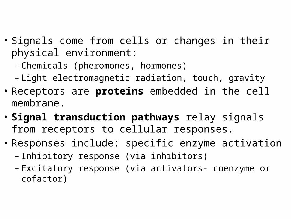
• Signals come from cells or changes in their physical environment: – Chemicals (pheromones, hormones)– Light electromagnetic radiation, touch, gravity
• Receptors are proteins embedded in the cell membrane.• Signal transduction pathways relay signals from
receptors to cellular responses.• Responses include: specific enzyme activation
– Inhibitory response (via inhibitors)– Excitatory response (via activators- coenzyme or cofactor)
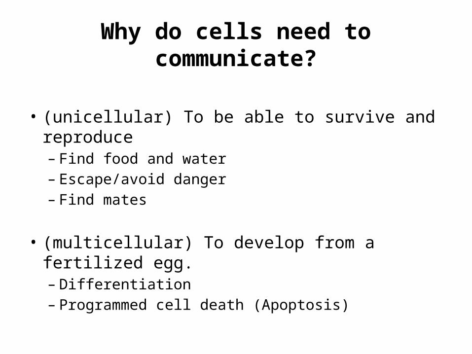
Why do cells need to communicate?
• (unicellular) To be able to survive and reproduce – Find food and water– Escape/avoid danger– Find mates
• (multicellular) To develop from a fertilized egg.– Differentiation– Programmed cell death (Apoptosis)
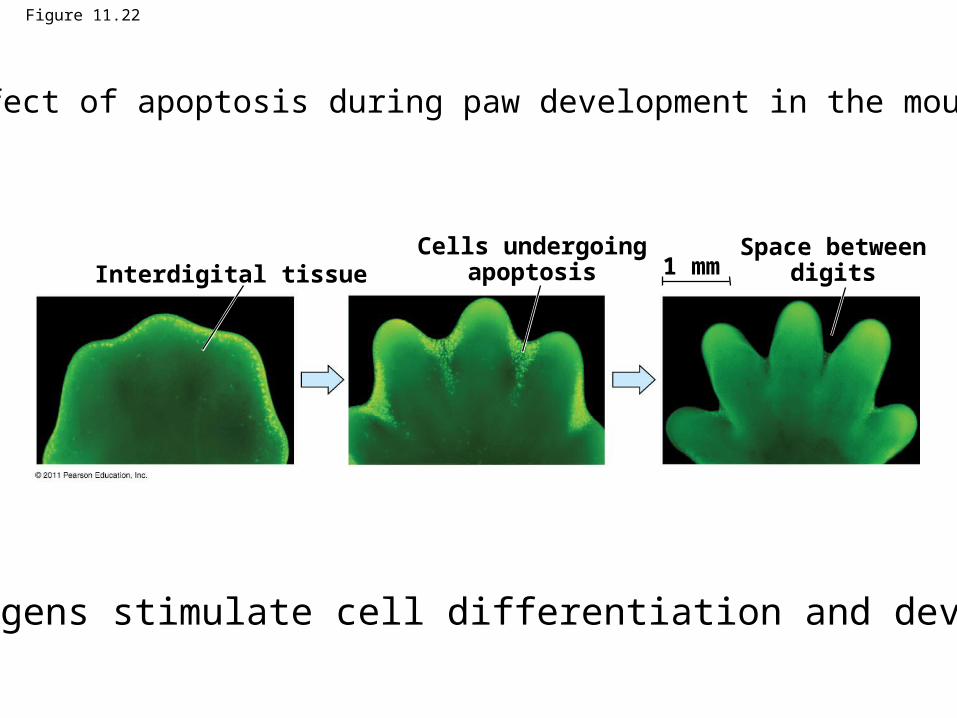
Figure 11.22
Interdigital tissueCells undergoing
apoptosisSpace between
digits1 mm
Effect of apoptosis during paw development in the mouse.
Ex.Morphogens stimulate cell differentiation and development.
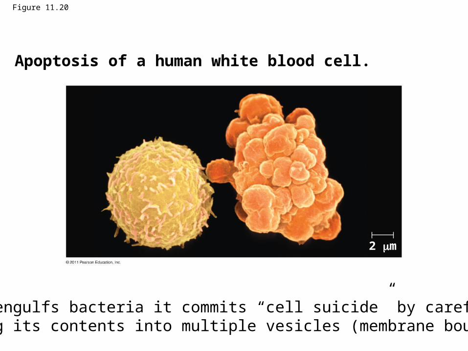
Figure 11.20
2 m
Apoptosis of a human white blood cell.
Once it engulfs bacteria it commits “cell suicide” by carefully packaging its contents into multiple vesicles (membrane bound bags).
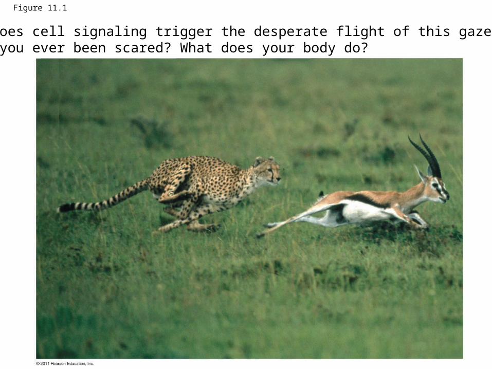
Figure 11.1
How does cell signaling trigger the desperate flight of this gazelle?Have you ever been scared? What does your body do?
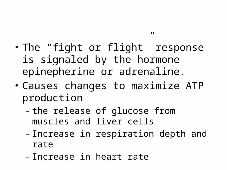
• The “fight or flight” response is signaled by the hormone epinepherine or adrenaline.
• Causes changes to maximize ATP production – the release of glucose from muscles and liver cells– Increase in respiration depth and rate– Increase in heart rate
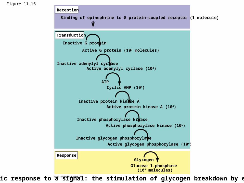
Figure 11.16
Reception
Transduction
Response
Binding of epinephrine to G protein-coupled receptor (1 molecule)
Inactive G protein
Active G protein (102 molecules)
Inactive adenylyl cyclaseActive adenylyl cyclase (102)
ATPCyclic AMP (104)
Inactive protein kinase AActive protein kinase A (104)
Inactive phosphorylase kinase
Active phosphorylase kinase (105)
Inactive glycogen phosphorylase
Active glycogen phosphorylase (106)
Glycogen
Glucose 1-phosphate (108 molecules)
Cytoplasmic response to a signal: the stimulation of glycogen breakdown by epinephrine.
How is communication different btwn single and multicellular organisms?
• In single-celled organisms, signal transduction pathways influence how the cell responds to its environment.
whereas
• In multicellular organisms, signal transduction pathways coordinate the activities within individual cells that support the function of the organism as a whole.– Temperature determination of sex in some vertebrate organisms – DNA repair mechanisms– Epinephrine stimulation of glycogen breakdown in mammals
How is communication different in multicellular organisms?
• In multicellular organisms, cell-to-cell and environment-to-cell chemical signaling pathways direct complex processes.– Ex. cell and organ differentiation to whole organism physiological
responses and behaviors.
• Certain signal pathways involve direct cell-to-cell contact, operate over very short distances, and may be determined by the structure of the organism or organelle, including– plasmodesmata in plants and – receptor-to-recognition protein interaction in the vertebrate
immune system.
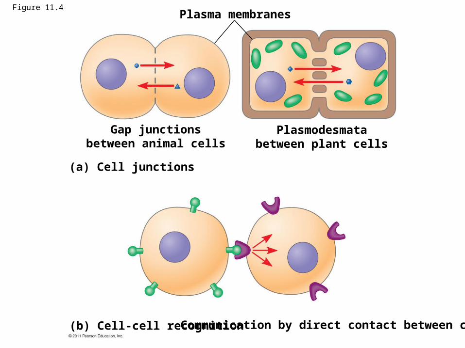
Figure 11.4Plasma membranes
Gap junctionsbetween animal cells
Plasmodesmatabetween plant cells
(a) Cell junctions
(b) Cell-cell recognition Communication by direct contact between cells.
Which life forms use cell communication?
• Cell-to-cell communication is ubiquitous in biological systems, from archaea and bacteria to multicellular organisms.
• The basic chemical processes by which cells communicate are shared across evolutionary lines of descent, and communication schemes are the products of evolution.
• For cells to function in a biological system, they must communicate with other cells and respond to their external environment.
When did cell communication evolve?
• Cell-to-cell communication is a component of higher-order biological organization and responses.
• Communication evolved billions of years ago among the most ancient bacteria.
OVERVIEW:
1) Cell communication evolved early in the history oflife.• Cell-to-cell communication is essential for multicellular
organisms. The trillions of cells in a human or an oak tree must communicate in order to develop from a fertilized egg.
• Additional evidence for the evolutionary relatedness of all life comes from discovering some universal mechanisms of cellular regulation.– Q: What molecular evidence suggests the unity of life on Earth
(descent from a common ancestor)?
• Knowledge of cell signaling mechanisms are answering questions in medicine and biology in these areas: embryological development, hormone action, cancer.– Ex. Changes in p53 activity can result in cancer.
• Examples of signals that can be received by cells and the possible responses:– Changes in light duration lead to changes in plants
(dropping leaves, flowering)– Light & phototropism (plant cell growth toward light)– Gravity or touch effects plant growth (shoots away from
gravity, roots toward gravity)
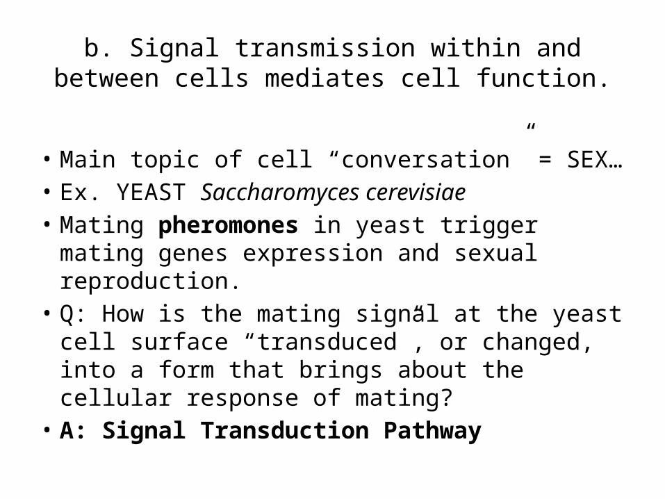
b. Signal transmission within and between cells mediates cell function.
• Main topic of cell “conversation” = SEX…• Ex. YEAST Saccharomyces cerevisiae• Mating pheromones in yeast trigger mating
genes expression and sexual reproduction. • Q: How is the mating signal at the yeast cell
surface “transduced”, or changed, into a form that brings about the cellular response of mating?
• A: Signal Transduction Pathway
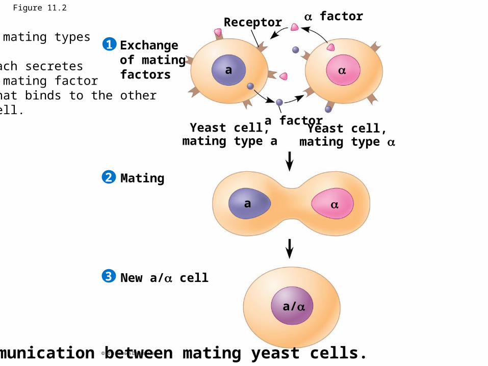
Figure 11.2
Exchange of mating factors
Receptor factor
a factorYeast cell,
mating type aYeast cell,
mating type
Mating
New a/ cell
1
2
3
a
a
a/
Communication between mating yeast cells.
2 mating types
Each secretesA mating factorThat binds to the otherCell.
• Scientists think signaling mechanisms evolved first in ancient prokaryotes and single celled eukaryotes and were then adopted for new uses by their multicellular descendants.
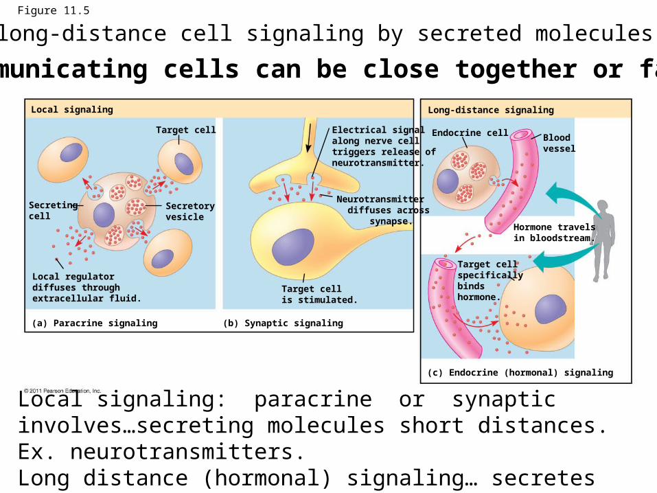
Figure 11.5
Local signaling Long-distance signaling
Target cell
Secretingcell
Secretoryvesicle
Local regulatordiffuses throughextracellular fluid.
(a) Paracrine signaling (b) Synaptic signaling
Electrical signalalong nerve celltriggers release ofneurotransmitter.
Neurotransmitter diffuses across synapse.
Target cellis stimulated.
Endocrine cell Bloodvessel
Hormone travelsin bloodstream.
Target cellspecificallybinds hormone.
(c) Endocrine (hormonal) signaling
2) Communicating cells can be close together or far apart.
Local signaling: paracrine or synaptic involves…secreting molecules short distances. Ex. neurotransmitters.Long distance (hormonal) signaling… secretes hormones for signaling at greater distances. Known as ENDOCRINE signaling.
Local and long-distance cell signaling by secreted molecules in animals.
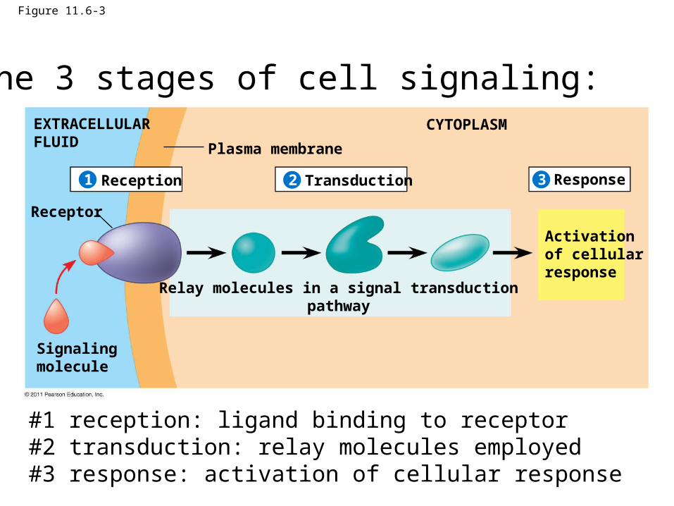
Figure 11.6-3
Plasma membrane
EXTRACELLULARFLUID
CYTOPLASM
Reception Transduction Response
Receptor
Signalingmolecule
Activationof cellularresponse
Relay molecules in a signal transductionpathway
321
3) The 3 stages of cell signaling:
#1 reception: ligand binding to receptor#2 transduction: relay molecules employed #3 response: activation of cellular response

THE DETAILS:
RECEPTION & THE INITIATION OF TRANSDUCTION
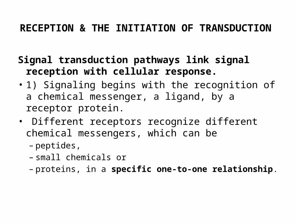
Signal transduction pathways link signal reception with cellular response.
• 1) Signaling begins with the recognition of a chemical messenger, a ligand, by a receptor protein.
• Different receptors recognize different chemical messengers, which can be – peptides, – small chemicals or – proteins, in a specific one-to-one relationship.
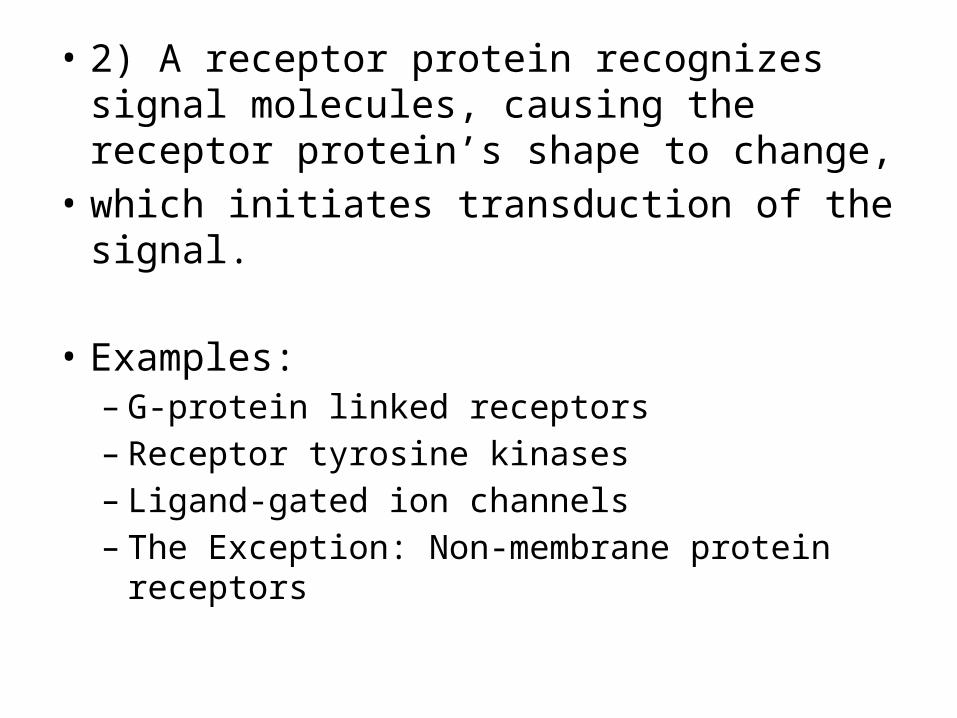
• 2) A receptor protein recognizes signal molecules, causing the receptor protein’s shape to change,
• which initiates transduction of the signal.
• Examples: – G-protein linked receptors– Receptor tyrosine kinases– Ligand-gated ion channels– The Exception: Non-membrane protein receptors
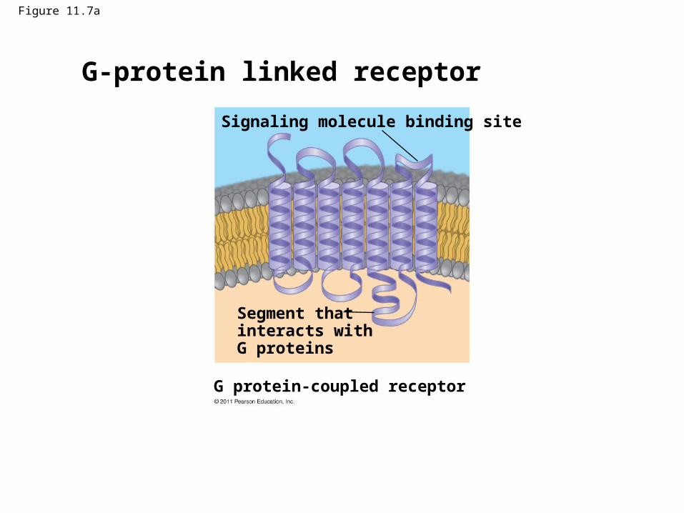
Figure 11.7a
G protein-coupled receptor
Signaling molecule binding site
Segment thatinteracts with G proteins
G-protein linked receptor
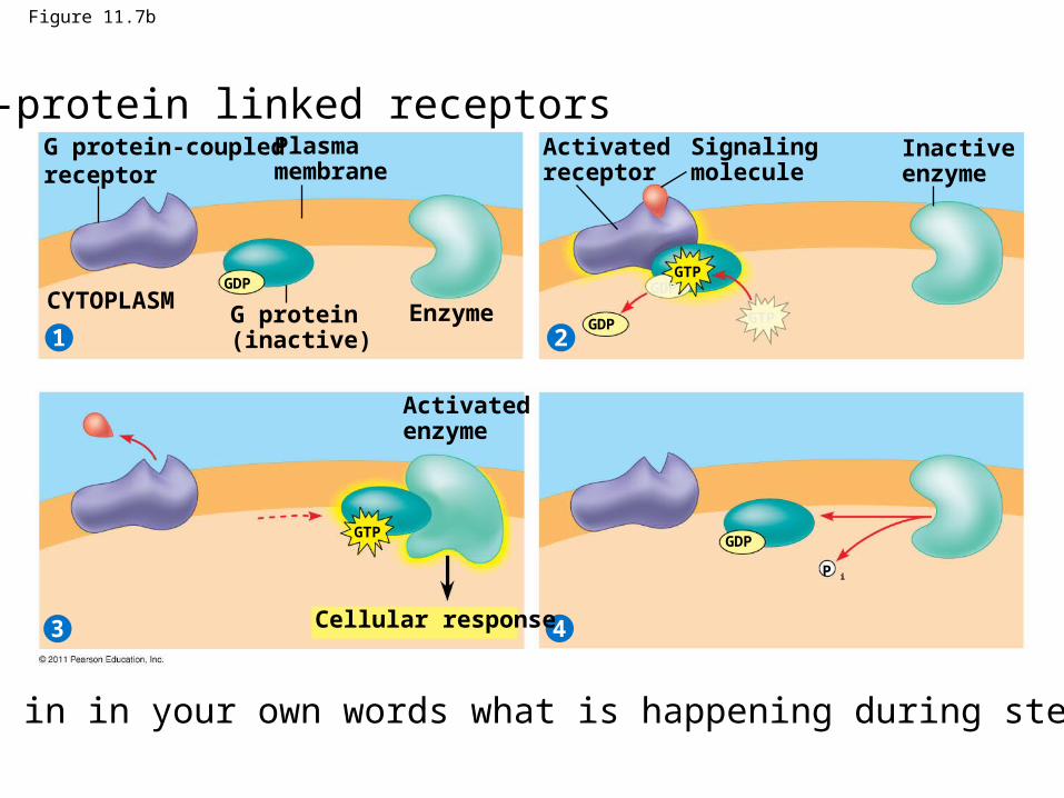
Figure 11.7b
G protein-coupledreceptor
21
3 4
Plasmamembrane
G protein(inactive)
CYTOPLASM Enzyme
Activatedreceptor
Signalingmolecule
Inactiveenzyme
Activatedenzyme
Cellular response
GDPGTP
GDPGTP
GTP
P i
GDP
GDP
G-protein linked receptors
Explain in in your own words what is happening during steps 1-4.
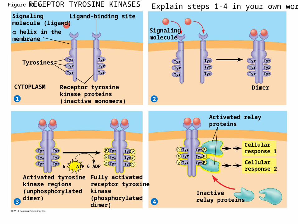
Figure 11.7c
Signalingmolecule (ligand)
21
3 4
Ligand-binding site
helix in themembrane
Tyrosines
CYTOPLASM Receptor tyrosinekinase proteins(inactive monomers)
Signalingmolecule
Dimer
Tyr
Tyr
Tyr
Tyr
Tyr
Tyr
Tyr
Tyr
Tyr
Tyr
Tyr
Tyr
Tyr
Tyr
Tyr
Tyr
Tyr
Tyr
Tyr
Tyr
Tyr
Tyr
Tyr
Tyr
Tyr
Tyr
Tyr
Tyr
Tyr
Tyr
Tyr
Tyr
Tyr
Tyr
Tyr
Tyr
P
P
P
P
P
P
P
P
P
P
P
P
Activated tyrosinekinase regions(unphosphorylateddimer)
Fully activatedreceptor tyrosinekinase(phosphorylateddimer)
Activated relayproteins
Cellularresponse 1
Cellularresponse 2
Inactiverelay proteins
6 ATP 6 ADP
RECEPTOR TYROSINE KINASES Explain steps 1-4 in your own words.
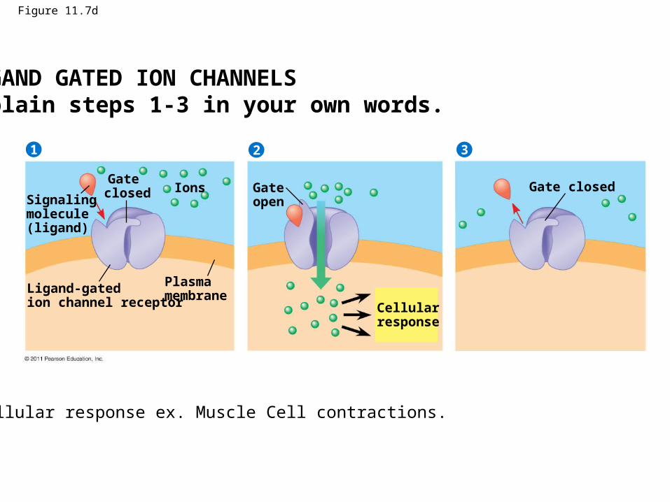
Figure 11.7d
Signalingmolecule (ligand)
21 3
Gate closed Ions
Ligand-gatedion channel receptor
Plasmamembrane
Gate open
Cellularresponse
Gate closed
LIGAND GATED ION CHANNELSExplain steps 1-3 in your own words.
Cellular response ex. Muscle Cell contractions.
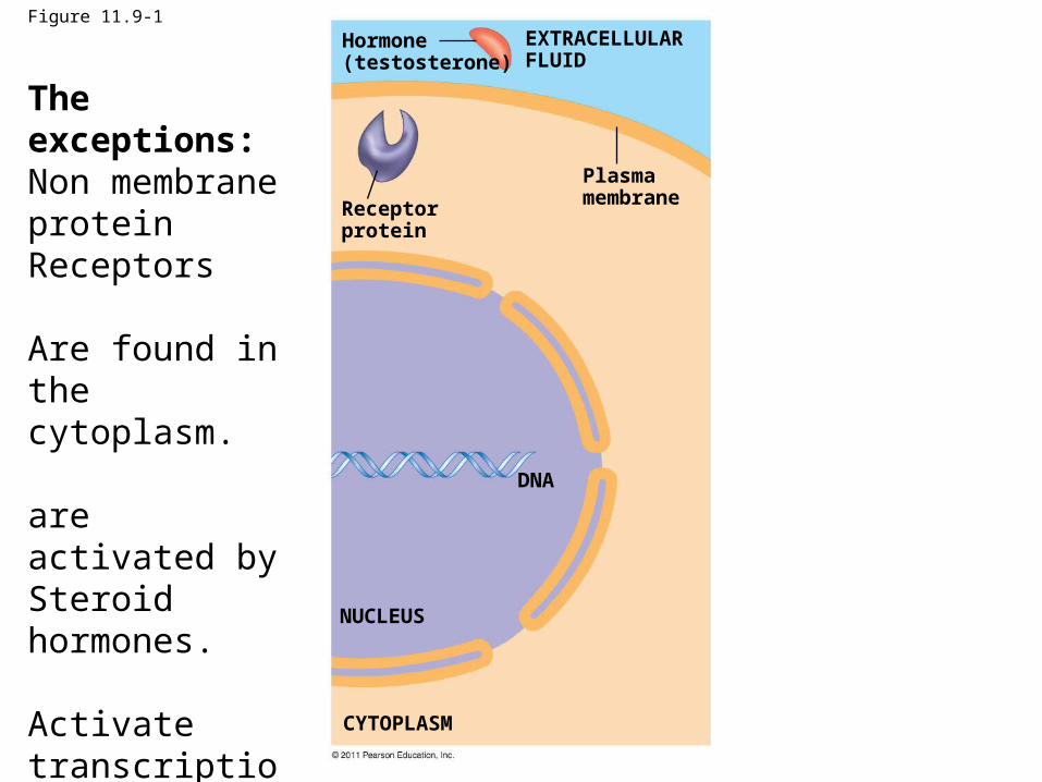
Figure 11.9-1
Hormone(testosterone)
Receptorprotein
Plasmamembrane
DNA
NUCLEUS
CYTOPLASM
EXTRACELLULARFLUID
The exceptions:Non membrane proteinReceptors
Are found in the cytoplasm.
are activated by Steroid hormones.
Activate transcription Factors in the nucleus.
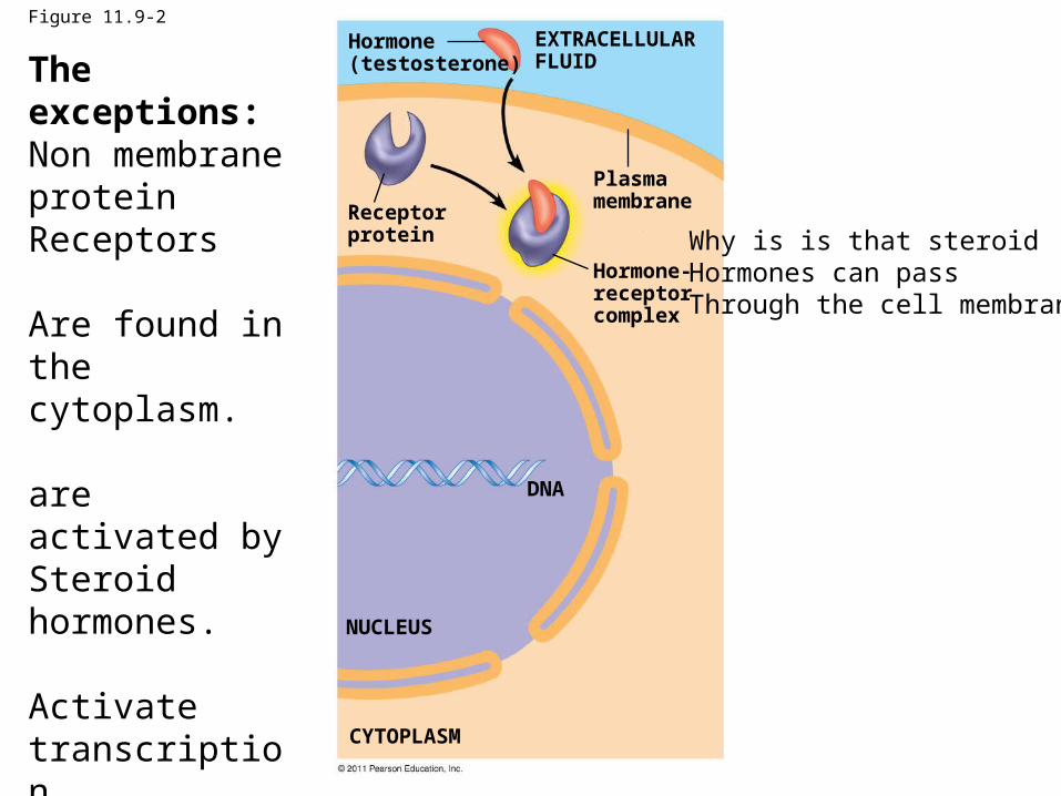
Figure 11.9-2
Hormone(testosterone)
Receptorprotein
Plasmamembrane
Hormone-receptorcomplex
DNA
NUCLEUS
CYTOPLASM
EXTRACELLULARFLUIDThe exceptions:
Non membrane proteinReceptors
Are found in the cytoplasm.
are activated by Steroid hormones.
Activate transcription Factors in the nucleus.
Why is is that steroidHormones can passThrough the cell membrane?
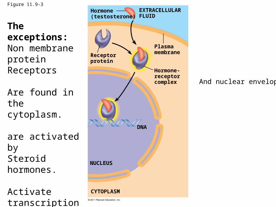
Figure 11.9-3
Hormone(testosterone)
Receptorprotein
Plasmamembrane
Hormone-receptorcomplex
DNA
NUCLEUS
CYTOPLASM
EXTRACELLULARFLUID
The exceptions:Non membrane proteinReceptors
Are found in the cytoplasm.
are activated by Steroid hormones.
Activate transcription Factors in the nucleus.
And nuclear envelope?
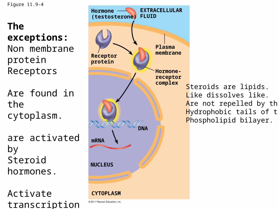
Figure 11.9-4
Hormone(testosterone)
Receptorprotein
Plasmamembrane
Hormone-receptorcomplex
DNA
mRNA
NUCLEUS
CYTOPLASM
EXTRACELLULARFLUID
The exceptions:Non membrane proteinReceptors
Are found in the cytoplasm.
are activated by Steroid hormones.
Activate transcription Factors in the nucleus.
Steroids are lipids.Like dissolves like.Are not repelled by theHydrophobic tails of thePhospholipid bilayer.
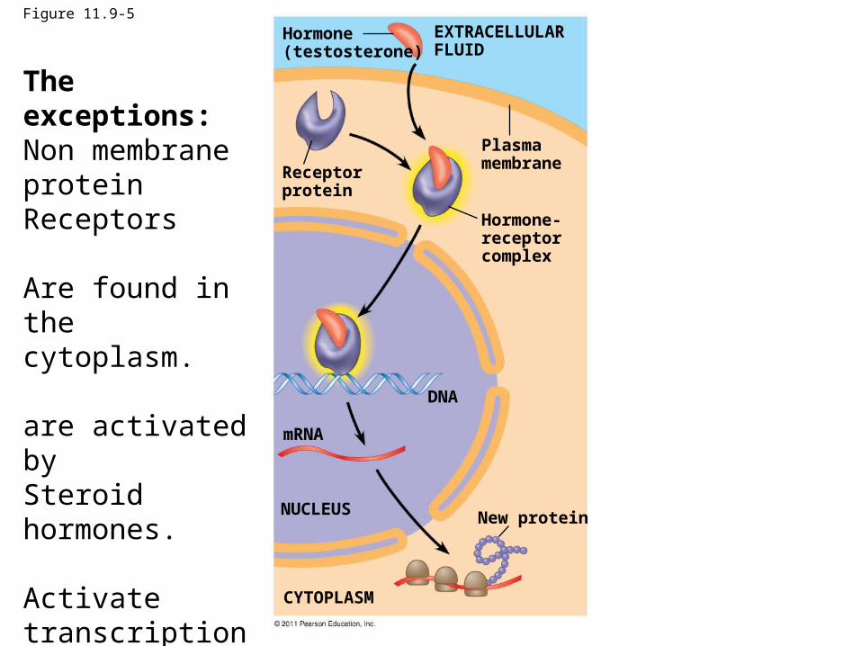
Figure 11.9-5
Hormone(testosterone)
Receptorprotein
Plasmamembrane
EXTRACELLULARFLUID
Hormone-receptorcomplex
DNA
mRNA
NUCLEUS
CYTOPLASM
New protein
The exceptions:Non membrane proteinReceptors
Are found in the cytoplasm.
are activated by Steroid hormones.
Activate transcription Factors in the nucleus.
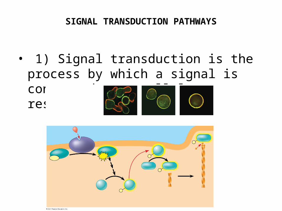
SIGNAL TRANSDUCTION PATHWAYS
• 1) Signal transduction is the process by which a signal is converted to a cellular response.
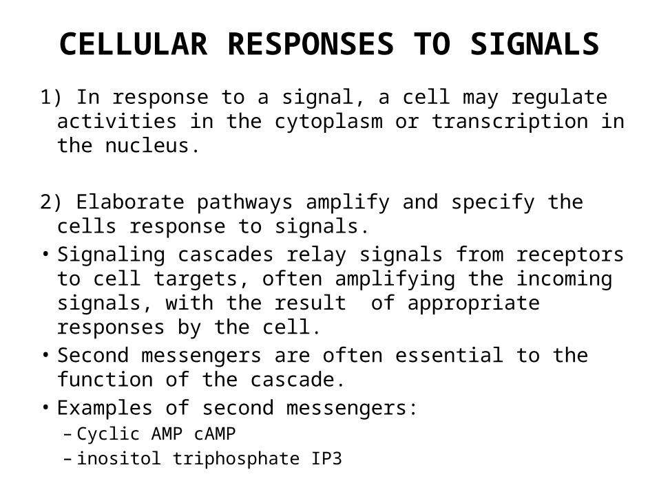
CELLULAR RESPONSES TO SIGNALS
1) In response to a signal, a cell may regulate activities in the cytoplasm or transcription in the nucleus.
2) Elaborate pathways amplify and specify the cells response to signals.
• Signaling cascades relay signals from receptors to cell targets, often amplifying the incoming signals, with the result of appropriate responses by the cell.
• Second messengers are often essential to the function of the cascade.
• Examples of second messengers:– Cyclic AMP cAMP– inositol triphosphate IP3
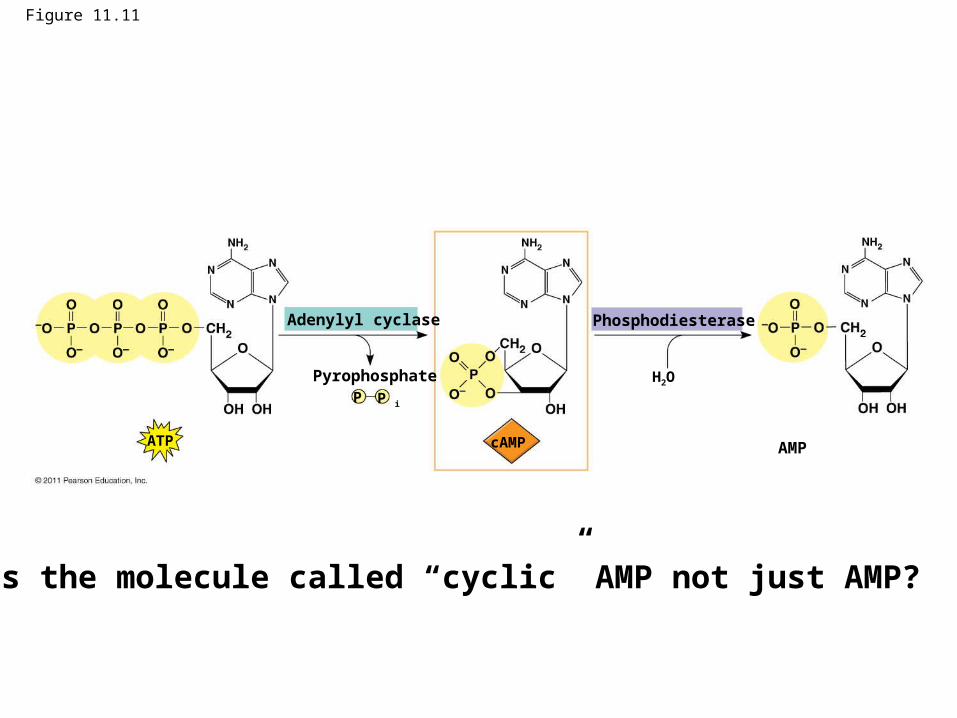
Figure 11.11
Adenylyl cyclase Phosphodiesterase
Pyrophosphate
AMP
H2O
ATP
P iP
cAMP
Why is the molecule called “cyclic” AMP not just AMP?
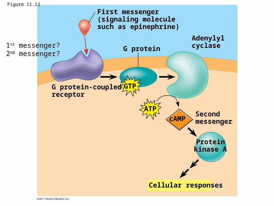
Figure 11.12
G protein
First messenger(signaling moleculesuch as epinephrine)
G protein-coupledreceptor
Adenylylcyclase
Second messenger
Cellular responses
Proteinkinase A
GTP
ATP
cAMP
1st messenger?2nd messenger?
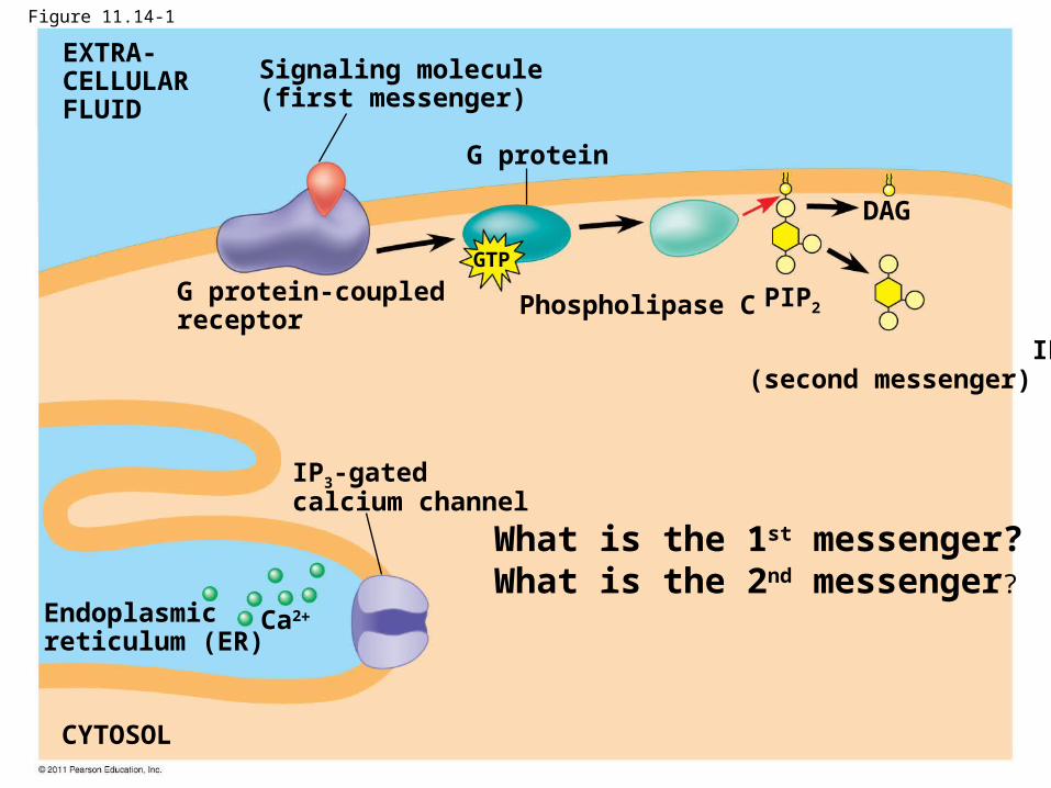
Figure 11.14-1
G protein
EXTRA-CELLULARFLUID
Signaling molecule(first messenger)
G protein-coupledreceptor
Phospholipase C
DAG
PIP2
IP3
(second messenger)
IP3-gatedcalcium channel
Endoplasmicreticulum (ER)
CYTOSOL
Ca2
GTP
What is the 1st messenger?What is the 2nd messenger?
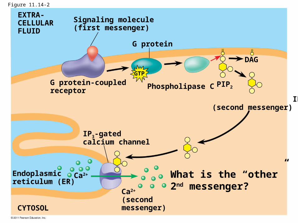
Figure 11.14-2
G protein
EXTRA-CELLULARFLUID
Signaling molecule(first messenger)
G protein-coupledreceptor
Phospholipase C
DAG
PIP2
IP3
(second messenger)
IP3-gatedcalcium channel
Endoplasmicreticulum (ER)
CYTOSOL
Ca2
(secondmessenger)
Ca2
GTP
What is the “other” 2nd messenger?
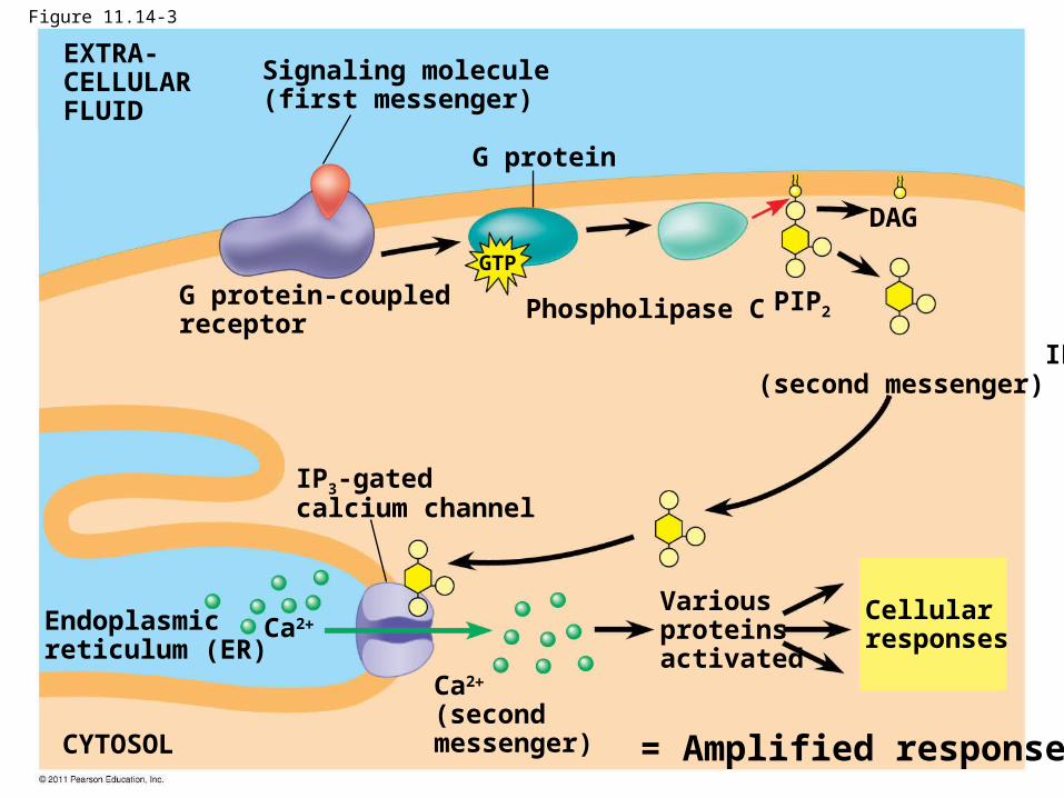
Figure 11.14-3
G protein
EXTRA-CELLULARFLUID
Signaling molecule(first messenger)
G protein-coupledreceptor
Phospholipase C
DAG
PIP2
IP3
(second messenger)
IP3-gatedcalcium channel
Endoplasmicreticulum (ER)
CYTOSOL
Variousproteinsactivated
Cellularresponses
Ca2
(secondmessenger)
Ca2
GTP
= Amplified response
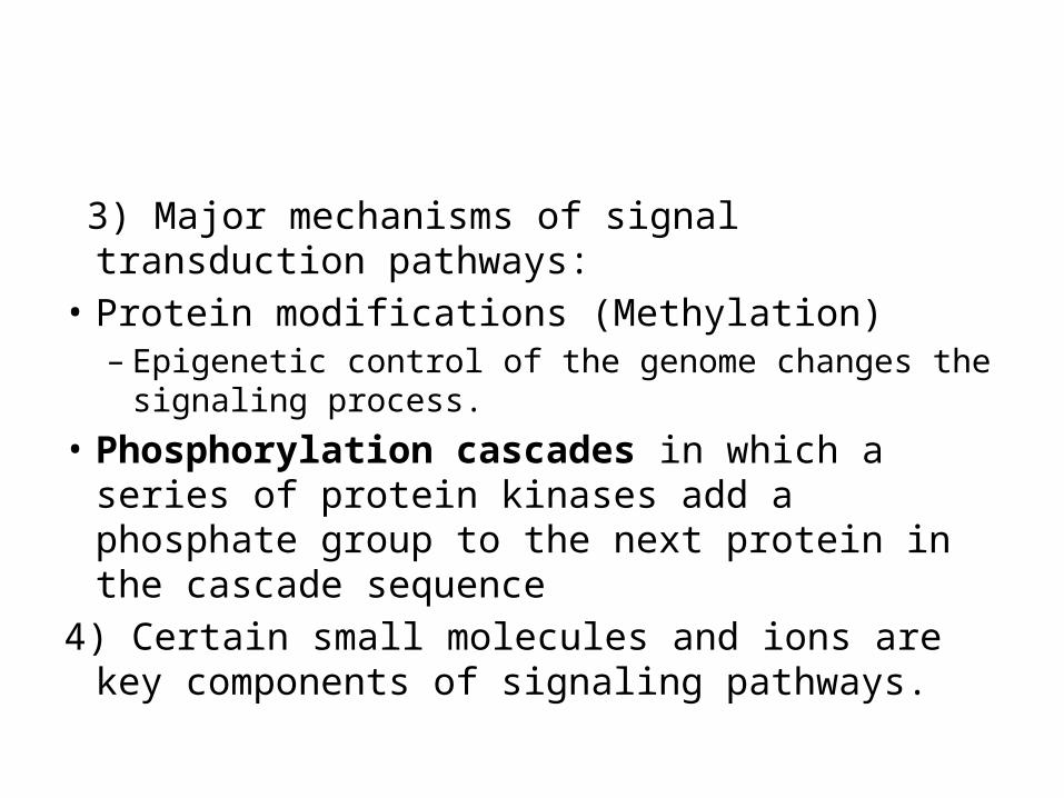
3) Major mechanisms of signal transduction pathways:
• Protein modifications (Methylation)– Epigenetic control of the genome changes the signaling
process.• Phosphorylation cascades in which a series of
protein kinases add a phosphate group to the next protein in the cascade sequence
4) Certain small molecules and ions are key components of signaling pathways.
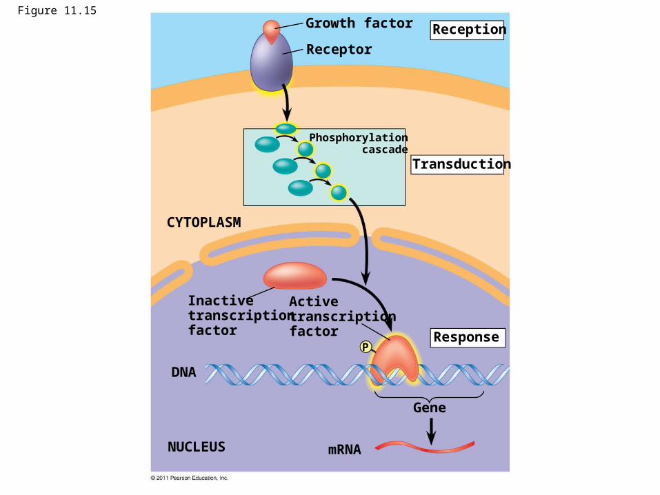
Figure 11.15Growth factor
Receptor
Reception
Transduction
CYTOPLASM
Response
Inactivetranscriptionfactor
Activetranscriptionfactor
DNA
NUCLEUS mRNA
Gene
Phosphorylationcascade
P
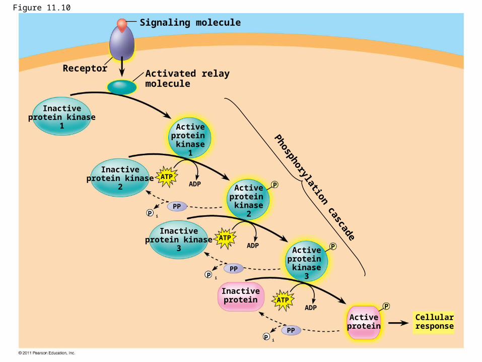
Figure 11.10
Receptor
Signaling molecule
Activated relaymolecule
Phosphorylation cascade
Inactiveprotein kinase
1 Activeprotein kinase
1
Activeprotein kinase
2
Activeprotein kinase
3
Inactiveprotein kinase
2
Inactiveprotein kinase
3
Inactiveprotein
Activeprotein
Cellularresponse
ATPADP
ATPADP
ATPADP
PP
PP
PP
P
P
P
P i
P i
P i
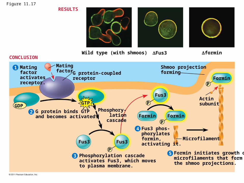
Figure 11.17
Wild type (with shmoos) Fus3 formin
Matingfactoractivatesreceptor.
Matingfactor G protein-coupled
receptor
Shmoo projectionforming
Formin
G protein binds GTPand becomes activated.
2
1
3
4
5
P
P
P
PForminFormin
Fus3
Fus3Fus3
GDPGTP
Phosphory- lation cascade
Microfilament
Actinsubunit
Phosphorylation cascadeactivates Fus3, which movesto plasma membrane.
Fus3 phos-phorylatesformin,activating it.
Formin initiates growth ofmicrofilaments that formthe shmoo projections.
RESULTS
CONCLUSION
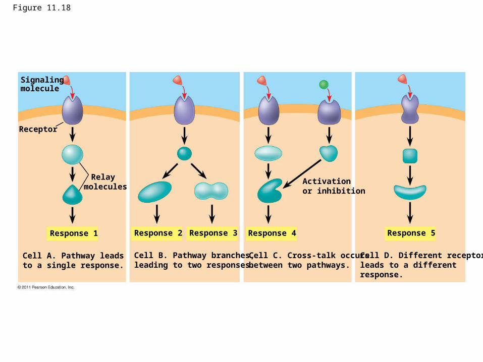
Figure 11.18
Signalingmolecule
Receptor
Relay molecules
Response 1
Cell A. Pathway leadsto a single response.
Response 2 Response 3 Response 4 Response 5
Activationor inhibition
Cell B. Pathway branches,leading to two responses.
Cell C. Cross-talk occursbetween two pathways.
Cell D. Different receptorleads to a differentresponse.
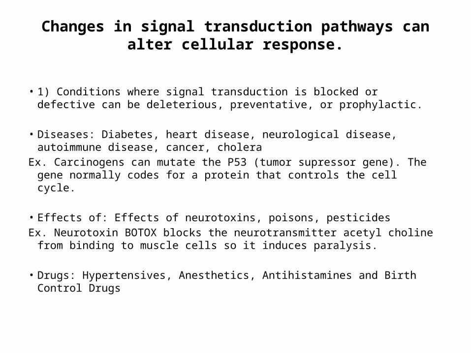
Changes in signal transduction pathways can alter cellular response.
• 1) Conditions where signal transduction is blocked or defective can be deleterious, preventative, or prophylactic.
• Diseases: Diabetes, heart disease, neurological disease, autoimmune disease,
cancer, cholera Ex. Carcinogens can mutate the P53 (tumor supressor gene). The gene normally
codes for a protein that controls the cell cycle. • Effects of: Effects of neurotoxins, poisons, pesticidesEx. Neurotoxin BOTOX blocks the neurotransmitter acetyl choline from binding to
muscle cells so it induces paralysis. • Drugs: Hypertensives, Anesthetics, Antihistamines and Birth Control Drugs

QUIZ
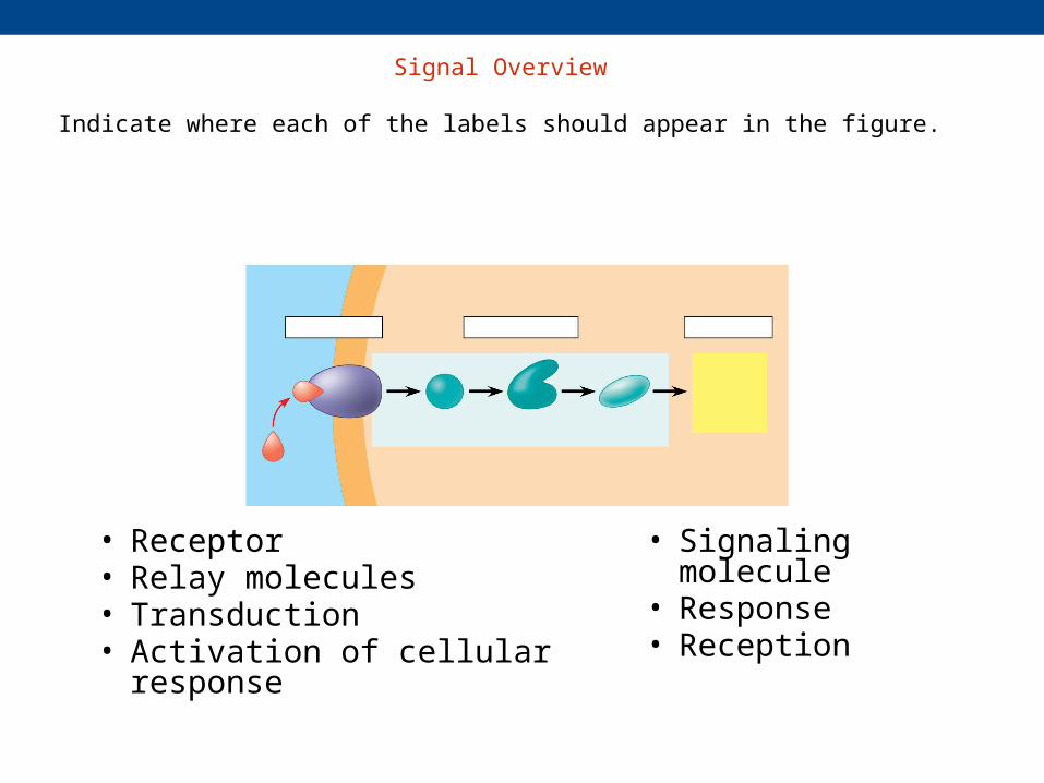
Signal Overview
Indicate where each of the labels should appear in the figure.
• Receptor• Relay molecules• Transduction• Activation of cellular response
• Signaling molecule• Response• Reception
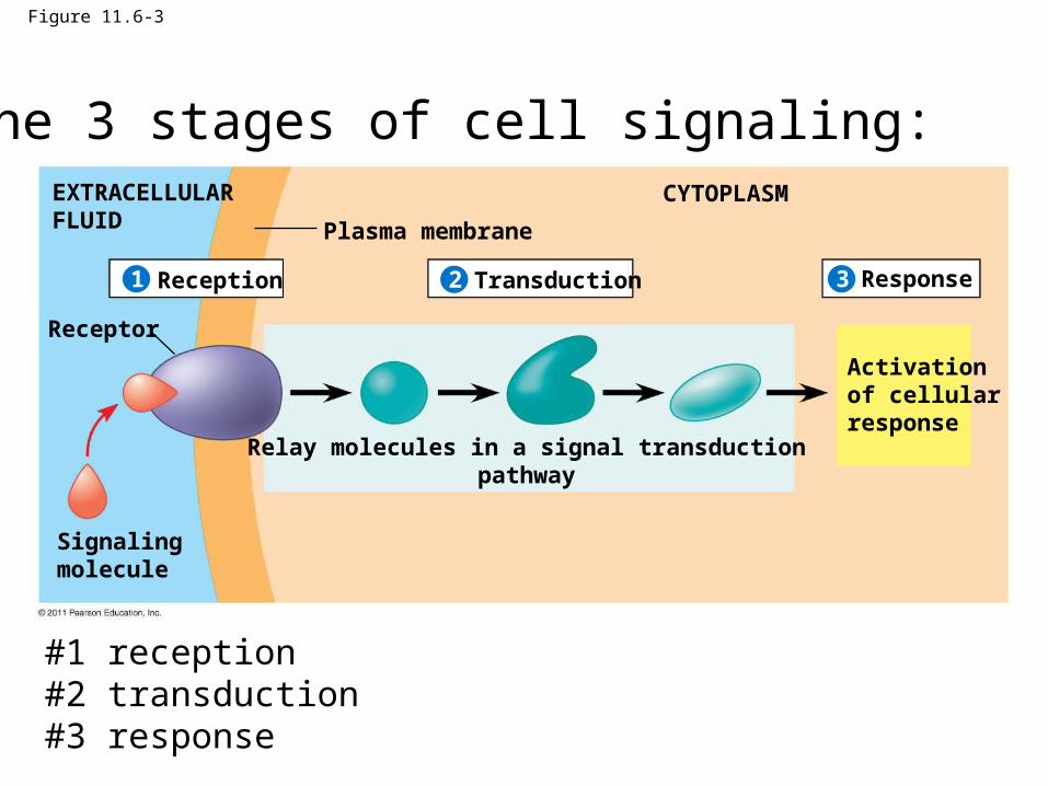
Figure 11.6-3
Plasma membrane
EXTRACELLULARFLUID
CYTOPLASM
Reception Transduction Response
Receptor
Signalingmolecule
Activationof cellularresponse
Relay molecules in a signal transductionpathway
321
3) The 3 stages of cell signaling:
#1 reception#2 transduction#3 response
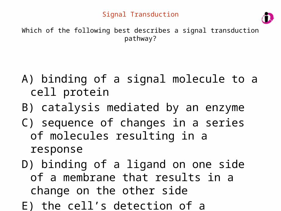
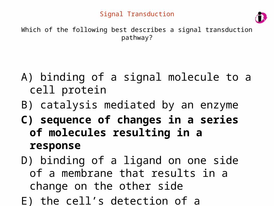
Signal Transduction
Which of the following best describes a signal transduction pathway?
A) binding of a signal molecule to a cell proteinB) catalysis mediated by an enzymeC) sequence of changes in a series of molecules
resulting in a responseD) binding of a ligand on one side of a membrane that
results in a change on the other sideE) the cell’s detection of a chemical or mechanical
stimulus
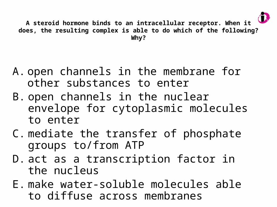
A steroid hormone binds to an intracellular receptor. When it does, the resulting complex is able to do which of the following? Why?
A. open channels in the membrane for other substances to enter
B. open channels in the nuclear envelope for cytoplasmic molecules to enter
C. mediate the transfer of phosphate groups to/from ATP
D. act as a transcription factor in the nucleusE. make water-soluble molecules able to diffuse across
membranes
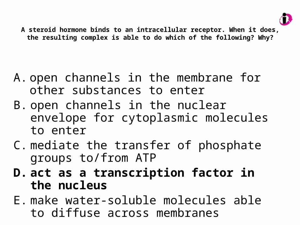
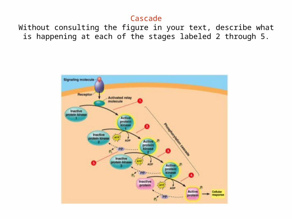
CascadeWithout consulting the figure in your text, describe what is
happening at each of the stages labeled 2 through 5.
Phosphorylation
In reactions mediated by protein kinases, what does phosphorylation of successive proteins do to drive the reaction?
A. make functional ATPB. change a protein from its inactive to its active formC. change a protein from its active to its inactive formD. alter the permeability of the cell’s membranesE. produce an increase in the cell’s store of inorganic
phosphates
Signal Molecules
What would happen to a cell whose receptors remain bound to the signal molecule(s)?
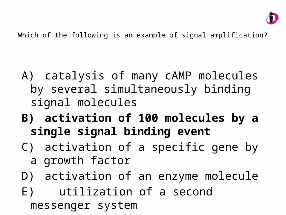
Which of the following is an example of signal amplification?
A) catalysis of many cAMP molecules by several simultaneously binding signal molecules
B) activation of 100 molecules by a single signal binding event
C) activation of a specific gene by a growth factor
D) activation of an enzyme moleculeE) utilization of a second messenger system
Cancer and Apoptosis
How could cancer result from a defect in apoptosis?
One of the important outcomes of apoptosis is protection of neighboring cells. Which of the following is responsible?
A. cell shrinkage and blebbingB. destruction of the cell’s DNAC. formation of numerous vesicles to be digestedD. action of tyrosine kinasesE. activation of specific proteins
One of the important outcomes of apoptosis is protection of neighboring cells. Which of the following is responsible?
A. cell shrinkage and blebbingB. destruction of the cell’s DNAC. formation of numerous vesicles to be
digestedD. action of tyrosine kinasesE. activation of specific proteins
What are the similarities among the following?
• G protein-coupled receptors
• receptor tyrosine kinases
• ion channel receptors