Embed Size (px)
Citation preview
Neurocritical Care Monitoring
Chad M. Miller • MiChel T. Torbey
Chad M. Miller, Md and Michel T. Torbey, Md
Neurocritical Care Monitoring
While damage resulting from a primary injury to the brain or spine may be unavoid-able, harm from secondary processes that cause further deterioration is not. This
practical, clinical resource describes the latest strategies for monitoring the brain after acute injury. With a focus on individualization of treatment, the book examines the role of various monitoring techniques in limiting disability and potentiating patient recovery during the acute phase of brain injury. International experts in diagnosis and treatment of secondary injury explain in detail the current utilization, benefits, nuances, and risks for each commercially available monitoring device as well as approaches vital to the care of brain and spine injured patients. They cover foundational strategies for neuromonitoring implementation and analysis, including proper catheter placement, duration of monitoring, and treatment thresholds that indicate the need for clinical intervention. The book also addresses multimodality monitoring and common programmatic challenges, and offers guidance on how to set up a successful multimodal monitoring protocol in the ICU. Also included is a chapter on the key role of nurses in neuromonitoring and effective bedside training for troubleshooting and proper execution of treatment protocols. Numerous illustrations provide further illumination.
“I commend the editors for their careful perspective on the current state of neuromonitoring. The individual chapters provide excellent overviews of specific neuromonitoring tools and paradigms.” – From the Foreword by J. Claude Hemphill III, MD, MAS, FNCS
9 781620 700259
Key Features: ■■ Presents state-of-the-art neuromonitoring techniques and clinical protocols for assessment and treatment
■■ Emphasizes practical implementation for successful patient outcomes
■■ Written by international experts at the forefront of neurocritical care monitoring
■■ Provides a framework for practitioners who wish to individualize patient care with an emphasis upon the needs of the critically ill brain
■■ Discusses the key role of nurses in neuromonitoring and effective bedside training for management and troubleshooting of devices
11 West 42nd Street New York, NY 10036 www.demosmedical.com
Recommended Shelving Category: Neurology
Neurocritical Care M
onitoring M
iller • Torbey
Miller_00259_PTR_01_1-17_12-09-14.indd 16 12/09/14 10:57 AM
Visit This Book’s Web Page / Buy Now / Request an Exam/Review CopyThis is a sample from Neurocritical Care Monitoring
© Demos Medical Publishing
Neurocritical Care Monitoring
Miller_00259_PTR_00_i-xii_FM_12-09-14.indd 1 12/09/14 11:12 AM

Miller_00259_PTR_01_1-17_12-09-14.indd 16 12/09/14 10:57 AM
Visit This Book’s Web Page / Buy Now / Request an Exam/Review CopyThis is a sample from Neurocritical Care Monitoring
© Demos Medical Publishing
Miller_00259_PTR_00_i-xii_FM_12-09-14.indd 2 12/09/14 11:12 AM
Miller_00259_PTR_01_1-17_12-09-14.indd 16 12/09/14 10:57 AM
Visit This Book’s Web Page / Buy Now / Request an Exam/Review CopyThis is a sample from Neurocritical Care Monitoring
© Demos Medical Publishing
Neurocritical Care Monitoring
Editors
Chad M. Miller, MDAssociate Professor of Neurology and Neurosurgery
Wexner Medical CenterOhio State University
Columbus, Ohio
Michel T. Torbey, MDProfessor of Neurology and Neurosurgery
Director, Division of Cerebrovascular Diseases and Neurocritical CareWexner Medical Center
Ohio State UniversityColumbus, Ohio
Miller_00259_PTR_00_i-xii_FM_12-09-14.indd 3 12/09/14 11:12 AM
Miller_00259_PTR_01_1-17_12-09-14.indd 16 12/09/14 10:57 AM
Visit This Book’s Web Page / Buy Now / Request an Exam/Review CopyThis is a sample from Neurocritical Care Monitoring
© Demos Medical Publishing
Visit our website at www.demosmedical.com
ISBN: 9781620700259e-book ISBN: 9781617051883
Acquisitions Editor: Beth BarryCompositor: Integra Software Services Pvt. Ltd.
© 2015 Demos Medical Publishing, LLC. All rights reserved. This book is protected by copyright. No part of it may be reproduced, stored in a retrieval system, or transmitted in any form or by any means, electronic, mechanical, photocopying, recording, or otherwise, without the prior written permission of the publisher.
Medicine is an ever-changing science. Research and clinical experience are continually expanding our knowl-edge, in particular our understanding of proper treatment and drug therapy. The authors, editors, and publisher have made every effort to ensure that all information in this book is in accordance with the state of knowledge at the time of production of the book. Nevertheless, the authors, editors, and publisher are not responsible for errors or omissions or for any consequences from application of the information in this book and make no warranty, expressed or implied, with respect to the contents of the publication. Every reader should examine carefully the package inserts accompanying each drug and should carefully check whether the dosage sched-ules mentioned therein or the contraindications stated by the manufacturer differ from the statements made in this book. Such examination is particularly important with drugs that are either rarely used or have been newly released on the market.
Library of Congress Cataloging-in-Publication Data
Neurocritical care monitoring / editors, Chad M. Miller, Michel T. Torbey. p. ; cm. Includes bibliographical references and index. ISBN 978-1-62070-025-9 (alk. paper) -- ISBN 978-1-61705-188-3 (e-book) I. Miller, Chad M., editor. II. Torbey, Michel T., editor. [DNLM: 1. Central Nervous System Diseases--diagnosis. 2. Neurophysiological Monitoring. 3. Critical Care--methods. 4. Nervous System Physiological Phenomena. WL 141] RC350.N49 616.8’0428--dc23
2014032210
Special discounts on bulk quantities of Demos Medical Publishing books are available to corporations, professional associations, pharmaceutical companies, health care organizations, and other qualifying groups. For details, please contact:
Special Sales DepartmentDemos Medical Publishing, LLC11 West 42nd Street, 15th FloorNew York, NY 10036Phone: 800-532-8663 or 212-683-0072Fax: 212-941-7842E-mail: [email protected]
Printed in the United States of America by Bradford and Bigelow.14 15 16 17 / 5 4 3 2 1
Miller_00259_PTR_00_i-xii_FM_12-09-14.indd 4 12/09/14 11:12 AM
Miller_00259_PTR_01_1-17_12-09-14.indd 16 12/09/14 10:57 AM
Visit This Book’s Web Page / Buy Now / Request an Exam/Review CopyThis is a sample from Neurocritical Care Monitoring
© Demos Medical Publishing
1. Intracranial Pressure Monitoring 1 Nessim Amin, MBBS and Diana Greene-Chandos, MD
2. Transcranial Doppler Monitoring 18 Maher Saqqur, MD, MPH, FRCPC, David Zygun, MD, MSc, FRCPC,
Andrew Demchuk, MD, FRCPC and Herbert Alejandro A. Manosalva, MD
3. Continuous EEG Monitoring 35 Jeremy T. Ragland, MD and Jan Claassen, MD, PhD
4. Cerebral Oxygenation 50 Michel T. Torbey, MD and Chad M. Miller, MD
5. Brain Tissue Perfusion Monitoring 59 David M. Panczykowski, MD and Lori Shutter, MD
6. Cerebral Microdialysis 70 Chad M. Miller, MD
7. Cerebral Autoregulation 85 Marek Czosnyka, PhD and Enrique Carrero Cardenal, PhD
8. Neuroimaging 102 Latisha K. Ali, MD and David S. Liebeskind, MD
Contents
Contributors vii
Foreword J. Claude Hemphill III, MD, MAS, FNCS ix
Preface xi
v
Miller_00259_PTR_00_i-xii_FM_12-09-14.indd 5 12/09/14 11:12 AM
Miller_00259_PTR_01_1-17_12-09-14.indd 16 12/09/14 10:57 AM
Visit This Book’s Web Page / Buy Now / Request an Exam/Review CopyThis is a sample from Neurocritical Care Monitoring
© Demos Medical Publishing
vi ■ Contents
9. Evoked Potentials in Neurocritical Care 124 Wei Xiong, MD, Matthew Eccher, MD, MSPH and Romergryko Geocadin, MD
10. Bioinformatics for Multimodal Monitoring 135 J. Michael Schmidt, PhD, MSc
11. Nursing: The Essential Piece to Successful Neuromonitoring 145 Tess Slazinski, RN, MN, CCRN, CNRN, CCNS
12. Multimodal Monitoring: Challenges in Implementation and Clinical Utilization 159 Chad M. Miller, MD
Index 167
Miller_00259_PTR_00_i-xii_FM_12-09-14.indd 6 12/09/14 11:12 AM
Miller_00259_PTR_01_1-17_12-09-14.indd 16 12/09/14 10:57 AM
Visit This Book’s Web Page / Buy Now / Request an Exam/Review CopyThis is a sample from Neurocritical Care Monitoring
© Demos Medical Publishing
vii
Latisha K. Ali, MD Assistant Professor, Department of Neurology, UCLA David Geffen School of Medicine, Los Angeles, California
Nessim Amin, MBBS Fellow of Neurosciences Critical Care, Departments of Neurological Surgery and Neurology, Wexner Medical Center, Ohio State University, Columbus, Ohio
Enrique Carrero Cardenal, PhD Professor, Department of Anesthesiology, Hospital Clinic, University of Barcelona, Barcelona, Spain
Jan Claassen, MD, PhD Assistant Professor of Neurology and Neurosurgery, Director, Neurocritical Care Training Program, New York Presbyterian Hospital, Division of Critical Care Neurology, Columbia University College of Physicians and Surgeons, New York, New York
Marek Czosnyka, PhD Professor, Department of Clinical Neurosciences, University of Cambridge, Cambridge, United Kingdom
Andrew Demchuk, MD, FRCPC Associate Professor, Department of Clinical Neurosciences, University of Calgary, Calgary, Alberta, Canada
Matthew Eccher, MD, MSPH Assistant Professor of Neurology and Neurosurgery, Case Western Reserve University School of Medicine, Cleveland, Ohio
Romergryko Geocadin, MD Associate Professor, Department of Anesthesiology and Critical Care Medicine, Department of Neurology, Department of Neurosurgery, Department of Medicine, Johns Hopkins University School of Medicine, Baltimore, Maryland
Contributors
Miller_00259_PTR_00_i-xii_FM_12-09-14.indd 7 12/09/14 11:12 AM
Miller_00259_PTR_01_1-17_12-09-14.indd 16 12/09/14 10:57 AM
Visit This Book’s Web Page / Buy Now / Request an Exam/Review CopyThis is a sample from Neurocritical Care Monitoring
© Demos Medical Publishing
viii ■ Contributors
Diana Greene-Chandos, MD Director of Education, Quality and Outreach for Neurosciences Critical Care, Wexner Medical Center, Ohio State University, Columbus, Ohio
David S. Liebeskind, MD Assistant Professor, Department of Neurology, UCLA David Geffen School of Medicine, Los Angeles, California
Herbert Alejandro A. Manosalva, MD Fellow in Cerebrovascular Diseases, Movement Disorders and Neurogenetics, Department of Neurology, University of Alberta, Edmonton, Canada
Chad M. Miller, MD Associate Professor of Neurology and Neurosurgery, Wexner Medical Center, Ohio State University, Columbus, Ohio
David M. Panczykowski, MD Resident, Neurological Surgery, Department of Neurological Surgery, University of Pittsburgh Medical Center, Pittsburgh, Pennsylvania
Jeremy T. Ragland, MD Fellow, Division of Neurocritical Care, Department of Neurology, Columbia University College of Physicians and Surgeons,New York Presbyterian Hospital/Columbia University Medical Center,New York, New York
Maher Saqqur, MD, MPH, FRCPC Associate Professor, Department of Medicine, Division of Neurology, University of Alberta, Edmonton, Alberta, Canada
J. Michael Schmidt, PhD, MSc Assistant Professor of Clinical Neuropsychology in Neurology, Informatics Director, Neurological Intensive Care Unit, Critical Care Neuromonitoring, Columbia University College of Physicians and Surgeons, New York, New York
Lori Shutter, MD Co-Director, Neurovascular ICU, UPMC Presbyterian Hospital, Director, Neurocritical Care Fellowship, Departments of Neurology, Neurosurgery, and Critical Care Medicine, University of Pittsburgh Medical Center, Pittsburgh, Pennsylvania
Tess Slazinski, RN, MN, CCRN, CNRN, CCNS Cedars Sinai Medical Center, Los Angeles, California
Michel T. Torbey, MD Professor of Neurology and Neurosurgery, Director, Division of Cerebrovascular Diseases and Neurocritical Care, Wexner Medical Center, Ohio State University, Columbus, Ohio
Wei Xiong, MD Assistant Professor of Neurology, Neurointensivist, Case Western Reserve University School of Medicine, Cleveland, Ohio
David Zygun, MD, MSc, FRCPC Professor and Divisional Director, Departments of Critical Care Medicine, Clinical Neurosciences, and Community Health Sciences, University of Calgary, Calgary, Alberta, Canada
Miller_00259_PTR_00_i-xii_FM_12-09-14.indd 8 12/09/14 11:12 AM
Miller_00259_PTR_01_1-17_12-09-14.indd 16 12/09/14 10:57 AM
Visit This Book’s Web Page / Buy Now / Request an Exam/Review CopyThis is a sample from Neurocritical Care Monitoring
© Demos Medical Publishing
ix
Foreword
When I was considering going into neurocritical care over 20 years ago, it was in large part because of an interest in the physiology (as opposed to anatomy) of acute brain catastro-phes (my term), and optimism that intervention must be possible. Patients in the pulmonary and cardiac intensive care units were active, and my colleagues routinely made treatment changes many times a day based on the physiology of the patient’s condition, a physiology that was identified by a monitor such as a flow-volume loop on the ventilator in an acute respiratory distress syndrome (ARDS) patient or a pulmonary-artery catheter in a patient with cardiogenic shock. As a neurology resident in an era when neurocritical care as a distinct discipline existed in very few places (my center was not one), it was interesting to watch general intensivists and neurologists alike walk past comatose patients, document an unchanged neurologic examination, declare them stable, and move on. Something nagged at me that these patients were also suffering from “active” conditions that deserved interven-tion. Many had suffered traumatic brain injury, ischemic stroke, intracerebral hemorrhage, and the like; if we would only identify the target, we could offer them the same level of care.
Sure, we had intracranial pressure monitoring and transcranial Doppler. I remember hearing about media reports of Dr. Randy Chesnut, who was pushing the concept that monitoring “the brain pressure” was important. We also had data from the Traumatic Coma Data Bank and Stroke Data Bank that suggested secondary brain insults were real and impacted our patients’ outcomes. The Brain Trauma Foundation Severe Head Injury Guidelines had not yet been published, the NINDS IV t-PA study was ongoing, and the idea of directly measuring cerebral metabolism in real time made sense, but I (and my col-leagues) had no idea how we might do it. Emboldened by the huge advances in basic and translational science in the 1980s and early 1990s that allowed understanding of the cellular mechanisms of acute ischemia and brain trauma, I realized that my patients were, in fact, undergoing active and potentially interveneable processes. The issue was now how to track these events and what to do.
Miller_00259_PTR_00_i-xii_FM_12-09-14.indd 9 12/09/14 11:12 AM
Miller_00259_PTR_01_1-17_12-09-14.indd 16 12/09/14 10:57 AM
Visit This Book’s Web Page / Buy Now / Request an Exam/Review CopyThis is a sample from Neurocritical Care Monitoring
© Demos Medical Publishing
x ■ Foreword
Whenever I think I have a good new idea, I first look to the past. The relevance of cere-bral metabolic function, blood flow, autoregulation, and other aspects of cerebral physiol-ogy to acute brain injury was not a new concept. Kety, Schmidt, Lassen, Fog, and others had been addressing this for nearly 60 years. It seemed that implementation science would be even more of a hurdle than the discovery of basic mechanisms had been for the emerg-ing world of neurocritical care I was entering. If I was going to act, I needed monitors to help direct me.
I wax philosophically because I think that my experience has been similar to many other colleagues. The last 20 years have sent us on a quest to understand more deeply the active processes that may be targets for intervention in our patients. In the neurocritical care unit, physiology matters. In fact, I believe that the principal focus of neurocritical care for acute central nervous system injuries is the prevention, identification, and treatment of secondary brain and spinal cord injury. Neuromonitoring is central to this and the last two decades have seen an explosion of technical advances that allows us to assess many of the processes that we knew were going on all along. This book is timely as it provides a current perspective on many of these tools, and the molecular and physiological underpinnings that they address.
The focus of this book, on the multimodal nature of monitoring, also emphasizes one of the most important lessons we have (re)learned: we are not monitoring an individual param-eter (such as cerebral blood flow, P
btO
2, or ICP). We are monitoring a patient. Our patients
are complex, with many interacting factors that all come together to define and direct their outcome from an acute neurologic catastrophe. I commend the editors for their careful per-spective on the current state of neuromonitoring. The individual chapters provide excellent overviews of specific neuromonitoring tools and paradigms. Attention is paid, throughout the book, from the introduction to the final chapters, to elucidating how multimodality neu-romonitoring is used by clinicians in a thoughtful way. Importantly, limitations of current technology are appropriately described and the essential role of nursing in neuromonitoring is emphasized. Also, the emerging importance of informatics technology in bringing clar-ity to the complexity of multimodal neuromonitoring is described.
We are at a very different place now than when I thought about going into neurocritical care. Advances in multimodal neuromonitoring have played an extremely important role in the development of the field. But as this book well describes, we are not at the end. The optimal tools and methods for improving patient outcomes remain elusive. We have made significant progress, but there is still a long way to go. I am very interested to see what I will write in the foreword to a book on Neurocritical Care Monitoring 20 years from now. Please enjoy this excellent book and help us all advance the field of neurocritical care.
J. Claude Hemphill III, MD, MAS, FNCSKenneth Rainin Endowed Chair in Neurocritical Care
Professor of Neurology and Neurological SurgeryUniversity of California, San Francisco
President, Neurocritical Care Society
Miller_00259_PTR_00_i-xii_FM_12-09-14.indd 10 12/09/14 11:12 AM
Miller_00259_PTR_01_1-17_12-09-14.indd 16 12/09/14 10:57 AM
Visit This Book’s Web Page / Buy Now / Request an Exam/Review CopyThis is a sample from Neurocritical Care Monitoring
© Demos Medical Publishing
xi
The specialty of neurocritical care arose from the identified need to provide brain-specific care to a subset of critically ill brain- and spine-injured patients. It was recognized that those patients with central nervous system injuries had unique requirements and that stan-dard provision of critical care protocols occasionally and inadvertently disregarded those needs. Furthermore, an appreciation arose that a patient’s ultimate clinical outcome often had as much to do with avoidance of clinical deterioration as it did upon the severity of the original insult. The first neurocritical care units were constructed on the premise that precise and expert physical examination could identify deterioration and allow interven-tion to alter the clinical course. As a result, these early units consisted of experienced and knowledgeable nurses and practitioners who focused on serial and methodical examination.
Over the past few decades, the breadth and complexity of secondary brain injury that results in patient deterioration have been better understood. Clear correlations began to be drawn between biochemical and cellular distress and eventual neuronal loss and disability. Furthermore, many of these changes were noted at stages where the patient’s condition remained amenable to therapy. Coincidentally, neurointensivists began to report that care, guided by recommended general treatment parameters (eg, blood pressure, systemic arte-rial oxygenation etc.) was not sufficient to identify and prevent a substantial portion of secondary worsening. While treatment that considered the demands of the brain had been a therapeutic improvement, it has become clear that care directed by the specific needs of the individual patient’s brain is required to optimize outcomes.
These goals have led to the heightened interest in neuromonitoring. Neuromonitoring is no longer simply a part of neurocritical care; it is essential for individualization of treat-ment and embodies the original intentions of the subspecialty. Utility of neuromonitoring is presently at a critical juncture, where the modifiable nature of injury is being defined and protocols utilizing the guidance of neuromonitoring devices are being tested. A detailed understanding of the various neuromonitoring devices and approaches is vital to those par-ticipating in the care of brain- and spine-injured patients.
Preface
Miller_00259_PTR_00_i-xii_FM_12-09-14.indd 11 12/09/14 11:12 AM
Miller_00259_PTR_01_1-17_12-09-14.indd 16 12/09/14 10:57 AM
Visit This Book’s Web Page / Buy Now / Request an Exam/Review CopyThis is a sample from Neurocritical Care Monitoring
© Demos Medical Publishing
xii ■ Preface
Neurocritical Care Monitoring has been written to comprehensively address the role of neuromonitoring in neurocritical care. Current utilization, benefits, and concerns for each commercially available neuromonitoring device are discussed within the book. Addition-ally, basic strategies for neuromonitoring implementation and analysis are included. The editors are indebted to the contributing authors, not only for their participation in the proj-ect, but also for their contributions in advancing the field of neuromonitoring.
Miller_00259_PTR_00_i-xii_FM_12-09-14.indd 12 12/09/14 11:12 AM
Miller_00259_PTR_01_1-17_12-09-14.indd 16 12/09/14 10:57 AM
Visit This Book’s Web Page / Buy Now / Request an Exam/Review CopyThis is a sample from Neurocritical Care Monitoring
© Demos Medical Publishing
1
Nessim Amin, MBBSDiana Greene-Chandos, MD
Intracranial Pressure Monitoring
IntroductIon
The roles of intracranial pressure (ICP) monitoring and control are both unique and vital to neurocritical care. When ICP rises above safe thresholds, serious consequences can ensue. As ICP rises, it decreases cerebral perfusion pressure (CPP) and may decrease cerebral blood flow (CBF) if not compensated by the intrinsic autoregulatory capacity of the brain. Additionally, persistent ICP elevations or pressure gradients bear the risk of tissue hernia-tion and subsequent neurologic decline. Maintaining an appropriate ICP is a therapeutic principle for critical neurologically injured patients. While radiologic imaging and clinical examination of the patient can provide valuable insight regarding ICP status, ICP moni-toring is required for definitive measurement and continuous tracking of this monitoring parameter.
The decision to place an invasive ICP monitor requires careful consideration, as it carries its own set of inherent risks. Furthermore, there has been recent debate regarding the appropriate indications for ICP monitoring as well as the role of ICP monitoring in improved clinical outcomes (1). Numerous noninvasive modalities have also been studied, including CT/MRI scans, fundoscopy, tympanic membrane displacement and transcranial Doppler (2), yet none have proven superior or as reliable as invasive monitoring. Despite its invasive nature, ICP monitoring via ventriculostomy has remained the gold standard for accurate measurement of ICP. Noninvasive modalities still have a place in the neuro-critical care setting, as they provide further information regarding the patient’s overall neurologic well-being. This chapter focuses on the invasive monitors of ICP. For critically ill brain-injured patients, ICP monitoring allows care to be tailored and individualized to meet the unique needs of the neurological or neurosurgical critical care patient.
Miller_00259_PTR_01_1-17_12-09-14.indd 1 12/09/14 10:56 AM
1
Miller_00259_PTR_01_1-17_12-09-14.indd 16 12/09/14 10:57 AM
Visit This Book’s Web Page / Buy Now / Request an Exam/Review CopyThis is a sample from Neurocritical Care Monitoring
© Demos Medical Publishing
2 ■ Neurocritical Care Monitoring
IntracranIal Pressure
Physiology of Intracranial Pressure Monitoring
The Monroe-Kellie doctrine states that the sum of the volume of blood, cerebrospinal fluid (CSF) and brain parenchyma must remain constant within the fixed dimensions of the rigid skull (3). These three components are essentially noncompressible and dis-place each other within the cranial vault to maintain a similar volume and pressure. While there is some variation in ICP and intracerebral volume associated with changes in the cardiac cycle, the ICP remains constant over the long term through compensatory decreases in the volume of one compartment when the volume of another compart-ment increases (4,5). This compensatory mechanism fails and intracranial hypertension ensues when an elevation in the volume of one compartment cannot be matched with an equal decrease in volume of the other two compartments.
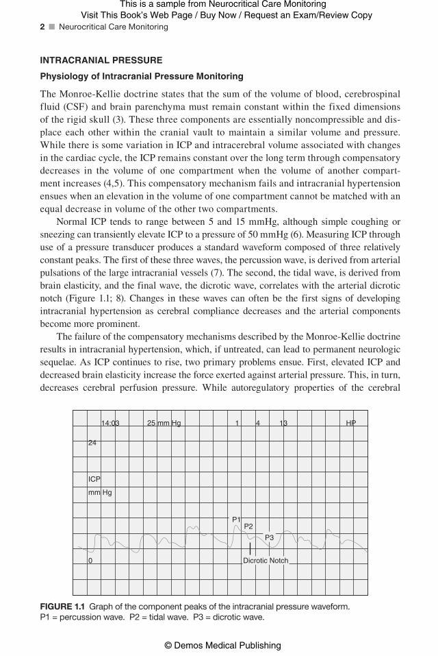
Normal ICP tends to range between 5 and 15 mmHg, although simple coughing or sneezing can transiently elevate ICP to a pressure of 50 mmHg (6). Measuring ICP through use of a pressure transducer produces a standard waveform composed of three relatively constant peaks. The first of these three waves, the percussion wave, is derived from arterial pulsations of the large intracranial vessels (7). The second, the tidal wave, is derived from brain elasticity, and the final wave, the dicrotic wave, correlates with the arterial dicrotic notch (Figure 1.1; 8). Changes in these waves can often be the first signs of developing intracranial hypertension as cerebral compliance decreases and the arterial components become more prominent.
The failure of the compensatory mechanisms described by the Monroe-Kellie doctrine results in intracranial hypertension, which, if untreated, can lead to permanent neurologic sequelae. As ICP continues to rise, two primary problems ensue. First, elevated ICP and decreased brain elasticity increase the force exerted against arterial pressure. This, in turn, decreases cerebral perfusion pressure. While autoregulatory properties of the cerebral
HP1
P1P2
P3
Dicrotic Notch
4 1314:03
24
ICP
mm Hg
0
25 mm Hg
Figure 1.1 Graph of the component peaks of the intracranial pressure waveform. P1 = percussion wave. P2 = tidal wave. P3 = dicrotic wave.
Miller_00259_PTR_01_1-17_12-09-14.indd 2 12/09/14 10:56 AM
Miller_00259_PTR_01_1-17_12-09-14.indd 16 12/09/14 10:57 AM
Visit This Book’s Web Page / Buy Now / Request an Exam/Review CopyThis is a sample from Neurocritical Care Monitoring
© Demos Medical Publishing
1: Intracranial Pressure Monitoring ■ 3
vasculature can compensate for this to an extent, perfusing pressures below the autoregula-tory curve can ultimately lead to cerebral ischemia (9). As the volume and pressure of the contents within the fixed cranial vault increase, displacement of brain tissue results. The most profound manifestation of this displacement is brain herniation.
Initiation of an Intracranial Pressure Monitoring device
Intracranial hypertension is found in 40% to 60% of severe head injuries and is a major fac-tor in 50% of all fatalities. Patients with suspected elevated ICP and a deteriorating level of consciousness are candidates for invasive ICP monitoring. The Glascow Coma Scale (GCS) level that requires ICP monitoring should be based on rate of decline and other clinical fac-tors such as CT evidence of mass effect and hydrocephalus. In general, ICP monitors should be placed in patients with a GCS score of less than 9 and in all patients whose condition is thought to be deteriorating due to elevated ICP (level of evidence V, grade C recommenda-tion). The type of monitor utilized depends on availability, experience, and the situation. Intraventricular ICP monitors and intraparenchymal fiberoptic ICP devices are the most commonly used methods of monitoring ICP.
ICP should be monitored in all salvageable patients with severe traumatic brain injury (TBI) with GCS 3 to 8 after resuscitation and:
(a) Abnormal CT scan of the head that reveals a hematoma, contusions, swelling, hernia-tion, or compressed basal cisterns
(b) A normal CT scan if two or more of the following features are noted at admission: age over 40 years, unilateral or bilateral motor posturing, and systolic blood pressure less than 90 mmHg (1)
In TBI patients with a GCS greater than 8, ICP monitoring should be considered if the CT demonstrates a significant mass lesion or if treatment or sedation is required for associated injuries (13). Although ICP monitoring is widely recognized as a standard of care for patients with severe TBI, care focused on maintaining monitored ICP at 20 mmHg or less was not shown to be superior to care based on imaging and clinical examination in a recent South American study by Chesnut et al. in 2012 (1). However, in that study, there were substantial ICP lowering therapies provided to the control group and overall patient management was much different than that provided at typical North American centers.
In non traumatic settings (eg, spontaneous intracranial hemorrhage [ICH], subarach-noid hemorrhage [SAH], status epilepticus, and cerebral infarction), the decision should be individualized and based on whether elevated ICP is expected. Examples include:
(a) Spontaneous ICH:
1. Patients with a GCS score of ≤ 8, those with clinical evidence of transtentorial herni-ation, or those with significant intraventricular hemorrhage (IVH) or hydrocephalus might be considered for ICP monitoring and treatment. A cerebral perfusion pressure of 50 to 70 mmHg may be reasonable to maintain depending on the status of cerebral autoregulation.
Miller_00259_PTR_01_1-17_12-09-14.indd 3 12/09/14 10:56 AM
Miller_00259_PTR_01_1-17_12-09-14.indd 16 12/09/14 10:57 AM
Visit This Book’s Web Page / Buy Now / Request an Exam/Review CopyThis is a sample from Neurocritical Care Monitoring
© Demos Medical Publishing
4 ■ Neurocritical Care Monitoring
2. Ventricular drainage as treatment for hydrocephalus is reasonable in patients with decreased level of consciousness (20).
(b) Aneurysmal SAH:
There are no definitive guidelines for methods and techniques for ICP management follow-ing aneurysmal SAH. Persistent ICP elevations have been correlated with poor outcomes after aneurysm rupture. Continuous ICP monitoring aids in the early detection of second-ary complications and guides therapeutic intervention. (24)
IcP thresholds
Current data support 20 to 25 mmHg as an upper threshold above which treatment is required for intracranial hypertension (21–23). There has been no difference in outcome between ICP thresholds of 20 and 25 mmHg (21). An opening ICP of 15 and higher has been identified as one of 5 factors associated with higher mortality. Brain shift and hernia-tion result from pressure differential rather than simply height of ICP elevation. As a result, the clinical exam and imaging result should be correlated with the ICP values obtained (13).
cerebral Perfusion threshold
CPP is calculated as mean arterial pressure (MAP) minus ICP. Optimal CPP is typically considered to range between 50 mmHg and 70 mmHg. The TBI guidelines support a CPP > 60 (level of evidence III). Low CPP (< 55 mmHg) and systemic hypotension have been well established as predictors for death and poor outcome (12). However, aggressive attempts to elevate CPP above 70 mmHg have shown no benefit and have been associ-ated with increased risk of acute respiratory distress syndrome (ARDS) related to the use of vasopressors and intravenous fluids (10,11). In addition, maintaining adequate CPP in patients with TBI tends to be more important than lowering ICP (11). However, it is pre-ferred to maintain both values within the goal ranges.
Intracranial Pressure Waveforms (lundeberg Pathological Waves)
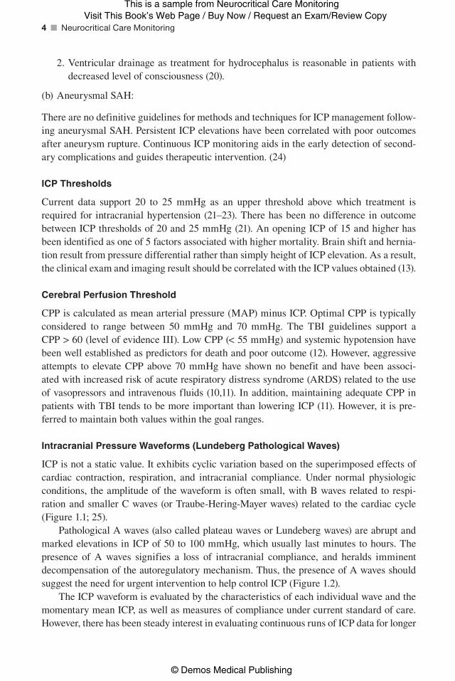
ICP is not a static value. It exhibits cyclic variation based on the superimposed effects of cardiac contraction, respiration, and intracranial compliance. Under normal physiologic conditions, the amplitude of the waveform is often small, with B waves related to respi-ration and smaller C waves (or Traube-Hering-Mayer waves) related to the cardiac cycle (Figure 1.1; 25).
Pathological A waves (also called plateau waves or Lundeberg waves) are abrupt and marked elevations in ICP of 50 to 100 mmHg, which usually last minutes to hours. The presence of A waves signifies a loss of intracranial compliance, and heralds imminent decompensation of the autoregulatory mechanism. Thus, the presence of A waves should suggest the need for urgent intervention to help control ICP (Figure 1.2).
The ICP waveform is evaluated by the characteristics of each individual wave and the momentary mean ICP, as well as measures of compliance under current standard of care. However, there has been steady interest in evaluating continuous runs of ICP data for longer
Miller_00259_PTR_01_1-17_12-09-14.indd 4 12/09/14 10:56 AM
Miller_00259_PTR_01_1-17_12-09-14.indd 16 12/09/14 10:57 AM
Visit This Book’s Web Page / Buy Now / Request an Exam/Review CopyThis is a sample from Neurocritical Care Monitoring
© Demos Medical Publishing
1: Intracranial Pressure Monitoring ■ 5
term trends and correlations using systems and waveform analysis techniques. Goals of this type of analysis include provision of a more sensitive assessment of the pathological state and an early indicator of impending system change. These techniques have included spec-tral analysis, waveform correlation coefficients, and system entropy.
These analytical techniques rely on the relationship between the ICP waveform and the arterial blood pressure (ABP) waveform. The correlation coefficient between changes in ABP and ICP is defined by Cosnyka et al. (1996) as the pressure reactivity index (PRx) (9). PRx varies from low values (no association) to values approaching 1.0 (strong positive association). With lower ABP, lower blood vessel wall tension results in an increase in transmission of the ABP waveform to the ICP. Also with elevated ICP, brain compliance is reduced, thereby increasing transmission of the ABP waveform. PRx has been implicated as a marker of autoregulatory reserve.
Approximate entropy (ApEn) is a measure of system regularity/randomness, devised for use in physiological systems (63). It measures the logarithmic likelihood that runs of patterns are similar over a given number of observations. Reductions in ApEn imply reduced randomness or increased order and have been associated with pathology in the cardiovascular, respiratory, and endocrine systems. Approximate entropy analysis has been successfully applied under conditions of raised ICP for measuring changes in transmission of system randomness between the heart rate and the ICP waveform.
ICP
ICP(mm Hg)
Minutes
50
50
40
40
30
30
A
B B20
20
10
100
0
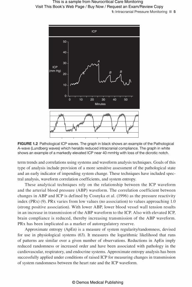
Figure 1.2 Pathological ICP waves. The graph in black shows an example of the Pathological A-wave (Lundberg waves) which heralds reduced intracranial compliance. The graph in white shows an example of a markedly elevated ICP near 40 mmHg with loss of the dicrotic notch.
Miller_00259_PTR_01_1-17_12-09-14.indd 5 12/09/14 10:56 AM
Miller_00259_PTR_01_1-17_12-09-14.indd 16 12/09/14 10:57 AM
Visit This Book’s Web Page / Buy Now / Request an Exam/Review CopyThis is a sample from Neurocritical Care Monitoring
© Demos Medical Publishing
6 ■ Neurocritical Care Monitoring
duration of Monitoring
A single ICP monitoring device is used as long as clinically necessary, with reinsertion of a new monitor only if a malfunction occurs, or if CSF cultures demonstrate an infection. Routine reinsertion of a new monitor increases risk of infection by unnecessarily reexpos-ing the patient to contamination at the time of insertion (19). There is an increased risk of infection with an external ventricular device after being in place for more than 5 days (26). Other ICP monitors (parenchymal and subdural) may begin to have measurement differ-ences (drift) due to inability to recalibrate over time (27,28).
External ventricular drain (EVD) is both a temporary monitor and treatment option for patients with increased ICP. An EVD is usually in place for 5 to 10 days. Indications of removal include: monitoring is no longer required, infection risk is increased, hydrocepha-lus is resolved, and/or ventriculoperitoneal or ventriculoatrial shunt is planned. Weaning of an EVD is done with the following steps as recommended by Varelas in 2006 (29):
■ Raise the drain height by 5 cm H2O every 12 hours only if ICP is not above the pre-
scribed parameter.
■ When the pressure level reaches 20 cm H2O and the EVD drains less than 200 mL/24
hours, clamp the EVD (written order obtained by neurosurgery or neurointensivist team). It is recommended to orient the stopcock “off” to drainage and “open” to the transducer. This technique is used to determine if the patient is continuing to tolerate weaning. The pressure level and the patient’s clinical status postclamping guide the neurosurgical or neurointensivist team’s decision to remove or unclamp the EVD.
Gradual, multistep weaning from external ventricular drainage in patients with aneurys-mal SAH (aSAH) provides no advantage over rapid weaning in preventing the need for shunts. Furthermore, gradual weaning prolongs intensive care unit and hospital stays. Con-sequently, for aSAH patients whose EVD was placed for reasons other than ICP elevation, rapid EVD weaning may be considered rather than gradual weaning.
tyPes of IntracranIal Pressure MonItorIng devIces
There are four main locations within the brain where ICP monitoring devices are fre-quently placed: fluid filled ventricle, brain parenchyma, subarachnoid, and epidural space. The decision of which location and device to use is based on the clinical scenario, appear-ance of the head CT (ie, size of cerebral lateral ventricles) and operator experience.
external ventricular drain evd
Clinical Utility
1. Cerebral edema with suspected elevated ICP: This utilization is best studied in patients with TBI. However, the clinical scenario and need for an EVD can be found with SAH, non traumatic ICH, IVH, ischemic stroke, hypoxic brain injury, cerebral venous throm-bosis (CVT), hepatic encephalopathy, cerebral neoplasm, and cerebral infections. EVDs not only allow monitoring of the ICP but also can serve as a treatment modality to allow drainage of CSF, which aids in lowering the ICP.
Miller_00259_PTR_01_1-17_12-09-14.indd 6 12/09/14 10:56 AM
Miller_00259_PTR_01_1-17_12-09-14.indd 16 12/09/14 10:57 AM
Visit This Book’s Web Page / Buy Now / Request an Exam/Review CopyThis is a sample from Neurocritical Care Monitoring
© Demos Medical Publishing
1: Intracranial Pressure Monitoring ■ 7
2. Hydrocephalus: Hydrocephalus occurs when there is an abnormality of production or resabsorption of CSF within and around the brain and spinal cord. The two types of hydrocephalus are communicating and obstructive. Communicating hydrocephalus occurs when CSF flow throughout the cisterns and the subarachnoid space is unim-peded. Obstructive hydrocephalus occurs when CSF flow within the ventricular sys-tem in blocked from either external compression or internal processes. In both forms of hydrocephalus, the result is an accumulation of CSF, which cannot be absorbed in a nor-mal fashion. In acute cases where the mental status is declining, an EVD is placed and remains until the cause of the hydrocephalus is resolved. If the need for CSF diversion is persistent, ventriculo-peritoneal shunting or ventriculo-atrial shunting may be needed.
3. Surgery: Some surgical procedures of the brain are aided by draining some CSF from the ventricles. In these cases, an external ventricular drain may be placed at the start or during the procedure to drain fluid for brain relaxation (eg, in aSAH, resection of Chiari malformations, or brain tumor).
4. Administering medication: There are some conditions that may require the direct admin-istration of medication into the cerebral ventricular system to bypass the blood–brain bar-rier. In order to do this, some patients may require a ventricular catheter, which enables intrathecal injection. Common clinical scenarios where the ventriculostomy has been used to inject medications include antibiotic administration for bacterial ventriculitis (31), intrathecal chemotherapy for brain cancer (32), and tissue plasminogen activator injection for clearance of IVH (33). These catheters can be used while the patient is in the hospital. However, when patients require long-term treatment, a permanent catheter can be placed, which is connected to a reservoir under the scalp called an Omaya reservoir. This is most commonly used for chemotherapeutic agents or antibiotics for refractory ventriculitis.
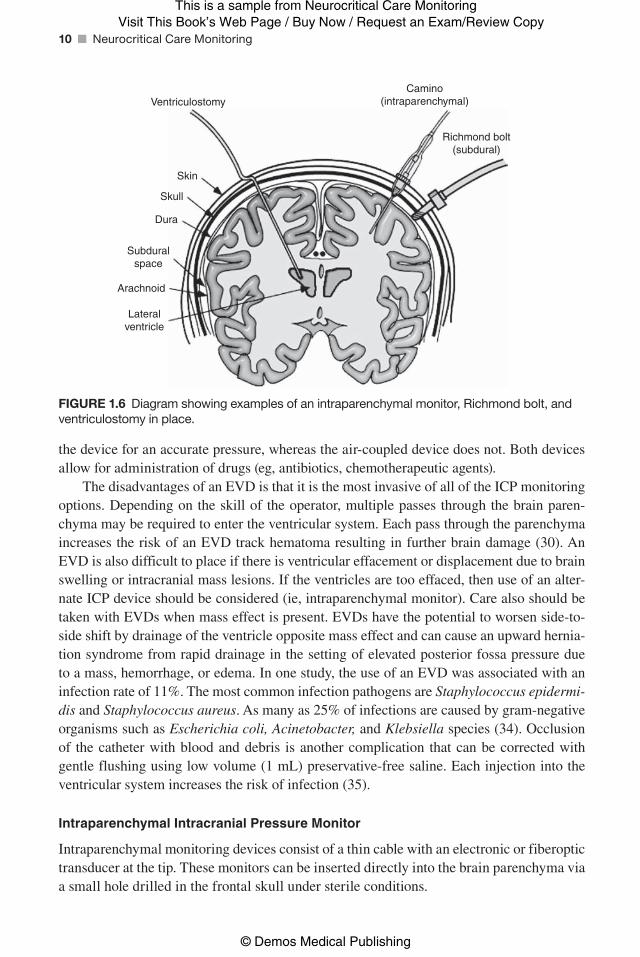
anatomy and Placement
The gold standard technique for ICP monitoring is a catheter inserted into the lateral ven-tricle, usually via a small right frontal burr hole. Under aseptic conditions, a scalp incision is made over the insertion site. Commonly, the Kocher’s point is used, which is located 2.5-cm lateral to the midline (or at the midpupillary line), 11-cm posterior to the nasion. To avoid the motor cortex, it should be at least 1-cm anterior to the coronal suture. A burr hole is then performed. After opening the dura, a ventricular catheter is passed into the ipsilat-eral lateral ventricle transcerebrally. This may be done free-handedly or under the guidance of ultrasound or neuronavigation software. After confirming CSF drainage, the distal end of the catheter is tunneled subcutaneously and allowed to exit the skin approximately 5 cm from the burr hole site. The catheter is connected to a closed external drainage system with an attached ICP monitoring transducer. Though clear benefit has not been demonstrated, prophylactic antibiotics can be given perioperatively to reduce the risk of infection.
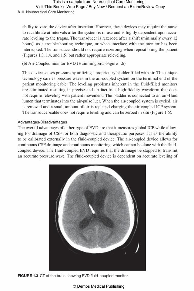
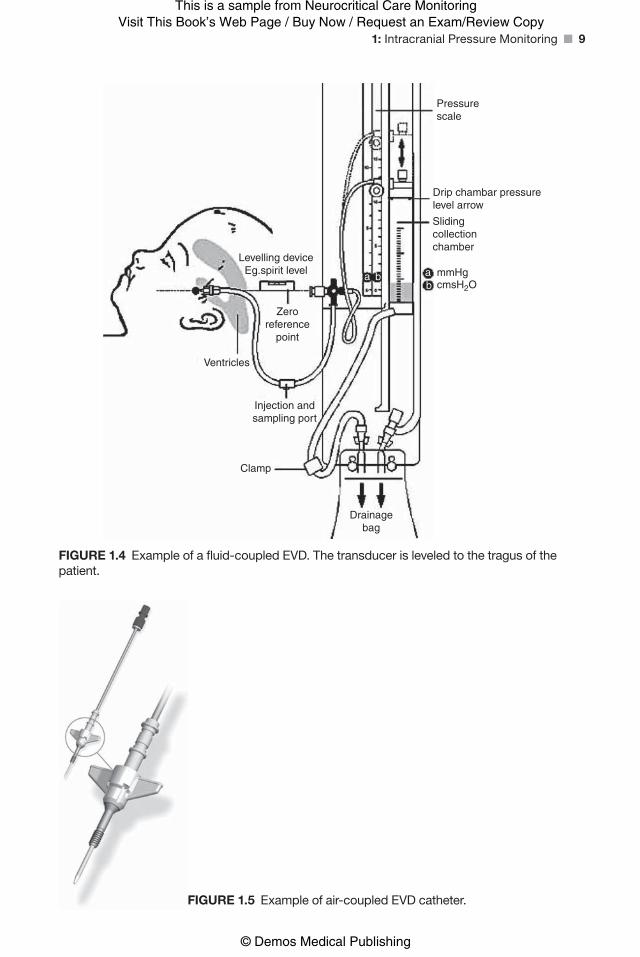
(a) Fluid-Coupled monitor EVD (detailed figure shown in Figure 1.4)
This monitoring system connects to the bedside patient monitor with a pressure cable plugged into a designated pressure module. The benefit of fluid-coupled systems is the
Miller_00259_PTR_01_1-17_12-09-14.indd 7 12/09/14 10:56 AM
Miller_00259_PTR_01_1-17_12-09-14.indd 16 12/09/14 10:57 AM
Visit This Book’s Web Page / Buy Now / Request an Exam/Review CopyThis is a sample from Neurocritical Care Monitoring
© Demos Medical Publishing
8 ■ Neurocritical Care Monitoring
ability to zero the device after insertion. However, these devices may require the nurse to recalibrate at intervals after the system is in use and is highly dependent upon accu-rate leveling to the tragus. The transducer is rezeroed after a shift (minimally every 12 hours), as a troubleshooting technique, or when interface with the monitor has been interrupted. The transducer should not require rezeroing when repositioning the patient (Figures 1.3, 1.4, and 1.5) but rather appropriate releveling.
(b) Air-Coupled monitor EVD (Hummingbird -Figure 1.6)
This device senses pressure by utilizing a proprietary bladder filled with air. This unique technology carries pressure waves in the air-coupled system on the terminal end of the patient monitoring cable. The leveling problems inherent in the fluid-filled monitors are eliminated resulting in precise and artifact-free, high-fidelity waveform that does not require releveling with patient movement. The bladder is connected to an air–fluid lumen that terminates into the air-pulse luer. When the air-coupled system is cycled, air is removed and a small amount of air is replaced charging the air-coupled ICP system. The transducer/cable does not require leveling and can be zeroed in situ (Figure 1.6).
Advantages/DisadvantagesThe overall advantages of either type of EVD are that it measures global ICP while allow-ing for drainage of CSF for both diagnostic and therapeutic purposes. It has the ability to be calibrated externally in the fluid-coupled device. The air-coupled device allows for continuous CSF drainage and continuous monitoring, which cannot be done with the fluid-coupled device. The fluid-coupled EVD requires that the drainage be stopped to transmit an accurate pressure wave. The fluid-coupled device is dependent on accurate leveling of
Figure 1.3 CT of the brain showing EVD fluid-coupled monitor.
Miller_00259_PTR_01_1-17_12-09-14.indd 8 12/09/14 10:57 AM
Miller_00259_PTR_01_1-17_12-09-14.indd 16 12/09/14 10:57 AM
Visit This Book’s Web Page / Buy Now / Request an Exam/Review CopyThis is a sample from Neurocritical Care Monitoring
© Demos Medical Publishing
1: Intracranial Pressure Monitoring ■ 9
Pressurescale
Drip chambar pressurelevel arrow
Slidingcollectionchamber
mmHgaab
bcmsH2O
Drainagebag
Clamp
Injection andsampling port
Ventricles
Zeroreference
point
Levelling deviceEg.spirit level
Figure 1.4 Example of a fluid-coupled EVD. The transducer is leveled to the tragus of the patient.
Figure 1.5 Example of air-coupled EVD catheter.
Miller_00259_PTR_01_1-17_12-09-14.indd 9 12/09/14 10:57 AM
Miller_00259_PTR_01_1-17_12-09-14.indd 16 12/09/14 10:57 AM
Visit This Book’s Web Page / Buy Now / Request an Exam/Review CopyThis is a sample from Neurocritical Care Monitoring
© Demos Medical Publishing
10 ■ Neurocritical Care Monitoring
the device for an accurate pressure, whereas the air-coupled device does not. Both devices allow for administration of drugs (eg, antibiotics, chemotherapeutic agents).
The disadvantages of an EVD is that it is the most invasive of all of the ICP monitoring options. Depending on the skill of the operator, multiple passes through the brain paren-chyma may be required to enter the ventricular system. Each pass through the parenchyma increases the risk of an EVD track hematoma resulting in further brain damage (30). An EVD is also difficult to place if there is ventricular effacement or displacement due to brain swelling or intracranial mass lesions. If the ventricles are too effaced, then use of an alter-nate ICP device should be considered (ie, intraparenchymal monitor). Care also should be taken with EVDs when mass effect is present. EVDs have the potential to worsen side-to-side shift by drainage of the ventricle opposite mass effect and can cause an upward hernia-tion syndrome from rapid drainage in the setting of elevated posterior fossa pressure due to a mass, hemorrhage, or edema. In one study, the use of an EVD was associated with an infection rate of 11%. The most common infection pathogens are Staphylococcus epidermi-dis and Staphylococcus aureus. As many as 25% of infections are caused by gram-negative organisms such as Escherichia coli, Acinetobacter, and Klebsiella species (34). Occlusion of the catheter with blood and debris is another complication that can be corrected with gentle flushing using low volume (1 mL) preservative-free saline. Each injection into the ventricular system increases the risk of infection (35).
Intraparenchymal Intracranial Pressure Monitor
Intraparenchymal monitoring devices consist of a thin cable with an electronic or fiberoptic transducer at the tip. These monitors can be inserted directly into the brain parenchyma via a small hole drilled in the frontal skull under sterile conditions.
Skin
VentriculostomyCamino
(intraparenchymal)
Richmond bolt(subdural)
Skull
Dura
Subduralspace
Arachnoid
Lateralventricle
Figure 1.6 Diagram showing examples of an intraparenchymal monitor, Richmond bolt, and ventriculostomy in place.
Miller_00259_PTR_01_1-17_12-09-14.indd 10 12/09/14 10:57 AM
Miller_00259_PTR_01_1-17_12-09-14.indd 16 12/09/14 10:57 AM
Visit This Book’s Web Page / Buy Now / Request an Exam/Review CopyThis is a sample from Neurocritical Care Monitoring
© Demos Medical Publishing
1: Intracranial Pressure Monitoring ■ 11
Anatomy and PlacementThe monitor is placed in the right or left prefrontal area. The most injured side should be selected in a focal injury. In diffuse brain injury or edema, the right hemisphere is gener-ally used.
Advantages/DisadvantagesThe advantages of an intraparenchymal ICP monitor include the ease of placement, low morbidity, and the ability to add additional monitoring probes such as brain tissue oxygen monitor (LICOX), cerebral blood flow (HEMEDEX), and cerebral microdiaysis probes to a multilumen bolt. It also carries lower risk of infection than EVDs and a lower nursing task burden.
The disadvantages include the inability to drain CSF for diagnostic or therapeutic pur-poses and the potential to lose accuracy (or “drift”) over several days, since the transducer cannot be recalibrated following initial placement. In addition, there is a greater risk of mechanical failure due to the complex design of these monitors (15–18).
subarachnoid Intracranial Pressure Monitor
This is another fluid-coupled system that connects the intracranial space to an external transducer at the bedside via saline-filled tubing. The subarachnoid bolt is actually a hollow screw that is inserted via a burr hole. The dura at the base of the bolt is perforated with a spinal needle, allowing the subarachnoid CSF to fill the bolt. Pressure tubing is then connected to establish communication with a pressure monitoring system. This method of ICP monitoring is no longer commonly used. The advantages include its minimally invasive nature and a low risk of infection.
The disadvantages include decreased accuracy compared to the intraventricular or intraparenchymal monitors; blockage of the system by tissue debris and increased cerebral edema; need for frequent recalibration; and increased risk of bleeding into the subarach-noid space.
epidural Intracranial Pressure Monitors
This device (the Gaeltec device) is inserted into the inner table of the skull and superficial to the dura. Typically, pressure is transduced by an optical sensor. These have a low infec-tion rate (approximately 1%) but are prone to malfunction, displacement, and baseline drift that can exceed 5 ± 10 mmHg after more than a few days of use. Much of the inaccuracy results from having the relatively inelastic dura between the sensor tip and the subarach-noid space.
Epidural monitors contain optical transducers that rest against the dura after passing through the skull. They often are inaccurate, as the dura dampens the pressure transmit-ted to the epidural space and, thus, are of limited clinical utility. They have been most commonly used in the setting of coagulopathic patients with hepatic encephalopathy whose course is complicated by cerebral edema. In this setting, use of these catheters is associated with a significantly lower risk of ICH (4% vs 20% and 22%, respectively, for intraparenchymal and intraventricular devices). It is also associated with decreased fatal
Miller_00259_PTR_01_1-17_12-09-14.indd 11 12/09/14 10:57 AM
Miller_00259_PTR_01_1-17_12-09-14.indd 16 12/09/14 10:57 AM
Visit This Book’s Web Page / Buy Now / Request an Exam/Review CopyThis is a sample from Neurocritical Care Monitoring
© Demos Medical Publishing
12 ■ Neurocritical Care Monitoring
hemorrhages (1% vs 5% and 4%, respectively, for intraparenchymal and intraventricular devices; 14). Otherwise, this device is rarely used in clinical practice.
lumbar catheter Intracranial Pressure Monitoring
Lumbar drainage devices (LDDs) are closed sterile systems that allow the drainage of CSF from the subarachnoid space. LDDs are inserted via a specialized spinal needle, known as a Touhy needle, into the lumbar subarachnoid space at the L2–L3 level or below. This placement avoids injury to the spinal cord, which ends at the conus medullaris at the level of the L1–L2 vertebral bodies. In the lumbar CSF space, the flexible spinal catheter will lie alongside the cauda equina, which consists of the ventral and dorsal spinal nerve roots that descend from the spinal cord and exit the spinal canal at lumbosacral levels. Insertion of the spinal catheter may cause transient radicular pain if the catheter brushes against one of the spinal nerve roots.
Occasionally, the pain can be persistent, especially if lumbar spinal stenosis causes the spinal catheter and the spinal nerve roots to remain in close contact. Placement of an LDD is an accepted medical therapy for the treatment of postoperative or traumatic dural fistulae (ie, CSF leak), treatment of shunt infections, and for the diagnostic evaluation of idiopathic normal pressure hydrocephalus. LDDs also are used to reduce ICP during a craniotomy and as adjuvant therapy in the management of traumatically brain-injured patients.
additional concerns With Intracranial Pressure Monitoring devices
Antibiotic ProphylaxisThe patients at greater risk for ICP monitor–related infection include those with the fol-lowing features: prolonged monitoring greater than 5 days, presence of ventriculostomy vs intraparenchymal monitor, CSF leak, concurrent infection, or serial ICP monitor place-ments. Multiple studies support the use of prophylactic systemic antibiotics throughout the duration of external ventricular drainage. However, prophylactic use of antibiotics raises concern for an increase in bacterial antimicrobial resistance. Recent studies have shown that antibiotics treatment given only during the insertion of the EVD may be associated with comparable infectious risks. The use of antibiotic-coated EVDs to prevent ventricu-litis has proven to be effective (36) in one study; however, the use of a silver-impregnated catheter was not proven to be beneficial (36,37).
Deep Venous Thrombosis ProphylaxisChemical prophylaxis has been shown to decrease rates of venous thrombosis formation and subsequent pulmonary embolism in neurologically critically ill patients. The incidence of bleeding was not different between early (24 hours) and delayed (72 hours) administra-tion of chemical prophylaxis in relation to insertion of either an EVD or intraparenchymal device, but there was reduced incidence of deep vein thrombosis (DVT)/pulmonary embo-lism (PE) with early administration. In addition, the early start of chemical prophylaxis did not show an increase in hemorrhagic complications (60).
Miller_00259_PTR_01_1-17_12-09-14.indd 12 12/09/14 10:57 AM
Miller_00259_PTR_01_1-17_12-09-14.indd 16 12/09/14 10:57 AM
Visit This Book’s Web Page / Buy Now / Request an Exam/Review CopyThis is a sample from Neurocritical Care Monitoring
© Demos Medical Publishing
1: Intracranial Pressure Monitoring ■ 13
Dressing and Dressing Changes of the EVDSkilled nursing is key to minimizing complications related to external ventricular drains. Dressing of the EVD site must be observed hourly to ensure that a CSF leak has not occurred. If a leak is identified, the insertion site should be inspected and the dressing should be replaced. Dressings should be changed using sterile technique when visibly soiled (61). Incor-rect or asterile dressing change has been associated with increased risk for ventriculitis (62).
Antiplatelet and Anticoagulant Use With EVDsThere is a trend toward higher bleeding complications from EVD placement in patients who are on antiplatelet or anticoagulant therapy. When starting these agents, one must weigh the indication for the agent with the risk of a ventriculostomy track hematoma or intraparenchymal monitor–associated bleed. The early use of chemical, subcutaneously injected, VTE chemoprophylaxis (first 24 hours) did not increase the incidence of bleeding complications but did not show better protection against venous thromboembolism when compared to delayed administration (64).
crItIcal care ManageMent of elevated IntracranIal Pressure
general Measures
The head and neck should be optimally positioned to minimize additional elevations in ICP. The head of the bed should be elevated to 30 degrees for patients with poor intracranial com-pliance. The neck should be free from compression, and the head should be positioned in the midline. When a cervical collar is present, it should be fitted just tight enough to provide stability but not so tight as to cause internal jugular vein compression.
Normorthermia (36–38°C) is strongly recommended in patients with cerebral edema, irrespective of the underlying etiology, to avoid the deleterious effects of fever on outcome (38). Numerous clinical trials have reported the value of induced moderate hypothermia for ICP control (39). Hyperthermia will increase ICP (40). Control of fever includes administra-tion of acetaminophen (325–650 mg orally or rectally every 6 hours) or ibuprofen (400 mg orally every 6 hours). In addition, surface cooling with ice packs, cool blankets, or surface devices (Artic Sun) is an effective noninvasive way to reduce fever. Intravascular cooling devices utilize a catheter with a balloon that circulates fluid internally; it is inserted into the inferior vena cava via the femoral vein, thereby allowing access to the body’s internal circulation to change temperature when that flow interacts with the catheter. It has been shown to be a highly effective, quick, and precise form of temperature control, but it does carry a procedural risk, increased risk of venous thrombosis (41), and risk of infection from a central venous catheter. (42)
Maintenance of euvolemia (with 0.9 % normal saline) is essential. Mild hypervolemia can be considered in order to maintain CPP, but this needs to be done judiciously to avoid pulmonary edema and ARDS. Hypotonic fluids such as 0.45% saline and dextrose in water should be avoided (43).
Normocarbia (PaCO2 35–45 mmHg) is preferred because hypercarbia will add to ele-
vations in ICP resulting from cerebral vasodilitation. In addition, avoidance of hypoxemia and maintenance of PaO
2 of 100 mg are recommended.
Miller_00259_PTR_01_1-17_12-09-14.indd 13 12/09/14 10:57 AM
Miller_00259_PTR_01_1-17_12-09-14.indd 16 12/09/14 10:57 AM
Visit This Book’s Web Page / Buy Now / Request an Exam/Review CopyThis is a sample from Neurocritical Care Monitoring
© Demos Medical Publishing
14 ■ Neurocritical Care Monitoring
Agitation contributes to elevated ICP. Care must be taken to ensure that pain is addressed adequately with short-acting narcotics such as morphine and fentanyl. Alcohol, illicit drug withdrawal, and delirium should be considered if pain is not responsible. When required, short-acting sedative agents are always preferred for the neurological population so that an adequate examination can still be obtained.
The use of prophylactic anti epileptic drugs (AEDs) remains controversial in the setting of acute brain injury. Seizures, whether they are clinically evident or nonconvulsive, result in elevations of ICP (44). AED prophylaxis in patients with TBI may be given for 1 week, but there is no evidence to support routine continued use (45). In patients with ICH or SAH, AEDs should not be routinely initiated, unless it is thought that a seizure might result in rehemorrhage or worsening of an unprotected aneurysm. Corticosteroids are beneficial only for patients with vasogenic edema related to abscess or brain tumors.
specific Measures
Hyperventilation is very effective in reducing ICPs related to cerebral edema acutely and for a short period of time (46). It works via the vasoconstrictor effect of decreased PaCO
2,
which persists only for 10 to 20 hours. PaCO2 levels below 25 can increase the risk of second-
ary cerebral ischemia from too much vasoconstriction (47). Sustained hyperventilation for 5 days has been shown to slow recovery of severe TBI at 3 and 6 months (48,49).
The use of osmotic therapy or hypertonic saline (HTS) is an effective way to reduce ICP from cerebral edema. For osmotic therapy, mannitol can be used with a target serum osmolality of 300 to 320 (50). It is administered as a 0.25 to 1.5 g/kg bolus intravenously. Mechanisms of action include acute dehydrating effect and secondary hyperosmolality (diuretic effect). Side effects include hypotension, hypovolemia, and renal tubular damage. Hypertonic saline boluses and infusions (3%, 7.5 %, 10%, and 23.4 %) have proven to be effective in numerous clinical scenarios marked by ICH. HTS administered in bolus form can resolve ICP episodes refractory to mannitol (51). The most effective concentrations and protocols for HTS use require further study.
The use of barbiturates results from their ability to reduce brain metabolism and cere-bral blood flow, thus lowering ICP. Barbiturate use may also exert a neuroprotective effect (52). Pentobarbital is most commonly used, with a loading dose of 5 to 20 mg/kg as a bolus, followed by 1 to 4 mg/kg per hr infusion. Barbiturate therapy can be complicated by hypo-tension, possibly requiring vasopressor support. The use of barbiturates is also associated with a loss of the neurologic examination, and requires accurate ICP, hemodynamic, and often EEG monitoring to guide therapy
As discussed earlier, a ventriculostomy should be inserted for very specific criteria for specific disease states. Rapid drainage of CSF should be avoided because this may lead to subdural hemorrhage (53). A lumbar drain is generally contraindicated in the setting of high ICP due to the risk of transtentorial herniation or central herniation.
When all medical measures to control ICH fail, decompressive hemi-craniectomy (DHC) can be considered. DHC removes the rigid confines of the bony skull, allowing noncompressive expansion of the volume of the intracranial contents. There is a growing body of literature supporting the efficacy of decompressive craniectomy in certain clinical
Miller_00259_PTR_01_1-17_12-09-14.indd 14 12/09/14 10:57 AM
Miller_00259_PTR_01_1-17_12-09-14.indd 16 12/09/14 10:57 AM
Visit This Book’s Web Page / Buy Now / Request an Exam/Review CopyThis is a sample from Neurocritical Care Monitoring
© Demos Medical Publishing
1: Intracranial Pressure Monitoring ■ 15
situations. Importantly, it has been demonstrated that in patients with elevated ICP, crani-ectomy alone lowered ICP 15%, but opening the dura in addition to the bony skull resulted in an average decrease in ICP of 70% (54,55). Decompressive craniectomy also appears to improve brain tissue oxygenation (56). It has been shown to improve outcomes in malignant MCA stroke syndromes (57), but has not been shown to improve outcomes in TBI (58,59).
references
1. Chesnut RM et al. A trial of intracranial-pressure monitoring in traumatic brain injury. N Engl J Med. 2012;367(26):2471–2481.
2. Raboel PH et al. Intracranial Pressure Monitoring: Invasive versus Non-Invasive Methods-A Review. Crit Care Res Pract. 2012;2012: 1–14.
3. Monro A. Observations on the Structure and Functions of the Nervous System 1783, Edinburgh: Printed for, and sold by, W. Creech. 176 p.
4. Greitz D et al. Pulsatile brain movement and associated hydrodynamics studied by magnetic reso-nance phase imaging. The Monro-Kellie doctrine revisited. Neuroradiology. 1992;34(5):370–380.
5. Neff S and Subramaniam RP, Monro-Kellie doctrine. J Neurosurg. 1996;85(6):1195. 6. Winn HR and Youmans JR. Youmans Neurological Surgery. 2004;5th:[4 v. (lxiv, 5296, cviii) ill.
(some col.) 28 cm. + 1 CD-ROM (4 3/4 in.)]. 7. Cardoso ER, Rowan JO, Galbraith S. Analysis of the cerebrospinal fluid pulse wave in intracranial
pressure. J Neurosurg. 1983;59(5):817–821. 8. Hamer J et al. Influence of systemic and cerebral vascular factors on the cerebrospinal fluid pulse
waves. J Neurosurg. 1977;46(1):36–45. 9. Marion DW, Darby J, Yonas H. Acute regional cerebral blood flow changes caused by severe head
injuries. J Neurosurg. 1991;74(3):407–414. 10. Schmidt JM et al. Cerebral perfusion pressure thresholds for brain tissue hypoxia and metabolic
crisis after poor-grade subarachnoid hemorrhage. Stroke. 2011;42(5):1351–1356. 11. Rosner MJ, Rosner SD, Johnson AH. Cerebral perfusion pressure: management protocol and clinical
results. J Neurosurg. 1995;83(6):949–962. 12. Balestreri M et al. Impact of intracranial pressure and cerebral perfusion pressure on severe disabil-
ity and mortality after head injury. Neurocrit Care. 2006;4(1):8–13. 13. Brain Trauma Foundation; American Association of Neurological Surgeons; Congress of Neurologi-
cal Surgeons; Joint Section on Neurotrauma and Critical Care, AANS/CNS, Bratton SL, Chestnut RM, Ghajar J, McConnell Hammond FF, Harris OA, Hartl R, Manley GT, Nemecek A, Newell DW, Rosenthal G, Schouten J, Shutter L, Timmons SD, Ullman JS, Videtta W, Wilberger JE, Wright DW. Guidelines for the management of severe traumatic brain injury. VI. Indications for intracranial pres-sure monitoring. J Neurotrauma. 2007;24 Suppl 1:S37–S44.
14. Blei AT, Olafsson S, Webster S, et al. Complications of intracranial pressure monitoring in fulminant hepatic failure. Lancet. 1993;341:157.
15. Ostrup RC, Luerssen TG, Marshall LF, et al. Continuous monitoring of intracranial pressure with a miniaturized fiberoptic device. J Neurosurg. 1987;67:206.
16. Gambardella G, d’Avella D, Tomasello F. Monitoring of brain tissue pressure with a fiberoptic device. Neurosurgery. 1992;31:918.
17. Bochicchio M, Latronico N, Zappa S, et al. Bedside burr hole for intracranial pressure monitoring performed by intensive care physicians. A 5-year experience. Intensive Care Med 1996;22:1070.
18. Kasotakis G, Michailidou M, Bramos A, et al. Intraparenchymal vs extracranial ventricular drain intracranial pressure monitors in traumatic brain injury: less is more? J Am Coll Surg. 2012 Jun;214(6):950–957. doi: 10.1016/j.jamcollsurg.2012.03.004. Epub 2012 Apr 26.
19. Kantar RK, Weiner LB, Patti AM, et al. Infectious complications and duration of intracranial pres-sure monitoring. Crit Care Med. 1985 Oct;13(10):837–839.
20. Morgenstern LB, Hemphill JC III, Anderson C, et al. American Heart Association Stroke Council and Council on Cardiovascular Nursing. Guidelines for the management of spontaneous intracerebral hem-orrhage: a guideline for healthcare professionals from the American Heart Association/American Stroke Association. Stroke. 2010 Sep;41(9):2108–2129. doi: 10.1161/STR.0b013e3181ec611b. Epub 2010 Jul 22.
Miller_00259_PTR_01_1-17_12-09-14.indd 15 12/09/14 10:57 AM
Miller_00259_PTR_01_1-17_12-09-14.indd 16 12/09/14 10:57 AM
Visit This Book’s Web Page / Buy Now / Request an Exam/Review CopyThis is a sample from Neurocritical Care Monitoring
© Demos Medical Publishing
16 ■ Neurocritical Care Monitoring
21. Ratanalert S, Phuenpathom N, Saeheng S, et al. ICP threshold in CPP management of severe head injury patients. Surg Neurol. 2004 May;61(5):429–434; discussion 434–435.
22. Saul TG, Ducker TB. Effect of intracranial pressure monitoring and aggressive treatment on mortal-ity in severe head injury. J Neurosurg. 1982 Apr;56(4):498–503.
23. Narayan RK et al. Intracranial pressure: to monitor or not to monitor? A review of our experience with severe head injury. J Neurosurg. 1982 May;56(5):650–659.
24. Mack WJ, King RG, Ducruet AF, et al. Intracranial Pressure Following Aneurysmal Subarachnoid Hemorrhage: Monitoring Practices and Outcome Data. Neurosurg Focus. 2003;14(4).
25. Hayashi M, Handa Y, Kobayashi H, et al. Plateau-wave phenomenon (I). Correlation between the appearance of plateau waves and CSF circulation in patients with intracranial hypertension. Brain. 1991;114 (Pt 6):2681.
26. Rebuck, K. Murry, D. Rhoney, D. et al. Infection related to intracranial pressure monitors in adults: analysis of risk factors and antibiotic prophylaxis. J Neurol Neurosurg Psychiatry. 2000 September;69(3):381–384.
27. Chen L, Du HG, Yin LC, et al. Zero drift of intraventricular and subdural intracranial pressure monitoring systems. Chin J Traumatol. 2013;16(2):99–102.
28. Rosa M Martínez-Mañasa, David Santamartab, José M de Camposb, et al. Camino® intracranial pressure monitor: prospective study of accuracy and complications. J Neurol Neurosurg Psychiatry 2000;69:82–86.
29. Varelas P, Helms A, Sinson G, et al. Clipping or coiling of ruptured cerebral aneurysms and shunt-dependent hydrocephalus. Neurocrit Care. 2006;4(3):223–228.
30. Maniker AH, Vaynman AY, Karimi RJ, et al. Hemorrhagic complications of external ventricular drainage. Neurosurgery. 2006 Oct;59(4 Suppl 2):ONS419-24; discussion ONS424-5.
31. Mueller SW, Kiser TH, Anderson TA, et al. Intraventricular daptomycin and intravenous linezolid for the treatment of external ventricular-drain-associatedventriculitis due to vancomycin-resistant Enterococcus faecium. Ann Pharmacother. 2012 Dec;46(12):e35. doi: 10.1345/aph.1R412. Epub 2012 Dec 11.
32. Birnbaum T, Baumgarten LV, Dudel C, et al. Successful long-term control of lymphomatous meningitis with intraventricular rituximab. J Clin Neurosci. 2013 Sep 17. pii: S0967-5868(13) 00287-7.
33. Ziai W, Moullaali T, Nekoovaght-Tak S, Ullman N, et al. No exacerbation of perihematomal edema with intraventricular tissue plasminogen activator in patients with spontaneous intraventricular hem-orrhage. Neurocrit Care. 2013 Jun;18(3):354–361.
34. Beer R, Lackner P, Pfausler B, et al. Nosocomial ventriculitis and meningitis in neurocritical care patients. J Neurol. 2008 Nov;255(11):1617–1624.
35. Hill M, Baker G, Carter D, et al. A multidisciplinary approach to end external ventricular drain infections in the neurocritical care unit. J Neurosci Nurs. 2012 Aug;44(4):188–193.
36. Sonabend AM, Korenfeld Y, Crisman C, et al. Prevention of ventriculostomy-related infections with prophylactic antibiotics and antibiotic-coated external ventricular drains: a systematic review. Neu-rosurgery. 2011 Apr;68(4):996–1005.
37. Xiang Wang, Yan Dong, Xiang-Qian Qi, et al. Clinical review: Efficacy of antimicrobial- impregnated catheters in external ventricular drainage—a systematic review and meta-analysis. Critical Care 2013, 17:234.
38. Rossi S, Zanier ER, Mauri I, et al. Brain temperature, body core temperature, and intracranial pres-sure in acute cerebral damage. J Neurol Neurosurg Psychiatry. 2001 Oct;71(4):448–454.
39. Polderman KH. Induced hypothermia and fever control for prevention and treatment of neurological injuries. Lancet 2008;371:1955–1969.
40. Jiang JY, Xu W, Li WP, et al. Effect of long-term mild hypothermia or short-term mild hypo-thermia on outcome of patients with severe traumatic brain injury. J Cereb Blood Flow Metab. 2006;26:771–776.
41. Simosa HF, Petersen DJ, Agarwal SK, et al. Increased risk of deep venous thrombosis with endovas-cular cooling in patients with traumatic head injury. Am Surg. 2007 May;73(5):461–464.
42. Patel N, Nair SU, Gowd P, et al. Central line associated blood stream infection related to cooling catheter in cardiac arrest survivors undergoing therapeutic hypothermia by endovascular cooling. Conn Med. 2013 Jan;77(1):35–41.
Miller_00259_PTR_01_1-17_12-09-14.indd 16 12/09/14 10:57 AM
Miller_00259_PTR_01_1-17_12-09-14.indd 16 12/09/14 10:57 AM
Visit This Book’s Web Page / Buy Now / Request an Exam/Review CopyThis is a sample from Neurocritical Care Monitoring
© Demos Medical Publishing
1: Intracranial Pressure Monitoring ■ 17
43. Zornow MH, Prough DS. Fluid management in patients with traumatic brain injury. New Horiz. 1995 Aug;3(3):488–498.
44. Gabor AJ, Brooks AG, Scobey RP, et al. Intracranial pressure during epileptic seizures. Electroen-cephalogr Clin Neurophysiol. 1984 Jun;57(6):497–506.
45. Chang BS, Lowenstein DH; Quality Standards Subcommittee of the American Academy of Neurol-ogy. Practice parameter: antiepileptic drug prophylaxis in severe traumatic brain injury: report of the Quality Standards Subcommittee of the American Academy of Neurology. Neurology. 2003 Jan 14;60(1):10–16.
46. Heffner JE, Sahn SA. Controlled hyperventilation in patients with intracranial hypertension. Application and management. Arch Intern Med. 1983 Apr;143(4):765–769.
47. Yundt KD, Diringer MN. The use of hyperventilation and its impact on cerebral ischemia in the treatment of traumatic brain injury. Crit Care Clin. 1997 Jan;13(1):163–184.
48. Diringer MN, Videen TO, Yundt K, et al. Regional cerebrovascular and metabolic effects of hyper-ventilation after severe traumatic brain injury. J Neurosurg. 2002 Jan;96(1):103–108.
49. Muizelaar JP, Marmarou A, Ward JD, et al. Adverse effects of prolonged hyperventilation in patients with severe head injury: a randomized clinical trial. J Neurosurg. 1991 Nov;75(5):731–739.
50. Roberts I, Schierhout G, Wakai A. Mannitol for acute traumatic brain injury. Cochrane Database Syst Rev. 2003;(2):CD001049
51. Fisher B, Thomas D and Peterson B. Hypertonic saline lowers raised intracranial pressure in chil-dren after head trauma. J Neurosurg Anesthesiol. 1992;4:4–10.
52. Roberts I. Barbiturates for acute traumatic brain injury. Cochrane Database Syst Rev. 2000;(2):CD000033.
53. Andrade AF, Paiva WS, Amorim RL, et al. Continuous ventricular cerebrospinal fluid drainage with intracranial pressure monitoring for management of posttraumatic diffuse brain swelling. Arq Neuropsiquiatr. 2011 Feb;69(1):79–84.
54. Timofeev I, Czosnyka M, Nortje J, et al. Effect of decompressive craniectomy on intracranial pressure and cerebrospinal compensation following traumatic brain injury. J Neurosurg. 2008 Jan;108(1):66–73.
55. Kunze E, Meixensberger J, Janka M, et al. Decompressive craniectomy in patients with uncontrol-lable intracranial hypertension. Acta Neurochir Suppl. 1998;71:16–18.
56. M Jaeger, M Soehle, and J Meixensberger. Effects of decompressive craniectomy on brain tissue oxygen in patients with intracranial hypertension. J Neurol Neurosurg Psychiatry. 2003 April; 74(4):513–515
57. Yang XF, Yao Y, Hu WW, et al. Is decompressive craniectomy for malignant middle cerebral artery infarction of any worth? J Zhejiang Univ Sci B. 2005 Jul;6(7):644–649.
58. D. J. Cooper, J. V. Rosenfeld, L. Murray, et al. Decompressive craniectomy in diffuse traumatic brain injury. The New England Journal of Medicine, vol. 364, no. 16, pp. 1493–1502, 2011.
59. J. Ma, C. You, L. Ma, et al. Is decompressive craniectomy useless in severe traumatic brain injury? Critical Care, vol. 15, no. 5, article 193, 2011.
60. Tanweer O, Boah A, Huang PP. Risks for hemorrhagic complications after placement of external ventricular drains with early chemical prophylaxis against venous thromboembolisms . J Neurosurg. 2013 Nov;119(5):1309–1313.
61. Slazinski. T. et al. Care of the patient undergoing intracranial pressure monitoring/external ventricu-lar drainage or lumbar drainage. American Association of Neuroscience Nurses: 1–37.
62. Korinek AM, Reina M, Boch AL, et al. Prevention of external ventricular drain-related ventriculitis. Acta Neurochir (Wien). 2005 Jan;147(1):39-45; discussion 45–46.
63. Pincus SM. Approximate entropy as a measure of system complexity. Proc Natl Acad Sci U S A 1991 March 15;88(6):2297–2301.
64. Tanweer O, Boah A, Huang PP. Risks for hemorrhagic complications after placement of external ventricular drains with early chemical prophylaxis against venous thromboembolisms. J Neurosurg. 2013 Nov;119(5):1309–1313.
Miller_00259_PTR_01_1-17_12-09-14.indd 17 12/09/14 10:57 AM