Embed Size (px)
Citation preview
Changes in intramitochondrial cardiolipin distribution inapoptosis-resistant HCW-2 cells, derived from the human promyelocytic
leukemia HL-60
Maria Garcia Fernandeza;b, Leonarda Troianob, Laura Morettib, Jessica Pedrazzib,Stefano Salviolic, Inma Castilla-Cortazara, Andrea Cossarizzab;*aDepartment of Human Physiology, University of Malaga, Campus Teatinos, 29080 Malaga, Spain
bDepartment of Biomedical Sciences, University of Modena and Reggio Emilia, via Campi 287, 41100 Modena, ItalycDepartment of Experimental Pathology, University of Bologna, via S. Giacomo 14, 40100 Bologna, Italy
Received 13 June 2000; revised 3 July 2000; accepted 10 July 2000
Edited by Vladimir Skulachev
Abstract Using a cytofluorimetric approach, we studiedintramitochondrial cardiolipin (CL) distribution in HCW-2 cells,an apoptosis-resistant clone of human HL-60 cells. In HL-60,about 50% of total CL is distributed in the outer leaflet ofmitochondrial inner membrane, while in HCW-2 a significantlyhigher amount of CL (about 65%) is in that site. In basalconditions, HSW-2 cells also show a reduced mitochondrialmembrane potential even if they are able to proliferate as theparental line. Taking into account the complex functions that CLplays in the regulation of mitochondrial activity, it is likely thatHCW-2 could produce ATP utilizing more glycolytic pathwaysrather than mitochondrial respiratory chain. ß 2000 Federa-tion of European Biochemical Societies. Published by ElsevierScience B.V. All rights reserved.
Key words: Mitochondrion; Cardiolipin; Mitochondrialmembrane potential ; Apoptosis; Flow cytometry
1. Introduction
Mitochondria are involved in many types of programmedcell death/apoptosis. In some experimental models, they pro-vide the energy required for the process to occur [1^3]. Inothers, they can trigger cell death in a number of ways: bydisrupting electron transport and energy metabolism, by re-leasing/activating proteins that mediate apoptosis, and by al-tering cellular redox potential [4^6]. As an abnormal resis-tance to apoptosis correlates with malformations,autoimmune diseases or cancer due to the persistence ofself-speci¢c immunocytes or mutated cells, the role of suchorganelles is crucial for the maintenance of the body homeo-stasis [7,8].
Recent studies have suggested that cardiolipin (CL), a di-acidic phospholipid that was isolated from beef heart and hasan unusually high content of linoleic acid ester residues [9],could be involved in apoptotic cell death [10,11]. CL is presentthroughout the eukaryotes including animals, plants and fun-gi. In mammalian tissues and in yeast, CL is found exclusively
in mitochondria [12,13], a site in which it is speci¢c and es-sential for the normal function of mitochondrial inner mem-brane system, and in particular for: (i) the activity of cyto-chrome bc1 and cytochrome c oxidase, key components of theelectron transport chain; (ii) the attachment of cytochrome cto the inner mitochondrial membrane; (iii) the activity ofmitochondrial transporters, including the ADP/ATP translo-case, the mono-, di-, tri-carboxylate carriers, the K-ketogluta-rate, aspartate/glutamate and palmitoylcarnitine carriers, andthe (acyl)carnitine translocase system; (iv) the functionality ofF0F1-ATP synthase [14].
In order to further investigate the role of such a molecule,we analyzed the apoptosis-resistant clone called HCW-2, de-rived from the human promyelocytic cell line HL-60. Thisclone has been selected from parental cells by treatmentwith a cytotoxic dose of 8-Cl-cAMP, and shows resistanceto cycloheximide- and staurosporine-induced cell death [15].
In the present paper, using a cyto£uorimetric approach thatallows the study of intact cells, we describe a direct assay foranalyzing intramitochondrial CL transverse distribution [16].The assay is based upon the following peculiar properties ofthe £uorescent dye 10-N-nonyl-3,6-bis(dimethylamino)acri-dine (nonyl acridine orange, NAO), i.e. : (i) two moleculesof NAO can bind with a high a¤nity one single CL molecule,forming NAO dimers; (ii) the dye is unable to bind zwitter-ionic phospholipids and has a low a¤nity for other anionicphospholipids; (iii) NAO is capable of changing its emissiondue to its spectral properties, as after dimer formation the£uorescence can shift from green (monomeric form) to red(dimeric form) [16,17]. Using such an assay, we could showthat the distribution of CL was markedly di¡erent in HCW-2cells when compared with the parental line HL-60.
2. Materials and methods
2.1. ChemicalsFetal calf serum and RPMI 1640 were from Life Technologies Ltd.
(Oxford, UK). NAO and 5,5P,6,6P-tetrachloro-1,1P,3,3P-tetraethylben-zimidazolcarbocyanine iodide (JC-1) were from Molecular Probes(Eugene, OR, USA). Valinomycin (Val) and other common chemicalswere from Sigma (St. Louis, MO, USA) and were of analytical grade.
2.2. Cell cultureMycoplasma-free HL-60 human promyelocytic leukemic cells and
its apoptosis-resistant clone HCW-2 (kindly provided by Drs. J.H.Wyche and Z. Han, Brown University, Providence, RI, USA) weregrown in suspension in complete culture medium (RPMI 1640 sup-
0014-5793 / 00 / $20.00 ß 2000 Federation of European Biochemical Societies. Published by Elsevier Science B.V. All rights reserved.PII: S 0 0 1 4 - 5 7 9 3 ( 0 0 ) 0 1 8 6 1 - 5
*Corresponding author. Fax: (39)-59-205 5426.E-mail: [email protected]
Abbreviations: CL, cardiolipin; NAO, nonyl acridine orange; v8,mitochondrial membrane potential ; JC-1, 5,5P,6,6P-tetrachloro-1,1P,3,3P-tetraethylbenzimidazolcarbocyanine iodide; Val, valinomycin
FEBS 23979 28-7-00
FEBS 23979FEBS Letters 478 (2000) 290^294
plemented with 10% heat-inactivated fetal calf serum, 2 mM L-gluta-mine, 100 UI/ml penicillin, 100 Wg/ml streptomycin) and kept at 37³Cin a humidi¢ed atmosphere (5% CO2 in air). Cells were collectedduring the log phase of growth, washed in phosphate-bu¡ered saline(PBS), counted and adjusted at a density of 1U106 cells/ml.
2.3. Analysis of CL distributionIn order to analyze CL intramitochondrial distribution, we took
advantage from the spectral characteristics of NAO [16,17]. In partic-ular, we used its capacity to form dimers when it interacts with diaci-dic phospholipid, and the fact that its £uorescence emission shiftsfrom 525 nM (monomeric form of the dye) to about 640 nm (underdimeric conditions, in the presence of a stoichiometric ratio of 2:1with CL). Cells were ¢xed in 1% formaldehyde (in PBS) for 15 min atroom temperature, then washed twice in cold PBS and adjusted at theconcentration of 0.5U106 cell/ml in PBS. Increasing amounts of NAO(0.1^35 WM) were added, and cells were kept at room temperature for15 min, washed twice with PBS and resuspended in a total volume of400 Wl of PBS and analyzed.
The red and green £uorescence emission intensity was plotted asfunction of the amount of NAO present in the incubation mixture;however, only the red £uorescence was used for the analysis of CLdistribution.
2.4. Analysis of mitochondrial membrane potentialMitochondrial membrane potential (v8) was measured by lipo-
philic cationic probe JC-1 [18], a dye we are using for this purposesince several years, and that reversibly changes its color from green toorange/red as v8 increases (over values of about 80^100 mV). Thisproperty is due to the reversible formation of JC-1 aggregates uponmembrane polarization that causes shifts in emitted light from 530 nm(i.e. emission of monomeric form) to 590 nm (i.e. emission of J-ag-gregates) when excited at 490 nm [19,20]. Cells were stained with 2.5Wg/ml JC-1 and kept at room temperature for 20 min, washed twicewith PBS, resuspended in a total volume of 400 Wl PBS and analyzedas described [21,22]. For a complete depletion of v8, the potassiumionophore Val (used at the concentration of 100 nM) was used as apositive control [18].
2.5. Flow cytometry and data analysisCyto£uorimetric analyses were performed using a FACScan cytom-
eter (Becton Dickinson, San Jose, CA, USA), equipped with an argonion laser tuned at 488 nm. Green £uorescence was detected throughthe standard band-pass ¢lter centered at 520 þ 10 nm, and red £uo-rescence through the long pass ¢lter (615 þ 15 nm). A standard cyto-gram based on the measurement of right angle scatter versus forwardangle scatter was de¢ned to eliminate cellular debris and aggregates.
Samples stained with scalar amounts of NAO were acquired inlinear scale, and the median £uorescence values from the resultinghistograms were then used to calculate the intensity of the £uores-cence mission. Median values were then plotted and analyzed by usingGra¢t 3.0 software.
Cells stained with JC-1 were acquired and analyzed in logarithmicscale; the FL1/FL2 ratio was calculated after linearization of data, asdescribed [23]. In all cases, a minimum of 104 events per sample wereacquired in list mode and analyzed with WinMDI 2.8 software (byDr. Trotter, Scripps Institute, La Jolla, CA, USA). Statistical analysiswas performed by a two-tailed Student's t test.
3. Results
3.1. Measurement of the distribution of CL between the lea£etsof mitochondrial inner membrane: the theoretical approach
Cells were labelled with di¡erent concentrations of NAO totake advantage of the capacity of this £uorescent dye to grad-ually bind CL with high a¤nity [16]. At low doses, NAObinds CL on the outer lea£et of mitochondrial inner mem-brane in a monomeric form; this phenomenon gives a green£uorescence emission after excitation at 490 nm. IncreasingNAO concentrations result in a qualitative change in £uores-cence emission because of the capacity of NAO to formdimers (i.e. two NAO molecules bind one CL molecule)
[16,17], and cells emit in the red channel. Simultaneously,while increasing the red £uorescence, they loose the capacityto emit in the green channel (as the number of NAO mono-mers decreases).
The curve of red emission shows a peculiar phenomenonrelated to the dose of dye and its distribution into the mito-chondrial inner membrane [17]. Indeed, when NAO dimersoccupy all the possible residues of CL that are present onthe outer lea£et of mitochondrial inner membrane, a plateauin £uorescence emission is reached. Then, the interactionsbetween NAO and CL induce modi¢cations of the inner mem-brane permeability, so that the dye may cross the membraneand may have free access to the CL present in the matrix side,with the subsequent formation of other dimers, and an in-crease in red £uorescence. Finally, when also the inner lea£etis saturated, i.e. when all the phosphate residues of CL havebound NAO, a new and ¢nal plateau is reached. The ¢rstplateau indicates the saturation of the CL residues exposedon the outer lea£et, the second the saturation of all the intra-mitochondrial CL, i.e. the maximal £uorescence. The ratiobetween the £uorescence intensity of the ¢rst plateau andthat of the second corresponds to the percentage of CLpresent on the cytoplasmic (outer) face of mitochondrial innermembrane.
Finally, it is noteworthy that, apart a low a¤nity, the stoi-chiometry of NAO binding to other phospholipids such as
Fig. 1. Analysis of intramitochondrial CL distribution in HL-60cells. Upper panel: £uorescence histograms after staining with di¡er-ent doses of NAO (only the red £uorescence is shown). Lower pan-el : analysis of green and red £uorescence intensity after stainingwith increased doses of NAO. Data are expressed as percentages ofthe maximum £uorescence intensity (i.e. that obtained with thehighest concentration of NAO), and were calculated on the medianvalue of each histogram. Arrow indicates the ¢rst plateau, i.e. thepoint of saturation of the CL residues present on the outer lea£etof the inner mitochondrial membrane. One experiment representa-tive of four is shown.
FEBS 23979 28-7-00
M. Garcia Fernandez et al./FEBS Letters 478 (2000) 290^294 291
phosphatidylserine and phosphatidylinositol (1 mol dye/1 molphospholipid) prevents the dimerization of the dye, and thesubsequent appearance of red £uorescence [24].
3.2. Distribution of CL into mitochondrial inner membraneNAO is a dye that has high a¤nity for CL, but it is also a
cationic probe which is sensitive to v8. Thus, cells were pre-treated with formaldehyde to collapse v8 and prevent theincorporation of NAO due to v8 rather than speci¢c bindingto CL [25]. Such a treatment also ensures a better preservationof cellular integrity, since high concentrations of NAO caninduce cell damages and disruptions [26].
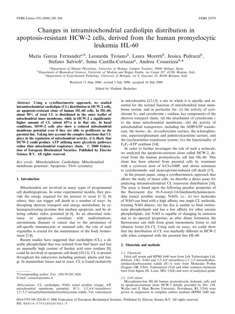
The binding curves obtained from the red £uorescence anal-ysis in both cell types (HL-60 and HCW-2) were biphasic,with two plateaus (arrows in the lower part of Figs. 1 and2, respectively). As described above, the ¢rst saturation levelcorresponds to the amount of CL located in the outer lea£etof mitochondrial internal membrane [17,27]. The second sat-uration level re£ects penetration of NAO into mitochondriaand titration of CL in both halves of inner membrane. Asexpected, increasing the NAO dose resulted either in an in-crease in red £uorescence or a decrease in the green £uores-cence (lower part of Figs. 1 and 2).
The distribution of CL on the cytoplasmic face (outer leaf-let) of the inner membrane in HL-60 was 50.2 þ 2.8% of thetotal mitochondrial CL content (mean of four independentexperiments). Such a value is similar to that reported in otherstudies on eukaryotic or mammalian cells [16,17,24,27,28].The apoptosis-resistant clone HCW-2 shows a signi¢canthigher percentage of CL present in the outer lea£et, i.e.64.8 þ 2.4%. The analysis of four separate experiments showedonly negligible variations, and the statistical analysis revealeda level of signi¢cance 6 0.01.
3.3. HCW-2 mitochondria respond normally to depolarizingstimuli
We then studied the mitochondrial membrane potential inHCW-2 stained with JC-1, and found that, in basal condi-tions, such cells had a slight reduction in orange £uorescence(FL2) and a parallel increase in green £uorescence (FL1), withrespect to the parental cell line HL-60. Consequently, afterlinearization of the median values obtained from the £uores-cence histograms, the FL1/FL2 ratio was higher in HCW-2compared to HL-60 cells (0.83 þ 0.05 vs. 0.67 þ 0.04, P6 0.05).Adding Val resulted in the same marked decrease in FL2 witha concomitant increase in FL1, which reveals a similar v8drop in both cell types (Fig. 3).
4. Discussion
CL appears to be involved, either directly or indirectly, inthe modulation of a variety of mitochondrial processes includ-ing the activation of mitochondrial enzymes, and hence pro-duction of energy by oxidative phosphorylation [29]. CL isspeci¢c and essential for the normal function of mitochondrialinner membrane system, even if its precise role is not com-pletely understood [14]. CL^protein interactions orient mem-brane proteins, matrix proteins, and, on the outer face ofinner mitochondrial membrane, receptors, enzymes andsome leader peptides for import. Moreover, they activate en-zymes or keep them inactive unless the inner membrane isdisrupted and modulate formation of non-bilayer HII-phases[30].
CL is greatly asymmetrical without involving proteins, hasthe only phospholipid headgroup that can collect and donatepumped protons to transmembrane porters at physiologicalpH levels, and mediates an adaptively responsive proton-se-lective leak that regulates non-phosphorylating oxidations[31]. The regulatory properties which govern CL biosynthesis,its remodelling and tra¤cking, are beginning to emerge [29].However, a consensus has not yet been established on the roleand meaning of the submitochondrial localization of CL. Thisquestion is important for understanding its biological func-tions, and to unravel the topographic relationships in themitochondrial structural framework [13]. Interestingly, recentdata indicate that, in yeast, the transmembrane asymmetry ofCL changes during the switch from fermentative to gluconeo-genic metabolism, and in£uences the transmembrane redoxpotential [27]. This could indicate that a di¡erent distributionof CL in the inner membrane could be related to a highernon-oxidative metabolism in apoptosis-resistant HCW-2 cells,compared to HL-60 cells.
We have investigated the distribution of CL in the mito-chondrial inner membrane in cells that were pre-treated withformaldehyde to prevent the incorporation of the dye due tov8 [25,32]. The higher amount of CL we found in the outerlea£et of apoptosis-resistant HCW-2 cells could suggest that,in comparison with the parental line HL-60, the glycolyticpathway gives a more relevant contribution to the establish-ment of the intracellular cellular ATP pool [33]. This is cor-roborated by preliminary data on the respiration of such cells,indicating that their metabolism is skewed towards anaerobicpathways (V. Bobyleva, personal communication).
To ascertain whether a relationship exists between CL to-pography and membrane redox potential [27], we then eval-uated v8 in both cell lines by using the speci¢c probe JC-1.
Fig. 2. Analysis of intramitochondrial CL distribution in HCW-2cells. Panels as in the legend to Fig. 1.
FEBS 23979 28-7-00
M. Garcia Fernandez et al./FEBS Letters 478 (2000) 290^294292
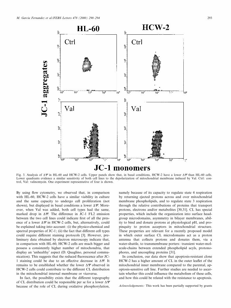
By using £ow cytometry, we observed that, in comparisonwith HL-60, HCW-2 cells have a similar viability in cultureand the same capacity to undergo cell proliferation (notshown), but displayed in basal conditions a lower v8. More-over, when Val was added, both cell types had the same,marked drop in v8. The di¡erence in JC-1 FL2 emissionbetween the two cell lines could indicate ¢rst of all the pres-ence of a lower v8 in HCW-2 cells, but, alternatively, couldbe explained taking into account: (i) the physico-chemical andspectral properties of JC-1; (ii) the fact that di¡erent cell typescould require di¡erent staining protocols [3]. However, pre-liminary data obtained by electron microscopy indicate that,in comparison with HL-60, HCW-2 cells are much bigger andpossess a consistently higher number of mitochondria, thatdisplay an `unhealthy' aspect (D. Quaglino, personal commu-nication). This suggests that the reduced £uorescence after JC-1 staining could be due to an e¡ective decrease in v8. Itremains to be established whether the lower v8 observed inHCW-2 cells could contribute to the di¡erent CL distributionin the mitochondrial internal membrane or viceversa.
In fact, the possibility exists that the di¡erent topographyof CL distribution could be responsible per se for a lower v8because of the role of CL during oxidative phosphorylation,
namely because of its capacity to regulate state 4 respirationby returning ejected protons across and over mitochondrialmembrane phospholipids, and to regulate state 3 respirationthrough the relative contributions of proteins that transportprotons, electrons and/or metabolites [30,31]. CL has specialproperties, which include the organization into surface head-group microdomains, asymmetry in bilayer membranes, abil-ity to bind and donate protons at physiological pH, and pro-pinquity to proton acceptors in mitochondrial structure.These properties are relevant for a recently proposed modelin which outer surface CL microdomains act as a protonantenna that collects protons and donates them, via awater-shuttle, to transmembrane porters: transient water-mol-ecule-chains between extended phospholipid acyls, protono-phores, and uncoupling proteins [31].
In conclusion, our data show that apoptosis-resistant cloneHCW-2 has a higher amount of CL in the outer lea£et of themitochondrial inner membrane compared to the parental, ap-optosis-sensitive cell line. Further studies are needed to ascer-tain whether this could in£uence the metabolism of these cells,and how this could be related with the resistance to apoptosis.
Acknowledgements: This work has been partially supported by grants
Fig. 3. Analysis of v8 in HL-60 and HCW-2 cells. Upper panels show that, in basal conditions, HCW-2 have a lower v8 than HL-60 cells.Lower quadrants evidence a similar sensitivity of both cell lines to the depolarization of mitochondrial membrane induced by Val. Ctrl : con-trol, Val: valinomycin. One experiment representative of four is shown.
FEBS 23979 28-7-00
M. Garcia Fernandez et al./FEBS Letters 478 (2000) 290^294 293
from Lilly to MGF; from MURST and Istituto Superiore di Sanita© ^Programma Nazionale di Ricerca sull'AIDS, 1998 (Progetto Patolo-gia, Clinica e Terapia dell'AIDS, ISS Grant 30B.23) to A.C. ProfessorValentina Bobyleva (University of Modena and Reggio Emilia) isacknowledged for useful comments and suggestions.
References
[1] Cossarizza, A., Kalashnikova, G., Grassilli, E., Chiappelli, F.,Salvioli, S., Capri, M., Barbieri, D., Troiano, L., Monti, D.and Franceschi, C. (1994) Exp. Cell Res. 214, 323^330.
[2] Cossarizza, A., Mussini, C., Mongiardo, N., Borghi, V., Sabba-tini, A., De Rienzo, B. and Franceschi, C. (1997) AIDS 11, 19^26.
[3] Salvioli, S., Dobrucki, J., Moretti, L., Troiano, L., Garcia Fer-nandez, M., Pinti, M., Pedrazzi, J., Franceschi, C. and Cossar-izza, A. (2000) Cytometry (in press).
[4] Kroemer, G., Zamzani, N. and Susin, S.A. (1997) Immunol. To-day 18, 44^51.
[5] Kroemer, G., Dallaporta, B. and Resche-Rigon, M. (1998) Ann.Rev. Physiol. 60, 619^642.
[6] Kiberstis, P.A. (1999) Science 283, 1475.[7] Skulachev, V.P. (1996) FEBS Lett. 397, 7^10.[8] Skulachev, V.P. (1999) Mol. Asp. Med. 20, 139^184.[9] Hatch, G.M. (1996) Mol. Cell. Biochem. 159, 139^148.
[10] Lieser, M.J., Park, J., Natori, S., Jones, B.A., Bronk, S.F. andGores, G.J. (1998) Gastroenterology 115, 693^701.
[11] Ushmorov, A., Ratter, F., Lehmann, V., Droge, W., Schirrmach-er, V. and Umansky, V. (1999) Blood 93, 2342^2352.
[12] Ioannou, P.V. and Golding, B.T. (1979) Prog. Lipid Res. 17,279^318.
[13] Schlame, M. and Hostetler, K.Y. (1997) Biochim. Biophys. Acta1348, 207^213.
[14] Gomez, B.J. and Robinson, N.C. (1999) Biochemistry 38, 9031^9038.
[15] Han, Z., Chatterjee, D., Early, J., Pantazis, P., Hendrickson,E.A. and Wyche, J.H. (1996) Cancer Res. 56, 1621^1628.
[16] Petit, J.M., Maftah, A., Ratinaud, M.H. and Julien, R. (1992)Eur. J. Biochem. 209, 267^273.
[17] Petit, J.M., Huet, O., Gallet, P.F., Maftah, A., Ratinaud, M.H.and Julien, R. (1994) Eur. J. Biochem. 220, 871^879.
[18] Cossarizza, A., Baccarani Contri, M., Kalashnikova, G. andFranceschi, C. (1993) Biochem. Biophys. Res. Commun. 197,40^45.
[19] Reers, M., Smith, T.W. and Chen, L.B. (1991) Biochemistry 30,4480^4486.
[20] Smiley, S.T., Reers, M., Mottola-Hartshorn, C., Lin, M., Chen,A., Smith, T.W., Steele, G.D. and Chen, L.B. (1991) Proc. Natl.Acad. Sci. USA 88, 3671^3675.
[21] Cossarizza, A., Cooper, E.L., Quaglino, D., Salvioli, S., Kalach-nikova, G. and Franceschi, C. (1995) Biochem. Biophys. Res.Commun. 214, 503^510.
[22] Bobyleva, V., Pazienza, T.L., Maseroli, R., Tomasi, A., Salvioli,S., Cossarizza, A., Franceschi, C. and Skulachev, V.P. (1998)FEBS Lett. 430, 409^413.
[23] Cossarizza, A., Ceccarelli, D. and Masini, A. (1996) Exp. CellRes. 222, 84^94.
[24] Gallet, P.F., Maftah, A., Petit, J.M., Denis Gray, M. and Julien,R. (1995) Eur. J. Biochem. 228, 113^119.
[25] Keij, J.F., Bell-Prince, C. and Steinkamp, J.A. (2000) Cytometry39, 203^210.
[26] Maftah, A., Petit, J.M. and Julien, R. (1990) FEBS Lett. 260,236^240.
[27] Gallet, P.F., Petit, J.M., Maftah, A., Zachowski, A. and Julien,R. (1997) Biochem. J. 324, 627^634.
[28] Gallet, P.F., Zachowski, A., Julien, R., Fellmann, P., Devaux,P.F. and Maftah, A. (1999) Biochim. Biophys. Acta 1418, 61^70.
[29] Hatch, G.M. (1998) Int. J. Mol. Med. 1, 33^41.[30] Hoch, F.L. (1992) Biochim. Biophys. Acta 1113, 71^133.[31] Hoch, F.L. (1998) J. Bioenerg. Biomembr. 30, 511^532.[32] Maftah, A., Petit, J.M., Ratinaud, M.H. and Julien, R. (1989)
Biochem. Biophys. Res. Commun. 164, 185^190.[33] Sweet, S. and Singh, G. (1999) J. Cell Physiol. 180, 91^96.
FEBS 23979 28-7-00
M. Garcia Fernandez et al./FEBS Letters 478 (2000) 290^294294


![Structural characterization of cardiolipin by tandem ......kingdom [4]. Cardiolipin is essential for the function of several enzymes of oxidative phosphorylation, and thus, for production](https://img.pdfslide.net/doc/110x75/5e9bda7953105a41956b711d/structural-characterization-of-cardiolipin-by-tandem-kingdom-4-cardiolipin.jpg)