Embed Size (px)
Citation preview
Changes in ocular aquaporin expression following optic nervecrush
Adnan Dibas,1 Hidehiro Oku,2 Masayuki Fukuhara,3 Takuji Kurimoto,3 Tsunehiko Ikeda,2
Rajkumar V. Patil,1,4 Najam A. Sharif,1,4 Thomas Yorio1
1Department of Pharmacology & Neuroscience, University of North Texas Health Science Center at Fort Worth, Fort Worth, TX;2Department of Ophthalmology, Osaka Medical College, Osaka, Japan; 3Department of Ophthalmology, Hyogo College ofMedicine, Hyogo, Japan; 4Pharmaceutical Research, Alcon Research Ltd, Fort Worth, TX
Purpose: Changes in the expression of water channels (aquaporins; AQP) have been reported in several diseases. However,such changes and mechanisms remain to be evaluated for retinal injury after optic nerve crush (ONC). This study wasdesigned to analyze changes in the expression of AQP4 (water selective channel) and AQP9 (water and lactate channel)following ONC in the rat.Methods: Rat retinal ganglion cells (RGCs) were retrogradely labeled by applying FluoroGold onto the left superiorcolliculus 1 week before ONC. Retinal injuries were induced by ONC unilaterally. Real-time PCR was used to measurechanges in AQP4, AQP9, thy-1, Kir4.1 (K+ channel), and β-actin messages. Changes in AQP4, AQP9, Kir4.1, B celllymphoma-x (bcl-xl), and glial fibrillary acidic protein (GFAP) expression were measured in total retinal extracts usingwestern blotting.Results: The number of RGCs labeled retrogradely from the superior colliculus was 2,090±85 cells/mm2 in rats withoutany treatment, which decreased to 1,091±78 (47% loss) and 497±87 cells/mm2 (76% loss) on days 7 and 14, respectively.AQP4, Kir4.1, and thy-1 protein levels decreased at days 2, 7, and 14, which paralleled a similar reduction in mRNAlevels, with the exception of Kir4.1 mRNA at day 2 showing an apparent upregulation. In contrast, AQP9 mRNA andprotein levels showed opposite changes to those observed for the latter targets. Whereas AQP9 mRNA increased at days2 and 14, protein levels decreased at both time points. AQP9 mRNA decreased at day 7, while protein levels increased.GFAP (a marker of astrogliosis) remained upregulated at days 2, 7, and 14, while bcl-xl (anti-apoptotic) decreased.Conclusions: The reduced expression of AQP4 and Kir4.1 suggests dysfunctional ion coupling in retina following ONCand likely impaired retinal function. The sustained increase in GFAP indicates astrogliosis, while the decreased bcl-xlprotein level suggests a commitment to cellular death, as clearly shown by the reduction in the RGC population anddecreased thy-1 expression. Changes in AQP9 expression suggest a contribution of the channel to retinal ganglion celldeath and response of distinct amacrine cells known to express AQP9 following traumatic injuries.
The glaucomas represent a heterogeneous group ofdiseases that result in a progressive optic neuropathycharacterized by functional and structural impairment ofocular tissues. Particularly affected are the trabecularmeshwork, the optic nerve head, and the retinal ganglion cells.
One of the risk factors in primary open angle glaucoma(POAG) is an associated elevation in intraocular pressure(IOP) [1]. Elevation of IOP results in the blockade of axonaltransport in animals as well as displacement of the optic nervehead [2-7]. Abnormalities in water balance play an importantrole in the pathophysiology of a variety of neurologicdisorders. Neuronal activity is associated with a redistributionof water—shrinkage of the extracellular space around activesynapses while enhancement of the extracellular spacevolume at more distant sites [8]. The discovery of aquaporins
Correspondence to: Dr. Adnan Dibas, Department of Pharmacology& Neuroscience, University of North Texas Health Science Centerat Fort Worth, 3500 camp Bowie Blvd., Fort Worth, TX, 76107;Phone: (817) 735-5036; FAX: (817) 735-0243; email:[email protected]
Molecular Vision 2010; 16:330-340 <http://www.molvis.org/molvis/v16/a39>Received 2 November 2009 | Accepted 26 February 2010 | Published 3 March 2010
© 2010 Molecular Vision
330
(AQPs) has provided a molecular basis for understandingwater transport in several tissues, including the ocular system[9]. Using Reverse-transcription-Polymerase Chain Reaction(RT–PCR), Tenckhoff et al. showed that human retinaexpresses mRNAs for AQP0 to AQP12 , whereas rat retina has the mRNAs for AQP0, AQP1, AQP3, AQP4, AQP5, AQP6, AQP7, AQP8, AQP9, and AQP11. The mRNAs for AQP2, AQP10, and AQP12 were not detected in the rat neural retina [10]. The same studyreported that the human Müller cell line
MIO-M1 [11] expressed mRNA for AQP0, AQP1, AQP3, AQP4, AQP5, AQP7, AQP8, AQP9, AQP10, and
AQP11 but not AQP2, AQP6, and AQP12 [10]. AQP0 is expressed in lens fiber [12] and rodent bipolar cells [13,14] and has been reported in distinct amacrines and ganglioncells of the rat retina [14]. While AQP1 is expressed by corneaendothelium, ciliary and lens epithelia, trabecular meshwork[15], pigment epithelial cells [16], rodent amacrine cells [17,18], and photoreceptor cells [19], AQP3 is detected inconjunctiva [20]. AQP4 is expressed by ciliary epithelium and

Müller cells and astrocytes [21-23], and AQP5 has beenreported in corneal and lacrimal gland epithelia [24]. AQP9has been detected in tyrosine hydroxylase-expressingamacrine cells in the rat retina [25], rat retinal ganglion cells(RGCs) [26], and human and rat pigment epithelial retinalcells [27].
Hypoxia and ischemia are associated with changes in thedensities of AQP expression and are also considered key riskfactors in the development of glaucomatous optic nerveneuropathy [1]. Interestingly, other insults, such as hypo-osmotic stress, known to affect AQP levels have also beenreported to cause glaucoma [28-30]. However, changes in theexpression of ocular AQPs upon retinal injuries have not beenfully characterized. In the current study we analyzed changesin AQP4 and AQP9 following optic nerve crush (ONC). Thecrush model closely mimics the damage that occurs intraumatic optic neuropathy, which results from indirecttrauma to the optic nerve. Such injuries appear to involve bothmechanical (primary) and ischemic-induced (secondary)processes that include degeneration of nerve axons and lossof myelin. Many studies have observed loss of retinal ganglioncells following crush that is comparable to glaucoma, with thecrush model regarded as an acute model of glaucoma[31-37].
METHODSMaterials: Monoclonal anti-thy-1 and rabbit anti-kir4.1antibodies were from Chemicon (Temecula, CA); AQP4(mouse and rabbit), AQP9 (goat and rabbit), B cell lymphoma-x (bcl-xl; mouse), and thy-1 (rabbit) antibodies were fromSanta Cruz Inc. (Santa Cruz, CA); monoclonal anti-glialfibrillary acidic protein antibodies were from Neomarkers(Fremont, CA); monoclonal anti-tubulin antibodies were fromUpstate Inc. (Lake Placid, NY); and secondary antibodies(donkey anti-mouse-conjugated Alexa 633, goat anti-rabbit-conjugated Alexa 633, and goat anti-mouse-conjugated Alexa488) were from Invitrogen Inc. (Carlsbad, CA).Animals: Nine-week-old male Wistar rats were purchasedfrom Japan SLC (Shizuoka, Japan). The rats were housed inan air-conditioned room with a temperature of approximately23 °C and 60% humidity and on a 12 h:12 h light–dark cycle.All animals were handled in accordance with the ARVOresolution for the Use of Animals in Ophthalmic and VisionResearch. The experimental protocol was approved by theCommittee of Animal Use and Care of the Osaka MedicalCollege.Optic nerve crush: Eight animals were anesthetized with 7%chloral hydrate solution (400 mg/kg). After making a skinincision along the superior orbital margin, the superior surfaceof the right eye was exposed. Removing the superior rectusmuscle exposed the optic nerve, which was crushed withforceps 2 mm behind the eye for 10 s, as described [38]. Carewas taken not to cause retinal ischemia, and we confirmed by
indirect ophthalmoscopy that the retinal circulation was notblocked. This procedure induced nearly complete damage tothe optic nerve, and none of the RGCs could be retrogradelylabeled from the superior colliculus [39]. A sham operationwas performed on the right eyes of other animals, and the opticnerve was exposed in the same way but not crushed as in theexperimental animals. The left eyes were not used as controlsbecause crushing one optic nerve affects the morphology ofthe contralateral retina [40].Rat retinal ganglion cells labeling and counting: Todetermine how the RGCs were lost following ONC, RGCswere retrogradely labeled by applying FluoroGold(hydroxystilbamidine; Biotium, Hayward, CA) onto the leftsuperior colliculus. Because no RGCs were labeled when thetracer was applied after the optic nerve was crushed, the tracerwas applied 1 week before crushing the right optic nerve. Afteranesthesia with 7% chloral hydrate, a skin incision was made,the skull was exposed, and the bone over the left hemispherewas drilled along the sagittal, coronal, and lambdoidal suturesand removed. After the dura was peeled off, the gray and whitematter of the left hemisphere was carefully removed byaspiration through the window, and the surface and brachiumof the superior colliculus were exposed. Then, a small spongesoaked with 2% FluoroGold solution in saline containing 10%DMSO (DMSO) was placed directly onto the superiorcolliculus, as described [41]. Seven days after FluoroGoldapplication, the right optic nerve was crushed. The operatedrats were perfused transcardially with 4% paraformaldehydein phosphate-buffered saline (PBS; 150 mM NaCl, 3.8 mMNaH2PO4, 16.2 mM Na2HPO4) on day 7 (n=4) and 14 (n=4)after the optic nerve crush, and the eyes were enucleated. Theretinas were dissected, flatmounted, and examined under afluorescence microscope (E800; Nikon, Tokyo, Japan) with aUV filter (365 nm).We counted the number of FluoroGold-labeled RGCs that had round-shaped somata and had fineregular dot-like fluorescent particles of FluoroGold in thecytoplasm. The number of RGCs was counted in 12 areas(0.48×0.48 mm2) at a distance of 1, 2, and 3 mm from the opticdisc along the nasotemporal and dorsoventral midlines (upper,lower, nasal, and temporal direction). The mean density ofRGC/mm2 was calculated, and the loss of RGCs was evaluatedby comparing the density in the retinas with ONC to that inuntreated retinas (n=4).Real-time analyses of AQP4/AQP9/Kir4.1/thy-1/ACTBmRNA levels: On days 2, 7, and 14, total RNA was isolatedfrom the retina after the ONC or sham operation with Trizol(Life Technology, Grand Island, NY) as described bymanufacturer’s instructions. Retina was removed and lyseddirectly by adding 1 ml Trizol and pipetting up and downseveral times. Two hundred μl of chloroform were addedfollowed by inverting tubes for several times then sampleswere put on ice for 5 min. Tubes were then centrifuged for 10min at 16,000× g and the upper 600 μl supernatant wasremoved to new tubes. Six hundred microliters of isopropanol
Molecular Vision 2010; 16:330-340 <http://www.molvis.org/molvis/v16/a39> © 2010 Molecular Vision
331
were added followed by inversion of samples several timesthen samples were put on ice for 10 min. RNA was recoveredby repeating centrifugation for 10 min at the same speed.Supernatant was discarded and RNA pellet was washed with1 ml of 70% ethanol and centrifugation was repeated (10 minat 16,000× g). The ethanol solution was removed and RNApellet was allowed to dry for 10 min at room temperature. Ineach tube, 50 μl TE buffer was added (10 mM Tris-HCl, pH8, 1 mM EDTA) following by pipetting up and down severaltimes to resuspend RNA. RNA concentration was determinedby adding 6 μl from each sample in 600 μl water andmeasuring the absorbance at 260 nm (when absorbance is 1,the RNA concentration is 40 ng/ml). In this study, 2 µg of totalRNA was reverse transcribed using the One-step reversetranscriptase (RT)–PCR kit (Qiagen, Valencia, CA),according to the manufacturers instructions. Controlquantative (Q)-PCR reactions were performed in the absenceof cDNA templates. β-actin (ACTB) was used as ahousekeeping gene. The primers for retinal AQP4 were 5′-CGG TTC ATG GAA ACC TCA CT-3′ (sense) and 5′-CATGCT GGC TCC GGT ATA AT-3′ (antisense), giving a 191-bp amplicon. The primers for retinal Kir4.1 were 5′-GCAAGA TCT CCC CCT CCG CAG-3′ (sense) and 5′-CAG ACGTTA CTA ATG CGC ACA CT-3′ (antisense), giving a 345-bp amplicon. The primers for ACTB were 5′-TGT GAT GGTGGG AAT GGG TCA G-3′ (sense) and 5′-TTT GAT GTCACG CAC GAT TTC C-3′ (antisense), giving a 514-bpamplicon. The primers for retinal AQP9 were 5′-CTC AGTCCC AGG CTC TTC AC-3′ (sense) and 5′-ATG GCT CTGCCT TCA TGT CT-3′ (antisense), giving a 184-bp amplicon.The primers for retinal thy-1 were 5′-CGC TTT ATC AAGGTC CTT ACT C-3′ (sense) and 5′-GCG TTT TGA GATATT TGA AGG T-3′ (antisense), giving a 344-bp amplicon.A 2.5-µl sample of cDNA from each treatment was used forRT–PCR amplification of each primer in a Cepheid smartcycler (Cepheid, Sunnyvale, CA). For retinal AQP4 andKir4.1, it was 45 cycles of denaturation at 95 °C for 30 s,annealing at 60 °C for 30 s, and extension at 72 °C for 1 min.For retinal AQP9, it was 45 cycles of denaturation at 95 °Cfor 30 s, annealing at 60 °C for 30 s, and extension at 72 °Cfor 1 min. For retinal thy-1, it was 50 cycles of denaturationat 95 °C for 15 s, annealing at 55 °C for 10 s, and extensionat 72 °C for 20 s. For ACTB, it was 40 cycles of denaturationat 95 °C for 1 min, annealing at 60 °C for 1 min, and extensionat 72 °C for 2 min. The melting curves were generated to detectthe melting temperatures of the specific products immediatelyafter the PCR run. The relative mRNA levels were determinedby the comparative cycle number at threshold (CT) method,as described in PE Biosystems User Bulletin #2. The foldchange was determined by the formula: fold change=2–Δ(ΔCt), where ΔCt=Ct,target-Ct,β-actin, where “target” is thegene of interest. To verify the sequence of products, regularPCR was performed, and the PCR products were run on a1.5% agarose gel in parallel with 100-bp DNA markers and
stained with ethidium bromide. Bands were cut and sequencedto verify identity. The authenticity of PCR products wasconfirmed using a BLAST search of the sequence through theNCBI.Western blotting: Retinas from eight rats that underwent ONCand eight rats that underwent a sham operation were dissected.Western blotting was performed on either total retinal lysatesor enriched plasma membrane and cytosolic fractions, asdescribed by Dibas et al. 2008 [42]. To prepare total retinallysate, retinas were dissected from eyes and solubilized in250 µl of a solution containing 20 mM Tris (pH 7.4), 10%sucrose, 2 mM Ethylenediamine-tetraacetic acid (EDTA),2 mM Ethylene glycol-bis(2-aminoethylether)-N,N,N′,N′-tetraacetic acid (EGTA), 50 mM NaF, 1% Triton X-100, 0.1%sodium dodecyl sulfate, and protease inhibitors (aprotonin(10 μg/ml), soybean trypsin inhibitor (10 μg/ml), leupeptin(10 μg/ml), and phenylmethanesulfonyl fluoride (100 μM)) at4 °C. Retinal lysates were incubated for 30 min on ice, brieflysonicated before centrifugation at 14,000× g for 15 min, andthe supernatant collected. Protein concentration wasmeasured using a bicinchoninic acid (BCA) protein assay kit(Sigma, St. Louis, MO) with bovine serum albumin (BSA) asthe standard. To enrich plasma membrane fractions, retinaswere dissected from eyes and homogenized into 700 µl of asolution containing 20 mM Tris (pH 7.4), 10% sucrose, 2 mMEDTA, 2 mM EGTA, 50 mM NaF, and protease inhibitors at4 °C, using a Potter homogenizer (Fisher Scientific, HoustonTX, 60 strokes) followed by centrifugation at 3,000× g for 5min. The unbroken tissue was sonicated 7X, andcentrifugation was repeated. The retinal supernatant was thencentrifuged for 30 min at 100,000× g at 4 °C. The resultantsupernatant (enriched cytosol) and pellet (enriched plasmamembrane) were collected for protein measurement using theBCA protein assay. Proteins were separated by 10% sodiumdodecyl sulfate- PAGE (PAGE), with 20–30 µg (enrichedfraction) or 100 µg (total retinal extract) of protein loaded ineach lane. Gels were equilibrated in transfer buffer (192 mMglycine, 20% methanol, and 25 mM Tris-HCl, pH 8.3) for 10min at room temperature and electroblotted on nitrocellulosemembranes for 75 min at 100 V using a Bio-Rad (Hercules,CA) electroblotting unit. The membranes were dried at roomtemperature. Western blotting was performed using achemiluminescent kit (Amersham Biosciences, Bedford,MA). Membranes were incubated with primary antibodies for60 min (1 µg/ml) and with the horseradish peroxidase-conjugated secondary antibody for 30 min (1:10,000).Membranes were exposed to an X-ray film for 30 s, then 1min then 5 min. X-ray film was removed at the end of eachtime point and immediately developed. Membranes werestripped and probed with anti-β-tubulin antibodies fornormalization. Experiments were repeated three times. Banddensities were quantified with image analysis software (Scion,Frederick, MD), and the intensity of AQP4/AQP9/thy-1/ glialfibrillary acidic protein (GFAP)/bcl-xl/kir4.1 bands was
Molecular Vision 2010; 16:330-340 <http://www.molvis.org/molvis/v16/a39> © 2010 Molecular Vision
332

normalized for every sample relative to the intensity of therespective tubulin bands.Statistical analyses: Data are reported as means±standarderror of the mean. All experiments were repeated at least threetimes with to three to four replicates per condition each time.Statistical significance was determined by using one-wayANOVA and Tukey multiple comparison tests at p<0.05.
RESULTSReduction of rat retinal ganglion cells after optic nerve crushand correlation with thy-1 mRNA and protein levels: ONC
caused a massive reduction in the RGC population with time.While the mean number of sham RGCs was 2,090±85 cells/mm2 in rats (n=4, Figure 1A), the number decreased to1,091±78 (n=4, 47% loss, Figure 1B) and 497±87 cells/mm2
(n=4, 76% loss versus sham, p<0.0001, Figure 1C) on days 7and 14, respectively. Thy-1, a known RGC stress markercommonly used to measure RGC populations, was quantified.There was a reduction in thy-1 protein (94±18%, 33±6%, and24±7% at days 2, 7, and 14, respectively, Figure 1D, p<0.005,n=7), although changes in thy-1 protein levels at day 2 were
Figure 1. Optic nerve crush decreasednumber of retinal ganglion cells(RGCs), thy-1 protein, and mRNAlevels. The mean number of RGCslabeled retrogradely from superiorcolliculus was 2090±85/mm2 in shamrats without any treatment (A), whichdecreased to 1091±78 (47% loss, B) and497±87/mm2 (76% loss, C) on day 7 and14, respectively. Thy-1 expressed byretinal ganglion cells is widely used asmarker for RGC stress and was shownto be reduced following optic nervecrush. Following retinal injuries withoptic nerve crush, retinas were dissectedand plasma membrane proteins wereisolated or total RNA was isolated andtranscribed into cDNA. Thirtymicrogram protein was loaded into eachlane. D: Immunoreactive bands forthy-1 and β-tubulin at 2, 7, and 14 daysfollowing crush showing a significantreduction in thy-1 protein levels at 7 and14 day but not at 2 days (94±18%,33±6%, 24±7% at 2, 7, and 14 day,respectively, n=7). Densitometricquantification is shown in E. Data areexpressed as a ratio of the control valueand each measurement represents mean±SEM *Denotes statistical significanceof thy-1 protein levels in optic nervecrushed retinas versus sham (p<0.005)as determined by one-way ANOVA andTukey multiple comparison test. Opticnerve crush also decreased thy-1transcripts significantly (58±7%, 6±2%,at 2 and 7 day, respectively, % p<0.001versus sham, n=8), as determined byquantitative real-time PCR (E). Thy1-mRNA at 2-weeks was below detectionlevels and considered zero. Geneexpression data of thy-1 is calculatedafter normalizing with β-actin.**Denote significant differencescompared with sham-retinas at p<0.05.Abbreviations: sham eye (C), andcrushed (R).
Molecular Vision 2010; 16:330-340 <http://www.molvis.org/molvis/v16/a39> © 2010 Molecular Vision
333

not statistically significant. In addition, ONC decreased thy-1mRNA significantly (58±7% and 6±2%, days 2 and 7,respectively, p<0.001 versus sham, Figure 1E; data expressedas a ratio of the control value, n=8). Thy-1 mRNA at 2 weekswas below detection levels. Figure 1E shows parallel changesin thy-1 mRNA to protein levels. When changes in thy-1expression were compared to the time course of RGC cell loss,it is clear that a significant loss of thy-1 mRNA and proteinoccurs well in advance of RGC cell loss. For example, whilethe RGC population decreased by approximately 47% at day7, thy-1 mRNA decreased by 94%, showing that the reductionof thy-1 mRNA is much greater than the loss of RGCs.Effect of optic nerve crush on AQP4 in rat retinas:Hypertrophy of the optic nerve has been reported in glaucomasubjects, and patients with normal tension glaucoma [43] orPOAG [44] have a larger optic disc compared to normalpatients. Therefore, the effect of ONC on AQP4 expression inretina was tested. As shown in Figure 2A, ONC resulted in adecrease in AQP4 protein (60±10, 63±10, and 38±10, p<0.001at days 2, 7, and 14, respectively, Figure 2A,B, n=7), and there
as a similar reduction in AQP4 mRNA (~40%), as determinedby Q-PCR (61±5%, 60±8%, and 58±6 at days 2, 7, and 14,respectively, p<0.001 versus control, Figure 2B, n=7). Figure2B shows parallel changes in AQP4 mRNA to protein levels.Data are expressed as a ratio of the control value, and eachmeasurement represents mean±SEM.Effect of optic nerve crush on Kir4.1 in rat retinas: In Müllercells, AQP4 co-localizes with an important retinal potassiumchannel known as Kir4.1 (mediates bidirectional K+ currents),and the co-expression of AQP4 with Kir4.1 suggests thatwater transport is coupled to the spatial buffering K+ currents[45,46]. The co-expression also suggests that osmoticgradients between the retina and the blood and vitreous canbe compensated by K+ and water influxes and effluxes fromMüller cells. Therefore, the effect of optic nerve crush onKir4.1 expression in retina was tested. As shown in Figure 3A,ONC caused a significant reduction in Kir4.1 protein levels atall time points (34±10, 72±6, and 22±9, p=0.001 at days 2, 7,and 14, respectively, Figure 3A,B, n=7). However, whileONC resulted in an increase in Kir4.1 mRNA at day 2
Figure 2. Optic nerve crush decreasedAQP4 protein and mRNA levels.Following retinal injuries with opticnerve crush, retinas were dissected andplasma membrane proteins wereisolated or total RNA was isolated andtranscribed into cDNA. Thirtymicrogram protein was loaded into eachlane. Immunoreactive bands for AQP4and β-tubulin 2, 7, and 14 days afteroptic nerve crush showing a significantreduction in AQP4 protein levels.Quantitative measurement usingwestern blot showed that optic nervecrush decreased AQP4 protein levels by~40% (60±10, 63±10, 38±10, at 2, 7,and 14 day, respectively, n=7, A).Densitometric quantification is shownin B. Data are expressed as a ratio of thetreated to control or sham value and eachmeasurement represents mean±SEM*Denote statistical significance ofAQP4 protein levels in optic nervecrushed retinas versus sham (p<0.005)as determined by one-way ANOVA andTukey multiple comparison test. Opticnerve crush also decreased AQP4transcripts significantly (61±5%,60±8%, 58±6, at 2, 7, and 14 day,respectively, n=7), as determined byquantitative real-time PCR (B). Geneexpression data of AQP4 is calculatedafter normalizing with ACTB. **Denotesignificant differences compared withsham-retinas at p<0.05. Abbreviations:sham eye (C), and crushed (R).
Molecular Vision 2010; 16:330-340 <http://www.molvis.org/molvis/v16/a39> © 2010 Molecular Vision
334

(~300%), as determined by Q-PCR (325±81%, p<0.001versus control, Figure 3B, n=6), it caused a significantdecrease at days 7 and 14 (55±7% and 41±9%, respectively,p<0.001 versus control, Figure 3B, n=6). Changes in Kir4.1mRNA and protein levels are shown in Figure 3B.
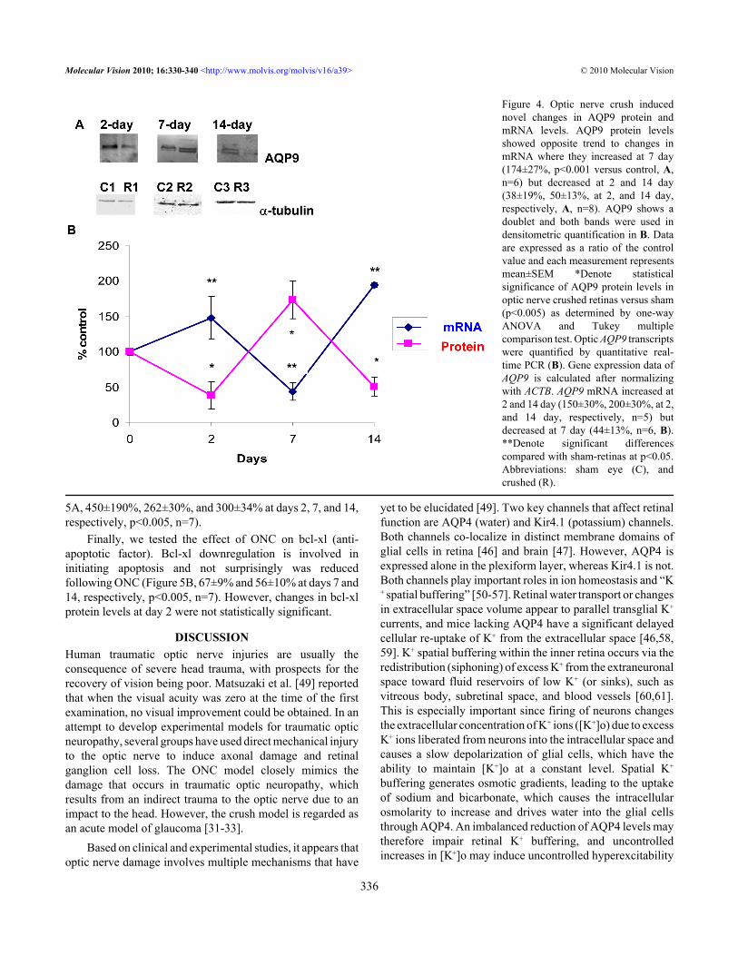
Effect of optic nerve crush on AQP9 in rat retinas: Severalstudies have shown that ONC induced hypoxia to the opticnerve [31-33]. A key channel that undergoes changes isAQP9, and its expression also appears to be upregulated afterischemic insult in the brain [47] and following IOP elevation(Dibas et al., unpublished observation). Therefore, the effectof ONC on AQP9 expression in retina was evaluated.Surprisingly, AQP9 protein levels showed an opposite trendto changes in message (mRNA) where they increased at day7 (174±27%, p<0.001 versus sham, Figure 4A,B, n=6) but
decreased at days 2 and 14 (38±19% and 50±13%,respectively, p<0.001 versus sham, Figure 4A,B, n=8). ONCresulted in novel changes in AQP9; its mRNA increased atdays 2 and 14 (150±30% and 200±30%, respectively, p<0.001versus control, Figure 4B, n=5) but decreased at day 7(44±13%, p<0.001 versus sham, Figure 4B, n=6). Therelationship between AQP9 mRNA and protein changes isshown in Figure 4B.Effect of optic nerve crush on glial fibrillary acidic proteinand bcl-xl in rat retinas: A universal early cellular marker forretinal injury is the upregulation of the intermediate filamentprotein GFAP, and its levels are commonly used as an indexof gliosis [48]. As expected, ONC also resulted in an increasein GFAP protein levels that persisted at all time points (Figure
Figure 3. Optic nerve crush decreasedKir4.1 protein and mRNA levels.Following retinal injuries with opticnerve crush, retinas were dissected andplasma membrane proteins wereisolated. Total RNA was isolated andtranscribed into cDNA. Real-time PCRwas performed using specific primers(see Methods). mRNA expression ofKir4.1 was adjusted to the mRNAcopies of ACTB (reference gene). Thirtymicrogram protein was loaded into eachlane. Immunoreactive bands for Kir4.1and β-tubulin 2, 7, and 14 days afteroptic nerve crush showing a significantreduction in Kir4.1 protein levels.Quantitative measurement usingwestern blot showed that elevation ofoptic nerve crush decreased Kir4.1protein levels (34±10, 72±6, 22±9, at 2,7, and 14 day, respectively, A, n=7).Densitometric quantification is shownin B. Data are expressed as a ratio of thecontrol value and each measurementrepresents mean±SEM *Denotestatistical significance of Kir4.1 proteinlevels in optic nerve crushed retinasversus sham (p<0.005) as determined byone-way ANOVA and Tukey multiplecomparison test. Results indicate thatmRNA expression level of Kir4.1 wassignificantly lower in optic nervecrushed retinas compared to sham at 7and 14 days (55±7%, 41±9%, at 7, and14 day, respectively, B, n=6). Bycontrast, optic nerve crush increasedKir4.1 mRNA at 2 days (325±81%, B,n=6). **Denote significant differencescompared with sham-retinas at p<0.05.Abbreviations: sham eye (C), andcrushed (R).
Molecular Vision 2010; 16:330-340 <http://www.molvis.org/molvis/v16/a39> © 2010 Molecular Vision
335
5A, 450±190%, 262±30%, and 300±34% at days 2, 7, and 14,respectively, p<0.005, n=7).
Finally, we tested the effect of ONC on bcl-xl (anti-apoptotic factor). Bcl-xl downregulation is involved ininitiating apoptosis and not surprisingly was reducedfollowing ONC (Figure 5B, 67±9% and 56±10% at days 7 and14, respectively, p<0.005, n=7). However, changes in bcl-xlprotein levels at day 2 were not statistically significant.
DISCUSSIONHuman traumatic optic nerve injuries are usually theconsequence of severe head trauma, with prospects for therecovery of vision being poor. Matsuzaki et al. [49] reportedthat when the visual acuity was zero at the time of the firstexamination, no visual improvement could be obtained. In anattempt to develop experimental models for traumatic opticneuropathy, several groups have used direct mechanical injuryto the optic nerve to induce axonal damage and retinalganglion cell loss. The ONC model closely mimics thedamage that occurs in traumatic optic neuropathy, whichresults from an indirect trauma to the optic nerve due to animpact to the head. However, the crush model is regarded asan acute model of glaucoma [31-33].
Based on clinical and experimental studies, it appears thatoptic nerve damage involves multiple mechanisms that have
yet to be elucidated [49]. Two key channels that affect retinalfunction are AQP4 (water) and Kir4.1 (potassium) channels.Both channels co-localize in distinct membrane domains ofglial cells in retina [46] and brain [47]. However, AQP4 isexpressed alone in the plexiform layer, whereas Kir4.1 is not.Both channels play important roles in ion homeostasis and “K+ spatial buffering” [50-57]. Retinal water transport or changesin extracellular space volume appear to parallel transglial K+
currents, and mice lacking AQP4 have a significant delayedcellular re-uptake of K+ from the extracellular space [46,58,59]. K+ spatial buffering within the inner retina occurs via theredistribution (siphoning) of excess K+ from the extraneuronalspace toward fluid reservoirs of low K+ (or sinks), such asvitreous body, subretinal space, and blood vessels [60,61].This is especially important since firing of neurons changesthe extracellular concentration of K+ ions ([K+]o) due to excessK+ ions liberated from neurons into the intracellular space andcauses a slow depolarization of glial cells, which have theability to maintain [K+]o at a constant level. Spatial K+
buffering generates osmotic gradients, leading to the uptakeof sodium and bicarbonate, which causes the intracellularosmolarity to increase and drives water into the glial cellsthrough AQP4. An imbalanced reduction of AQP4 levels maytherefore impair retinal K+ buffering, and uncontrolledincreases in [K+]o may induce uncontrolled hyperexcitability
Figure 4. Optic nerve crush inducednovel changes in AQP9 protein andmRNA levels. AQP9 protein levelsshowed opposite trend to changes inmRNA where they increased at 7 day(174±27%, p<0.001 versus control, A,n=6) but decreased at 2 and 14 day(38±19%, 50±13%, at 2, and 14 day,respectively, A, n=8). AQP9 shows adoublet and both bands were used indensitometric quantification in B. Dataare expressed as a ratio of the controlvalue and each measurement representsmean±SEM *Denote statisticalsignificance of AQP9 protein levels inoptic nerve crushed retinas versus sham(p<0.005) as determined by one-wayANOVA and Tukey multiplecomparison test. Optic AQP9 transcriptswere quantified by quantitative real-time PCR (B). Gene expression data ofAQP9 is calculated after normalizingwith ACTB. AQP9 mRNA increased at2 and 14 day (150±30%, 200±30%, at 2,and 14 day, respectively, n=5) butdecreased at 7 day (44±13%, n=6, B).**Denote significant differencescompared with sham-retinas at p<0.05.Abbreviations: sham eye (C), andcrushed (R).
Molecular Vision 2010; 16:330-340 <http://www.molvis.org/molvis/v16/a39> © 2010 Molecular Vision
336

and abnormal synchronization of retinal neurons. In thepresent study using the rat ONC model, we have shown forthe first time that such injury causes a significant decrease inAQP4 and Kir4.1 protein and mRNA levels in retina,suggesting impaired ion homeostasis and K+ spatial buffering.
However, AQP4 downregulation may be initiallyneuroprotective. AQP4 deletion in mice is neuroprotective ina transient ischemia model of retinal injury [62], and thepermanent middle cerebral artery occlusion model in rodents(similar to ischemic hemispheric stroke in humans)demonstrated that wild-type mice had a higher mortality rateand a significantly greater neurologic deficit at 24 h comparedwith AQP4 null mice [63].
Several studies have suggested that AQP4 levels varydepending on the insult being studied. For example, whilemiddle cerebral artery occlusion and hyperosmotic stressinduced by intraperitoneal infusion of mannitol increasedAQP4 in rodent brain [64,65], hypoxia evoked a markeddecrease in AQP4 in astrocytes in vitro, and subsequent re-oxygenation elicited the restoration of the expression of AQP4to its basal levels [66].
Another key channel is AQP9. Although lactate can betransported by special proteins known as monocarboxylatetransporters, it can also be transported by AQP9. AQP9possesses general features of a water channel, but in additionit is permeable to a wide variety of noncharged solutes, suchas lactate, β-hydroxybutyrate, glycerol, purines, pyrimidines,urea, mannitol, and sorbitol [67]. ONC exerted novel changesin AQP9 mRNA and protein levels where it appears thatincreased mRNA slowed protein translation, and whenever
mRNA decreased, protein production increased. Although theexact mechanism for such a relationship is unknown, it mayreflect the retinal fuel supply and demand changes followinga massive injury, such as crush, or an attempt atosmoregulation. It has been accepted that glucose conversionby Müller cells and astrocytes into lactate followed by itsrelease into the extracellular space can serve as a fuel forneurons [68-72]. It is also thought that the glial–neuronallactate shuttle is important for recovery of neurons afterhypoxic injury as the blockade of lactate transport exacerbatesneuronal damage in a rat model of cerebral ischemia [73].While AQP9 expression was upregulated after an ischemicinsult [67], hypoxia evoked a significant decrease in AQP9 inastrocytes [66].
A universal early cellular marker for retinal injury is theupregulation of the intermediate filament protein GFAP.Although the exact function of GFAP is unknown, itsimmunoreactivity is commonly used as an index of gliosis[74]. The current study has shown an increase in GFAP proteinlevels (by western blot) following the ONC procedure. GFAPwas upregulated following elevation of IOP [75-79], andincreased GFAP staining was observed in astrocytes at theoptic nerve head; this staining correlated strongly with theseverity of glaucoma in POAG patients [74]. Furthermore,retinal injuries induced by intravitreal enthothelin injectionincreased GFAP expression in rat retina [42,80], which furtherconfirms that GFAP is a universal indicator for cellular/tissueinjuries and where glia replace dead neurones. When changesin thy-1 expression were compared to the time course of RGCcell loss, it is clear that a significant loss of thy-1 mRNA and
Figure 5. Glial fibrillary acidic proteinis upregulated following optic nervecrush, while B cell lymphoma-x (bcl-xl)is downregulated. Following retinalinjuries with optic nerve crush, retinaswere dissected and cytosolic proteinswere isolated. Fifty micrograms proteinwas loaded into each lane. Glialfibrillary acidic protein (GFAP), acellular marker for retinal injury, wasupregulated (A: 450 ±190%, 262±30%,and 300±34%, days 2, 7, and 14,respectively, p<0.001, n=7). Bcl-xl(anti-apoptotic factor) downregulationis involved in initiating apoptosis andnot surprisingly was reduced followingoptic nerve crush (B: 67±9% and56±10%, at days 7 and 14, respectively,p<0.005, n=7). However, changes inbcl-xl protein levels at day 2 were notstatistically significant.
Molecular Vision 2010; 16:330-340 <http://www.molvis.org/molvis/v16/a39> © 2010 Molecular Vision
337

protein precedes RGC cell loss. Our results are in agreementwith earlier reports showing that thy-1 is an early marker ofRGC stress but not a marker of RGC loss and that thy-1 mRNAand protein levels do not reflect the number of RGCs presentin models of retinal damage [81-83].
In summary, in the current study we report changes in keyAQPs (AQP4 and AQP9) and the K+ channel that may explainmultiple mechanisms mediating optic nerve degenerationfollowing mechanical injury that resembles human traumaticoptic nerve injuries resulting from a severe head trauma. Abetter understanding of the cascade of events following opticnerve injuries will help provide better medical interventionand pioneer new therapeutics for prevention or restoringirreversible vision loss in patients after a traumatic optic nerveinjury.
ACKNOWLEDGMENTSThis research was supported in part by a grant from the AlconResearch, Ltd to A.D and T.Y.
REFERENCES1. Clark AF, Yorio T. Ophthalmic drug discovery. Nat Rev Drug
Discov 2003; 2:448-59. [PMID: 12776220]2. Anderson DR, Hendrickson A. Effect of intraocular pressure on
rapid axoplasmic transport in monkey optic nerve. InvestOphthalmol 1974; 13:771-83. [PMID: 4137635]
3. Coleman AL, Quigley HA, Vitale S, Dunkelberger G.Displacement of the optic nerve head by acute changes inintraocular pressure in monkey eyes. Ophthalmology 1991;98:35-40. [PMID: 2023730]
4. Levy NS. The effects of elevated intraocular pressure on slowaxonal protein flow. Invest Ophthalmol 1974; 13:691-5.[PMID: 4137262]
5. Levy NS, Crapps EE, Bonney RC. Displacement of the opticnerve head. Response to acute intraocular pressure elevationin primate eyes. Arch Ophthalmol 1981; 99:2166-74. [PMID:7305717]
6. Levy NS, Crapps EE. Displacement of optic nerve head inresponse to short-term intraocular pressure elevation inhuman eyes. Arch Ophthalmol 1984; 102:782-6. [PMID:6721773]
7. Knox DL, Eagle RC Jr, Green WR. Optic nerve hydropic axonaldegeneration and blocked retrograde axoplasmic transport:histopathologic features in human high-pressure secondaryglaucoma. Arch Ophthalmol 2007; 125:347-53. [PMID:17353405]
8. Holthoff K, Witte OW. Intrinsic optical signals in ratneocortical slices measured with near-infrared dark-fieldmicroscopy reveal changes in extracellular space. J Neurosci1996; 16:2740-9. [PMID: 8786449]
9. Agre P, King LS, Yasui M, Guggino WB, Ottersen OP,Fujiyoshi Y, Engel A, Nielsen S. Aquaporin water channelsfrom atomic structure to clinical medicine. J Physiol 2002;542:3-16. [PMID: 12096044]
10. Tenckhoff S, Hollborn M, Kohen L. Wolf s, S Wiedemann p,Bringmann A. Diversity of aquaporin mRNA expressed byrat and human retinas. Neuroreport 2005; 16:53-6. [PMID:15618890]
11. Limb GA, Salt TE, Munro PM, Moss SE, Khaw PT. In vitrocharacterization of a spontaneously immortalized humanMüller cell line (MIO-M1). Invest Ophthalmol Vis Sci 2002;43:864-9. [PMID: 11867609]
12. Ball LE, Little M, Nowak MW, Garland DL, Crouch RK, ScheyKL. Water permeability of C-terminally truncated aquaporin0 (AQP0 1–243) observed in the aging human lens. InvestOphthalmol Vis Sci 2003; 44:4820-8. [PMID: 14578404]
13. Farjo R, Peterson WM, Naash MI. Expression Profiling afterRetinal Detachment and Reattachment: A Possible Role forAquaporin-0. Invest Ophthalmol Vis Sci 2008; 49:511-21.[PMID: 18234993]
14. Iandiev I, Pannicke T, Härtig W, Grosche J, Peter WiedemannP, Reichenbach A, Bringmann A. Localization of aquaporin-0immunoreactivity in the rat retina. Neurosci Lett 2007;426:81-6. [PMID: 17881123]
15. Verkman AS. Role of aquaporin water channels in eye function.Exp Eye Res 2003; 76:137-43. [PMID: 12565800]
16. Stamer WD, Bok D, Hu J, Jaffe GJ. B.S. McKay BS.Aquaporin-1 channels in human retinal pigment epithelium:role in transepithelial water movement. Invest OphthalmolVis Sci 2003; 44:2803-8. [PMID: 12766090]
17. Kang TH, Choi YK, Kim IB, Oh SJ, Chun MH. Identificationand characterization of an aquaporin 1 immunoreactiveamacrine-type cell of the mouse retina. J Comp Neurol 2005;488:352-67. [PMID: 15952169]
18. Kim IB, Lee EJ, Oh SJ, Park CB, Pow DV, Chun MH. Lightand electron microscopic analysis of aquaporin 1-like-immunoreactive amacrine cells in the rat retina. J CompNeurol 2002; 452:178-91. [PMID: 12271491]
19. Iandiev I, Pannicke T, Reichel MB, Wiedemann P, ReichenbachA, Bringmann A. Expression of aquaporin-1immunoreactivity by photoreceptor cells in the mouse retina.Neurosci Lett 2005; 388:96-9. [PMID: 16039047]
20. Levin MH, Verkman AS. Aquaporin-Dependent WaterPermeation at the Mouse Ocular Surface: In VivoMicrofluorimetric Measurements in Cornea and Conjunctiva.Invest Ophthalmol Vis Sci 2004; 45:4423-32. [PMID:15557451]
21. Hamann S, Zeuthen T, La Cour M, Nagelhus EA, Ottersen OP,Agre P, Nielsen S. Aquaporins in complex tissues:distribution of aquaporins 1–5 in human and rat eye. Am JPhysiol 1998; 274:C1332-45. [PMID: 9612221]
22. Zou YY, Lu J, Poon DJ, Kaur C, Cao Q, Teo AL, Ling EA.Combustion smoke exposure induces up-regulatedexpression of vascular endothelial growth factor, aquaporin4, nitric oxide synthases and vascular permeability in theretina of adult rats. Neuroscience 2009; 160:698-709. [PMID:19285541]
23. Kaur C, Sivakumar V, Yong Z, Lu J, Foulds WS, Ling EA.Blood-retinal barrier disruption and ultrastructural changes inthe hypoxic retina in adult rats: the beneficial effect ofmelatonin administration. J Pathol 2007; 212:429-39. [PMID:17582234]
24. Funaki H, Yamamoto T, Koyama Y, Kondo D, Yaoita E,Kawasaki K, Kobayashi H, Sawaguchi S, Abe H, Kihara I.Localization and expression of AQP5 in cornea, seroussalivary glands, and pulmonary epithelial cells. Am J Physiol1998; 275:C1151-7. [PMID: 9755069]
Molecular Vision 2010; 16:330-340 <http://www.molvis.org/molvis/v16/a39> © 2010 Molecular Vision
338

25. Iandiev I, Biedermann B, Reichenbach A, Wiedemann P,Bringmann A. Expression of aquaporin-9 immunoreactivityby catecholaminergic amacrine cells in the rat retina.Neurosci Lett 2006; 398:264-7. [PMID: 16446030]
26. Dibas A, Yang M-H, Bobich J, Yorio T. Stress-induced changesin neuronal Aquaporin-9 (AQP9) in a retinal ganglion cell-line. Pharmacol Res 2007; 55:378-84. [PMID: 17337204]
27. Dibas A, Yorio T. Regulation of Transport in the RPE. In:Barnstable CJ, Tombran-Tink J, editors. Ocular Transportersin Ophthalmic Diseases and Drug Delivery. Philadelphia:Humana Press; 2008. p. 157–184.
28. Van Brussel MS, Koppius PW, Schut NH. Headache duringhemodialysis-an uncommon cause for a common problem.Clin Nephrol 2008; 69:219-20. [PMID: 18397722]
29. Song WK, Ha SJ, Yeom HY, Seoung GJ, Hong YJ. Recurrentintraocular pressure elevation during hemodialysis in apatient with neovascular glaucoma. Korean J Ophthalmol2006; 20:109-12. [PMID: 16892647]
30. Tawara A. Intraocular pressure during hemodialysis. J UOEH2000; 22:33-43. [PMID: 10736823]
31. Zalish M, Lavie V, Duvdevani R, Yoles E, Schwartz M.Gangliosides attenuate axonal loss after optic nerve injury.Retina 1993; 13:145-7. [PMID: 8337497]
32. Minzenberg M, Berkelaar M, Bray G, McKerracher L. Changesin retinal ganglion cell axons after optic nerve crush:neurofilament expression is not the sole determinant ofcalibre. Biochem Cell Biol 1995; 73:599-604. [PMID:8714678]
33. Yoles E, Schwartz M. Elevation of intraocular glutamate levelsin rats with partial lesion of the optic nerve. Arch Ophthalmol1998; 116:906-10. [PMID: 9682704]
34. Klocker N, Zerfowski M, Gellrich NC, Bahr M. Morphologicaland functional analysis of an incomplete CNS fiber tractlesion: graded crush of the rat optic nerve. J Neurosci Methods2001; 110:147-53. [PMID: 11564535]
35. Ohlsson M, Westerlund U, Langmoen IA, Svensson M.Methylprednisolone treatment does not influence axonalregeneration or degeneration following optic nerve injury inthe adult rat. J Neuroophthalmol 2004; 24:11-8. [PMID:15206432]
36. Levkovitch-Verbin H, Harris-Cerruti C, Groner Y, WheelerLA, Schwartz M, Yoles E. RGC death in mice after opticnerve crush injury: oxidative stress and neuroprotection.Invest Ophthalmol Vis Sci 2000; 41:4169-74. [PMID:11095611]
37. Klöcker N, Zerfowski M, Gellrich NC, Bähr M. Morphologicaland functional analysis of an incomplete CNS fiber tractlesion: graded crush of the rat optic nerve. J Neurosci Methods2001; 110:147-53. [PMID: 11564535]
38. Berry M, Carlile J, Hunter A. Peripheral nerve explants graftedinto the vitreous body of the eye promote the regeneration ofretinal endothelin-1 after optic nerve injury retinal ganglioncell axons severed in the optic nerve. J Neurocytol 1996;25:147-70. [PMID: 8699196]
39. Kurimoto T, Ishii M, Tagami Y, Nishimura M, Miyoshi T,Tsukamoto Y, Mimura O. Xylazine promotes axonalregeneration in the crushed optic nerve of adult rats.Neuroreport 2006; 17:1525-9. [PMID: 16957602]
40. Panagis L, Thanos S, Fischer D, Dermon CR. Unilateral opticnerve crush induces bilateral retinal glial cell proliferation.Eur J Neurosci 2005; 21:2305-9. [PMID: 15869529]
41. Mey J, Thanos S. Intravitreal injections of neurotrophic factorssupport the survival of axotomized retinal ganglion cells inadult rats in vivo. Brain Res 1993; 602:304-17. [PMID:8448673]
42. Dibas A, Yang M-H, He S, Bobich J, Yorio Y. Changes in ocularAquaporin-4 (AQP4) following retinal injury. Mol Vis 2008;14:1770-83. [PMID: 18836575]
43. Jonas JB. Size of glaucomatous optic discs. Ger J Ophthalmol1992; 1:41-4. [PMID: 1477617]
44. Pernet V, Di Polo A. Synergistic action of brain-derivedneurotrophic factor and lens injury promotes retinal ganglioncell survival but leads to optic nerve dystrophy in vivo. Brain2006; 129:1014-26. [PMID: 16418178]
45. Nagelhus EA, Veruki ML, Torp R, Haug FM, Laake JH, NielsenS, Agre P, Ottersen OP. Aquaporin-4 water channel proteinin the rat retina and optic nerve: polarized expression inMuller cells and fibrous astrocytes. J Neurosci 1998;18:2506-19. [PMID: 9502811]
46. Nagelhus EA, Horio Y, Inanobe A, Fujita A, Haug FM, NielsenS, Kurachi Y, Ottersen OP. Immunogold evidence suggeststhat coupling of K+ siphoning and water transport in rat retinalMuller cells is mediated by a coenrichment of Kir4.1 andAQP4 in specific membrane domains. Glia 1999; 26:47-54.[PMID: 10088671]
47. Badaut J, Hirt L, Granziera C, Bogousslavsky J, Magistretti PJ,Regli L. Astrocyte-specific expression of aquaporin-9 inmouse brain is increased after transient focal cerebralischemia. J Cereb Blood Flow Metab 2001; 21:477-82.[PMID: 11333357]
48. Woldemussie E, Wijono M, Ruiz G. Müller cell response tolaser-induced increase in intraocular pressure in rats. Glia2004; 47:109-19. [PMID: 15185390]
49. Matsuzaki H, Kunita M, Kawai K. Optic nerve damage in headtrauma: clinical and experimental studies. Jpn J Ophthalmol1982; 26:447-61. [PMID: 6820094]
50. Newman EA. Distribution of potassium conductance inmammalian Müller (glial) cells: a comparative study. JNeurosci 1987; 7:2423-32. [PMID: 2441009]
51. Newman E, Reichenbach A. The Müller cell: a functionalelement of the retina. Trends Neurosci 1996; 19:307-17.[PMID: 8843598]
52. Newman EA, Frambach DA, Odette LL. Control ofextracellular potassium levels by retinal glial cell K+siphoning. Science 1984; 225:1174-5. [PMID: 6474173]
53. Walz W. Role of astrocytes in the clearance of excessextracellular potassium. Neurochem Int 2000; 36:291-300.[PMID: 10732996]
54. Kofuji P, Newman EA. Potassium buffering in the centralnervous system. Neuroscience 2004; 129:1045-56. [PMID:15561419]
55. Judd MG, Nagaraja TN, Brookes N. Potassium-inducedstimulation of glutamate uptake in mouse cerebral astrocytes:the role of intracellular pH. J Neurochem 1996; 66:169-76.[PMID: 8522950]
56. Chesler M, Kraig RP. Intracellular pH transients of mammalianastrocytes. J Neurosci 1989; 9:2011-9. [PMID: 2723764]
Molecular Vision 2010; 16:330-340 <http://www.molvis.org/molvis/v16/a39> © 2010 Molecular Vision
339

57. Brookes N, Turner RJ. Extracellular potassium regulates theglutamine content of astrocytes: mediation by intracellularpH. Neurosci Lett 1993; 160:73-6. [PMID: 8247337]
58. Amiry-Moghaddam M, Otsuka T, Hurn PD, Traystman RJ,Haug FM, Froehner SC, Adams ME, Neely JD, Agre P,Ottersen OP, Bhardwaj A. An syntrophin-dependent pool ofAQP4 in astroglial end-feet confers bidirectional water flowbetween blood and brain. Proc Natl Acad Sci USA 2003;100:2106-11. [PMID: 12578959]
59. Amiry-Moghaddam M, Williamson A, Palomba M, Eid T, deLanerolle NC, Nagelhus EA, Adams ME, Froehner SC, AgreP, Ottersen OP. Delayed K+ clearance associated withaquaporin-4 mislocalization: phenotypic defects in brains ofalpha-syntrophin-null mice. Proc Natl Acad Sci USA 2003;100:13615-20. [PMID: 14597704]
60. Pannicke T, Iandiev I, Uckermann O, Biedermann B, KutzeraF, Wiedemann P, Wolburg H, Reichenbach A, Bringmann A.A potassium channel-linked mechanism of glial cell swellingin the postischemic retina. Mol Cell Neurosci 2004;26:493-502. [PMID: 15276152]
61. Nielsen S, Nagelhus EA, Amiry-Moghaddam M, Bourque C,Agre P, Ottersen OP. Specialized membrane domains forwater transport in glial cells: high-resolution immunogoldcytochemistry of aquaporin-4 in rat brain. J Neurosci 1997;17:171-80. [PMID: 8987746]
62. Da T, Verkman AS. Aquaporin-4 gene disruption in miceprotects against impaired retinal function and cell death afterischemia. Invest Ophthalmol Vis Sci 2004; 45:4477-83.[PMID: 15557457]
63. Manley GT, Binder DK, Papadopoulos MC, Verkman A. Newinsights into water transport and edema in the central nervoussystem from phenotype analysis of aquaporin-4 null mice.Neuroscience 2004; 129:983-91. [PMID: 15561413]
64. Taniguchi M, Yamashita T, Kumura E, Tamatani M, KobayashiA, Yokawa T, Maruno M, Kato A, Ohnishi T, Kohmura E,Tohyama M, Yoshimine T. Induction of aquaporin-4 waterchannel mRNA after focal cerebral ischemia in rat. Brain ResMol Brain Res 2000; 78:131-7. [PMID: 10891592]
65. Arima H, Yamamoto N, Sobue K, Umenishi F, Tada T, KatsuyaH, Asai K. Hyperosmolar mannitol stimulates expression ofaquaporin 4 and 9 through a p38 mitogen activated proteinkinase-dependent pathway in rat astrocytes. J Biol Chem2003; 278:44525-34. [PMID: 12944406]
66. Yamamoto N, Sobue K, Miyachi T, Inagaki M, Miura Y,Katsuya H, Asai K. Differential regulation of aquaporinexpression in astrocytes by protein kinase C. Brain Res MolBrain Res 2001; 95:110-6. [PMID: 11687282]
67. Badaut J, Hirt L, Granziera C, Bogousslavsky J, Magistretti PJ,Regli L. Astrocyte-specific expression of aquaporin-9 inmouse brain is increased after transient focal cerebralischemia. J Cereb Blood Flow Metab 2001; 21:477-82.[PMID: 11333357]
68. Winkler BS, Starnes CA, Sauer MW, Firouzgan Z, Chen SC.Cultured retinal neuronal cells and Müller cells both show netproduction of lactate. Neurochem Int 2004; 45:311-20.[PMID: 15145547]
69. Winkler BS, Sauer MW, Starnes CA. Effects of L-glutamate/D-aspartate and monensin on lactic acid production in retina
and cultured retinal Müller cells. J Neurochem 2004;89:514-25. [PMID: 15056294]
70. Poitry S, Poitry-Yamate C, Ueberfeld J, MacLeish PR,Tsacopoulos M. Mechanisms of glutamate metabolicsignaling in retinal glial (Müller) cells. J Neurosci 2000;20:1809-21. [PMID: 10684882]
71. Magistretti PJ, Pellerin L. Cellular mechanisms of brain energymetabolism and their relevance to functional brain imaging.Philos Trans R Soc Lond B Biol Sci 1999; 354:1155-63.[PMID: 10466143]
72. Dringen R, Schmoll D, Cesar M, Hamprecht B. Incorporationof radioactivity from [14C]lactate into the glycogen ofcultured mouse astroglial cells. Evidence for gluconeogenesisin brain cells. Biol Chem Hoppe Seyler 1993; 374:343-7.[PMID: 8338635]
73. Schurr A, Payne RS, Miller JJ, Tseng MT, Rigor BM. Blockadeof lactate transport exacerbates delayed neuronal damage ina rat model of cerebral ischemia. Brain Res 2001;895:268-72. [PMID: 11259789]
74. Hernandez MR. The optic nerve head in glaucoma: role ofastrocytes in tissue remodeling. Prog Retin Eye Res 2000;19:297-321. [PMID: 10749379]
75. Johnson EC, Deppmeier LM, Wentzien SK, Hsu I, MorrisonJC. Chronology of optic nerve head and retinal responses toelevated intraocular pressure. Invest Ophthalmol Vis Sci2000; 41:431-42. [PMID: 10670473]
76. Prasanna G, Hulet C, Desai D, Krishnamoorthy RR, NarayanS, Anne-Marie Brun A, Suburo AM, Yorio T. Effect ofelevated intraocular pressure on endothelin-1 in a rat modelof glaucoma. Pharmacol Res 2005; 51:41-50. [PMID:15519534]
77. Tanihara H, Hangai S, Sawaguchi H, Abe M, Kageyama F,Nakazawa F, Shirasawa E, Honda Y. Up-regulation of glialfibrillary acidic protein in the retina of primate eyes withexperimental glaucoma. Arch Ophthalmol 1997; 115:752-6.[PMID: 9194727]
78. Yu S, Tanabe T, Yoshimura N. A rat model of glaucomainduced by episcleral vein ligation. Exp Eye Res 2006;83:758-70. [PMID: 16707124]
79. Ju KR, Kim HS, Kim JH, Lee NY, Park CK. Retinal glial cellresponses and Fas/FasL activation in rats with chronic ocularhypertension. Brain Res 2006; 1122:209-21. [PMID:17045251]
80. Lau J, Dang M, Hockmann K, Ball AK. Effects of acute deliveryof endothelin-1 on retinal ganglion cell loss in the rat. ExpEye Res 2006; 82:132-45. [PMID: 16045909]
81. Nash MS, Osborne NN. Assessment of thy-1 mRNA levels asan index of retinal ganglion cell damage. Invest OphthalmolVis Sci 1999; 40:1293-8. [PMID: 10235569]
82. Schlamp CL, Johnson EC, Li Y, Morrison JC, Nickells RW.Changes in Thy1 gene expression associated with damagedretinal ganglion cells. Mol Vis 2001; 7:192-201. [PMID:11509915]
83. Huang W, Fileta J, Guo Y, Grosskreutz CL. Downregulation ofThy1 in retinal ganglion cells in experimental glaucoma. CurrEye Res 2006; 31:265-71. [PMID: 16531284]
Molecular Vision 2010; 16:330-340 <http://www.molvis.org/molvis/v16/a39> © 2010 Molecular Vision
The print version of this article was created on 1 March 2010. This reflects all typographical corrections and errata to the articlethrough that date. Details of any changes may be found in the online version of the article.
340