Embed Size (px)
Citation preview
Vol. 115, No. 2, 1983
September 15, 1983
BIOCHEMICAL AND BIOPHYSICAL RESEARCH COMMUNICATIONS
Pages 518-524
CHANGES OF THE PATTERN OF BILIARY BILE ACIDS DURING ISOLATED RAT LIVER PERFUSION
Juergen Schoelmerich I , $higehiro Kitamura and Katsumi Miyai
Department of Pathology University of California, San Diego
La Jolla, California
Received August 3, 1983
/ In spite of the extensive use of isolated perfused liver systems fo~bibil~ele
acid related studies, the composition of biliary bile acids during liver per- fusion is not well known. Using recently developed bioluminescence assays for 3a-OH-, 7~-OH-, and 12~-0H- bile acids we studied the hydroxylation pattern of bile acids in bile during 90 minutes perfusion of isolated rat liver without added bile acid load. At the beginning 7~-hydroxylated bile acids comprised more than 50% of total bile acids from male livers and more than 90% from female livers, this percentage declined rapidly during the first 40-50 minutes of perfusion to values between 10 and 20%. 12~-hydroxylated bile acids comprised between 15 and 30% of the total at beginning of the perfusion and decreased to about 10% after 40 minutes. Sex differences as well as the influence of the duration of perfusion should be considered when the isolated perfused rat liver is used for bile acid related studies.
The perfused rat liver is a well accepted system to study liver metabol-
ism and secretion (1,2,3). In spite of its use for bile acid secretion and
uptake studies (4,5,6,7) little is known about the pattern of bile acids
secreted during perfusion. This may, however, be of importance for the known
decrease of bile acid secretion and bile flow during perfusion (7,8). This
knowledge may also shed some light on the bile acid synthesis of the rat liver
during perfusion without bile acid load. Since sex differences of hepatic
bile acids in rats have also been reported (9), it seems to be necessary to be
aware of possible differences in the excretion of different bile acids from
livers of male and female rats.
Recently, bioluminescence assays have been developed for 3e-0H-, 7~-OH-
and 12e-0H- bile acids, thus allowing their differentiation by the position of
the hydroxy groups within the molecule (10,11). Using simple equations it is
I. Present address: Department of Internal Medicine, University of Freiburg, West Germany.
0006-291 X/83 $1.50 Copyright © 1983 by Academic Press, Inc. All rights of reproduction in any form reserved. 518
Vol. 115, No. 2, 1983 BIOCHEMICAL AND BIOPHYSICAL RESEARCH COMMUNICATIONS
also possible to calculate the rates of excretion of some individual bile
acids (12). Using these assays we have studied the bile acid secretion pat-
tern during isolated~r.~iyer p2~r~igD, .....
MATERIALS AND METHODS
Materials:
Male and female rats (Fisher 344) with a body weight of 200-300 g were used. Rats were fed a standard diet (Purina Rat Chow, St. Louis, MO). Chem- icals of analytical grade were purchased from Mallinckrodt (Paris, KE), lactic acid, acetoacetic acid and 6-hydroxybutyric acid from Sigma (St. Louis, MO), Pyruvic acid, NAD, NADH, NADP and other biochemicals from Boehringer Mannheim (West Germany). Enzymes for the bioluminescence assays were partly supplied by Sigma (7~-hydroxysteroid dehydrogenase, 3~-hydroxysteroid dehydrogenase), and from Boehringer (bacterial diaphorase). The remaining enzymes were not commercially available and were supplied by Dr. Marlene DeLuca, Department of Chemistry, University of California, San Diego, CA (bacterial luciferase (13)) and by Dr. Ian A. MacDonald, Department of Medicine, Dalhousie University, Halifax, Canada (12~-hydroxysteroid dehydrogenase (15)).
Methods:
Perfusion: Livers were perfused as described by others (3,15,16) in a nonrecirculating system. The perfusate consisted of Krebs Ringer bicarbonate buffer (KBR, pH 7.4, gassed with carbogen) containing glucose (5 mE), lactate (2.1 mE), pyruvate (0.3 mM), acetoacetate (O.1 mM) and 6-hydroxybutyrate (0.08 mE). Bile was collected after cannulation of the common bile duct with a PE- 50 polyethylene catheter at 5-10 minute intervals during 90 minutes of perfu- sion. Perfusate samples were obtained at 10 minute intervals to assess meta- bolic parameters. Or uptake and pH of the liver outflow were recorded as well as pressure and flowZrate (4ml/g/min) of the perfusate. After 90 minutes livers were fixed by perfusion with a mixture of paraformaldehyde (1.5%) and glutaraldehyde (0.25%) in phosphate buffer (pH 7.3), embedded in JB4 and exam- ined by light microscopy.
Assay methods: Lactate, pyruvate, acetoacetate, 6-hydroxybutyrate and glucose were measured as described (I 7). Urea was determined with aid of a test kit of Boehringer, LDH with a test kit of Sigma. Bile acid analysis was performed using the appropriate triple of enzymes coimmobilized on Sepharose 4B (10,11,18). A test tube with 350 ~l of sodium phosphate buffer (0.1 M, pH 7.0) containing decanal (O.0OO1%), 10 91 of FMN (0.5 mE), 10 ~l of the immo- bilized enzyme system, and 10 ~l sample (bile, diluted 1:10-1:20) was inserted into an Aminco Glow Chem photometer. By injection of 1OO ~I NAD(P) (0.5 mE) the reaction was started and the resulting light emission peak recorded. The assay systems are described in detail elsewhere (10,11). By comparing the light emission with a standard curve bile acid concentrations were obtained for 3~-0H-, 7a-0H-, and 12~-0H- bile acids. Recovery studies with standards added to bile revealed recoveries between 90 and 105%, thus confirming the applicability of the assay on bile.
RESULTS
The general perfusion parameters were in good agreement with those
reported in the literature (1,2,7, 15,16). The livers were also morphologi-
cally intact, as judged by light microscopy.
5 1 9
Vol. 115, No. 2, 1983 BIOCHEMICAL AND BIOPHYSICAL RESEARCH COMMUNICATIONS
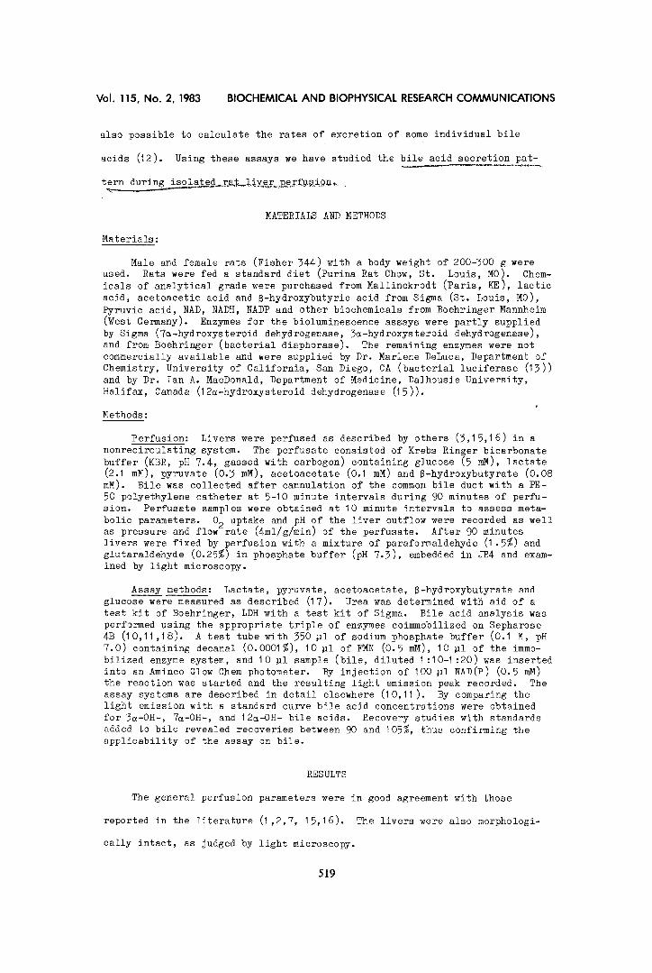
Bile flow as well as total bile acid excretion as measured by the 5~-
assay decreased during perfusion as expected. There was a good correlation of
bile acid excretion and bile flow either when eight perfused animals were com-
pared or when the average at each time point of perfusion was used (Figure I).
There was no difference between male and female rats concerning bile flow
(0.57+O.14 vs. 0.60+0.08 mg/min/g liver) and total bile acid excretion aver-
age (2.5+1.2 vs. 2.8+0.4 nmoles/min/g liver) although the females excreted
slightly greater amounts in the first 30 minutes of perfusion than the males.
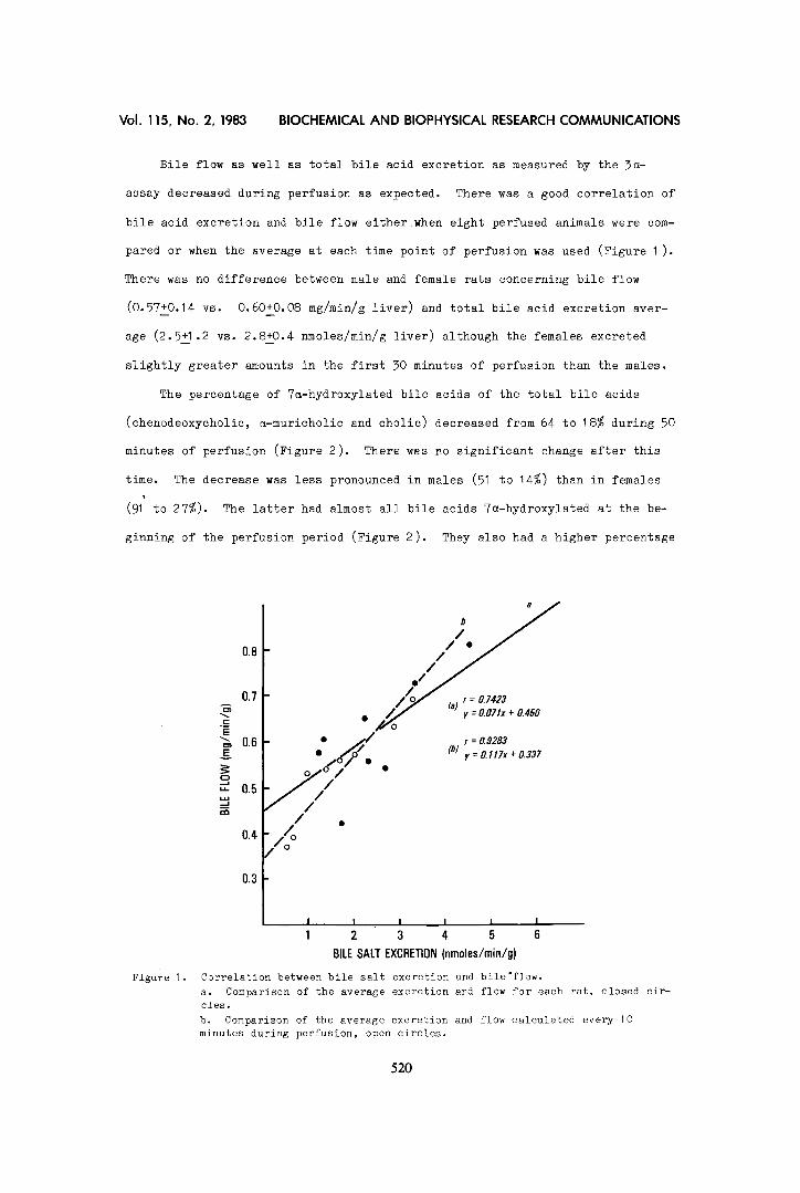
The percentage of 7~-hydroxylated bile acids of the total bile acids
(chenodeoxycholie, ~-muricholic and cholie) decreased from 64 %o 18% during 50
minutes of perfusion (Figure 2). There was no significant change after this
time. The decrease was less pronounced in males (51 to 14%) than in females
(91' to 27%). The latter had almost all bile acids 7~-hydroxylated at the be-
ginning of the perfusion period (Figure 2). They also had a higher percentage
Figure 1.
0.8
07 A
._= E
0.6 E
"-" 0.5
0.4
03
//
, , ~ Z r = 0.7423
r = 0.9283 : /o// (b) y = ~ l l T x + 0 . 3 3 7
/// /
/o °
I I I I I I 1 2 3 4 5 6
BILE SALT EXCRETION (nmoles/min/g)
Correlation between bile salt excretion and bile'flow. a. Comparison of the average excretion and flow for each rat, closed cir- cles. b. Comparison of the average excretion and flow calculated every 10 minutes during perfusion, open circles.
520
Vol. 115, No. 2, 1983 BIOCHEMICAL AND BIOPHYSICAL RESEARCH COMMUNICATIONS
90
7O I .--- z
,,.,..
~_ 5O
30
INIMALS
"S
LES
10
Figure 2.
10 20 30 40 50
TIME (minutes)
Percentage of total bile acids as 7a-hydroxylated or 12a-hydroxylated at several time points during perfusion for all animals (n=9), males (n=6) and females (n=3).
(27%) of 12a-hydroxylated bile acids than the males at the beginning which
then decreased during the subsequent 50 minutes while no decrease occurred in
males. After 50 minutes of perfusions the ratios of 7~/3~ and 12~/3a were
almost identical in males and females. Using simple equations i12) neglecting
deoxycholic and lithocholic acid, since they have been shown to be present in
rat bile in trace amounts, and assuming that bile acids neither 7~- nor 12a-
hydroxylated were ~-muricholic or hyodeoxycholic acid, we calculated the
excretion of bile acid groups given in Table I. Females had a large amount of
chenodeoxycholic + a-muricholic acids at the beginning and also a higher
TABLE I
Bile acids all rats males females 20' 40' 20' 40' 20' 40'
Cholic 0.59 0.24 O.51 0.20 0.90 0.34
B-muricholic + hyodeoxycholic 1.51 1.48 2.02 1.50 0.30 1.60
a-muricholic + chenodeoxycholic 1.40 0.33 0.72 0.20 2.33 O.51
Total 3.50 2.05 3.25 1.90 3.53 2.45
Biliary bile acid excretion in nmoles/minute/g liver at 20 and 40 minutes of perfusion for males (n=6) and females (n=3) as calculated from the results of the three assays assuming that bile acids having neither a 7~- nor a 12~-hydroxy group were 6-muricholic or hyodeoxycholic acid.
521
Vol. 115, No. 2, 1983 BIOCHEMICAL AND BIOPHYSICAL RESEARCH COMMUNICATIONS
amount of cholic acid than males which, in turn, had predominantly 6-and 76-
hydroxylated bile acids. These differences, however, almost disappeared after
40 minutes of perfusion.
DISCUSSION
Methodology:
The pattern of bile acids in bile during isolated rat liver perfusion has
rarely been studied since no simple and rapid methods were available. The
bioluminescence assays are rapid and sensitive and, as such, make sequential
monitoring of bile acid concentrations during perfusion feasible. Since they
are also very sensitive, they can be used in similar studies with isolated
hepatocytes where only small amounts of bile acids are present as has been
recently described using a radioimmunoassay (20). Accuracy and specificity
are also very good (10,11,12) thus making the results very reproducible. The
addition of an assay using a 7 ~-hydroxysteroid dehydrogenase currently under
investigation will make their application on experimental systems using rats
even more useful.
Bile acid pattern:
Since the perfusion parameters as well as bile flow and total bile acid
excretion were rather similar to the data described by others (3,4,5,7,8,16),
the changes observed in the bile acid pattern in bile do not seem to be the
result of perfusion irregularities.
The correlation obtained between bile flow and bile acid excretion was
also in good agreement with that found by others (20,21,22). The pattern of
bile acids in bile observed at the beginning of perfusion resembled that found
in vivo (23,24). However, the amount of 6- or 76-hydroxylated bile acids was
somewhat greater and that of cholic acid smaller than reported. This may be
due to species and sex differences. The sex difference found in our small
number of animals resembled that recently reported by others (9) very closely,
chenodeoxycholic and ~-muricholic acid being the predominant bile acids in
female livers. Since this sex difference disappears during perfusion it may
be caused by systemic factors (e.g. hormones, bacterial flora) in the animals
and not by differences in hepatic metabolic pathways.
522
Vol. 115, No. 2, 1983 BIOCHEMICAL AND BIOPHYSICAL RESEARCH COMMUNICATIONS
During the perfusion period no enterohepatic circulation is present.
Therefore, the bile acids excreted are thought to be derived mainly from de
novo synthesized pools with progression of perfusion time. It has been shown
that the perfused rat liver reduces 7 keto bile acids considerably less than 3
keto bile acids to ~-hydroxy bile acids. Only 30% of 7-keto bile acids are
reduced to 7a-OH acids. The amount of B-hydroxy acids could not be determined
in that study (7). Also a decrease of 7a-hydroxylated bile acids in bile dur-
ing starvation, a condition in some aspects similar to the isolated perfusion,
has been reported (23). We cannot decide if the small amount of 7a-hydroxy-
lated bile acids after 50 minutes of perfusion is due to an almost complete
transformation of chenodeoxycholic into muricholic acids (25), to the use of
other cholesterol pools (26), or to pathways from cholesterol to bile acids
bypassing 7~-hydroxylation (27,28,29,30,31). The data are also in accordance
with the results of others showing the rate of synthesis for cholic acid to
6-muricholic acid to be 1:3 in rats (24,32).
In summary, it is important to realize that the pattern of biliary bile
acids changes during isolated rat liver perfusion, and that sex differences
also exist. This might also be true for intracellular bile acids in perfused
livers and isolated hepatocytes as well. Monitoring of bile acid excretion
during the experiment with the aid of bioluminescence assays may help to
understand the changing pattern further and may facilitate the use of the per-
fused liver and presumably isolated hepatocytes as well in bile acid related
studies.
ACKNOWLEDGMENTS
We wish to thank M. DeLuca for the possibility of developing the biolumines- cence asays. We thank I.A. MacDonald for the supply of 12G-hydroxysteroid dehydrogenase, J. Keeney for help in preparation and A.F. Hofmann for critical reading of the manuscript.
REFERENCES
I. Ross, B.C. (I 972) Perfusion Techniques in Biochemistry, pp. 133-213, Clarendon Press, Oxford.
2. Schimassek, H. (I 962) Life. Sci. 11, 629-634. 3. H~ussinger, D., Gerok, W., Sies H. (1982) Eur. J. Biochem. 126, 69-76. 4. Brauer, R.W., Pessotti, R.L., Pizzolato, P. (1951) Proc. Soc. Exp. Biol.
Med. 78, 174-I~. 5. Herz, R., Paumgartner, G., Preisig, R. (1975) Scand. J. Gastroenterol.
11, 741-746.
523
Vol. 115, No. 2, 1983 BIOCHEMICAL AND BIOPHYSICAL RESEARCH COMMUNICATIONS
6. Utili, R., Abernathy, C.O., Zimmerman, H.J. (1977) J. Lab. Clin. Med. 89, 471 -482.
7. Anwer, M.S., Hegner D. (I 982 ) Hoppe-Seyler's Z. Physiol. Chem. 363, 731- 735.
8. Van Dyke, R.W., Stephens, J.E., Scharschmidt, B.V. (1982) J. Clin. Invest. 70, 505-51 7.
9. Kurtz, W., Leuschner, U., Hellstern, A., Janka, P. (I 982)Hepato- gastroenterol. 29, 227-231.
10. Roda, A., Kricka, L.J., DeLuca, M., Hofmann, A.F. (1952) J. Lipid Res. 23, 1354-I 361 .
11. Schoelmerich, J., Hinkley, J.E., MacDonald, I.A., Hofmann, A.F., DeLuca, M. (in press) Anal. Biochem.
12. MacDonald, I.A., Williams, C.N., Musial, B.C. (1980) J. LiPid Res. 21, 381 -385.
13. Wienhausen, G.K., Kricka, L.J., Hinkley, J.E., DeLuea, M. (1982)Appl. Biochem. Biotechnol. 7, 463-473.
14. MacDonald, I.A., Jellett, J.F., Mahony, D.E. (1979) J. Lipid Res. 20, 234 -473.
15. Sies, H. (I 978)Methods Enzymol. 52, 48-59. 16. Brunengraber, H., Boutry, M., Daikuhara, Y., Kopelovich, L., Lowenstein,
J.M. (I 975) Methods Enzymol. 35, 597-607. 17. Maughan, R.J. (1982) Clin. Chim. Acta. 122, 231-240. 18. Ford, J., DeLuca, M. (1981)Anal. Biochem. 110, 43-48. 19. Botham, K.M., Boyd, G.S., Williamson, D., Beckett, G.J. (1983) F.E.B.S.
Lett. 151 , 19-21. 20. Javitt, N.B., Emerman, S. (1968) J. Clin Invest. 47, 1002-1014. 21. Hardison, W.G.M., Wood, C.A. (1978) Am. J. Physiol 235, EI58-E164. 22. Blitzer, B.L., Boyer, L.L. (1982) Gastroenterol 82, 346-357. 23. lllman, R.J., Beins, D.M., Balasubramanian, S., Topping, D.L. (19$2)
Biochem. Intern. 5, 367-374. 24. Kinugasa, T., Uchida, K., Kadowaki, M., Takase, H., Nomura, Y., Saito, Y.
(I 981 ) J. Lipid Res. 22, 201-207. 25. Kellogg, T.F. (I 973) The Bile Acids, Chemistry, Physiology, and Metabol-
ism, Vol. 2: Physiology and Metabolism (Nair, P.P., Kritchevsky, D., eds.) pp. 283-306, Plenum Press, New York.
26. Kempen, H.J., Holstein M.V., de Lange, J. (1983) J. Lipid Res. 24, 316- 323.
27. Kok, E., Burstein, S., Javitt, N.B., Gut, M., Byon, C.Y. (I 981 ) J. Biol. Chem. 256, 61 55-61 59.
28. Bostrom, H., Wikvall, K. (I 982) J. Biol. Chem. 257, 11755-11759. 29. Palmer, R.H. (I 952) Progress in Liver Disease, Vol. VII (Popper, H.,
Schaffner, F., eds.) pp. 221-242, Grune & Stratton, New York. 30. Botham, K.M., Becket, G.J., Percy-Robb, I.W., Boyd, G.S. (1980) Eur. J.
Biochem. 103, 299-305. 31. Freese, D.K., Hanson, R.F., Sharp, R.L. (1983) Gastroenterol. 84, 1159. 32. Vlahcevic, Z.R., Kubaska, W.M., Gurley, E.C., Whitehead, T.R., Guzelain,
P.S., Hylemon, P.B. (1983) Gasteroenterol. 84, 1401.
524