Embed Size (px)
Citation preview

Channel Catfish Follicle-Stimulating Hormone andLuteinizing Hormone: Complementary DNA Cloning andExpression During Ovulation
Zhanjiang Liu,* Soonhag Kim, and Attila Karsi
Fish Molecular Genetics and Biotechnology Laboratory, Department of Fisheries and Allied Aquacultures, and Program of Cell
and Molecular Biosciences, Auburn University, Auburn, AL 36849, U.S.A.
Abstract: Gonadotropins are important regulators of reproduction. To develop molecular resources for pro-
duction of recombinant gonadotropins, we have cloned and sequenced complementary DNA encoding the
channel catfish (Ictalurus punctatus) follicle-stimulating hormone (FSH) and luteinizing hormone (LH), which
encode 132 and 140 amino acid proteins, respectively. The deduced amino acid sequences of the catfish FSH
and LH are highly conserved with those cloned from other teleosts. Both the FSH and the LH were highly
induced during ovulation after injection of carp pituitary extract. Taken together with our previous findings of
enhanced expression of growth hormone and other pituitary hormones, this research suggests that a hormonal
cocktail may be needed for more efficient manipulation of catfish reproduction. The availability of the catfish
FSH and LH cDNAs provides the opportunity for production of immunologically or biologically active re-
combinant gonadotropins for the study of catfish reproductive physiology and the manipulation of artificial
spawning for aquaculture.
Key words: spawning, induction, gene, expression, pituitary, reproduction.
INTRODUCTION
A family of related glycoprotein hormones are produced in
mammals: follicle-stimulating hormone (FSH), luteinizing
hormone (LH), thyroid-stimulating hormone (TSH), and
the chorionic gonadotropin (CG). These glycoproteins all
share the same � subunit in a given species in a functional
heterodimer composed of one � subunit and one � subunit.
The different � subunit confers distinct functions to each of
the 4 glycoproteins (Pierce and Parsons, 1981).
In teleosts, 2 types of gonadotropin were first identified
in chum salmon (Oncorhynchus keta) (Kawauchi et al.,
1986), then referred to as gonadotropin I and II (GTH-I
and GTH-II), and now generally accepted as homologues of
mammalian FSH and LH, respectively. In the last decade,
FSH and LH complementary DNAs have been cloned from
various teleost species (Sekine et al., 1989; Lin et al., 1992;
Kato et al., 1993; Hassin et al., 1995; Elizur et al., 1996;
Nagae et al., 1996; Rosenfeld et al., 1997; Yoshiura et al.,
1997). Although FSH and LH are similar in structure, they
are synthesized in 2 different cell types (Nozaki et al., 1990a,
b; Naito et al., 1991, 1993) and at different stages of the
reproductive cycle (Naito et al., 1991). FSH is expressed inReceived March 12, 2001; accepted June 27, 2001
*Corresponding author: fax 334-844-9208; e-mail [email protected].
Mar. Biotechnol. 3, 590–599, 2001DOI: 10.1007/s1012601-0067-5
© 2001 Springer-Verlag New York Inc.

the periphery of the glandular cords of the proximal pars
distalis (PPD), in close association with somatotrophs; LH
is expressed in the central parts of the glandular cords of the
PPD. Temporally, FSH is expressed first in ontogeny and
involved in early reproductive events such as steroidogen-
esis, vitellogenesis, and spermatogenesis, whereas LH is ex-
pressed in later stages of reproductive cycle and regulates
spermiation and ovulation (Naito et al., 1991).
Mechanisms controlling ovulation and spawning have
not been well studied in channel catfish (Ictalurus puncta-
tus) despite its significance for aquaculture (Dunham,
1993). Channel catfish spawn once a year during late spring
and early summer when water temperature is 24° to 27°C.
Under natural conditions, only a small percentage of chan-
nel catfish spawn each year. The majority carry their oocytes
through the summer but fail to spawn. The major environ-
mental regulator of catfish spawning is temperature (Liu et
al., 1997), while photoperiod appears to be more important
for other teleosts. This spawning behavior should result
from the complex interaction along the brain-pituitary-
gonad axis, but the hormone profile during oogenesis is not
known (reviewed by Patino 1995, 1997).
For aquaculture, carp pituitary extract (CPE) has been
used most successfully for induction of spawning in catfish
(Dunham, 1993; Liu et al., 1997; Lambert et al., 1999), but
a low hatching rate has been routinely observed for embryos
obtained from females treated with artificial spawning in-
duction reagents. This problem is most prominent for pro-
duction of channel catfish × blue catfish F1 hybrids. Al-
though the F1 hybrid catfish derived from the female chan-
nel catfish × male blue catfish perform better in essentially
all important traits such as growth rate, feed conversion
efficiency, disease resistance, fillet yield, and harvestability,
mass production of the hybrid fingerlings has not been
feasible for the catfish industry. The major problem ap-
peared to be the inconsistent results with induction of using
CPE, which often gave very low hatching rates. Two factors
have been proposed as the causes of the failure in CPE-
induced spawning: first, great variation in the quality of
CPE from the commercial sources because of the age, sea-
son of harvest, sex, and physiological condition of carps
may greatly affect the composition of CPE; second, the het-
erologous nature of induction hormones may not be opti-
mal for catfish. One could argue that homogeneous hor-
mones should work better for their own systems. In addi-
tion, if recombinant FSH and LH can be produced, then the
amount of these hormones can be precisely controlled. To-
ward this end, we report here isolation and sequence analy-
sis of the cDNAs encoding the channel catfish FSH and LH
and their expression during spawning induced by CPE.
Along with the previous cloning of � subunit, this work
should provide molecular resources for production of re-
combinant GTHs for application in catfish.
MATERIALS AND METHODS
Animals and Treatments
Channel catfish were obtained from the Fish Genetics Fa-
cility of Auburn University. Gravid fish were selected for
experiments and randomized in 7 groups of 5 fish each and
placed in indoor tanks. The water temperature was kept at
27° to 28°C. Tissues from fish in each treatment group were
collected at different times for isolation of RNA. At the
beginning of the experiment, tissues from the first group of
fish were harvested as 0-hour controls. CPE (Stollers, Spirit
Lake, Iowa) was then injected into the fish of the remaining
6 groups at a priming dose of 2 mg/kg of body weight and
at a resolving dose of 8 mg/kg of body weight after 12 hours
when appropriate (these doses are routinely used for in-
duced spawning in catfish hatcheries). Pituitaries were col-
lected at 4, 8, and 12 hours (before second injection), 16
hours (4 hours after the second injection), 20 hours, and 36
hours after the initial injection. All fish were treated with
tricaine methane sulfonate (MS-222) at 300 ppm before
harvesting the pituitaries to euthanize the fish.
RNA Isolation
Pituitary glands from the 5 noninjected fish were pooled,
and 30 pituitaries after injection with carp pituitary extracts
were pooled prior to RNA extraction. The sample of 30
pituitaries included 5 pituitaries each at 4, 8, 12, 16, 20, and
36 hours after the initial injection. The pituitary glands were
harvested from freshly killed fish, frozen in liquid nitrogen,
then transferred for storage in a −80°C freezer. At the time
of RNA extraction, pituitaries were frozen in liquid nitrogen
again, pulverized with a mortar and pestle, and then ho-
mogenized (model 985-370, Biospec Products, Inc., Wis.) in
RNA extraction buffer following the guanidinium thiocya-
nate method (Chomczynski and Sacchi, 1987). Poly(A)+
RNA was purified by using the Oligotex Spin Column Kit
according to the manufacturer’s instructions (Qiagen Inc.,
Chatsworth, Calif.).
Channel Catfish FSH and LH 591

Complementary DNA Library Construction andMass In Vivo Excision
The Unizap � cDNA library was constructed from the
poly(A)+ RNA isolated from channel catfish pituitaries ac-
cording to the manufacturer’s instructions (Stratagene, La
Jolla, Calif.) and has been previously described (Liu et al.,
1997). The � Unizap cDNA library was converted into a
plasmid library in order to simplify DNA preparation and
other procedures, by using the mass in vivo excision pro-
cedure (Stratagene) according to the manufacturer’s in-
structions, and as previously described (Karsi et al., 1998).
Amplification and Molecular Cloning
The fixed orientation of cDNAs in the plasmid vector allows
use of vector-anchored polymerase chain reaction (PCR)
using one primer specific to the gene of interest and one
primer with vector sequence (Liu et al., 1997). The up-
stream portion of a cDNA can be amplified by using the
reverse sequencing primer in the cloning vector and a gene-
specific primer that is complementary to the RNA se-
quences. Similarly, the downstream portion of a cDNA can
be amplified using the universal primer and a gene-specific
primer that has the same sequences as the RNA. To obtain
probes, this approach was used to amplify the downstream
portions of cDNAs for both FSH and LH. The gene-specific
primers used to amplify the FSH and LH downstream se-
quences were 5�-CCAATCTCCATCACCGTGG-3� and 5�-
CCAAGGAACCTGTGTACAAGAGC-3�, respectively. The sequence
designs of these primers were based on conserved regions of
alignments of cloned counterparts from other teleosts. After
successful amplification of a portion of the cDNAs, the
aplicon was cloned in a pCR2.1 (Invitrogen, Carlsbad, Ca-
lif.) vector, and only one strand was sequenced to determine
the identity of the amplicon by BLAST searches. The veri-
fied fragments were then used as probes to screen the cDNA
library in order to obtain the complete cDNA clone (Sam-
brook et al., 1989).
PCR reactions (50 µl) were carried out in 50-mM KCl,
10-mM Tris (pH 9.0 at 25°C), 0.1% Triton X-100, 0.25-mM
each of deoxynucleotide triphosphates (dNTPs), 1.5-mM
MgCl2, 20-µM each of the upper and lower PCR primers,
approximately 10 ng of plasmid library DNA, and 2.5 U of
Taq DNA polymerase. The temperature profiles used to
amplify the cDNAs were 94°C for 30 seconds, 45°C for 1
minute, and 72°C for 2 minutes, for 35 cycles.
DNA Sequencing and Sequence Analysis
All cDNA clones were sequenced by the dideoxy chain-
termination method (Sanger et al., 1977) using the cycle
sequencing kit (Applied Biosystems, Inc., Foster City, Ca-
lif.). Minipreparation plasmid DNA (1 µl, about 200–500
ng) was used for each reaction. The profiles for cycling were
94°C for 1 minute, 72°C for 1 minute, 55°C for 1 minute,
for 30 cycles. An initial 2 minutes of extra denaturation at
94°C was always used. After initial sequencing with either
the universal or the reverse sequencing primers, the primer-
walking method was used to generate complete sequences.
Sequencing primers were ordered from Gibco (Bethesda,
Md.). DNA sequences were compiled by using microcom-
puter software packages, DNASIS (Hitachi, version 2.0), or
DNASTAR (DNA Star Inc., Madison, Wis.) Protein se-
quences were aligned by using the MEGALIGN program of
the DNASTAR package with CLUSTAL method and residue
weight table set at PAM250 (Hein, 1990). Sequences used
for alignments other than reported here were extracted
from the nonredundant GenBank+EMBL+DDBJ+PDB da-
tabases from the National Center for Biotechnology Infor-
mation using BLAST searches.
Northern Hybridization Analysis
Total RNA isolated from pituitary was electrophoresed on a
1% agarose gel with formaldehyde (Sambrook et al., 1989),
then transferred to a piece of Zetabind nylon membrane
(Wattman, Wis.) by capillary transfer with 10× SSC over-
night. The complete FSH and LH cDNA fragments were gel
purified and used as probes. The probes were made using
the random primer method (Sambrook et al., 1989) with a
labeling kit from Boehringer Mannheim (Indianapolis,
Ind.) Membranes were dried at 80°C under vacuum for 2
hours and prehybridized in 50% formamide, 5× SSPE, 0.1%
sodium dodecylsulfate (SDS), 5× Denhardt’s, and 100 µg/
ml sonicated and denatured calf thymus DNA overnight.
Hybridizations were carried out at 42°C overnight in the
same solution with probes added (more than 109 cpm/µg
DNA). The Zetabind membranes were washed first in 2×
SSC for 10 minutes at room temperature, followed by 3
washes in 0.2 × SSC with SDS at 0.2% at 50°C for 15
minutes each. The membranes were then wrapped by Saran
wrap and exposed to Kodak BioMax MS film for 30 minutes
to overnight depending on signal intensities. Hybridization
signals were quantified using the GS-525 Molecular Imag-
ing System (Molecular Dynamics, Sunnyvale, Calif.). The
592 Zhanjiang Liu et al.

magnitude of enhanced gene expression after CPE induc-
tion was calibrated by measuring also the levels of �-actin
messenger RNA (Liu et al., 1990). In other words, the RNA
loading differences were corrected by using a factor that
brought � actin to the same level before and after CPE
induction.
RESULTS AND DISCUSSION
Cloning and Sequencing of the FSH cDNA
Part of the FSH subunit cDNA was initially isolated by PCR
amplification using vector-anchored PCR (Liu et al., 1997).
The PCR reaction produced a product with the expected
size of 670 bp. The PCR product was cloned and partially
sequenced to verify its identity by BLAST searches of the
GenBank databases. A complete clone was then obtained by
screening the pituitary cDNA library.
The nucleotide sequences and the deduced amino acid
sequences for the FSH cDNA are shown in Figure 1. The
sequences were submitted to GenBank with accession num-
ber AF112191. The cDNA consisted of a 399-bp coding
region (including the termination codon), an 18-bp 5� un-
translated region (UTR), and a 360-bp 3� UTR. Three poly-
adenylation signal sequences AATAAA exist in the 3� UTR,
but all are located about 160 bp upstream from the poly-
adenylation sequences. This is somewhat longer than the
usual distance of approximately 30 bp observed for most
genes. The 3 potential poly(A)+ signals are part of a simple
sequence repeat located at the nontranslated region with an
imperfect sequence of (CA)n(AAAT)n(CCA)n (Figure 1). At 25
bp upstream of poly(A)+ site there are sequences ATTAAA,
similar to the consensus polyadenylation signal sequences.
It is likely that this sequence is actually used as the poly(A)+
signal. The coding region encodes a putative peptide of 132
amino acids, 17 amino acids of which are a signal peptide,
as determined by the alignment of amino acid sequences to
other teleosts (Lin et al., 1992; Hassin et al., 1995; Power et
al., 1997; Rosenfeld et al., 1997).
Until recently it was widely believed that catfish had
only one GTH, although both FSH and LH were found in
several teleosts (Hassin et al., 1995; Elizur et al., 1996; Gar-
cia-Hernandez et al., 1997; Yoshiura et al., 1997). The only
indication that 2 GTHs are present in catfish was the recent
cloning of the FSH and LH receptors (Kumar et al., 2001a,
b). This research provided the first concrete evidence for the
presence of both FSH and LH in catfish. This finding indi-
cates that the molecular architecture in regulation of catfish
gonadal development is similar to that of other teleost
fishes.
Cloning and Sequencing of the LH cDNA
With an approach similar as to that used to isolate the FSH,
LH cDNA fragments were obtained by vector-anchored
PCR. PCR amplification of the downstream portion of the
LH yielded a fragment with the expected size of 460 bp, as
determined by agarose gel electrophoresis. This fragment
was cloned, sequenced, and determined to be the LH. The
fragment was then used as a probe to obtain a complete
cDNA clone by screening the pituitary cDNA library. The
nucleotide sequences and the deduced amino acid se-
quences of LH are shown in Figure 2. The sequences have
been submitted to GenBank with accession number
AF112192. The cDNA sequences contained a coding se-
Figure 1. The nucleotide and deduced amino acid sequences of
cDNA encoding channel catfish FSH (GenBank accession number
AF112191). Nucleotide sequences are numbered on the top, far
right, and the amino acid sequence on the bottom, far right. The
coding region is in uppercase letters and the noncoding region in
lowercase letters. The first amino acid in the mature peptide is
underlined. The stop codon is indicated by a bold asterisk. Three
AATAAA sequences and an ATTAAA potential poly(A)+ sequence are
underlined. The simple sequence repeats are placed between ver-
tical bars (|). A long arrow indicates the position of the PCR
primer.
Channel Catfish FSH and LH 593
quence of 423 bp, a 5� UTR of 14 bp, and a 3� UTR of 123
bp. The LH cDNA encodes a polypeptide of 140 amino
acids, with 24 amino acids of signal sequence. The mature
peptide of LH in channel catfish, therefore, is predicted to
be 116 amino acids. A putative poly(A)+ signal sequence
(AATATA) was found at 17 bp upstream from the poly(A)+
site.
Structural and Evolutionary Conservation
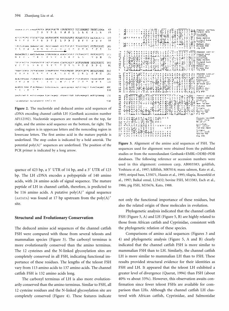
The deduced amino acid sequences of the channel catfish
FSH were compared with those from several teleosts and
mammalian species (Figure 3). The carboxyl terminus is
more evolutionarily conserved than the amino terminus.
The 12 cysteines and the N-linked glycosylation sites are
completely conserved in all FSH, indicating functional im-
portance of these residues. The lengths of the teleost FSH
vary from 113 amino acids to 137 amino acids. The channel
catfish FSH is 132 amino acids long.
The carboxyl terminus of LH is also more evolution-
arily conserved than the amino terminus. Similar to FSH, all
12 cysteine residues and the N-linked glycosylation site are
completely conserved (Figure 4). These features indicate
not only the functional importance of these residues, but
also the related origin of these molecules in evolution.
Phylogenetic analysis indicated that the channel catfish
FSH (Figure 5, A) and LH (Figure 5, B) are highly related to
those from African catfish and Cyprinidae, consistent with
the phylogenetic relation of these species.
Comparisons of amino acid sequences (Figures 3 and
4) and phylogenetic analysis (Figure 5, A and B) clearly
indicated that the channel catfish FSH is more similar to
mammalian FSH than to LH. Similarly, the channel catfish
LH is more similar to mammalian LH than to FSH. These
results provided structural evidence for their identities as
FSH and LH. It appeared that the teleost LH exhibited a
greater level of divergence (Querat, 1994) than FSH (about
40% vs about 33%). However, this observation awaits con-
firmation since fewer teleost FSHs are available for com-
parison than LHs. Although the channel catfish LH clus-
tered with African catfish, Cyprinidae, and Salmonidae
Figure 2. The nucleotide and deduced amino acid sequences of
cDNA encoding channel catfish LH (GenBank accession number
AF112192). Nucleotide sequences are numbered on the top, far
right, and the amino acid sequence on the bottom, far right. The
coding region is in uppercase letters and the noncoding region in
lowercase letters. The first amino acid in the mature peptide is
underlined. The stop codon is indicated by a bold asterisk. The
potential poly(A)+ sequences are underlined. The position of the
PCR primer is indicated by a long arrow.
Figure 3. Alignment of the amino acid sequences of FSH. The
sequences used for alignment were obtained from the published
studies or from the nonredundant Genbank+EMBL+DDBJ+PDB
databases. The following reference or accession numbers were
used in this alignment: common carp, AB003583; goldfish,
Yoshiura et al., 1997; killifish, M87014; masu salmon, Kato et al.,
1993; striped bass, L35071, Hassin et al., 1995; tilapia, Rosenfeld et
al., 1997; Baikal omul, L23432; bovine FSH, M13383, Esch et al.,
1986; pig FSH, M35676, Kato, 1988.
594 Zhanjiang Liu et al.

(with about 20% divergence), several separate clusters ap-
peared to be prominent: the Pacific herring by itself stands
as a group (about 32% divergence), and striped bass, porgy,
bonito, tuna, and killifish appeared to be a distinct cluster in
the phylogenetic tree (Figure 5, B).
Similarity of the Catfish FSH and LH Subunits
A BLAST search using the channel catfish FSH as a query
revealed significant similarities between channel catfish FSH
and LH from other organisms, in addition to the expected
similarities to FSH (e.g., smallest accumulative probability
for similarity between the channel catfish FSH and the Af-
rican catfish LH is 1.7 × 10⁻²⁹). Thus, in terms of molecular
evolution, it is likely that the FSH and LH of higher organ-
isms both evolved from the teleost FSH (Power et al., 1997).
This prompted us to compare the channel catfish FSH with
LH. A comparison of the channel catfish FSH and LH
amino acid sequences indicated 41% similarity between
them. All 12 cysteine residues are conserved with fixed po-
sitions. Querat (1994) suggested that the cysteines are nec-
essary to establish disulfide bonds with the � subunit and to
determine tertiary structure essential for receptor binding.
In addition to the 12 cysteine residues, a block of 10 amino
acids located between the 7th cysteine and the 10th cysteine
are identical between the channel catfish FSH and LH. This
block of amino acids contain Pro-Val-Ala (PVA) sequences
suggested for LH binding to the � subunit. In addition, all
other key residues in LH suggested for binding to the �
subunit (Querat, 1994; Power et al., 1997) are also shared by
the FSH: the Cys (C) and Gly (G) in the teleost-specific
sequence (CSGH), the N-glycosylation site, and the Glu (E)
at the fourth position before the second cysteine. The his-
tidine (H) residue in the so-called teleost-specific sequence
(CSGH) is replaced by a tyrosine (Y) residue in killifish,
frog, and human. Other identical blocks include the YXSP
between the fifth and the sixth cysteine, the YET between
the sixth and the seventh cysteine, and the GVD between
Figure 4. Alignment of the amino acid sequences of LH. The
following references or accession numbers were used in this align-
ment: African catfish, X97761, Rebers et al., 1997; striped bass,
L35096; tilapia, Rosenfeld et al., 1997; Chinook salmon, Xiong and
Hew, 1993; Chum salmon, M27154, Sekine et al., 1989; European
eel, X61039, Querat et al., 1990; killifish, M87015; carp, X59889,
Chang et al., 1992; Masu salmon, S69276; yellowfin porgy, L11722;
Pacific herring, Power et al., 1997; grass carp, X61051; human LH,
X00264, Talmadge et al., 1984; goldfish, D88024; bullfrog, 126481;
tuna, 585227, Okada et al. 1994; bonito, 298263, Koide et al., 1993;
arctic cisco, 1346202.
Figure 5. Cluster analysis of vertebrate pituitary glycoprotein FSH
(A) and LH (B). The dendrograms were generated by the MEGA-
LIGN program of the DNASTAR package using the CLUSTAL
method. With the balanced display, MEGALIGN averages the dis-
tances between ancestors in the tree. The dotted lines indicate a
negative branch length introduced by averaging the tree. The ac-
cession numbers were as described for Figures 3 and 4.
Channel Catfish FSH and LH 595

the seventh and the eighth cysteine residues. These similari-
ties indicate 2 possibilities: the structural requirement or the
functional requirement. The � subunits must couple to �
subunits in the form of heterodimers to function. Thus,
these shared blocks of amino acids may directly be involved
in binding to the � subunit. However, extensive identity of
amino acid sequences may suggest highly similar functions.
In this view, most earlier studies concerning functions of
FSH and LH were based on temporal difference in expres-
sion and perhaps also on inference from the mammalian
FSH and LH. Systematic determination of FSH expression
during ovulation was conducted in few teleosts. In fact,
most studies used immunoassays to measure pituitary and
plasma levels of GTH during sexual maturation and the
subsequent reproductive cycles. The cross-reactivity of the
antisera used in these studies is unknown (Hassin et al.,
1995), especially considering the structural similarities be-
tween the FSH and LH. The possibility that FSH may also
function for ovulation and spawning activities needs to be
studied. In view of this notion, the channel catfish FSH was
also activated during carp-pituitary-induced ovulation as
discussed below.
Enhanced Expression of FSH and LH DuringCarp-Pituitary-Induced Ovulation
Expression of FSH and LH before and after injection of CPE
was determined by Northern blot hybridization. Drastic in-
creases in expression levels of the FSH and LH RNAs were
observed during ovulation induced by injection of CPE
(Figure 6). FSH and LH RNAs were expressed 4.3-fold and
10-fold, respectively, compared with those before CPE-
induced ovulation. Although the enhanced gene expression
was determined by one set of experiments in this research,
similar levels of enhanced expression of FSH and LH were
revealed by expressed sequence tag analysis (Karsi et al.,
1998). Expression of the GTH � subunits was more dra-
matically activated during ovulation by CPE than the �
subunit. We previously found only a 40% increase of the
�-subunit RNA using the same animals and pituitaries (Liu
et al., 1997). The absolute expression level of the � subunit,
however, was much higher than the � subunits.
This research may have significant practical implica-
tions for manipulation of catfish spawning. Currently, CPE
is the most frequently used reagent for induction of spawn-
ing in catfish hatcheries, but the level of success is incon-
sistent. CPE contains the full multiplicity of pituitary hor-
mones that may directly work on the gonad. In addition to
the direct roles of GTHs, growth hormone and IGF are
known to be vital for gamete development and maturation
(Kagawa et al., 1994; Izadyar et al. 1997; Maestro et al.,
1997; Huang et al., 1998; Weil et al., 1999; Baker et al., 2000;
Tacon et al., 2000). We also found significant induction of
endogenous growth hormone gene expression during CPE-
induced ovulation (Karsi et al., 1998). Furthermore, recep-
tors for prolactin, TSH, and gonadotropin-releasing hor-
mone have been recently reported to be present in the go-
nad tissues of fish, suggesting direct action of these
hormones on the gonad (Alok et al., 2000; Kumar et al.,
2000, 2001a, b). Endogenous GTH expression may also be
involved in CPE-induced spawning as evidenced by the
drastic activation of GTH gene expression. In a similar
mechanism, luteinizing hormone–releasing hormone can
effectively induce catfish spawning as well (Lin et al., 1988),
although not as effectively as carp pituitary extract.
The drastic induction of FSH and LH, as well as growth
hormone (Karsi et al., 1998), during CPE-induced spawn-
ing suggests that multiple pituitary hormones may be
needed for optimal induction of catfish spawning activities.
In a practical sense, this research suggests that a cocktail or
sequential administration of FSH and LH, or perhaps
growth hormone and other pituitary hormones may pro-
duce better results for artificially induced spawning. Even
though CPE may contain all these hormones, the exact
amount of the hormones contained within CPE may vary
greatly from batch to batch. Therefore, the results of CPE-
induced spawning may be unpredictable. As a matter of
Figure 6. Expression of the channel catfish FSH and LH. Total
RNA was used in the Northern hybridization analysis. For FSH
(A), 2 separate gels with equal loading were run, transferred, and
probed with �-actin probe (Liu et al., 1990) (top) and the FSH
probe (bottom). For LH (B), the blot was simultaneously probed
with both �-actin and LH probes. Pituitary RNA before injection
with the carp pituitary extract (lane 1) and after induced ovulation
with carp pituitary extract (lane 2) was used to determine activa-
tion of gene expression.
596 Zhanjiang Liu et al.

fact, different preparations of CPE have produced highly
variable results (unpublished results). It is, therefore, im-
perative to produce biologically active recombinant FSH
and LH and other hormones for the study of reproductive
physiology of catfish. The availability of the channel catfish
FSH and LH cDNAs would make it possible to produce
biologically active hormonal cocktails for studying regula-
tion of spawning in channel catfish and for manipulation of
their reproductive capacity. Such cocktails are particularly
important for the production of interspecific hybrids of
channel catfish females and blue catfish males, which ex-
hibit superior aquaculture traits; however, mass production
of their fingerlings is not yet feasible.
ACKNOWLEDGMENTS
This project was supported by a USDA NRICGP grant to
Z.L. and R.D. (98-35205-6738), by the Auburn University
Competitive BioGrant (Biogrant J. Liu 99), and in part by a
USDA BARD grant to Z.L., R.D., and B. Moav (US2954-
97). S. Kim was supported by a graduate research assistant-
ship from the Department of Fisheries and Allied Aquacul-
tures. We thank Dr. Bernhart Kaltenback for allowing us to
use his DNASTAR sequence analysis package and Dr. Skip
Bartol for his GS-525 Molecular Imaging System to quantify
our Northern blots.
REFERENCES
Alok, D., Hassin, S., Kumar, R.S., Trant, J.M., Yu, K., and Zohar,
Y. (2000). Characterization of a pituitary GnRH-receptor from a
perciform fish, Morone saxatilis: functional expression in a fish cell
line. Mol Cell Endocrinol 168:65–75.
Baker, D.M., Davies, B., Dickhoff, W.W., and Swanson, P. (2000).
Insulin-like growth factor I increases follicle-stimulating hormone
(FSH) content and gonadotropin-releasing hormone-stimulated
FSH release from coho salmon pituitary cells in vitro. Biol Reprod
63:865–871.
Chang, Y.S., Huang, F.L., and Lo, T.B. (1992). Isolation and se-
quence analysis of carp gonadotropin beta-subunit gene. Mol Mar
Biol Biotechnol 1:97–105.
Chomczynski, P., and Sacchi, N. (1987). Single-step method of
RNA isolation by acid guanidinium thiocyanate–phenol–
chloroform extraction. Anal Biochem 162:156–159.
Dunham, R.A. (1993). Observations on controlled artificial
spawning of channel catfish. Prog Fish Cult 55:60–61.
Elizur, A., Zmora, N., Rosenfeld, H., Meiri, I., Hassin, S., Gordin,
H., and Zohar, Y. (1996). Gonadotropins beta-GtHI and beta-
GtHII from the gilthead seabream, Sparus aurata. Gen Com En-
docrinol 102:39–46.
Esch, F.S., Mason, A.J., Cooksey, K., Mercado, M., and Shimasaki,
S. (1986). Cloning and DNA sequence analysis of the cDNA for the
precursor of the beta chain of bovine follicle stimulating hormone.
Proc Natl Acad Sci USA 83:6618–6621.
Garcia-Hernandez, M.P., Koide, Y., Diaz, M.V., and Kawauchi, H.
(1997). Isolation and characterization of two distinct gonadotro-
pins from the pituitary gland of Mediterranean yellowtail, Seriola
dumerilii. Gen Comp Endocrinol 106:389–399.
Hassin, S., Elizur, A., and Zohar, Y. (1995). Molecular cloning and
sequence analysis of striped bass (Morone saxatilis) gonadotropins
I and II subunits. J Mol Endocrinol 15:23–35.
Hein, J. (1990). Unified approach to alignment and phylogenies.
Methods Enzymol 183:626–645.
Huang, Y.S., Rousseau, K., Le Belle, N., Vidal, B., Burzawa-Gerard,
E., Marchelidon, J., and Dufour, S. (1998). Insulin-like growth
factor-I stimulates gonadotrophin production from eel pituitary
cells: a possible metabolic signal for induction of puberty. J En-
docrinol 159:43–52.
Izadyar, F., Van Tol, H.T., Colenbrander, B., and Bevers, M.M.
(1997). Stimulatory effect of growth hormone on in vitro matu-
ration of bovine oocytes is exerted through cumulus cells and not
mediated by IGF-I. Mol Reprod Dev 47:175–180.
Kagawa, H., Kobayashi, M., Hasegawa, Y., and Aida, K. (1994).
Insulin and insulin-like growth factors I and II induce final matu-
ration of oocytes of red seabream, Pagrus major, in vitro. Gen
Comp Endocrinol 95:293–300.
Karsi, A., Li, P., Dunham, R.A., and Liu, Z.J. (1998). Transcrip-
tional activities in the pituitaries of channel catfish (Ictalurus punc-
tatus) before and after induced ovulation as revealed by expressed
sequence tag analysis. J Mol Endocrinol 21:121–129.
Kato, Y. (1988). Cloning and DNA sequence analysis of the cDNA
for the precursor of porcine follicle stimulating hormone (FSH)
beta-subunit. Mol Cell Endocrinol 55:107–112.
Kato, Y., Gen, K., Maruyama, O., Tomizawa, K., and Kato, T.
(1993). Molecular cloning of cDNAs encoding two gonadotrophin
beta subunits (GTH-I beta and -II beta) from the masu salmon,
Oncorhynchus masou: rapid divergence of the GTH-I beta gene.
J Mol Endocrinol 11:275–282.
Kawauchi, H., Suzuki, K., Nagahama, Y., Adach, S., Naito, N., and
Nakai, Y. (1986). Occurrence of two distinct gonadotropins in
chum salmon pituitary. In: Pars Distalis of the Pituitary Gland-
Structure, Function, and Regulation, F. Yoshimura, A., and Gorb-
Channel Catfish FSH and LH 597
man, A., (eds.). New York, N.Y.: Elsevier Science Publishers BV
(Biomedical Division), 383–390.
Koide, Y., Itoh, H., and Kawauchi, H. (1993). Isolation and char-
acterization of two distinct gonadotropins, GTH I and GTH II,
from bonito (Katsuwonus plelamis) pituitary glands. Int J Pept
Protein Res 41:52–65.
Kumar, R.S., Ijiri, S., Kight, K., Swanson, P., Dittman, A., Alok, D.,
Zohar, Y., and Trant, J.M. (2000). Cloning and functional expres-
sion of a thyrotropin receptor from the gonads of a vertebrate
(bony fish): potential thyroid-independent role for thyrotropin in
reproduction. Mol Cell Endocrinol 167:1–9.
Kumar, R.S., Ijiri, S., and Trant, J.M. (2001a). Molecular biology
of channel catfish gonadotropin receptors, 1: cloning of a func-
tional luteinizing hormone receptor and preovulatory induction of
gene expression. Biol Reprod 64:1010–1018.
Kumar, R.S., Ijiri, S., and Trant, J.M. (2001b). Molecular biology
of the channel catfish gonadotropin receptors, 2: complementary
DNA cloning, functional expression, and seasonal gene expression
of the follicle-stimulating hormone receptor. Biol Reprod 65:710–
717.
Lambert, D., Argue, B., Liu, Z.J., and Dunham, R.A. (1999). Effects
of seasonal variations, thyroid and steroid hormones, and carp
pituitary extract on the artificial production of channel catfish
Ictalurus punctatus × blue catfish I. furcatus hybrids. J World Aqua-
culture Soc 30:80–89.
Lin, H.R., Van der Kraak, G., Zhou, X.J., Liang, J.Y., Peter, R.E.,
Rivier J.E., and Vale, W.W. (1988). Effects of [D-Arg6, Trp7, Leu8,
Pro9NEt]-luteinizing hormone-releasing hormone (sGnRH-A)
and [D-Ala6, Pro9NEt]-luteinizing hormone-releasing hormone
(LHRH-A), in combination with pimozide or domperidone, on
gonadotropin release and ovulation in the Chinese loach and com-
mon carp. Gen Comp Endocrinol 69:31–40.
Lin, Y., Rupnow, B.A., Price, D.A., Greenberg, R.M., and Wallace
R.A. (1992). Fundulus heteroclitus gonadotropin, 3: cloning and
sequencing of gonadotropic hormone (GTH) I and II �-subunits
using the polymerase chain reaction. Mol Cell Endocrinol 85:127–
139.
Liu, Z.J., Zhu, Z., Roberg, K., Faras, A., Guise, K., Kapuscinski, A.,
and Hackett, P. (1990). Isolation and characterization of the �-ac-
tin gene of carp (Cyprinus carpio). DNA Seq 1:125–136.
Liu, Z.J., Li, P., Argue, B., and Dunham, R.A. (1997). Gonadotro-
pin � subunit glycoprotein from channel catfish (Ictalurus punc-
tatus) and its expression during hormone-induced ovulation. Mol
Mar Biol Biotechnol 6:221–231.
Maestro, M.A., Planas, J.V., Moriyama, S., Gutierrez, J., Planas, J.,
and Swanson, P. (1997). Ovarian receptors for insulin and insulin-
like growth factor I (IGF-I) and effects of IGF-I on steroid pro-
duction by isolated follicular layers of the preovulatory coho
salmon ovarian follicle. Gen Comp Endocrinol 106:189–201.
Nagae, M., Todo, T., Gen, K., Kato, Y., Young, G., Adachi, S., and
Yamauchi, K. (1996). Molecular cloning of the cDNAs encoding
pituitary glycoprotein hormone alpha- and gonadotropin II beta-
subunits of the Japanese eel, Anguilla japonica, and increase in
their mRNAs during ovarian development induced by injection of
chum salmon pituitary homogenate. J Mol Endocrinol 16:171–181.
Naito, N., Hyodo, S., Okumoto, N., Urano, A., and Nakai, Y.
(1991). Differential production and regulation of gonadotropins
(GtH-I and GtH-II) in the pituitary gland of rainbow trout, On-
corhynchus mykiss, during ovarian development. Cell Tissue Res
266:457–467.
Naito, N., Suzuki, K., Swanson, P., Nozaki, M., Kawauchi, H., and
Nakai, Y. (1993). Ultrastructural characteristics of two distinct
gonadotrophs (GtH-I and GtH-II cells) in the pituitary of the
rainbow trout (Oncorhynchus mykiss). Fish Physiol Biochem 11:
241–246.
Nozaki, M., Naito, N., Swanson, P., Dickhoff, W.W., Nakai, Y.,
Suzuki, K., and Kawauchi, H. (1990a). Salmonid pituitary gonad-
otropins, II: ontogeny of GtH-I and GtH-II cells in the rainbow
trout (Salmo gairdneri irideus). Gen Com Endocrinol 77:358–367.
Nozaki, M., Naito, N., Swanson, P., Miyata, K., Nakai, Y., Oota, Y.,
Suzuki, K., and Kawauchi, H. (1990b). Salmonid pituitary gonad-
otropins, I: distinct cellular distributions of two gonadotropins,
GtH I and II. Gen Com Endocrinol 77:348–357.
Okada, T., Kawazoe, I., Kimura, S., Sasamoto, Y., Aida, K., and
Kawauchi, H. (1994). Purification and characterization of gonad-
otropin I and II from pituitary glands of tuna. Int J Pept Protein
Res 43:69–80.
Patino, R. (1995). Mechanisms of maturational competence in fish
gonads. In: Proceedings of the Fifth International Symposium on the
Reproductive Physiology of Fish. Austin: University of Texas at Aus-
tin, 143–145.
Patino, R. (1997). Manipulations of the reproductive system of
fishes by means of exogenous chemicals. Prog Fish Cult 59:118–
128.
Pierce, J.G., and Parsons, T.F. (1981). Glycoprotein hormones:
structure and function. Annu Rev Biochem 50:465–495.
Power, M.E., Carolsfeld, J., Wallis, G.P., and Sherwood, N.M.
(1997). Isolation and characterization of a cDNA for gonadotropin
II-� of Pacific herring, an ancient teleost. J Fish Biol 50:315–323.
Querat, B. (1994). Molecular evolution of the glycoprotein hor-
mones in vertebrates. In: Perspectives in Comparative Endocrinol-
ogy, Davey, K.G., Peter, R.E., and Tobe, S.S. (eds.). Ottawa: Na-
tional Research Council of Canada, 27–35.
598 Zhanjiang Liu et al.

Querat, B., Moumni, M., Jutisz, M., Fontaine, Y.A., and Counis, R.
(1990). Molecular cloning and sequence analysis of the cDNA for
the putative beta subunit of the type-II gonadotrophin from the
European eel. J Endocrinol 4:257–264.
Rebers, F.E.M., Tensen, C.P., Schulz, R.W., Goos, H.J.T., and Bo-
gerd, J. (1997). Modulation of glycoprotein hormone alpha- and
gonadotropin II beta-subunit mRNA levels in the pituitary gland
of mature male African catfish, Clarias gariepinus. Fish Physiol
Biochem 17:99–108.
Rosenfeld, H., Levavi-sivan, B., Melamed, P., Yaron, Z., and Eli-
zur, A. (1997). The GTH � subunits of tilapia: gene cloning and
expression. Fish Physiol Biochem 17:85–92.
Sambrook, J., Frisch, E.F., and Maniatis, T. (1989). Molecular
Cloning: A Laboratory Manual. Cold Spring Harbor, N.Y.: Cold
Spring Harbor Laboratory Press.
Sanger, F., Nicklen, S., and Coulson, A.R. (1977). DNA sequencing
with chain-terminating inhibitors. Proc Natl Acad Sci USA 74:
5463–5467.
Sekine, S., Saito, A., Itoh, H., Kawauchi, H., and Itoh, S. (1989).
Molecular cloning and sequence analysis of chum salmon gonad-
otropin cDNAs. Proc Natl Acad Sci USA 86:8645–8649.
Tacon, P., Baroiller, J.F., Le Bail, P.Y., Prunet, P., Jalabert, B.
(2000). Effect of egg deprivation on sex steroids, gonadotropin,
prolactin, and growth hormone profiles during the reproductive
cycle of the mouthbrooding cichlid fish Oreochromis niloticus. Gen
Comp Endocrinol 117:54–65.
Talmadge, K., Vamvakopoulos, N.C., and Fiddes, J.C. (1984). Evo-
lution of the genes for the beta subunits of human chorionic
gonadotropin and luteinizing hormone. Nature 307:37–40.
Weil, C., Carre, F., Blaise, O., Breton, B., Le Bail, P.Y. (1999).
Differential effect of insulin-like growth factor I on in vitro go-
nadotropin (I and II) and growth hormone secretions in rainbow
trout (Oncorhynchus mykiss) at different stages of the reproductive
cycle. Endocrinology 140:2054–2062.
Xiong, F., and Hew, C.L. (1993). Chinook salmon (Oncorhynchus
tschawytscha) gonadotropin II beta-subunit gene encodes multiple
messenger ribonucleic acids. Can J Zool 69:2572–2578.
Yoshiura, Y., Kobayashi, M., Kato, Y., and Aida, K. (1997). Mo-
lecular cloning of the cDNAs encoding two gonadotropin beta
subunits (GTH-I beta and -II beta) from the goldfish, Carassius
auratus. Gen Comp Endocrinol 105:379–389.
Channel Catfish FSH and LH 599