Embed Size (px)
Citation preview
Chaperone Hsp27 inhibits translationduring heat shock by binding eIF4Gand facilitating dissociation of cap-initiationcomplexesRafael Cuesta, Gaurav Laroia, and Robert J. Schneider1
Department of Microbiology, New York University School of Medicine and Kaplan Cancer Center, New York,New York 10016 USA
Inhibition of protein synthesis during heat shock limits accumulation of unfolded proteins that might damageeukaryotic cells. We demonstrate that chaperone Hsp27 is a heat shock-induced inhibitor of cellular proteinsynthesis. Translation of most mRNAs requires formation of a cap-binding initiation complex known aseIF4F, consisting of factors eIF4E, eIF4A, eIF4E kinase Mnk1, poly(A)-binding protein, and adaptor proteineIF4G. Hsp27 specifically bound eIF4G during heat shock, preventing assembly of the cap-initiation/eIF4Fcomplex and trapping eIF4G in insoluble heat shock granules. eIF4G is a specific target of Hsp27, as eIF4E,eIF4A, Mnk1, poly(A)-binding protein, eIF4B, and eIF3 were not bound by Hsp27 and were not recruited intoinsoluble complexes. Dissociation of eIF4F was enhanced during heat shock by ectopic overexpression ofHsp25, the murine homolog of human Hsp27. Overexpression of Hsc70, a constitutive homolog of Hsp70,prevented loss of cap-initiation complexes and maintained eIF4G solubility. Purified Hsp27 specifically boundpurified eIF4G in vitro, prevented in vitro translation, eliminated eIF4G interaction with protein bindingfactors, and promoted eIF4G insolubilization. These results therefore demonstrate that Hsp27 is aheat-induced inhibitor of eIF4F-dependent mRNA translation.
[Key Words: Heat shock; translation; eIF4G; chaperones; eIF4F]
Received February 4, 2000; revised version accepted May 1, 2000.
Mammalian cells respond to heat stress in a highly or-chestrated manner. Heat shock activates a specific tran-scriptional response and largely inhibits protein synthe-sis, establishing conditions that favor the exclusivetranslation of heat shock mRNAs (for review, see Dun-can 1996; Morimoto 1998). Translational inhibition ofnon-heat shock mRNAs during heat stress prevents syn-thesis of nascent proteins that might misfold because ofelevated temperature and impair the cell (Morimoto1998). mRNAs that are specifically translated in mam-malian cells during heat shock generally encode molecu-lar chaperones, such as heat shock proteins (Hsps)Hsp100, Hsp90, Hsp70, Hsp60, Hsp40, and Hsp27 (Mo-rimoto 1998), which increase in abundance through pref-erential translation of their mRNAs. Heat shock chaper-ones facilitate native protein refolding and increase cellsurvival following thermal stress (Parsell and Lindquist1993). In mammalian cells, the small Hsp family (Hsp25in mouse, Hsp27 in human) are ATP-independent chap-erones that physically complex with certain unfoldingprotein intermediates, limiting denaturation and en-
hancing thermoresistance of the cell (Jakob et al. 1993;Ehrnsperger et al. 1997). During recovery from heatshock, ATP-dependent chaperones Hsp40, Hsp60,Hsp70, and the Hsp100 family, participate in refoldingprotein intermediates (Craig et al. 1994; Glover andLindquist 1998). Misfolded or excessively aggregated pro-teins are degraded by the ubiquitin–proteasome path-way, which is linked to the heat shock chaperone net-work (Morimoto 1998). Currently, only a few identifiedproteins have been shown to be specifically trapped byHsp27.
The ability of the translation apparatus to withstandirreversible inactivation during heat stress is associatedin an unknown way with expression of Hsp25/27 andHsp70/Hsc70 proteins (Liu et al. 1992; Li et al. 1995;Carper et al. 1997; for review, see Duncan 1996). It is alsogenerally thought that these chaperones protect the pro-tein synthetic apparatus during translational inhibitioninduced by heat shock (Mizzen and Welch 1988; Liu etal. 1992; Carper et al. 1997). It is not well understoodhow heat shock blocks protein synthesis in mammaliancells. Heat shock induces dephosphorylation and possi-bly inactivation of cap-binding protein eIF4E, in a widevariety of cell types, leading to inhibition of cap-depen-
1Corresponding author.E-MAIL [email protected]; FAX (212) 263-8276.
1460 GENES & DEVELOPMENT 14:1460–1470 © 2000 by Cold Spring Harbor Laboratory Press ISSN 0890-9369/00 $5.00; www.genesdev.org
Cold Spring Harbor Laboratory Press on September 11, 2018 - Published by genesdev.cshlp.orgDownloaded from
dent mRNA translation (Duncan and Hershey 1989;Lamphear and Panniers 1990). eIF4E forms a cap-initia-tion complex with the RNA helicase eIF4A and eIF4G,collectively referred to as translation initiation factoreIF4F. eIF4G also binds poly(A)-binding protein (PABP).The cap-initiation complex unwinds 58 secondary struc-ture (Sonenberg 1996), facilitating cap- and poly(A)-de-pendent mRNA translation (Imataka et al. 1998). Cap-dependent mRNA translation correlates with phos-phorylation of eIF4E, which is carried out by the kinaseMnk1 (Fukunaga and Hunter 1997; Waskiewicz et al.1997), a member of the cap-initiation complex. Mnk1facilitates phosphorylation eIF4E in vivo when both arebound to eIF4G (Pyronnet et al. 1999; Waskiewicz et al.1999). Inhibition of cap-dependent translation correlateswith decreased phosphorylation of eIF4E, which occursduring heat shock and infection with certain viruses(Huang and Schneider 1991; Feigenblum and Schneider1993, 1996; Feigenblum et al. 1998; Yueh and Schneider1996, 2000).
A family of small eIF4E-binding proteins known as4E-BP-1, 4E-BP-2, and 4E-BP-3 bind and sequester eIF4Ein a phosphorylation-dependent manner (Lin et al. 1994;Pause et al. 1994; Haghighat et al. 1995; Beretta et al.1996). Hypophosphorylated 4E-BP1 binds eIF4E duringheat shock (Feigenblum and Schneider 1996; Vries et al.1997; Wang et al. 1998). Studies indicate that although4E-BP binding of eIF4E participates in inhibition of eIF4Ephosphorylation and shutoff of protein synthesis duringheat shock, it is not sufficient, and other uncharacterizedcomponents of the heat shock response have been impli-cated (Feigenblum and Schneider 1996; Vries et al. 1997;Wang et al. 1998).
Here we show that inhibition of eIF4F(cap)-dependentprotein synthesis during heat shock involves Hsp27.Hsp27 was found to specifically bind eIF4G, which iscoupled to dissociation of the eIF4F(cap)-complex andinsolubilization of eIF4G into heat shock granules, con-sistent with inhibition of non-heat shock mRNA trans-lation. These results define eIF4G as a physiological tar-get of Hsp27, and they describe a novel molecularmechanism for inhibition of protein synthesis duringheat shock.
Results
Heat shock does not impair Mnk1 kinase activity
The eIF4E kinase, Mnk1, is a member of the eIF4F com-plex, which facilitates phosphorylation of eIF4E andtranslation of eIF4F(cap)-dependent mRNAs (Pyronnet etal. 1999; Waskiewicz et al. 1999). Because inhibition ofmRNA translation during heat shock is strongly associ-ated with a block in eIF4E phosphorylation, we investi-gated whether the activity of Mnk1 or its interactionwith eIF4G are targets for control of protein synthesisduring heat shock. Studies first determined whether heatshock blocks eIF4F(cap)-dependent translation by block-ing Mnk1 kinase activity. 293T cells were transfectedwith plasmids expressing GST–Mnk1, or a kinase-defec-
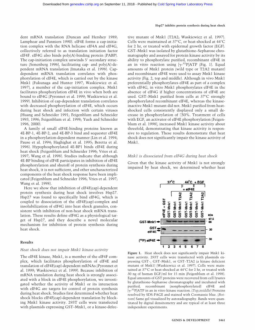
tive mutant of Mnk1 (T2A2; Waskiewicz et al. 1997).Cells were maintained at 37°C, or heat-shocked at 44°Cfor 2 hr, or treated with epidermal growth factor (EGF).GST–Mnk1 was isolated by glutathione–Sepharose chro-matography and assayed for protein kinase activity by itsability to phosphorylate purified, recombinant eIF4E inan in vitro reaction using [g-32P]ATP (Fig. 1). Equalamounts of Mnk1 protein (wild type or T2A2 mutant)and recombinant eIF4E were used to assay Mnk1 kinaseactivity (Fig. 2, top and middle). Although in vivo Mnk1preferentially phosphorylates eIF4E as part of a complexwith eIF4G, in vitro Mnk1 phosphorylates eIF4E in theabsence of eIF4G if higher concentrations of eIF4E areused. GST–Mnk1 purified from cells at 37°C stronglyphosphorylated recombinant eIF4E, whereas the kinase-inactive Mnk1 mutant did not. Mnk1 purified from heat-shocked cells consistently displayed only a small de-crease in phosphorylation of ∼50%. Treatment of cellswith EGF, an activator of eIF4E phosphorylation (Feigen-blum et al. 1998), increased Mnk1 kinase activity aboutthreefold, demonstrating that kinase activity is respon-sive to regulation. These results demonstrate that heatshock does not significantly impair the kinase activity ofMnk1.
Mnk1 is dissociated from eIF4G during heat shock
Given that the kinase activity of Mnk1 is not stronglyimpaired by heat shock, we determined whether heat
Figure 1. Heat shock does not significantly impair Mnk1 ki-nase activity. 293T cells were transfected with plasmids ex-pressing GST−, GST–Mnk1, or GST–T2A2 (a kinase deficientmutant of Mnk1) (Waskiewicz et al. 1997). Cells were main-tained at 37°C or heat-shocked at 44°C for 2 hr, or treated with30 ng of human EGF/ml for 15 min (Feigenblum et al. 1998).Equal amounts of GST proteins were recovered from cell lysatesby glutathione–Sepharose chromatography and incubated withpurified, recombinant (nonphosphorylated) eIF4E and[g-32P]ATP in an in vitro kinase reaction. (Top,middle) Proteinsresolved by SDS-PAGE and stained with Coomassie blue. (Bot-tom) Same gel visualized by autoradiography. Bands were quan-titated by digital densitometry and are typical of at least threeindependent experiments.
Hsp27 inhibits protein synthesis during heat shock
GENES & DEVELOPMENT 1461
Cold Spring Harbor Laboratory Press on September 11, 2018 - Published by genesdev.cshlp.orgDownloaded from
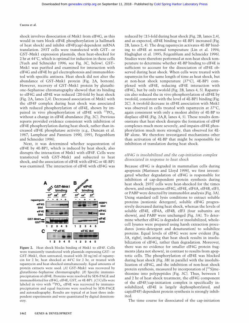
shock involves dissociation of Mnk1 from eIF4G, as thiswould in turn block eIF4E phosphorylation (a hallmarkof heat shock) and inhibit eIF4F(cap)-dependent mRNAtranslation. 293T cells were transfected with GST− orGST–Mnk1 expression plasmids, then heat-shocked for2 hr at 44°C, which is optimal for induction in these cells(Yueh and Schneider 1996; see Fig. 3C, below). GST–Mnk1 was purified and examined for interaction witheIF4G and eIF4E by gel electrophoresis and immunoblot-ted with specific antisera. Heat shock did not alter theabundance of GST–Mnk1 protein (Fig. 2A, bottom).However, recovery of GST–Mnk1 protein by glutathi-one–Sepharose chromatography showed that its bindingto eIF4G and eIF4E was reduced ∼20-fold by heat shock(Fig. 2A, lanes 2,4). Decreased association of Mnk1 withthe eIF4F complex during heat shock was associatedwith reduced phosphorylation of eIF4E, shown by im-paired in vivo phosphorylation of eIF4E with 32PO4,without a change in eIF4E abundance (Fig. 2C). Previousreports provided evidence consistent with inhibition ofeIF4E phosphorylation during heat shock, rather than in-creased eIF4E phosphatase activity (e.g., Duncan et al.1987; Lamphear and Panniers 1990, 1991; Feigenblumand Schneider 1996).
Next, it was determined whether sequestration ofeIF4E by 4E-BP1, which is induced by heat shock, alsodisrupts the interaction of Mnk1 with eIF4F. Cells weretransfected with GST–Mnk1 and subjected to heatshock, and the association of eIF4E with eIF4G or 4E-BP1was examined. The interaction of eIF4E with eIF4G was
reduced by ∼15-fold during heat shock (Fig. 2B, lanes 2,4),and as expected, eIF4E binding to 4E-BP1 increased (Fig.2B, lanes 2, 4). The drug rapamycin activates 4E-BP bind-ing to eIF4E at normal temperature (Lin et al. 1994;Haghighat et al. 1995; Feigenblum and Schneider 1996).Studies were therefore performed at non-heat shock tem-perature to determine whether 4E-BP binding to eIF4E issufficient to account for the dissociation of eIF4E ob-served during heat shock. When cells were treated withrapamycin for the same length of time as heat shock, butat non-heat shock temperature (37°C), 4E-BP1 com-plexed with eIF4E, reducing eIF4E interaction witheIF4G, but by only twofold (Fig. 2B, lanes 4, 5). Rapamy-cin also reduced the in vivo phosphorylation of eIF4E bytwofold, consistent with the level of 4E-BP1 binding (Fig.2C). A twofold decrease in eIF4E association with Mnk1was observed in cells treated with rapamycin at 37°C,again consistent with only a modest ability of 4E-BP todisplace eIF4E (Fig. 2A,B, lanes 4, 5). These results dem-onstrate that heat shock disrupts the formation of eIF4Fcomplexes much more severely, and impairs eIF4E phos-phorylation much more strongly, than observed for 4E-BP alone. We therefore investigated mechanisms otherthan activation of 4E-BP that might be responsible forinhibition of translation during heat shock.
eIF4G is insolubilized and the cap-initiation complexdissociated in response to heat shock
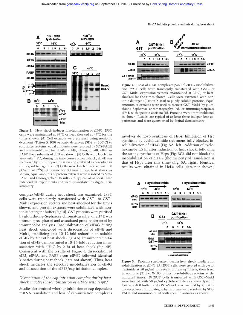
Because eIF4G is degraded in mammalian cells duringapoptosis (Marissen and Lloyd 1998), we first investi-gated whether degradation of eIF4G is responsible forinhibition of cap-dependent protein synthesis duringheat shock. 293T cells were heat-shocked for the timesshown, and endogenous eIF4G, eIF4E, eIF4A, eIF4B, eIF3,or PABP were detected by immunoblot analysis (Fig. 3A).Using standard cell lysis conditions to extract solubleproteins (nonionic detergent), soluble eIF4G progres-sively decreased during heat shock, whereas the levels ofsoluble eIF4E, eIF4A, eIF4B, eIF3 (four subunits areshown), and PABP were unchanged (Fig. 3A). To deter-mine whether eIF4G is degraded or insolubilized, whole-cell lysates were prepared using harsh extraction proce-dures (ionic-detergent and denaturation) to solubilizeproteins. Equal levels of eIF4G were now evident (Fig.3A, right), indicating that heat shock results in insolu-bilization of eIF4G, rather than degradation. Moreover,there was no evidence for smaller eIF4G protein frag-ments (data not shown), in contrast to results from apop-totic cells. The phosphorylation of eIF4E was blockedduring heat shock (Fig. 3B) in parallel with the insolubi-lization of eIF4G, and the inhibition of non-heat shockprotein synthesis, measured by incorporation of [35S]me-thionine into polypeptides (Fig. 3C). Thus, between 1and 2 hr of heat shock treatment, the eIF4G componentof the eIF4F/cap-initiation complex is specifically in-solubilized, eIF4E is largely dephosphorylated, andcap(eIF4F)-dependent protein synthesis is strongly inhib-ited.
The time course for dissociated of the cap-initiation
Figure 2. Heat shock blocks binding of Mnk1 to eIF4F. Cellswere transiently transfected with plasmids expressing GST− orGST–Mnk1, then untreated, treated with 20 ng/ml of rapamy-cin for 2 hr, heat shocked at 44°C for 2 hr, or treated withrapamycin and heat-shocked simultaneously. Equal amounts ofprotein extracts were used. (A) GST–Mnk1 was recovered byglutathione–Sepharose chromatography. (B) Specific immuno-precipitation of eIF4E. Proteins were resolved by SDS-PAGE andimmunoblotted for eIF4G, eIF4E, GST, or 4E-BP1. (C) Cells werelabeled in vivo with 32PO4, eIF4E was recovered by immuno-precipitation and equal fractions were resolved by SDS-PAGEand autoradiography. Results are typical of at least three inde-pendent experiments and were quantitated by digital densitom-etry.
Cuesta et al.
1462 GENES & DEVELOPMENT
Cold Spring Harbor Laboratory Press on September 11, 2018 - Published by genesdev.cshlp.orgDownloaded from
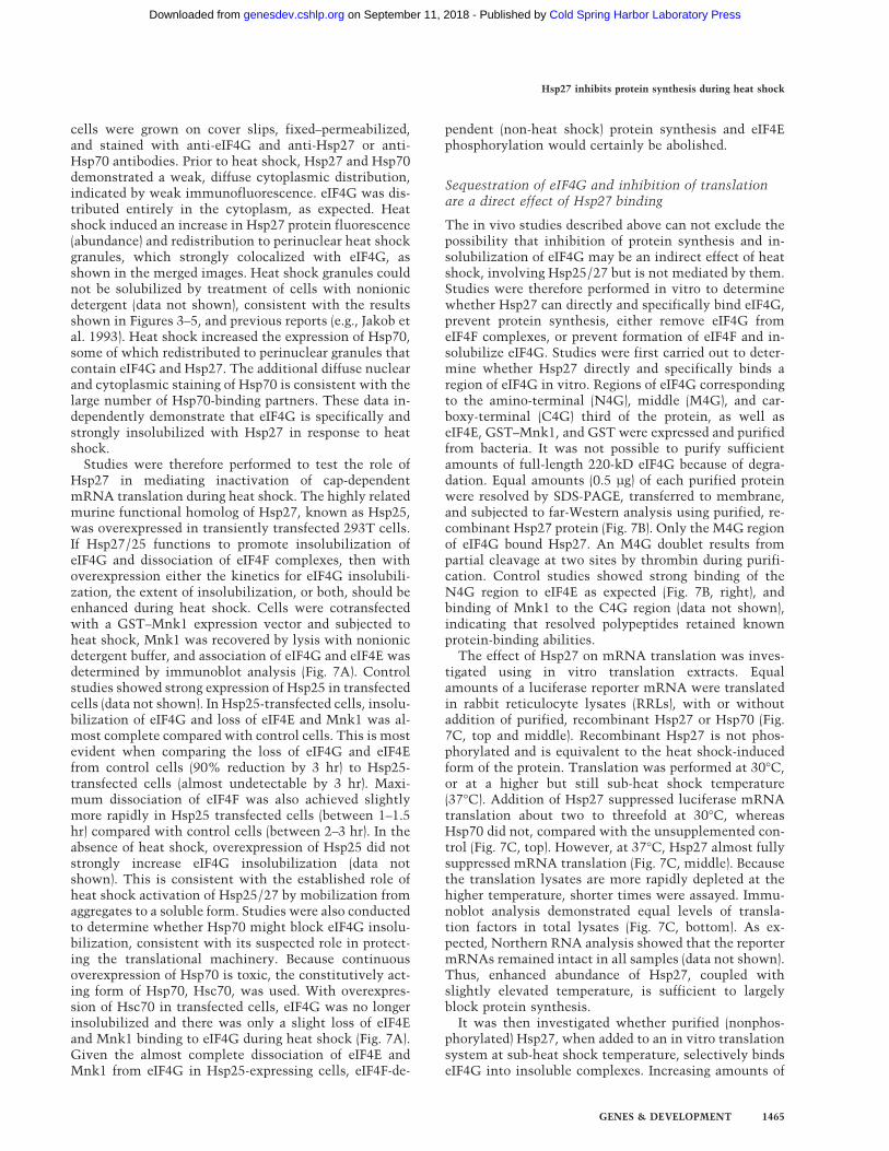
complex/eIF4F during heat shock was examined. 293Tcells were transiently transfected with GST− or GST–Mnk1 expression vectors and heat-shocked for the timesshown, and protein extracts were solubilized with non-ionic detergent buffer (Fig. 4). GST proteins were purifiedby glutathione–Sepharose chromatography, or eIF4E wasimmunoprecipitated and associated proteins detected byimmunoblot analysis. Insolubilization of eIF4G duringheat shock coincided with dissociation of eIF4E andMnk1, stabilizing at a 10–12-fold reduction in solubleeIF4G by 2 hr of heat shock (Fig. 4A). Immunoprecipita-tion of eIF4E demonstrated a 10–15-fold reduction in as-sociation with eIF4G by 2 hr of heat shock (Fig. 4B).Consistent with the results of Figure 3, dissociation ofeIF3, eIF4A, and PABP from eIF4G followed identicalkinetics during heat shock (data not shown). Thus, heatshock mediates the selective insolubilization of eIF4Gand dissociation of the eIF4F/cap-initiation complex.
Dissociation of the cap-initiation complex during heatshock involves insolubilization of eIF4G with Hsp27
Studies determined whether inhibition of cap-dependentmRNA translation and loss of cap-initiation complexes
involves de novo synthesis of Hsps. Inhibition of Hspsynthesis by cycloheximide treatment fully blocked in-solubilization of eIF4G (Fig. 5A, left). Addition of cyclo-heximide 1.5 hr after induction of heat shock, followingthe strong synthesis of Hsps (Fig. 3C), did not block theinsolubilization of eIF4G (the majority of translation isthat of Hsps after this time) (Fig. 5A, right). Identicalresults were obtained in HeLa cells (data not shown),
Figure 3. Heat shock induces insolubilization of eIF4G. 293Tcells were maintained at 37°C or heat shocked at 44°C for thetimes shown. (A) Cell extracts were prepared using nonionicdetergent (Triton X-100) or ionic detergent (SDS at 100°C) tosolubilize proteins, equal amounts were resolved by SDS-PAGEand immunoblotted for eIF4G, eIF4E, eIF4A, eIF4B, eIF3, orPABP. Four subunits of eIF3 are shown. (B) Cells were labeled invivo with 32PO4 during the time course of heat shock, eIF4E wasrecovered by immunoprecipitation and analyzed as described inthe legend to Figure 2. (C) Cells were labeled in vivo with 50µCi/ml of [35S]methionine for 30 min during heat shock asshown, equal amounts of protein extracts were resolved by SDS-PAGE and fluorographed. Results are typical of at least threeindependent experiments and were quantitated by digital den-sitometry.
Figure 4. Loss of eIF4F complexes parallel eIF4G insolubiliza-tion. 293T cells were transiently transfected with GST− orGST–Mnk1 expression vectors, maintained at 37°C, or heat-shocked for the times shown. Cells were extracted with non-ionic detergent (Triton X-100) to purify soluble proteins. Equalamounts of extracts were used to recover GST–Mnk1 by gluta-thione–Sepharose chromatography (A), or immunoprecipitateeIF4E with specific antisera (B). Proteins were immunoblottedas shown. Results are typical of at least three independent ex-periments and were quantitated by digital densitometry.
Figure 5. Proteins synthesized during heat shock mediate in-solubilization of eIF4G. (A) 293T cells were treated with cyclo-heximide at 50 µg/ml to prevent protein synthesis, then lysedin nonionic (Triton X-100) buffer to solubilize proteins at theindicated times. (B) 293T cells transfected with GST–Mnk1were treated with 50 µg/ml cycloheximide as shown, lysed inTriton X-100 buffer, and GST–Mnk1 was purified by glutathi-one–Sepharose chromatography. Proteins were resolved by SDS-PAGE and immunoblotted with specific antisera as shown.
Hsp27 inhibits protein synthesis during heat shock
GENES & DEVELOPMENT 1463
Cold Spring Harbor Laboratory Press on September 11, 2018 - Published by genesdev.cshlp.orgDownloaded from

demonstrating that heat shock insolubilization of eIF4Gis not cell-type specific and involves the nascent synthe-sis of one or more Hsps. Studies next determinedwhether the dissociation of cap-initiation complexesalso requires synthesis of Hsps. 293T cells transfectedwith GST–Mnk1 were treated with cycloheximide dur-ing heat shock, GST–Mnk1 was recovered, and associ-ated proteins were detected by immunoblot analysis. Inthe absence of Hsp synthesis, the abundance of eIF4G–eIF4E–Mnk1 complexes was reduced by twofold at 2–3hr of heat shock, compared with 10-fold with ongoingHsp synthesis (cf. Figs. 4A and 5B). Because the vast ma-jority of protein synthesis during heat shock correspondsto Hsps, we determined whether particular Hsps specifi-cally promote loss of cap-initiation complexes and in-solubilization of eIF4G.
Heat shock induces formation of heat shock granules,which consist largely of Hsp27 and smaller amounts ofHsp70, in combination with a small number of non-Hspsidentified to date. We therefore asked whether eIF4G isspecifically insolubilized during heat shock by bindingHsp27, and possibly Hsp70. Studies first determinedwhether eIF4G is coinsolubilized during heat shock withHsp27 and possibly Hsp70. 293T cells were heat-shockedfor 2 hr, and cell lysates were prepared in ionic detergentto preserve strong protein interactions (RIPA buffer).Whole-cell lysates showed a four- to sixfold increasedabundance of Hsp70 and Hsp27 with heat shock (Fig. 6A,compare lysate samples). During heat shock, Hsp27 andeIF4G were predominantly (∼90%) coisolated in the in-soluble protein fraction, whereas insoluble Hsp70 levelsincreased by only approximately twofold (Fig. 6A, com-pare insoluble protein lanes and total protein). Studieswere therefore carried out to examine the proteins asso-ciated with eIF4G during heat shock. HA–eIF4G was ex-pressed in transiently transfected cells at 37°C or duringheat shock. Whole-cell lysates were then prepared usingan ionic detergent (RIPA buffer, see Materials and Meth-ods), but without abolishing protein–protein interac-tions. The total abundance of eIF4G, Hsp105, Hsc70, andHsp40 proteins changed little during heat shock (Fig. 6B,left). The levels of Hsp90, Hsp70, Hsp60, and Hsp27 pro-teins increased variously from three- to sixfold with heatshock. Examination of HA–eIF4G during heat shock (Fig.6B, right) showed no specific interaction with Hsp105,Hsp90, Hsp40, or Hsp60. Hsc70 coimmunoprecipitatedwith eIF4G at a low but constitutive level, as observedpreviously (Laroia et al. 1999). Hsp27 interaction witheIF4G was almost undetectable in non-heat-shockedcells, but increased strongly by 30 min of heat shock, andmaximally by 1.5–2 hr. Hsp70 interaction with eIF4Gincreased about tenfold by 1–2 hr of heat shock. Theseresults demonstrate a strong and specific coassociationbetween eIF4G and Hsp27, with kinetics that parallelthe dissociation of cap-initiation complexes, dephos-phorylation of eIF4E, insolubilization of eIF4G and inhi-bition of non-Hsp synthesis.
eIF4G was found to colocalize in heat shock granuleswith Hsp27 during heat shock (Fig. 6C), consistent withthe biochemical interaction results shown above. HeLa
Figure 6. Chaperones associated with eIF4G. (A) 293T cellswere heat shocked at 44°C for 2 hr. Cells were lysed in RIPAdetergent buffer. Whole-cell lysates containing total cell protein(total) were normalized for equal protein levels, insoluble pro-tein pellets were recovered by glycerol gradient centrifugation,pellets were resolubilized in SDS with heating to 100°C andproteins were resolved by SDS-PAGE and immunoblotted asindicated (insoluble). (B) 293T cells were transfected with anHA-epitope-tagged eIF4G expression vector and maintained at37°C or heat-shocked at 44°C. (Left) Cells were lysed as above;equal amounts were resolved by SDS-PAGE and immunoblot-ted, as shown. (Right) eIF4G was immunoprecipitated with an-tisera to the HA-epitope, or preimmune serum (Preim.), andprecipitates were resolved by SDS-PAGE and immunoblottedwith antisera as shown. Results were quantified by digital den-sitometry. (C) HeLa cells were grown on cover slips, heat-shocked for 2 hr at 44°C or maintained at 37°C, fixed-perme-abilized, and reacted with primary antibodies to eIF4G, Hsp27,or Hsp70, followed by staining with the following secondaryantibodies: eIF4G, green fluorescence, Hsp27 or Hsp70, red fluo-rescence. Cells were visualized and photographed using a ZeissAxiophot microscope. Coimaging analysis was performed bydouble-exposure using fluorescein and rhodamine-specific filters.
Cuesta et al.
1464 GENES & DEVELOPMENT
Cold Spring Harbor Laboratory Press on September 11, 2018 - Published by genesdev.cshlp.orgDownloaded from
cells were grown on cover slips, fixed–permeabilized,and stained with anti-eIF4G and anti-Hsp27 or anti-Hsp70 antibodies. Prior to heat shock, Hsp27 and Hsp70demonstrated a weak, diffuse cytoplasmic distribution,indicated by weak immunofluorescence. eIF4G was dis-tributed entirely in the cytoplasm, as expected. Heatshock induced an increase in Hsp27 protein fluorescence(abundance) and redistribution to perinuclear heat shockgranules, which strongly colocalized with eIF4G, asshown in the merged images. Heat shock granules couldnot be solubilized by treatment of cells with nonionicdetergent (data not shown), consistent with the resultsshown in Figures 3–5, and previous reports (e.g., Jakob etal. 1993). Heat shock increased the expression of Hsp70,some of which redistributed to perinuclear granules thatcontain eIF4G and Hsp27. The additional diffuse nuclearand cytoplasmic staining of Hsp70 is consistent with thelarge number of Hsp70-binding partners. These data in-dependently demonstrate that eIF4G is specifically andstrongly insolubilized with Hsp27 in response to heatshock.
Studies were therefore performed to test the role ofHsp27 in mediating inactivation of cap-dependentmRNA translation during heat shock. The highly relatedmurine functional homolog of Hsp27, known as Hsp25,was overexpressed in transiently transfected 293T cells.If Hsp27/25 functions to promote insolubilization ofeIF4G and dissociation of eIF4F complexes, then withoverexpression either the kinetics for eIF4G insolubili-zation, the extent of insolubilization, or both, should beenhanced during heat shock. Cells were cotransfectedwith a GST–Mnk1 expression vector and subjected toheat shock, Mnk1 was recovered by lysis with nonionicdetergent buffer, and association of eIF4G and eIF4E wasdetermined by immunoblot analysis (Fig. 7A). Controlstudies showed strong expression of Hsp25 in transfectedcells (data not shown). In Hsp25-transfected cells, insolu-bilization of eIF4G and loss of eIF4E and Mnk1 was al-most complete compared with control cells. This is mostevident when comparing the loss of eIF4G and eIF4Efrom control cells (90% reduction by 3 hr) to Hsp25-transfected cells (almost undetectable by 3 hr). Maxi-mum dissociation of eIF4F was also achieved slightlymore rapidly in Hsp25 transfected cells (between 1–1.5hr) compared with control cells (between 2–3 hr). In theabsence of heat shock, overexpression of Hsp25 did notstrongly increase eIF4G insolubilization (data notshown). This is consistent with the established role ofheat shock activation of Hsp25/27 by mobilization fromaggregates to a soluble form. Studies were also conductedto determine whether Hsp70 might block eIF4G insolu-bilization, consistent with its suspected role in protect-ing the translational machinery. Because continuousoverexpression of Hsp70 is toxic, the constitutively act-ing form of Hsp70, Hsc70, was used. With overexpres-sion of Hsc70 in transfected cells, eIF4G was no longerinsolubilized and there was only a slight loss of eIF4Eand Mnk1 binding to eIF4G during heat shock (Fig. 7A).Given the almost complete dissociation of eIF4E andMnk1 from eIF4G in Hsp25-expressing cells, eIF4F-de-
pendent (non-heat shock) protein synthesis and eIF4Ephosphorylation would certainly be abolished.
Sequestration of eIF4G and inhibition of translationare a direct effect of Hsp27 binding
The in vivo studies described above can not exclude thepossibility that inhibition of protein synthesis and in-solubilization of eIF4G may be an indirect effect of heatshock, involving Hsp25/27 but is not mediated by them.Studies were therefore performed in vitro to determinewhether Hsp27 can directly and specifically bind eIF4G,prevent protein synthesis, either remove eIF4G fromeIF4F complexes, or prevent formation of eIF4F and in-solubilize eIF4G. Studies were first carried out to deter-mine whether Hsp27 directly and specifically binds aregion of eIF4G in vitro. Regions of eIF4G correspondingto the amino-terminal (N4G), middle (M4G), and car-boxy-terminal (C4G) third of the protein, as well aseIF4E, GST–Mnk1, and GST were expressed and purifiedfrom bacteria. It was not possible to purify sufficientamounts of full-length 220-kD eIF4G because of degra-dation. Equal amounts (0.5 µg) of each purified proteinwere resolved by SDS-PAGE, transferred to membrane,and subjected to far-Western analysis using purified, re-combinant Hsp27 protein (Fig. 7B). Only the M4G regionof eIF4G bound Hsp27. An M4G doublet results frompartial cleavage at two sites by thrombin during purifi-cation. Control studies showed strong binding of theN4G region to eIF4E as expected (Fig. 7B, right), andbinding of Mnk1 to the C4G region (data not shown),indicating that resolved polypeptides retained knownprotein-binding abilities.
The effect of Hsp27 on mRNA translation was inves-tigated using in vitro translation extracts. Equalamounts of a luciferase reporter mRNA were translatedin rabbit reticulocyte lysates (RRLs), with or withoutaddition of purified, recombinant Hsp27 or Hsp70 (Fig.7C, top and middle). Recombinant Hsp27 is not phos-phorylated and is equivalent to the heat shock-inducedform of the protein. Translation was performed at 30°C,or at a higher but still sub-heat shock temperature(37°C). Addition of Hsp27 suppressed luciferase mRNAtranslation about two to threefold at 30°C, whereasHsp70 did not, compared with the unsupplemented con-trol (Fig. 7C, top). However, at 37°C, Hsp27 almost fullysuppressed mRNA translation (Fig. 7C, middle). Becausethe translation lysates are more rapidly depleted at thehigher temperature, shorter times were assayed. Immu-noblot analysis demonstrated equal levels of transla-tion factors in total lysates (Fig. 7C, bottom). As ex-pected, Northern RNA analysis showed that the reportermRNAs remained intact in all samples (data not shown).Thus, enhanced abundance of Hsp27, coupled withslightly elevated temperature, is sufficient to largelyblock protein synthesis.
It was then investigated whether purified (nonphos-phorylated) Hsp27, when added to an in vitro translationsystem at sub-heat shock temperature, selectively bindseIF4G into insoluble complexes. Increasing amounts of
Hsp27 inhibits protein synthesis during heat shock
GENES & DEVELOPMENT 1465
Cold Spring Harbor Laboratory Press on September 11, 2018 - Published by genesdev.cshlp.orgDownloaded from
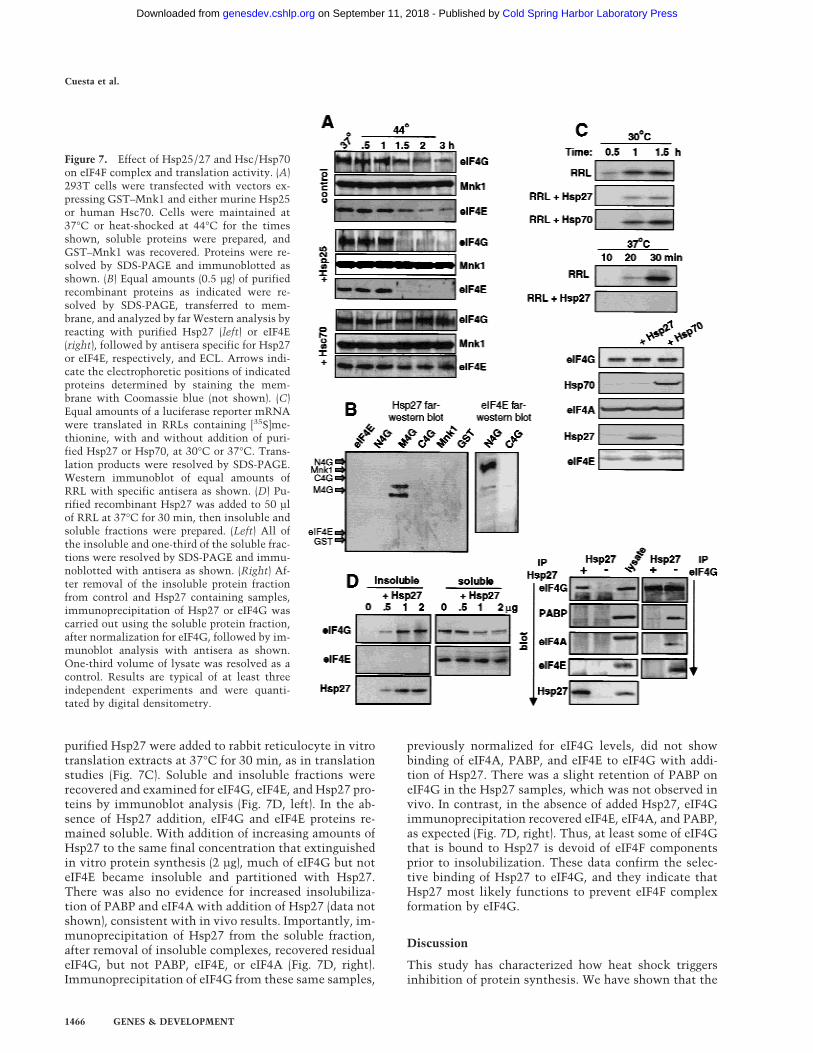
purified Hsp27 were added to rabbit reticulocyte in vitrotranslation extracts at 37°C for 30 min, as in translationstudies (Fig. 7C). Soluble and insoluble fractions wererecovered and examined for eIF4G, eIF4E, and Hsp27 pro-teins by immunoblot analysis (Fig. 7D, left). In the ab-sence of Hsp27 addition, eIF4G and eIF4E proteins re-mained soluble. With addition of increasing amounts ofHsp27 to the same final concentration that extinguishedin vitro protein synthesis (2 µg), much of eIF4G but noteIF4E became insoluble and partitioned with Hsp27.There was also no evidence for increased insolubiliza-tion of PABP and eIF4A with addition of Hsp27 (data notshown), consistent with in vivo results. Importantly, im-munoprecipitation of Hsp27 from the soluble fraction,after removal of insoluble complexes, recovered residualeIF4G, but not PABP, eIF4E, or eIF4A (Fig. 7D, right).Immunoprecipitation of eIF4G from these same samples,
previously normalized for eIF4G levels, did not showbinding of eIF4A, PABP, and eIF4E to eIF4G with addi-tion of Hsp27. There was a slight retention of PABP oneIF4G in the Hsp27 samples, which was not observed invivo. In contrast, in the absence of added Hsp27, eIF4Gimmunoprecipitation recovered eIF4E, eIF4A, and PABP,as expected (Fig. 7D, right). Thus, at least some of eIF4Gthat is bound to Hsp27 is devoid of eIF4F componentsprior to insolubilization. These data confirm the selec-tive binding of Hsp27 to eIF4G, and they indicate thatHsp27 most likely functions to prevent eIF4F complexformation by eIF4G.
Discussion
This study has characterized how heat shock triggersinhibition of protein synthesis. We have shown that the
Figure 7. Effect of Hsp25/27 and Hsc/Hsp70on eIF4F complex and translation activity. (A)293T cells were transfected with vectors ex-pressing GST–Mnk1 and either murine Hsp25or human Hsc70. Cells were maintained at37°C or heat-shocked at 44°C for the timesshown, soluble proteins were prepared, andGST–Mnk1 was recovered. Proteins were re-solved by SDS-PAGE and immunoblotted asshown. (B) Equal amounts (0.5 µg) of purifiedrecombinant proteins as indicated were re-solved by SDS-PAGE, transferred to mem-brane, and analyzed by far Western analysis byreacting with purified Hsp27 (left) or eIF4E(right), followed by antisera specific for Hsp27or eIF4E, respectively, and ECL. Arrows indi-cate the electrophoretic positions of indicatedproteins determined by staining the mem-brane with Coomassie blue (not shown). (C)Equal amounts of a luciferase reporter mRNAwere translated in RRLs containing [35S]me-thionine, with and without addition of puri-fied Hsp27 or Hsp70, at 30°C or 37°C. Trans-lation products were resolved by SDS-PAGE.Western immunoblot of equal amounts ofRRL with specific antisera as shown. (D) Pu-rified recombinant Hsp27 was added to 50 µlof RRL at 37°C for 30 min, then insoluble andsoluble fractions were prepared. (Left) All ofthe insoluble and one-third of the soluble frac-tions were resolved by SDS-PAGE and immu-noblotted with antisera as shown. (Right) Af-ter removal of the insoluble protein fractionfrom control and Hsp27 containing samples,immunoprecipitation of Hsp27 or eIF4G wascarried out using the soluble protein fraction,after normalization for eIF4G, followed by im-munoblot analysis with antisera as shown.One-third volume of lysate was resolved as acontrol. Results are typical of at least threeindependent experiments and were quanti-tated by digital densitometry.
Cuesta et al.
1466 GENES & DEVELOPMENT
Cold Spring Harbor Laboratory Press on September 11, 2018 - Published by genesdev.cshlp.orgDownloaded from

chaperone Hsp27 participates in inhibition of eIF4F(cap)-dependent translation initiation by binding eIF4G,which is associated with a significant reduction in for-mation of eIF4F complexes and insolubilization ofeIF4G. The results largely account for the inhibition ofeIF4E phosphorylation, the disassembly of initiation fac-tor eIF4F, and impaired eIF4F-dependent mRNA transla-tion that is a hallmark of heat shock (Duncan et al. 1987;Duncan and Hershey 1989; Lamphear and Panniers 1990;Zapata et al. 1991; Feigenblum and Schneider 1996). Thecontinued translation of heat shock mRNAs occurs be-cause they require minimal amounts of eIF4F, either byvirtue of initiation through internal ribosome entry, orby ribosome shunting mechanisms (Yueh and Schneider2000; for review, see Schneider 2000).
It is significant that heat shock, in the absence of Hspsynthesis, was not sufficient to strongly insolubilizeeIF4G (Fig. 5). These results implicated overexpression ofcertain Hsps in dissociation of the cap-initiation com-plex (eIF4F). Hsp27 was the only Hsp to significantlycoisolate with eIF4G, and with kinetics coincident withinhibition of translation and insolubilization of eIF4G(Fig. 6B). Moreover, overexpression of Hsp25, the murinehomolog of human Hsp27, enhanced the magnitude ofeIF4G insolubilization during heat shock, whereas over-expression of Hsp70 prevented it (Fig. 7A). These dataindicate that some members of the small Hsp family(Hsp25–27) play an important role in regulating the levelof eIF4F availability for protein synthesis.
Studies have shown previously that in normal cellsHsp25/27 are found in large insoluble complexes. Heatshock induces rapid phosphorylation of these proteins,which is linked to solubilization, followed by dephos-phorylation and insolubilization into perinuclear heatshock granules (Landry et al. 1991; Lambert et al. 1999;Rogalla et al. 1999). New synthesis of Hsp25/27 alsooccurs. The chaperone function of Hsp25–27, the abilityto bind to unfolding proteins and to protect unfoldingproteins against irreversible denaturation, involves thetemporally late insolubilization of Hsp25–27 during heatshock (Lambert et al. 1999; Rogalla et al. 1999). The in-solubilization of eIF4G (Fig. 3) and the loss of eIF4F me-diated by Hsp27 (Fig. 6B) is consistent with this kineticprofile. Insolubilization of Hsp27 during heat shock isassociated with a dramatic increase in cell survival (Ja-kob et al. 1993; Carper et al. 1997; Ehrnsperger et al.1997), linked in part to the ability of Hsp27 to protect thetranslation machinery during recovery from heat shock(Liu et al. 1992; Li et al. 1995; Carper et al. 1997). Hsp27traps unfolding protein intermediates, preventing irre-versible denaturation and aggregation, but it does notactually chaperone protein refolding during recoveryfrom heat shock. In our studies, the inhibition of non-Hsp synthesis during heat shock was in accord with theinsolubilization of eIF4G by Hsp27. There was no evi-dence for insolubilization of other eIF4G-associated fac-tors, including eIF4E, eIF4A, eIF3, Mnk1, and PABP (Figs.3,4,7). There was also little evidence for insolubilizationof eIF4G with any other Hsp chaperones, other thanHsp27 (Fig. 6B). This indicates a specificity to the inter-
action, as opposed to the mere binding of Hsp27 to de-natured eIF4G. This is also supported by the specific invitro binding of Hsp27 to the middle region of eIF4G (Fig.7B). Finally, as some soluble eIF4G was complexed withHsp27 and was devoid of eIF4E, eIF4A, and PABP (Fig.7D), typical components of the eIF4F complex, thisraises two possible mechanism of action whereby trans-lation is inhibited by Hsp27. Hsp27 may preferentiallyinteract with eIF4G that is within the eIF4F complex,thereby acting to remove associated proteins. However,we believe that it is most likely that Hsp27 preferen-tially binds to eIF4G that is no longer within the eIF4Fcomplex, thereby acting to shift the equilibrium toeIF4G removal. It is thought that factors frequently cycleon and off eIF4G, making this second possibility moreattractive. It is also not known whether Hsp27 bindingto the central region of eIF4G leads directly to its insolu-bilization, or represents a primary interaction site fol-lowed by other sites of interaction with Hsp27, leadingto eIF4G insolubilization.
An observation of this work that will require addi-tional study is the possibility that Mnk1 might be freedof eIF4G to then participate in a heat shock signal trans-duction response. Mnk1 retains kinase activity duringheat shock (Fig. 1), despite displacement from eIF4G.Mnk1 is a member of the MAP kinase-activated proteinkinase (MAPKAPK) family (Fukunaga and Hunter 1997;Waskiewicz et al. 1997), and like other members of thisfamily, it is activated by the cell stress-activated p38kinases. It is significant that Mnk1 shares about 30%identity to MAPKAPKs and to a newly identified p38-regulated/activated protein kinase (PRAK), which canphosphorylate Hsp27 in response to cell stress (New etal. 1998). It is possible that Mnk1 might participate inphosphorylation of insolubilized Hsp27 protein, therebypromoting the disaggregation of eIF4G from heat shockgranules and the reactivation of eIF4F(cap)-dependentprotein synthesis.
Materials and methods
Plasmids
Plasmids pEBG–Mnk1, PGEX–Mnk1, pEBG–Mnk1 (T2A2)(Waskiewicz et al. 1997) (kindly provided by J.A. Cooper),pcDNA–HA–eIF4GI (Imataka et al. 1997) (a gift of N. Sonen-berg), pcDNA–Hsc70 (gift from C. Daly), and pCIneo-Hsp25 (giftfrom J. Landry) were described previously. Plasmids expressingGST-fusion amino-terminal (157–626), middle (627–1045), andcarboxy-terminal (1045–1560) segments of eIF4GI were con-structed as follows. An EcoRI–XhoI fragment was prepared byPCR amplification of the corresponding eIF4GI sequence usingspecific primers and cloned into an EcoRI/XhoI-digested pGEX-4T-I vector (Amersham Pharmacia Biotech). The primers usedto amplify each segment were 58-CCCCGAATTCATGTCTG-GGGCCCGC-38 and 58-CAACCTCGAGTCAGAAGTCTGG-GCC-38 for the amino-terminal; 58-CAACGAATTCACTCCA-TCCTTTGCC-38 and 58-AAAACTCGAGTCAGAGCTGGTT-GTTAG-38 for the middle fragment and 58-CAACGAATTCCT-CTTTGCACCTGGAG-38 and 58-CAACCTCGAGTCAGACT-CCTCCTCTG-38 for the carboxy-terminal segment of eIF4GI.
Hsp27 inhibits protein synthesis during heat shock
GENES & DEVELOPMENT 1467
Cold Spring Harbor Laboratory Press on September 11, 2018 - Published by genesdev.cshlp.orgDownloaded from
Cell culture and transient transfection
293 cells are a human embryonic kidney cell line transformedwith the E1 region of Ad5. 293T cells express the SV40 TAg.293, 293T, and HeLa cells were grown in Dulbecco’s modifiedEagle’s medium (DMEM; Gibco) supplemented with 10% calfserum (Hyclone). For transient transfection, 1 × 10⁶ 293 or 293Tcells were passaged onto 10-cm plates 24 hr prior to calciumphosphate precipitation with 5 µg of each plasmid. Cells wereharvested and lysed 48–72 hr later. Heat shock of cells wascarried out at 44°C for 2 hr unless otherwise stated. To stimu-late cells with EGF (Sigma), they were first serum starved for 24hr in serum-free medium then treated with EGF (30 ng/ml) for15 min. Rapamycin (Calbiochem) was added to the cells at afinal concentration of 20 ng/ml and incubation was continuedeither at 37°C or 44°C. Cycloheximide (Calbiochem) was usedat 50 µg/ml.
GST protein purification
To obtain GST fusion proteins from mammalian cells, 293 or293T cells were transiently transfected for 48–72 hr with pEBG–Mnk1, pEBG–Mnk1 (T2A2), or pEBG, lysed in Triton lysisbuffer (1% Triton X-100, 50 mM NaF, 10 mM HEPES at pH 7.4,2 mM EDTA, 2 mM sodium orthovanadate, 0.1% b-mercapto-ethanol, 1 µg/ml aprotinin, 1 µg/ml leupeptin, 1 mM PMSF),and lysates clarified by centrifugation for 15 min at 14,000g at4°C. GST proteins were purified with glutathione–Sepharose 4B(Pharmacia) for 1 hr at 4°C, beads were collected by centrifuga-tion and washed 3× with Triton lysis buffer (for copurificationexperiments) or once with lysis buffer containing 0.5 M LiCl and3× with lysis buffer without LiCl (for in vitro kinase assays).GST fusion proteins were purified from Escherichia coli BL21 asdescribed Smith and Johnson (1988). Removal of the GST affin-ity tail from GST–N4G, GST–M4G, and GST–C4G was carriedout by thrombin cleavage.
Immunoprecipitation and insoluble-soluble protein analysis
293, 293T, or HeLa cells were lysed 48–72 hr post-transfectionin Nonidet P-40 lysis buffer (0.5% NP-40, 50 mM HEPES at pH7.0, 250 mM NaCl, 2 mM EDTA, 2 mM sodium orthovanadate,25 mM glycerophosphate, 1 µg/ml aprotinin, 1 µg/ml leupeptin,1 mM PMSF). Lysates were clarified by centrifugation for 15 minat 14,000g. The pellet was retained as the insoluble fraction, thesupernatant as soluble protein. Pellets were resuspended inRIPA buffer as below. Harsh lysis conditions were carried out inRIPA buffer (1% NP-40, 0.5% sodium deoxycholate, 0.1% SDS,150 mM NaCl, 50 mM Tris-HCl at pH 8.0, 2 mM EDTA, 2 mM
sodium orthovanadate, 50 mM NaF, 1 µg/ml aprotinin, 1 µg/mlleupeptin, 1 mM PMSF). If used directly for Western blotting,cells were incubated at 100°C for 2 min in SDS lysis buffer(0.5% SDS, 50 mM Tris-HCl at pH 8.0,, 1 mM DTT) followed bySDS–polyacrylamide gel electrophoresis. Lysates were incu-bated for 2 hr at 4°C with 2 µg/ml RNase A and rabbit poly-clonal serum against eIF4E (Feigenblum and Schneider 1996), ormouse anti-HA monoclonal antibody (12CA5, Roche MolecularBiochem.). Either protein A- or G–agarose was added and incu-bation continued for 1 hr at 4°C. Precipitates were washed 3×with lysis buffer, boiled in Laemmli sample buffer, and analyzedby SDS-PAGE.
Immunoblotting
Gels were electroblotted onto Immobilon-P membrane (Milli-pore), blocked 12–15 hr in blocking buffer (150 mM NaCl, 10 mM
Tris-HCl at pH 8.0, 5% nonfat dry milk (NFDM)] at 4°C. Im-mune reagents were rabbit polyclonal antisera to eIF4G (gift ofDr. L. Carrasco, CSK/Universidad Autonoma, Madrid, Spain),polyclonal goat antisera to eIF3 (gift of W. Merrick, Case West-ern Reserve University), antisera to eIF4B (gift of N. Sonenberg,McGill University), affinity-purified rabbit polyclonal antibodyto GST (no. Z-5, Santa Cruz Biotech.), rabbit polyclonal anti-eIF4E antiserum, rabbit anti-BP1 polyclonal antibody, goat poly-clonal antibody to Hsp70 (no. K-20, Santa Cruz Biotech.), mousemonoclonal antibody to Hsp27 (Stressgen Biotech.), goat anti-Hsc70 polyclonal antibody (no. K-19, Santa Cruz Biotech.),mouse anti-Hsp90 monoclonal antibody (StressGen Biotech),goat polyclonal antibody to Hsp40 (no. N-19, Santa Cruz Bio-tech), guinea pig anti-Hsp60 polyclonal antibody (gift of N.Cowan, New York University), rabbit anti-Hsp105 polyclonalantibody (no. N-187, Santa Cruz Biotech), horseradish peroxi-dase-conjugated donkey anti-rabbit or sheep anti-mouse second-ary antibodies (Amersham), horseradish peroxidase-conjugateddonkey anti-goat (no. SC-2033, Santa Cruz Biotech), mouseanti-HA monoclonal antibody (12CA5, Roche Molecular Bio-chem.), and enhanced chemiluminescence system (ECL; Amer-sham).
Far Western analysis
Purified N4G, M4G, C4G, eIF4E, GST–Mnk1, and GST wereresolved by electrophoresis in a 10% SDS–polyacrylamide gelsand transferred to Immobilon-P membrane (Millipore). Mem-brane was blocked for 16 hr at 4°C in TBS containing 5% nonfatdry milk and then incubated for 2 hr at RT in blocking buffercontaining 1 µg/ml of either human Hsp27 (Stressgen Biotech.)or eIF4E. Following 3 washes with TBS, complexes were de-tected as described above for immunoblotting.
Metabolic labeling of cells
Cells were labeled with 50 µCi of [35S]methionine per ml (Ea-sytag Express Protein Labeling Mix, Dupont/NEN) in DMEMwithout methionine for 30 min. Cells were lysed in 0.5% NP-40lysis buffer at 4°C and sonicated 2× for 30 sec, and equalamounts of protein were analyzed by SDS-PAGE and fluorogra-phy. Specific activity was determined by trichloroacetic acidprecipitation and liquid scintillation. For 32PO4 labeling, cellswere washed 2× with phosphate-free DMEM (Gibco) incubatedat 37°C for 30 min in this medium supplemented with 1%dialyzed fetal bovine serum, then labeled for 2 hr in [32P]ortho-phosphate (0.1 mCi/ml; DuPont/NEN). Cells were lysed in0.5% NP-40 buffer and eIF4E was immunoprecipitated as de-scribed above.
Kinase assays
Wild-type Mnk1 and a catalytically inactive Mnk1 (T2A2) wereassayed for kinase activity in vitro by addition of 1 µg purifiedeIF4E to a purified GST-bead mixture. Kinase–substrate reac-tions were carried out at 30°C for 20 min in kinase buffer [20mM HEPES at pH 7.4, 10 mM MgCl2, 10 mM b-glycerophosphate,2 mM sodium orthovanadate, 1 mM DTT, 25 µM ATP, 10 µCi[g-32P]ATP (3,000 Ci/mmol)]. Products were resolved by SDS-PAGE and visualized by autoradiography.
Immunofluorescence microscopy
HeLa cells grown on coverslips were heat-shocked for the indi-cated times, 24 hr after passage. Cells were fixed with acetone/methanol (7:3) for 7 min at −20°C, blocked in 1× PBS–1%NFDM for 30 min at 25°C, then incubated with antibodiesagainst eIF4G, Hsp70, or Hsp27 in PBS–1% NFDM for 1 hr at
Cuesta et al.
1468 GENES & DEVELOPMENT
Cold Spring Harbor Laboratory Press on September 11, 2018 - Published by genesdev.cshlp.orgDownloaded from
37°C in a humidified chamber. Fixed cells were washed 4× withPBS–1% NFDM, reacted with anti-rabbit IgG-fluorescein,F(ab8)2 fragment and anti-mouse Ig(polyvalent)–rhodamine,F(ab8)2 fragment for 1 hr at 37°C in a humidified chamber, thenwashed 4× with PBS–1% NFDM and mounted using Victashieldmounting media (Vector Laboratories).
In vitro translation studies
Equal amounts of in vitro transcribed luciferase mRNA weretranslated in 40 µl of RRL (Promega) supplemented with 20 µCiof [35S]methionine, or in RRL containing 2 µg of purified recom-binant Hsp27 or Hsp70 (Stressgen) at 30°C for up to 2 hr or at37°C for up to 30 min. Equal aliqouts were removed at differenttime points, resolved by SDS-PAGE, and visualized by autora-diography or immunoblotting. Equal aliqouts were partitionedinto insoluble protein by centrifugation in a microfuge at14,000g for 5 min at 4°C, and the supernatant was retained asthe soluble fraction.
Acknowledgments
We thank N. Sonenberg for the HA–eIF4GI clone and antisera toeIF4B, J. Landry for the Hsp25 clone, J. Cooper for GST–Mnk1clones, I. Novoa and L. Carrasco for antisera to eIF4G, G. Drey-fuss for PABP antibody, and N. Cowan for Hsp60 antibodies,and W. Merrick for eIF3 antisera. This work was supported byNIH grant CA 42357 (to R.J.S.) and by a fellowship from Fun-dacion Ramon Areces, Spain (to R.C.).
The publication costs of this article were defrayed in part bypayment of page charges. This article must therefore be herebymarked “advertisement” in accordance with 18 USC section1734 solely to indicate this fact.
References
Beretta, L., A.-C. Gingras, Y.V. Svitkin, M.N. Hall, and N.Sonenberg. 1996. Rapamycin blocks the phosphorylation of4E-BP1 and inhibits cap-dependent initiation of translation.EMBO J. 15: 658–664.
Carper, S.W., T. Rocheleau, D. Cimino, and F.K. Storm. 1997.Hsp 27 stimulates recovery of RNA and protein synthesisfollowing a heat shock. J. Cell. Biochem. 66: 153–164.
Craig, E.A., J.S. Weissman, and A.L. Horwich. 1994. Hsps andmolecular chaperones: mediators of protein conformationand turnover in the cell. Cell 78: 365–372.
Duncan, R.F. 1996. Translational control during heat shock. InTranslational control (ed. J.W.B. Hershey, M.B. Mathews,and N. Sonenberg), pp. 271–294. Cold Spring Harbor Labora-tory Press, Cold Spring Harbor, NY.
Duncan, R.F. and J.W.B. Hershey. 1989. Protein synthesis andprotein phosphorylation during heat stress, recovery, and ad-aptation. J. Cell. Biol. 109: 1467–1481.
Duncan, R., S.C. Milburn, and J.W.B. Hershey. 1987. Regulatedphosphorylation and low abundance of Hela cell initiationfactor eIF-4F suggest a role in translational control. J. Biol.Chem. 262: 380–388.
Ehrnsperger, M., S. Graber, M. Gaestel, and J. Buchner. 1997.Binding of nonnative protein to Hsp25 during heat shockcreates a reservoir of folding intermediates for reactivation.EMBO J. 16: 221–229.
Feigenblum, D. and R.J. Schneider. 1993. Modification of eu-karyotic initiation factor 4F during infection by influenzavirus. J. Virol. 67: 3027–3035.
———. 1996. Cap-binding protein (eukaryotic initiation factor
4E) and 4E-inactivating protein BP-1 independently regulatecap-dependent translation. Mol. Cell. Biol. 16: 5450–5457.
Feigenblum, D., R. Walker, and R.J. Schneider. 1998. Adenovi-rus induction of an interferon-regulatory factor during entryinto the late phase of infection. J. Virol. 72: 9257–9266.
Fukunaga, R. and T. Hunter. 1997. MNK1, a new MAP kinase-activated protein kinase, isolated by a novel expressionscreening method for identifying protein kinase substrates.EMBO J. 16: 1921–1933.
Glover, J.R. and S. Lindquist. 1998. Hsp104, Hsp70, and Hsp40:A novel chaperone system that rescues previously aggre-gated proteins. Cell 94: 73–82.
Haghighat, A., S. Mader, A. Pause, and N. Sonenberg. 1995.Repression of cap-dependent translation by 4E-binding pro-tein 1: Competition with p220 for binding to eukaryotic ini-tiation factor-4E. EMBO J. 14: 5701–5709.
Huang, J. and R.J. Schneider. 1991. Adenovirus inhibition ofcellular protein synthesis involves inactivation of cap bind-ing protein. Cell 65: 271–280.
Imataka, H., H.S. Olsen, and N. Sonenberg. 1997. A new trans-lational regulator with homology to eukaryotic translationinitiation factor 4G. EMBO J. 16: 817–825.
Imataka, H., A. Gradi, and N. Sonenberg. 1998. A newly iden-tified N-terminal amino acid sequence of human eIF4Gbinds poly(A)-binding protein and functions in poly(A)-de-pendent translation. EMBO J. 17: 7480–7489.
Jakob, U., M. Gaestal, K. Engel, and J. Buchner. 1993. Small heatshock proteins are molecular chaperons. J. Biol. Chem. 268:1517–1520.
Lambert, H., S.J. Charette, A.F. Bernier, A. Guimond, and J.Landry. 1999. HSP27 multimerization mediated by phos-phorylation-sensitive intermolecular interactions at theamino terminus. J. Biol. Chem. 274: 9378–9385.
Lamphear, B.J. and R. Panniers. 1990. Cap binding protein com-plex that restores protein synthesis in heat shocked Ehrlichcell lysates contains highly phosphorylated eIF-4E. J. Biol.Chem. 265: 5333–5336.
———. 1991. Heat shock impairs the interaction of cap bindingprotein complex with 58 mRNA cap. J. Biol. Chem. 266:2789–2794.
Landry, J., P. Chretien, A. Laszlo, and H. Lambert. 1991. Phos-phorylation of HSP27 during development and decay of ther-motolerance in Chinese hamster cells. J. Cell. Physiol.147: 93–101.
Laroia, G., R. Cuesta, and R.J. Schneider. 1999. Control ofmRNA decay by the ubiquitinproteasome-heat shock net-work. Science 284: 499–502.
Li, L., G. Shen, and G.C. Li. 1995. Effects of expressing humanHsp70 and its deletion derivatives on heat killing and onRNA and protein synthesis. Exp. Cell. Res. 217: 460–468.
Lin, T.-A., X. Kong, T.A.J. Haystead, A. Pause, G. Belsham, N.Sonenberg, and J.C. Lawrence. 1994. PHAS-1 as a link be-tween mitogen-activated protein kinase and translation ini-tiation. Science 266: 653–656.
Liu, R.Y., X. Li, L. Li, and G.C. Li. 1992. Expression of humanHsp70 in rat fibroblasts enhances cell survival and facilitatesrecovery from translational and transcriptional inhibitionfollowing heat shock. Cancer Res. 52: 3667–3673.
Marissen, W.E. and R.E. Lloyd. 1998. Eukaryotic translation ini-tiation factor 4G is targeted for proteolytic cleavage bycaspase 3 during inhibition of translation in apoptotic cells.Mol. Cell. Biol. 18: 7565–7574.
Mizzen, L.A. and W.J. Welch. 1988. Characterization of thethermotolerant cell. I. Effects on protein synthesis activityand the regulation of heat-shock protein 70 expression. J.Cell. Biol. 106: 1105–1116.
Hsp27 inhibits protein synthesis during heat shock
GENES & DEVELOPMENT 1469
Cold Spring Harbor Laboratory Press on September 11, 2018 - Published by genesdev.cshlp.orgDownloaded from
Morimoto, R.I. 1998. Regulation of the heat shock transcrip-tional response: Cross talk between a family of heat shockfactors, molecular chaperones, and negative regulators.Genes & Dev. 12: 3788–3796.
New, L., Y. Jiang, M. Zhao, K. Liu, W. Zhu, L.J. Flood, Y. Kato,G.C. Parry, and J. Han. 1998. PRAK, a novel protein kinaseregulated by the p38 MAP kinase. EMBO J. 17: 3372–3384.
Parsell, D.A. and S. Lindquist. 1993. The function of heat shockproteins in stress tolerance: Degradation and reactivation ofdamaged proteins. Annu. Rev. Genet. 27: 437–496.
Pause, A., G.J. Belsham, A.-C. Gingras, O. Donze, T.-A. Lin, J.C.Lawrence, and N. Sonenberg. 1994. Insulin-dependentstimulation of protein synthesis by phosphorylation of aregulator of 58-cap function. Nature 371: 762–767.
Pyronnet, S., H. Imataka, A.C. Gingras, R. Fukunaga, T. Hunter,and N. Sonenberg. 1999. Human eukaryotic translation ini-tiation factor 4G (eIF4G) recruits Mnk1 to phosphorylateeIF4E. EMBO J. 18: 270–279.
Rogalla, T., M. Ehrnsperger, X. Preville, A. Kotlyarov, G.Lutsch, C. Ducasse, C. Paul, M. Wieske, A.P. Arrigo, J. Buch-ner, and M. Gaestel. 1999. Regulation of Hsp27 oligomeriza-tion, chaperone function, and protective activity against oxi-dative stress/tumor necrosis factor alpha by phosphoryla-tion. J. Biol. Chem. 274: 18947–18956.
Schneider, R.J. 2000. Translational control during heat shock. InTranslational control (ed. J.W.B. Hershey, M. Mathews, andN. Sonenberg) Cold Spring Harbor Laboratory Press, ColdSpring Harbor, NY. (In press.)
Smith, D.B. and K.S. Johnson. 1988. Single-step purification ofpolypeptides expressed in Escherichia coli as fusions withglutathione S-transferase. Gene 67: 31–40.
Sonenberg, N. 1996. mRNA 58 cap-binding protein eIF-4E andcontrol of cell growth. In Translational Control (ed. J.W.B.Hershey, M. Mathews, and N. Sonenberg), pp. 245–270. ColdSpring Harbor Laboratory Press, Cold Spring Harbor, NY.
Vries, R.G., A. Flynn, J.C. Patel, X. Wang, R.M. Denton, andC.G. Proud. 1997. Heat shock increases the association ofbinding protein-1 with initiation factor 4E. J. Biol. Chem.272: 32779–32784.
Wang, X., A. Flynn, A.J. Waskiewicz, B.L.J. Webb, R.G. Vries,I.A. Baines, J.A. Cooper, and C.G. Proud. 1998. The phos-phorylation of eukaryotic initiation factor eIF-4E in responseto phorbol esters, cell stresses and cytokines is mediated bydistinct MAP kinase pathways. J. Biol. Chem. 273: 9373–9377.
Waskiewicz, A.J., A. Flynn, C.G. Proud, and J.A. Cooper. 1997.Mitogen-activated protein kinases activate the serine/threo-nine kinases Mnk1 and Mnk2. EMBO J. 16: 1909–1920.
Waskiewicz, A.J., J.C. Johnson, B. Penn, M. Mahalingham, S.R.Kimball, and J.A. Cooper. 1999. Phosphorylation of the cap-binding protein eukaryotic translation initiation factor 4E byprotein kinase Mnk1 in vivo. Mol. Cell. Biol. 19: 1871–1880.
Yueh, A. and R.J. Schneider. 1996. Selective translation by ri-bosome jumping in adenovirus infected and heat shockedcells. Genes & Dev. 10: 1557–1567.
———. 2000.Translation by ribosome shunting on adenovirusand Hsp70 mRNAs facilitated by complementarity to 18SrRNA. Genes & Dev. 14: 414–421.
Zapata, J.M., F.G. Maroto, and J.M. Sierra. 1991. Inactivation ofmRNA cap-binding protein complex in Drosophila melano-gaster embryos under heat shock. J. Biol. Chem. 266: 16007–16014.
Cuesta et al.
1470 GENES & DEVELOPMENT
Cold Spring Harbor Laboratory Press on September 11, 2018 - Published by genesdev.cshlp.orgDownloaded from

10.1101/gad.14.12.1460Access the most recent version at doi: 14:2000, Genes Dev.
Rafael Cuesta, Gaurav Laroia and Robert J. Schneider eIF4G and facilitating dissociation of cap-initiation complexesChaperone Hsp27 inhibits translation during heat shock by binding
References
http://genesdev.cshlp.org/content/14/12/1460.full.html#ref-list-1
This article cites 40 articles, 29 of which can be accessed free at:
License
ServiceEmail Alerting
click here.right corner of the article or
Receive free email alerts when new articles cite this article - sign up in the box at the top
Cold Spring Harbor Laboratory Press
Cold Spring Harbor Laboratory Press on September 11, 2018 - Published by genesdev.cshlp.orgDownloaded from