Embed Size (px)
Citation preview

9/19/11
1
Chapter 9
The Autonomic Nervous System
Lecture PowerPoint
Copyright © The McGraw-Hill Companies, Inc. Permission required for reproduction or display.
I. Neural Control of Involuntary Effectors
Autonomic Motor Nerves
• Innervate organs not under voluntary control
• Effectors include: – Cardiac muscle – Smooth muscle of visceral organs and blood
vessels – Glands
Autonomic Neurons • Somatic motor neurons have cell bodies in
the spinal cord and just one neuron traveling from spinal cord to effector.
• The autonomic motor system has two sets of neurons in the PNS. – The first has cell bodies in the brain or spinal
cord and synapses in an autonomic ganglion. – The second has cell bodies in the ganglion
and synapses on the effector.
Autonomic Neurons
• Preganglionic neurons: originate in the midbrain or hindbrain or from the thoracic, lumbar, or sacral spinal cord
• Postganglionic neurons: originate in ganglion
Autonomic Ganglia
• Located in the head, neck, and abdomen as well as in chains along either side of the spinal cord
9/19/11
2
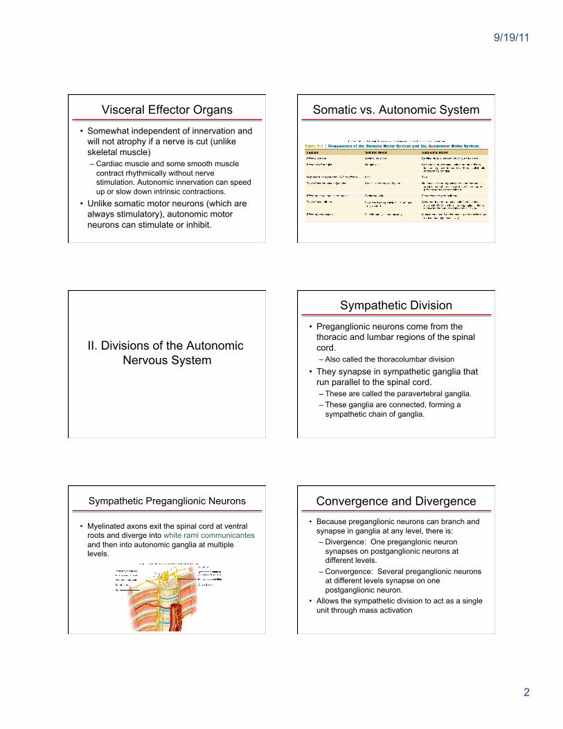
Visceral Effector Organs • Somewhat independent of innervation and
will not atrophy if a nerve is cut (unlike skeletal muscle) – Cardiac muscle and some smooth muscle
contract rhythmically without nerve stimulation. Autonomic innervation can speed up or slow down intrinsic contractions.
• Unlike somatic motor neurons (which are always stimulatory), autonomic motor neurons can stimulate or inhibit.
Somatic vs. Autonomic System
II. Divisions of the Autonomic Nervous System
Sympathetic Division • Preganglionic neurons come from the
thoracic and lumbar regions of the spinal cord. – Also called the thoracolumbar division
• They synapse in sympathetic ganglia that run parallel to the spinal cord. – These are called the paravertebral ganglia. – These ganglia are connected, forming a
sympathetic chain of ganglia.
Sympathetic Preganglionic Neurons
• Myelinated axons exit the spinal cord at ventral roots and diverge into white rami communicantes and then into autonomic ganglia at multiple levels.
Convergence and Divergence • Because preganglionic neurons can branch and
synapse in ganglia at any level, there is: – Divergence: One preganglonic neuron
synapses on postganglionic neurons at different levels.
– Convergence: Several preganglionic neurons at different levels synapse on one postganglionic neuron.
• Allows the sympathetic division to act as a single unit through mass activation

9/19/11
3
Sympathetic Postganglionic Neurons
• Unmyelinated axons of the postganglionic neurons form the gray rami communicantes, which return to the spinal nerve and travel with other spinal neurons to their effectors.
Collateral Ganglia • Many of the sympathetic neurons that exit the
spinal cord below the diaphragm do not synapse in the sympathetic chain of ganglia.
• Instead, they form splanchnic nerves, which synapse in collateral ganglia. – Collateral ganglia include celiac, superior
mesenteric, and inferior mesenteric ganglia. – Postganglionic neurons innervate organs of
the digestive, urinary, and reproductive systems.
Sympathetic Neuron Pathways Collateral Sympathetic Ganglia
Adrenal Glands
• The adrenal medulla secretes epinephrine and norepinephrine when stimulated by the sympathetic nervous system.
• Embryologically, the adrenal medulla is a modified ganglion and is innervated directly by preganglionic sympathetic neurons.
Summary of the Sympathetic Division

9/19/11
4
Parasympathetic Division
• Preganglionic neurons come from the brain or sacral region of the spinal cord. – Also called the craniosacral division
• They synapse on ganglia located near or in effector organs. – Called terminal ganglia
Parasympathetic Division
• Preganglionic neurons do not travel with somatic neurons (as sympathetic postganglionic neurons do).
– Effectors in the skin and skeletal muscles (sweat glands, blood vessels) receive sympathetic but not parasympathetic innervation.
Cranial Nerves and the Parasympathetic Division
• The occulomotor, facial, glosso-pharyngeal, and vagus nerves carry parasympathetic preganglionic neurons.
– Occulomotor: Preganglionic fibers exit midbrain and synapse on the ciliary ganglion.
– Postganglionic fibers innervate the ciliary muscle of the eye.
Cranial Nerves and the Parasympathetic Division
• Facial nerve: Preganglionic fibers exit the pons and synapse in: – Pterygopalatine ganglion: Postganglionic
fibers synapse on nasal mucosa, pharynx, palate, and lacrimal glands.
– Submandibular ganglion: Postganglionic fibers synapse on salivary glands.
– Glossopharyngeal: Preganglionic fibers synapse on otic ganglion. Postganglionic fibers innervate salivary gland.
Cranial Nerves and the Parasympathetic Division
Vagus: Preganglionic fibers exit medulla, branch into several plexi and nerves, and travel to ganglia within effector organs (heart, lungs, esophagus, stomach, pancreas, liver, intestines).
Cranial Nerves and the Parasympathetic Division

9/19/11
5
Sacral Nerves
• Preganglionic nerves from the sacral region of the spinal cord provide innervation to the lower part of the large intestine, rectum, urinary and reproductive organs.
• Terminal ganglia are located within these organs.
Summary of Parasympathetic Division
Comparison of the Sympathetic and Parasympathetic Divisions
III. Functions of the Autonomic Nervous System
Sympathetic Functions
• The sympathetic division activates the body for “fight or flight” through the release of norepinephrine from postganglionic neurons and the secretion of epinephrine from the adrenal medulla. – Prepares the body for intense physical
activity in emergencies by increasing heart rate and blood glucose levels and by diverting blood to skeletal muscles
Parasympathetic Functions
• The parasympathetic division is antagonistic to the sympathetic division.
• Allows the body to “rest and digest” through the release of ACh from postganglionic neurons – Slows heart rate, dilates visceral blood
vessels, increases digestive activities

9/19/11
6
Summary of Autonomic Functions Cholinergic Synaptic Transmission
• Acetylcholine (ACh) is the neurotransmitter used by all preganglionic neurons. – It is also the neurotransmitter released from most parasympathetic postganglionic neurons.
– Some sympathetic postganglionic neurons (those that innervate sweat glands and skeletal muscle blood vessels) release ACh.
– These synapses are called cholinergic.
Adrenergic Synaptic Transmission
• Norepinephrine is the neurotransmitter released by most sympathetic postganglionic neurons.
– These synapses are called adrenergic.
Adrenergic Synaptic Transmission
Neurotransmitters of the Autonomic Nervous System Varicosities
• Axons of postganglionic neurons have various swellings called varicosities that release neurotransmitter along the length of the axon.
• They form “synapses en passant” in passing.
• Sympathetic and parasympathetic neurons innervate the same tissues.

9/19/11
7
Varicosities Response to Adrenergic Stimulation
• Can stimulate or inhibit, depending on receptors
– Stimulation: heart, dilatory muscles of the iris, smooth muscles of some blood vessels (causes vessel constriction)
– Inhibition: Bronchioles in lungs, other blood vessels; inhibits contraction and causes dilation of these structures
α and β Adrenergic Receptors
• Two types of α (α1 and α2) • Two types of β (β1 and β2) • All act using G-proteins and second
messenger systems. – β receptors use cAMP. – α receptors use a Ca2+ second messenger
system.
α and β Adrenergic Receptors • Adrenergic effects in different organs
α2 Receptors
• Located on presynaptic axons • When stimulated, result in inhibition of
norepinephrine release in the synapse – May be a negative-feedback system – Some drugs to lower blood pressure act on
these α2 receptors to inhibit presynaptic neurons in the brain, inhibiting the whole sympathetic nervous system.
Response to Cholinergic Stimulation
• ACh released from preganglionic neurons of both the sympathetic and parasympathetic division is stimulatory.
• ACh from postganglionic neurons of the parasympathetic division can be stimulatory or inhibitory, depending on receptors.

9/19/11
8
Cholinergic Receptors
• Nicotinic: found in autonomic ganglia – Stimulated by ACh – Serve as ion channels
• Muscarinic: found in visceral organs – Five types identified; can be stimulatory or
inhibitory (opening K+ or Ca2+ channels) – Use G-proteins and second messenger
system
ACh Receptor Function
ACh Receptor Structure Summary of Receptor Activity in the
Autonomic Nervous System
Other Autonomic Neurotransmitters
• Some postganglionic autonomic neurons are not inactivated by drugs that block ACh or norepinephrine activity.
• Called “nonadrenergic, noncholinergic fibers”
• Proposed neurotransmitters include ATP, vasoactive intestinal peptide, and nitric oxide.
Nonadrenergic, Noncholinergic Fibers
• Important for erection of the penis. • Parasympathetic neurons innervate blood
vessels, causing relaxation and vasodilation using NO.
• NO can also produce smooth muscle relaxation in the stomach, intestines, and urinary bladder.

9/19/11
9
Organs with Dual Innervation
• Most visceral organs are innervated by both sympathetic and parasympathetic neurons.
• Most of the time these systems are antagonists: – Heart rate – Digestive functions – Pupil diameter
Cooperative Effects • Occur when both divisions produce
different effects that work together to promote a single action: – Erection and ejaculation: Parasympathetic
division causes vasodilation and erection; sympathetic causes ejaculation
– Urination: Parasympathetic division aids in urinary bladder contraction; sympathetic helps with bladder muscle tone to control urination.
Complementary Effects
• Occur when both divisions produce similar effects on the same target
– Salivary gland secretion: Parasympathetic division stimulates secretion of watery saliva; sympathetic constricts blood vessels so the secretion is thicker.
Organs Without Dual Innervation
• The following organs are innervated by the sympathetic division only: – Adrenal medulla – Arrector pili muscles in skin – Sweat glands in skin – Most blood vessels
• Regulated by increase and decrease in sympathetic nerve activity
• Important for body temperature regulation
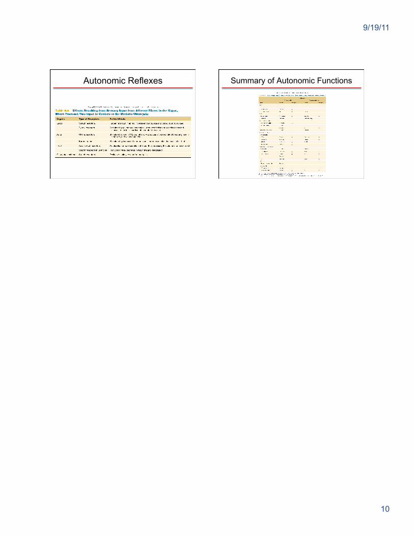
Control of ANS by the Brain
• Many visceral functions are regulated by autonomic reflexes. – Sensory input is sent to brain centers (usually
by the vagus nerve), which integrate the information and modify the activity of preganglionic neurons.
– Medulla oblongata controls many cardiovascular, pulmonary, urinary, reproductive, and digestive functions.
Regulation of the Medulla
• Higher brain regions regulate the medulla.
– Hypothalamus: major regulatory center of the ANS
– Limbic system: responsible for autonomic responses during emotional states (blushing, pallor, fainting, sweating, racing heart rate)
9/19/11
10
Autonomic Reflexes Summary of Autonomic Functions