Embed Size (px)
Citation preview
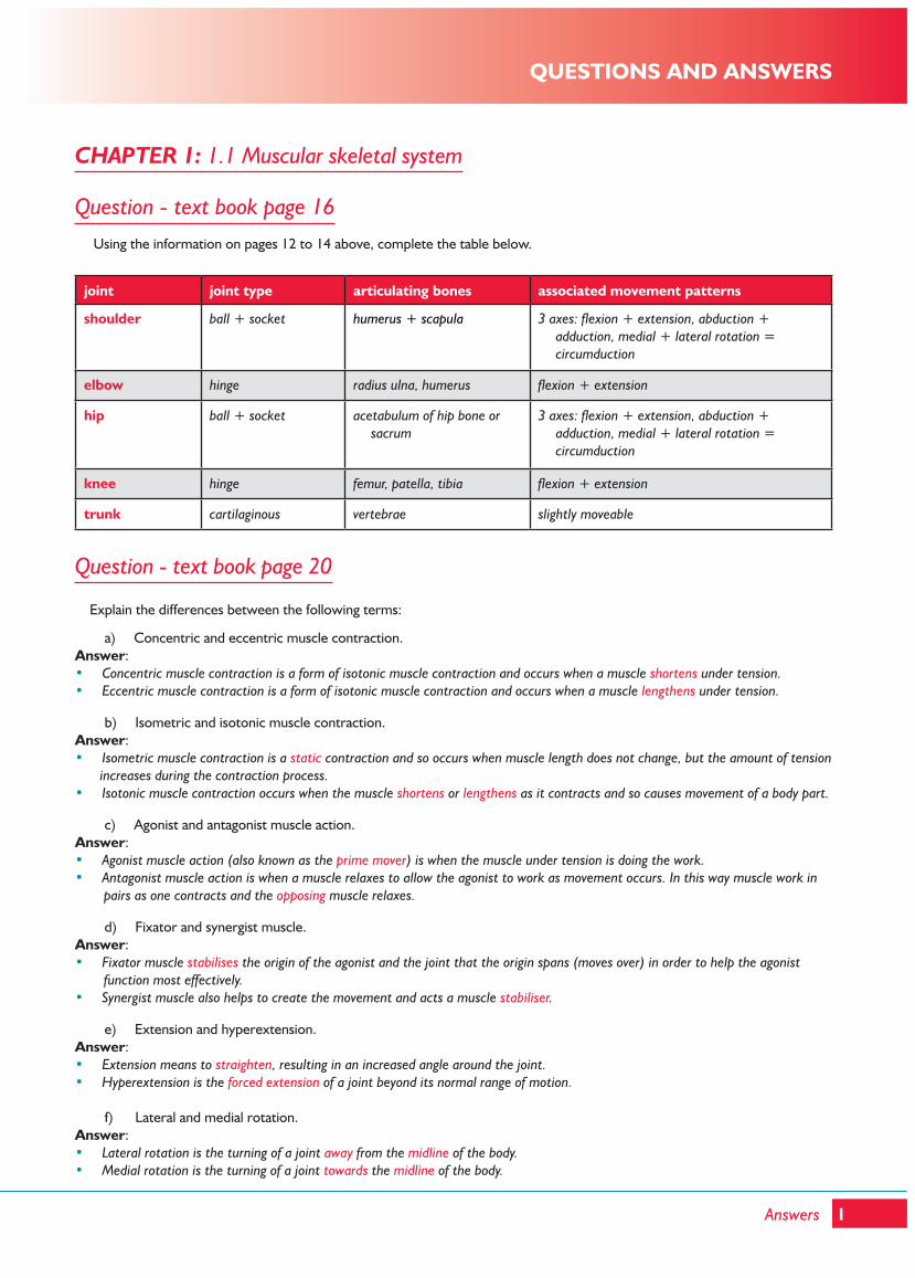
CHAPTER 1: 1.1 Muscular skeletal system
Question - text book page 16 Using the information on pages 12 to 14 above, complete the table below.
joint joint type articulating bones associated movement patterns
shoulder ball + socket humerus + scapula 3 axes: flexion + extension, abduction + adduction, medial + lateral rotation = circumduction
elbow hinge radius ulna, humerus flexion + extension
hip ball + socket acetabulum of hip bone or sacrum
3 axes: flexion + extension, abduction + adduction, medial + lateral rotation = circumduction
knee hinge femur, patella, tibia flexion + extension
trunk cartilaginous vertebrae slightly moveable
Question - text book page 20
Explain the differences between the following terms:
a) Concentric and eccentric muscle contraction.Answer:• Concentric muscle contraction is a form of isotonic muscle contraction and occurs when a muscle shortens under tension. • Eccentric muscle contraction is a form of isotonic muscle contraction and occurs when a muscle lengthens under tension.
b) Isometric and isotonic muscle contraction.Answer:• Isometric muscle contraction is a static contraction and so occurs when muscle length does not change, but the amount of tension
increases during the contraction process.• Isotonic muscle contraction occurs when the muscle shortens or lengthens as it contracts and so causes movement of a body part.
c) Agonist and antagonist muscle action.Answer:• Agonist muscle action (also known as the prime mover) is when the muscle under tension is doing the work.• Antagonist muscle action is when a muscle relaxes to allow the agonist to work as movement occurs. In this way muscle work in
pairs as one contracts and the opposing muscle relaxes.
d) Fixator and synergist muscle.Answer:• Fixator muscle stabilises the origin of the agonist and the joint that the origin spans (moves over) in order to help the agonist
function most effectively. • Synergist muscle also helps to create the movement and acts a muscle stabiliser.
e) Extension and hyperextension.Answer:• Extension means to straighten, resulting in an increased angle around the joint.• Hyperextension is the forced extension of a joint beyond its normal range of motion.
f) Lateral and medial rotation. Answer:• Lateral rotation is the turning of a joint away from the midline of the body.• Medial rotation is the turning of a joint towards the midline of the body.
1Answers
QUESTIONS AND ANSWERS
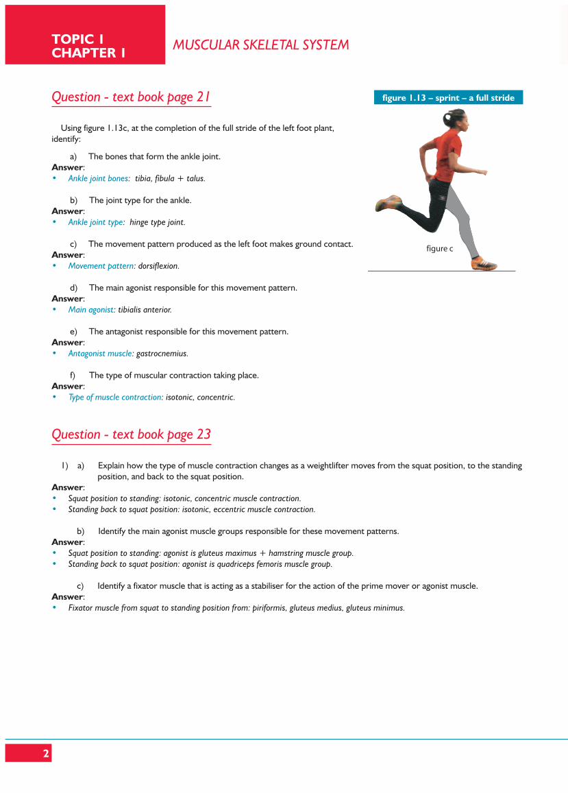
Question - text book page 21 Using figure 1.13c, at the completion of the full stride of the left foot plant, identify:
a) The bones that form the ankle joint.Answer:• Ankle joint bones: tibia, fibula + talus.
b) The joint type for the ankle.Answer:• Ankle joint type: hinge type joint.
c) The movement pattern produced as the left foot makes ground contact.Answer:• Movement pattern: dorsiflexion.
d) The main agonist responsible for this movement pattern.Answer:• Main agonist: tibialis anterior.
e) The antagonist responsible for this movement pattern.Answer:• Antagonist muscle: gastrocnemius.
f) The type of muscular contraction taking place.Answer:• Type of muscle contraction: isotonic, concentric.
Question - text book page 23 1) a) Explain how the type of muscle contraction changes as a weightlifter moves from the squat position, to the standing position, and back to the squat position.Answer:• Squat position to standing: isotonic, concentric muscle contraction. • Standing back to squat position: isotonic, eccentric muscle contraction.
b) Identify the main agonist muscle groups responsible for these movement patterns.Answer:• Squat position to standing: agonist is gluteus maximus + hamstring muscle group. • Standing back to squat position: agonist is quadriceps femoris muscle group.
c) Identify a fixator muscle that is acting as a stabiliser for the action of the prime mover or agonist muscle.Answer:• Fixator muscle from squat to standing position from: piriformis, gluteus medius, gluteus minimus.
MUSCULAR SKELETAL SYSTEMTOPIC 1 CHAPTER 1
2
figure 15 - sprint - a full stride
figure a figure b figure c
figure 1.13 – sprint – a full stride
Questions - text book page 24 1) Hockey involves movement at many joints in the body. Identify which bones articulate at each of the following joints:
a) Shoulder.Answer:• Shoulder: Scapula, humerus.
b) Knee.Answer:• Knee: Femur, tibia, patella.
c) Elbow.Answer:• Elbow: Radius, ulna, humerus.
d) Hip.Answer:• Hip: Femur, acetabulum of hip bone or sacrum. e) Ankle.Answer:• Ankle: Talus, tibia and fibula
2) Complete gaps in the table below naming the main agonist and antagonist in the named activity:
action main agonist main antagonist
elevating the shoulder trapezius lattissimus dorsi
extending the elbow joint triceps biceps
flexing the knee joint hamstrings group quadriceps group
dorsiflexing the ankle joint tibialis anterior calf muscles - gastrocnemeus, soleus
flexing the trunk rectus abdominus erector spinae
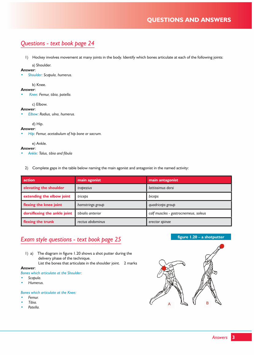
Exam style questions - text book page 25
1) a) The diagram in figure 1.20 shows a shot putter during the delivery phase of the technique. List the bones that articulate in the shoulder joint. 2 marksAnswer:Bones which articulate at the Shoulder: • Scapula.• Humerus.
Bones which articulate at the Knee:• Femur.• Tibia.• Patella.
QUESTIONS AND ANSWERS
3Answers
figure 1.20 – a shotputter
1) b) Briefly explain the movement sequence of the right arm during the delivery phase of the shot put. 3 marksAnswer:From diagram A: • Shoulder is extended and abducted, elbow is flexed, wrist is pronated and hyper extended.
From diagram B: • Shoulder is flexed and elevated, elbow extended,.• Wrist is pronated and extended, and phalanges are extended.
2) Describe the following movement terminology, and give a physical activity for each movement: Abduction, circumduction, rotation and plantarflexion. 8 marksAnswer:• Abduction is a movement pattern in which a body segment moves away from the midline of the body. • Example: leg action going into a cartwheel.
• Circumduction is a movement pattern which consists of flexion, abduction, extension and adduction to create a cone shape in space.
• Example: a small circular hand dance gesture.
• Rotation is a movement pattern around the axis down the centre of a long bone. • Example: delivery phase of a discus throw.
• Plantarflexion is a movement pattern of the foot that results in the toe being pointed downwards. • Example: diving off a springboard.
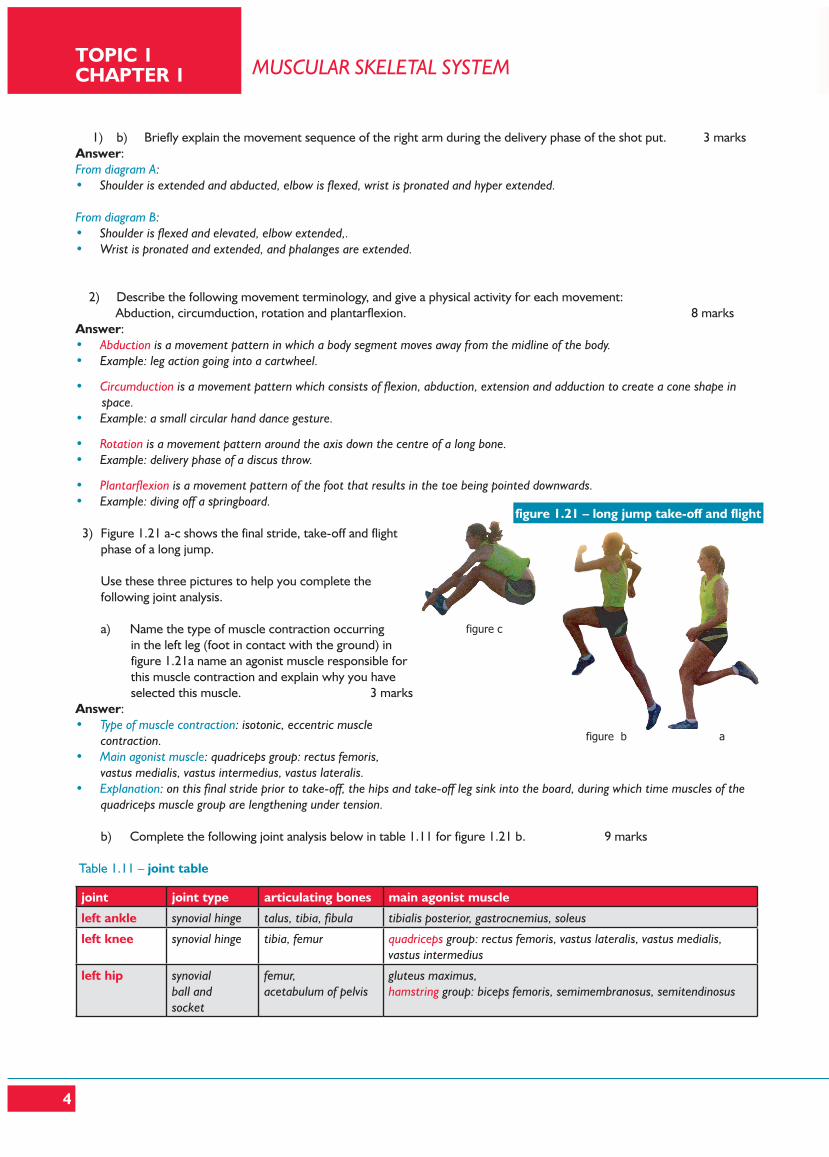
3) Figure 1.21 a-c shows the final stride, take-off and flight phase of a long jump.
Use these three pictures to help you complete the
following joint analysis.
a) Name the type of muscle contraction occurring in the left leg (foot in contact with the ground) in figure 1.21a name an agonist muscle responsible for this muscle contraction and explain why you have selected this muscle. 3 marks
Answer:• Type of muscle contraction: isotonic, eccentric muscle
contraction.• Main agonist muscle: quadriceps group: rectus femoris,
vastus medialis, vastus intermedius, vastus lateralis.• Explanation: on this final stride prior to take-off, the hips and take-off leg sink into the board, during which time muscles of the
quadriceps muscle group are lengthening under tension.
b) Complete the following joint analysis below in table 1.11 for figure 1.21 b. 9 marks
Table 1.11 – joint table
joint joint type articulating bones main agonist muscle
left ankle synovial hinge talus, tibia, fibula tibialis posterior, gastrocnemius, soleus
left knee synovial hinge tibia, femur quadriceps group: rectus femoris, vastus lateralis, vastus medialis,vastus intermedius
left hip synovialball andsocket
femur,acetabulum of pelvis
gluteus maximus,hamstring group: biceps femoris, semimembranosus, semitendinosus
figure 30 - long jump take-off and flight
figure b a
figure c
figure 1.21 – long jump take-off and flight
MUSCULAR SKELETAL SYSTEMTOPIC 1 CHAPTER 1
4
3) c) Describe the changes in movement patterns in the left ankle, knee, hip and trunk from figures 1.21 b to c. 4 marksAnswer:• Left ankle changes from plantarflexion to dorsiflexion.• Left knee changes from extension to flexion.• Left hip changes from extension to flexion.• Trunk changes from extension to flexion.
d) Suggest two factors which could affect the range of movement at the hip joint. 2 marksAnswer:• Shape of ball and socket joint, or the shape of bones.• Extensibility of attached muscles or muscle tendons.• Elasticity of attached ligaments.• Length of muscle or bulk of muscle. e) Identify the predominant fibre type (refer to page 63) stressed during the take-off and give two reasons why this fibre type would be used. Identify the type of muscle contraction occurring during the take-off phase of the long jump Answer: 4 marks• Fast twitch glycolytic fibre (type IIb) during the take-off phase. Reasons: • Fast contraction speed.• High power output/force exerted.• High anaerobic capacity. Type of muscle contraction:• Isotonic concentric muscle contraction.
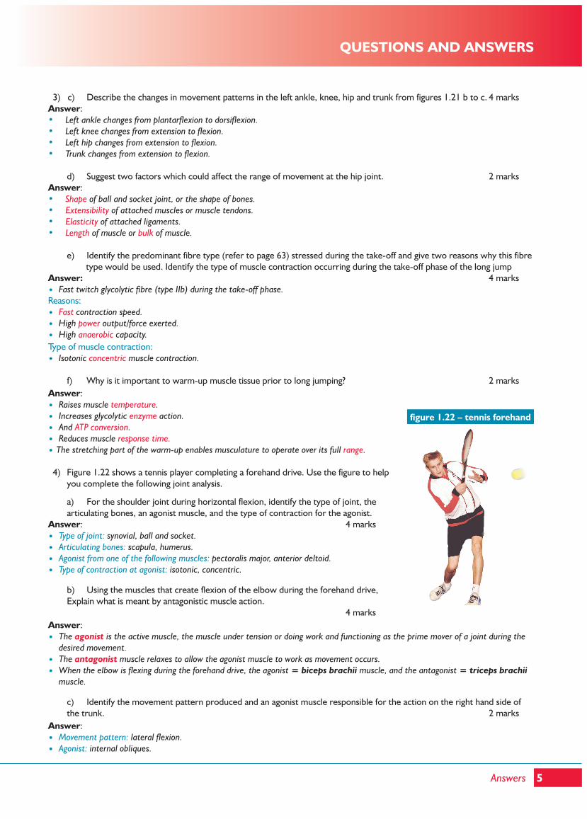
f) Why is it important to warm-up muscle tissue prior to long jumping? 2 marksAnswer:• Raises muscle temperature.• Increases glycolytic enzyme action.• And ATP conversion.• Reduces muscle response time.• The stretching part of the warm-up enables musculature to operate over its full range. 4) Figure 1.22 shows a tennis player completing a forehand drive. Use the figure to help
you complete the following joint analysis.
a) For the shoulder joint during horizontal flexion, identify the type of joint, the articulating bones, an agonist muscle, and the type of contraction for the agonist.
Answer: 4 marks• Type of joint: synovial, ball and socket.• Articulating bones: scapula, humerus.• Agonist from one of the following muscles: pectoralis major, anterior deltoid.• Type of contraction at agonist: isotonic, concentric.
b) Using the muscles that create flexion of the elbow during the forehand drive, Explain what is meant by antagonistic muscle action. 4 marks
Answer:• The agonist is the active muscle, the muscle under tension or doing work and functioning as the prime mover of a joint during the
desired movement.• The antagonist muscle relaxes to allow the agonist muscle to work as movement occurs. • When the elbow is flexing during the forehand drive, the agonist = biceps brachii muscle, and the antagonist = triceps brachii
muscle.
c) Identify the movement pattern produced and an agonist muscle responsible for the action on the right hand side of the trunk. 2 marks
Answer:• Movement pattern: lateral flexion.• Agonist: internal obliques.
figure 31 - tennis forehandfigure 1.22 – tennis forehand
QUESTIONS AND ANSWERS
5Answers
6
4) d) For the right wrist, identify the articulating bones, a fixator or stabilising muscle, and the movement pattern at the completion of the forehand drive. 3 marks
Answer:• Articulating bones: ulna, radius and carpals.• Agonist: wrist extensors.• Movement pattern: extension. 5) Differentiate between concentric, eccentric and isometric muscle contraction, using practical examples to support your
answer. 6 marksAnswer:• Concentric muscle contraction: a muscular contraction which shortens whilst producing tension.• Example: driving upward phase in a jump or squat.
• Eccentric muscle contraction: a muscular contraction which lengthens whilst producing tension.• Example: landing from a jump off a box.
• Isometric muscle contraction: a muscle contraction which stays the same length whilst producing tension.• Example: holding a plank position.
MUSCULAR SKELETAL SYSTEMTOPIC 1 CHAPTER 1