Embed Size (px)
Citation preview
3
Julie A. Douthwaite and Ronald H. Jackson (eds.), Ribosome Display and Related Technologies: Methods and Protocols, Methods in Molecular Biology, vol. 805, DOI 10.1007/978-1-61779-379-0_1, © Springer Science+Business Media, LLC 2012
Chapter 1
Ribosome Display: A Perspective
Andreas Plückthun
Abstract
Ribosome display is an in vitro evolution technology for proteins. It is based on in vitro translation, but prevents the newly synthesized protein and the mRNA encoding it from leaving the ribosome . It thereby couples phenotype and genotype. Since no cells need to be transformed, very large libraries can be used directly in selections, and the in vitro amplifi cation provides a very convenient integration of random mutagenesis that can be incorporated into the procedure. This review highlights concepts, mechanisms, and different variations of ribosome display and compares it to related methods. Applications of ribosome display are summarized, e.g., the directed evolution of proteins for higher binding affi nity, for higher stability or other improved biophysical parameters and enzymatic properties. Ribosome display has developed into a robust technology used in academia and industry alike, and it has made the cell-free Darwinian evolution of proteins over multiple generations a reality.
Key words: Directed evolution , Cell-free translation , Ribosome display , Protein engineering , Antibody engineering , DARPins , Designed ankyrin repeat proteins , Affi nity maturation
All technologies of molecular evolution must couple phenotype and genotype. There are two fundamental possibilities for achieving this. The fi rst one is compartmentalization. Nature’s compartments are cells: they secure that the superior phenotype expressed by one cell’s mutant genotype can be replicated, without the gene products from the wild type interfering. All selections based on microbial phenotypes use this principle. The second possibility is a direct physical coupling of genetic material to the protein product. Nature’s example would be viruses: the virus coat and its receptor-binding properties are the phenotype whose genetic information is encoded on the viral genome, inside the virion.
1. Introduction: Ribosome Display in Context
4 A. Plückthun
In order to use selection methods in biotechnology, i.e., for the enrichment of binders from libraries and their evolutionary improvement, one can exploit both compartmentalization and physical linkages. The most frequently used compartments are microbial cells, where intracellular interactions can be detected by genetic means. As exemplifi ed by the yeast two hybrid system ( 1 ) or by the protein fragment complementation assay ( 2 ) , only those cells grow in which a desired molecular interaction restores the critical factor, which has previously been split in two pieces. Alternatively, one can use enzymatic or optical means for detecting interactions ( 3– 5 ) . These examples are only meant to be illustrative – a large research fi eld has developed around exploiting such phenomena.
Most popular, however, have been systems where the interac-tion itself occurs outside of the cell, even though the polypeptides are still being produced by cells. Thus, expression on the surface of bacteria ( 6 ) or yeast ( 7 ) has been used successfully, as has been the display of peptides ( 8– 11 ) and polypeptides ( 12– 14 ) on the surface of fi lamentous phages – probably still the most widely used display technology. In phage display, the bacterial cells are producing the phages, and thus the bacterial cells must fi rst be transformed with the library, limiting the diversity. However, this will not be a comprehensive review on display technologies; such reviews can be found elsewhere ( 15– 20 ) .
In all these technologies that involve cells at any step, including phage display, the genetic information needs to be introduced into cells. This will usually limit the diversity present in the cells to below what has been present in the actual library of DNA molecules. The diversity reached in practice after transformation will depend on the host system (bacteria vs. yeast) and on the efforts made to transform the cells. This large-scale transformation step, while not diffi cult, can be quite laborious. It is this step that is avoided in technologies that are performed fully in vitro.
This chapter will summarize ribosome display, and mention other technologies that operate without any cells during the library selection. They are not restricted by the effort spent on the trans-formation step. This advantage is apparent in the primary library, which can be of bigger size, as all DNA (or mRNA) molecules present can in principle give rise to proteins that take part in the selection.
The most important advantage of ribosome display and other “full” in vitro technologies is, however, the easy combination with PCR-based randomization techniques, and thus the creation of a true Darwinian evolution process – in contradistinction to a mere selection from an existing “constant” library. In all technologies that require transformation of cells, after each randomization step the cells have to be transformed with the new library, and the workload is thus potentiated by the number of evolution steps.
51 Ribosome Display: A Perspective
In ribosome display, the workload to select binders in the absence or presence of randomization is almost identical. Thus, the more randomization steps are to be carried out in succession, the more attractive ribosome display becomes.
Of course, technologies have been developed to carry out ran-domization directly in cells as well ( 21– 23 ) . Nonetheless, in vitro methods give the user full control over where mutations should occur in the sequence (by using, e.g., a randomized cassette), which residue types are to be introduced (by using trinucleotide building blocks ( 24 ) or suitable mononucleotide mixtures), or how many random mutations should occur on average ( 25 ) – a level of control not yet within reach in cellular systems. Furthermore, the “shuffl ing” of the library ( 26, 27 ) can easily be introduced into the procedure if desired, to recombine mutants.
Key to the development of ribosome display was the observation that rare mRNAs coding for a particular protein can be isolated from a pool of mRNAs by immunoprecipitation of stalled ribo-somal complexes containing the nascent polypeptides ( 28, 29 ) . Apparently, ribosomal stalling is frequent enough to be experimen-tally exploited.
To create RNA-based “aptamers”, Tuerk and Gold ( 30 ) had developed a technology called SELEX (Systematic Evolution of Ligands by Exponential Enrichment), where multiple rounds of in vitro transcription of random nucleic acid pools, followed by affi nity selection of the RNA aptamers and subsequent RT-PCR, lead to the selection of target-binding RNAs. In SELEX, genotype and phenotype are simultaneously represented by the same RNA molecule, since it exerts its function through its three-dimensional structure, which is in turn determined by its nucleotide sequence.
In their original publication about SELEX, Tuerk and Gold ( 30 ) already speculated that a similar approach might be adapted to protein selection, referring to the isolation of stalled translation complexes ( 28, 29 ) .
The fi rst experimental demonstration of the ribosome display technology was the selection of short peptides from a library using an Escherichia coli S30 in vitro translation system ( 31, 32 ) . Kawasaki ( 33 ) had proposed a similar approach to enrich peptides from libraries in a patent application, however, without giving a detailed example, which was only published in 1997 ( 34 ) .
Meanwhile, Hanes and Plückthun ( 35 ) had developed and fi rst reported ribosome display for whole proteins, initially antibody scFv fragments, after signifi cantly improving and modifying the system to increase its effi ciency and to allow folding of scFv frag-ments during in vitro translation ( 36 ) . The key observation with
2. Development of Ribosome Display
6 A. Plückthun
these longer open reading frames (ORFs) was that mutations were found at great frequency ( 35 ) due to the large number of PCR cycles that every gene had undergone after several rounds. Because of the short encoded peptide sequences, Mattheakis et al. ( 31, 32 ) did not observe mutations. To play out its strengths, ribosome display needs to be carried out with whole proteins. The appearance of mutations, just by using a normal polymerase without proof-reading activity, then made it clear that the unique advantage of ribosome display lies in its potential to do true Darwinian evolu-tion , as opposed to mere selection , from a given library. This poten-tial was demonstrated in the following year ( 37 ) by striving to select antibodies for improved affi nity, and by adding additional diversity through random mutagenesis. The fi rst selection from a non-immune synthetic antibody library followed soon thereafter ( 38 ) .
In the above experiments, the bacterial cell-free translation extracts were home-made, as they needed to be free of reducing agents (because of the intramolecular disulfi de bonds within the scFv domains), and they were required in large amounts. The feasibil-ity of carrying out ribosome display in a eukaryotic cell-free translation extracts was subsequently demonstrated with an scFv-kappa fusion ( 39 ) , and then by selecting an antibody in the same format from immunized transgenic mice ( 40 ) .
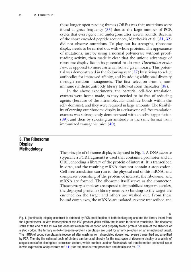
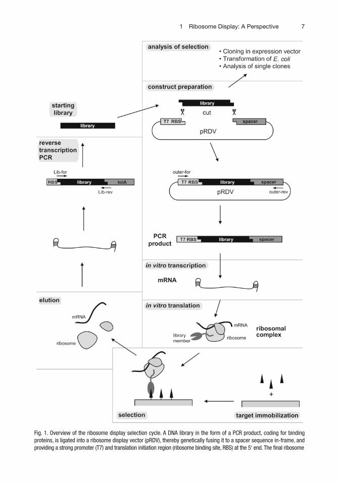
The principle of ribosome display is depicted in Fig. 1 . A DNA cassette (typically a PCR fragment) is used that contains a promoter and an ORF, encoding a library of the protein of interest. It is transcribed in vitro, and the resulting mRNA does not contain a stop codon. Cell-free translation can run to the physical end of this mRNA, and complexes consisting of the protein of interest, the ribosome, and mRNA are formed. The ribosome itself serves as the connector. These ternary complexes are exposed to immobilized target molecules, the displayed proteins (library members) binding to the target are enriched on the target and others are washed out. From these bound complexes, the mRNAs are isolated, reverse transcribed and
3. The Ribosome Display Methodology
Fig. 1. (continued) display construct is obtained by PCR amplifi cation of both fl anking regions and the library insert from the ligated vector. In vitro transcription of this PCR product yields mRNA that is used for in vitro translation. The ribosome stalls at the end of the mRNA and does not release the encoded and properly folded protein because of the absence of a stop codon. The ternary mRNA–ribosome–protein complexes are used for affi nity selection on an immobilized target. The mRNA of bound complexes is recovered after washing from dissociated ribosomes, reverse transcribed and amplifi ed by PCR. Thereby the selected pools of binders can be used directly for the next cycle of ribosome display or analysis of single clones after cloning into expression vectors, which are then used for Escherichia coli transformation and small-scale in vivo expression. Adapted from ref. 111 ; for the most current procedure and details see ref. 97.
71 Ribosome Display: A Perspective
Fig. 1. Overview of the ribosome display selection cycle. A DNA library in the form of a PCR product, coding for binding proteins, is ligated into a ribosome display vector (pRDV), thereby genetically fusing it to a spacer sequence in-frame, and providing a strong promoter (T7) and translation initiation region (ribosome binding site, RBS) at the 5 ¢ end. The fi nal ribosome
8 A. Plückthun
PCR amplifi ed to serve as the input of another round. After 3–5 rounds, the resulting DNA fragments are ligated into an expression vector and E. coli are transformed. The different proteins made by individual E. coli clones can then be further evaluated.
The logic of the individual steps will now be discussed. In vitro methods cannot rely on cells to link phenotype and genotype. Instead, a direct physical link between the genetic material and the protein product must be made. (An in vitro alternative is to create compartments in the form of a water-in-oil emulsion, which will be discussed below.) During protein biosynthesis, the encoding mRNA is read by the ribosome and parts of it are almost engulfed by the small subunit, which mediates codon/anti-codon contact to the tRNA. The protein emerges from the ribosomal tunnel within the large ribosomal subunit. During all these steps, the protein chain is covalently connected through an ester bond to the peptidyl-tRNA within the P-site, and thereby tightly maintained within the ribosome. Thus, during protein biosynthesis, neither protein nor mRNA can leave.
Translation normally ends at a stop codon. In E. coli ribosomes, the UAG and UAA stop codons are directly recognized by the Release Factor 1 (RF1), the UGA and UAA stop codons by RF2 ( 41 ) . With the help of RF3, the ester bond between the synthesized protein and the last tRNA is positioned such that it is hydrolyzed, and the last amino acids of the fi nished protein, still within the exit tunnel, can slide out, leaving the empty tRNA behind. Now the ribosome recycling factor (RRF) and elongation factor G (EF-G) together help separate the large from the small subunit: they remove the tRNA, and after subunit dissociation, the mRNA can then leave as well ( 42 ) . A similar mechanism also functions in eukaryotic ribosomes ( 43 ) .
The absence of a stop codon in mRNA for ribosome display thus ensures that this “normal” course of events does not take place. The experimental conditions must furthermore minimize any early unwanted spontaneous hydrolysis of the ester bond between polypep-tide and tRNA or any other spontaneous falling apart of the ternary complex. This is usually achieved by rather short translation times, subsequent cooling of the solution and addition of Mg 2+ . These short translation times are also a compromise between effi cient translation and degradation of the mRNA by nucleases present in the extract. It is believed that high Mg 2+ “condenses” the ribosome by binding to the rRNA, making it diffi cult for the peptidyl-tRNA to dissociate or be hydrolyzed. While the details differ, the general strategy is the same for eukaryotic ribosomes ( 44 ) .
The absence of a stop codon then causes “stalled” ribosomes. Once the ribosomes are at the end of the mRNA, they would have a peptidyl-tRNA in the P site and (presumably) an empty A site. As this situation can appear in the bacterial cell as well (if an mRNA molecule is missing its 3 ¢ end containing the stop codon), bacteria
91 Ribosome Display: A Perspective
have devised a mechanism called trans -translation to rescue such stalled ribosomes, which would clog up protein biosynthesis ( 45, 46 ) . The key molecule to act on the stalled ribosome is the trans-fer-messenger RNA (tmRNA, also called 10S RNA or SsrA), which has a tRNA-like domain charged with alanine and an mRNA-like domain. Peptide synthesis is resumed, with the additional help of the protein SmpB, by incorporating Ala, and thereby transferring the chain to the tmRNA. Up to now, the tmRNA molecule has acted like tRNA. Now its mRNA domain is being read, and the sequence ANDENYALAA* is appended to the protein, which ends with a stop codon (!), thus leading to regular termination and recy-cling of the ribosome. Even worse for ribosome display, this C-terminal sequence which has been added serves as a degradation tag in E. coli . Thus, the action of tmRNA would be detrimental for ribosome display. Starting from our very fi rst experiments, an antisense oligonucleotide has always been added to titrate out tmRNA ( 35 ) .
In order for the protein of interest to fold and be able to inter-act with a target, the whole protein of interest must be outside of the tunnel once the ribosomes have come to the physical end of the mRNA. Thus, to remain connected to the tRNA at the same time, the protein of interest must be fused to an unstructured region at the C-terminus, occupying the ribosomal tunnel. We have called this the “spacer” or “tether”. This protein tail, which is the same in all library members, is thus fused in frame to the C-terminus of the randomized protein of interest in the ribosome display construct.
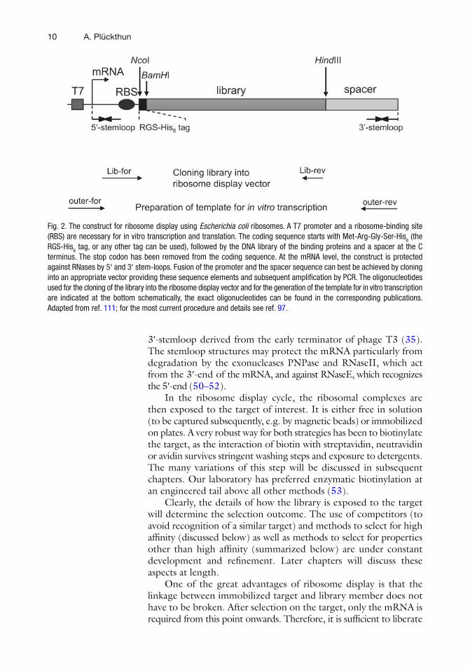
The features of the ribosome display construct are summarized in Fig. 2 . On the DNA level, the construct requires a strong promoter for effi cient in vitro transcription to mRNA. On the mRNA level, the construct contains, as a regulatory sequence for translation, either a prokaryotic ribosome-binding site ( 47, 48 ) if the E. coli system is used, or a Kozak consensus and enhancer sequence ( 49 ) if the eukaryotic ribosome display system is used. This sequence is followed by the ORF encoding the protein to be displayed, followed by a spacer sequence fused in frame to the protein of interest, as described above.
At both ends of the mRNA, the ribosome display construct should include stemloops. 5 ¢ - and 3 ¢ -stemloops are known to stabilize mRNA against RNases in vivo as well as in vitro. The presence of stemloops is important, especially in the E. coli ribosome display system because the extract used for in vitro translation contains high RNase activities. The effi ciency of ribosome display was increased approximately 15-fold ( 35 ) , when a 5 ¢ -stemloop derived from the T7 gene 10 upstream region and a 3 ¢ -stemloop derived from the terminator of the E. coli lpp lipoprotein were introduced into the ribosome display construct ( 35 ) . A similar improvement in effi ciency was observed when using the same 5 ¢ -stemloop and the
10 A. Plückthun
3 ¢ -stemloop derived from the early terminator of phage T3 ( 35 ) . The stemloop structures may protect the mRNA particularly from degradation by the exonucleases PNPase and RNaseII, which act from the 3 ¢ -end of the mRNA, and against RNaseE, which recognizes the 5 ¢ -end ( 50– 52 ) .
In the ribosome display cycle, the ribosomal complexes are then exposed to the target of interest. It is either free in solution (to be captured subsequently, e.g. by magnetic beads) or immobilized on plates. A very robust way for both strategies has been to biotinylate the target, as the interaction of biotin with streptavidin, neutravidin or avidin survives stringent washing steps and exposure to detergents. The many variations of this step will be discussed in subsequent chapters. Our laboratory has preferred enzymatic biotinylation at an engineered tail above all other methods ( 53 ) .
Clearly, the details of how the library is exposed to the target will determine the selection outcome. The use of competitors (to avoid recognition of a similar target) and methods to select for high affi nity (discussed below) as well as methods to select for properties other than high affi nity (summarized below) are under constant development and refi nement. Later chapters will discuss these aspects at length.
One of the great advantages of ribosome display is that the linkage between immobilized target and library member does not have to be broken. After selection on the target, only the mRNA is required from this point onwards. Therefore, it is suffi cient to liberate
Fig. 2. The construct for ribosome display using Escherichia coli ribosomes. A T7 promoter and a ribosome-binding site (RBS) are necessary for in vitro transcription and translation. The coding sequence starts with Met-Arg-Gly-Ser-His 6 (the RGS-His 6 tag, or any other tag can be used), followed by the DNA library of the binding proteins and a spacer at the C terminus. The stop codon has been removed from the coding sequence. At the mRNA level, the construct is protected against RNases by 5 ¢ and 3 ¢ stem-loops. Fusion of the promoter and the spacer sequence can best be achieved by cloning into an appropriate vector providing these sequence elements and subsequent amplifi cation by PCR. The oligonucleotides used for the cloning of the library into the ribosome display vector and for the generation of the template for in vitro transcription are indicated at the bottom schematically, the exact oligonucleotides can be found in the corresponding publications. Adapted from ref. 111 ; for the most current procedure and details see ref. 97.
111 Ribosome Display: A Perspective
it by the dissociation of small and large ribosomal subunits by the addition of EDTA. This has the advantage that it is not more diffi cult to isolate complexes of very high affi nity (which would be hard to dissociate) than those of lower affi nity. Nonetheless, more specifi c elution procedures can be of interest for selecting binders to particular epitopes or with particular properties.
The mRNA needs to be reverse transcribed and the resulting DNA then amplifi ed and brought back to the initial format containing a promoter for transcription of the next round. In the initial rounds, very few mRNA molecules will be obtained after selection. This step is perhaps one of the few technically demanding steps, as it requires attention to the fragility of RNA (in the presence of nucleases, which can also be introduced by careless laboratory handling). When designing a new ribosome display cassette (with different promoter, ribosome binding site, N-terminal tag on the protein, ORF, C-terminal tether and 5 ¢ and 3 ¢ stemloops), care must be taken in designing the primers needed for the PCR. Obviously, they must bind with very high specifi city. These reverse transcription and PCR steps appear to be the most frequent focus of troubleshooting, when designing a new system from scratch. Even when taking a well-working system, and merely replacing the ORF, it must be considered that new hairpins might form unintentionally, e.g., engaging the start codon or the ribosome binding site, which would compromise translation effi ciency. Fortunately, these issues are easily and rapidly evaluated.
The most frequently used methods in ribosome display include the use of E. coli S-30 extracts for translation, which do contain ribosome-associated factors important for protein folding such as the trigger factor ( 54 ) . In addition, molecular chaperones that are not associated to the ribosome are present in the E. coli extract, such as DnaJ/K/GrpE and GroEL/ES, as well as small heat shock proteins and others ( 55 ) . Additional factors can be added, depend-ing on the requirements of the proteins to be displayed ( 36 ) . Antibody scFv fragments required the addition of eukaryotic protein disulfi de isomerase ( 36 ) .
Ribosome display has also been used as a tool to defi ne a binding epitope, making use of the somewhat surprising fi nding that, while still on the ribosome, aggregation of a protein seems to be effi ciently prevented. It was found that proteins, such as eukaryotic receptors that could not be expressed in functional form in E. coli nor effi ciently refolded from inclusion bodies, nor expressed in functional form by in vitro translation, would fold while still attached to the E. coli ribosome ( 56 ) . Perhaps the ribosome enhances solubility of the
4. Protein Folding in Ribosome Display
12 A. Plückthun
ternary complex and sterically blocks aggregation. To defi ne a binding epitope, a cell surface receptor was subjected to several rounds of random mutagenesis at high error rate, and this ribosome display library of receptor point mutants was selected on the target. The epitope could be recognized as an area devoid of surface mutations – evolutionary pressure in the experiment apparently maintained the residues in the epitope ( 57 ) .
When using a eukaryotic translation system ( 39, 44, 58 ) , the corresponding eukaryotic proteins would be expected to be present in the extract. On the other hand, if a system from pure components is used ( 59– 62 ) , it may be necessary to add these proteins relevant for folding, depending on the protein to be displayed ( 36 ) .
The ribosome display method can also be carried out with eukaryotic extracts, using a reticulocyte lysate ( 39, 58 ) . Different methods of sequence recovery have been compared, and it was concluded that a similar procedure as used in the prokaryotic system also performs best in the eukaryotic system ( 44 ) , even though in situ recovery can also be carried out ( 39, 58 ) . The wheat germ in vitro translation system has also been used ( 34 ) . While one might specu-late that a eukaryotic translation system should perform better with eukaryotic proteins, there is actually no evidence for this ( 63 ) . If particular factors are needed, such as, e.g., molecular chaperones and protein disulfi de isomerase, they can (and have to) be added to either system ( 36 ) .
The use of a ribosome display system based on in vitro translation with purifi ed components (PURE system) has also been described ( 59– 62 ) . In this system, no release factor is present. Matsuura et al. examined the effi ciency of ribosome display in the absence and presence of a stop codon, as well as when using the secM stalling sequence ( 64 ) as an alternative means of trapping the ternary protein–ribosome–mRNA complex ( 60 ) . Interestingly, the effi ciency of display was almost identical in all cases. Another encouraging fi nding from the use of the PURE system is that the intrinsic stability of the ternary complexes is actually very high. Even after an incubation of the ternary complexes for 1 h at 50 °C, the display effi ciency drops by less than a factor of 10. Presumably, both in the prokaryotic and eukaryotic extracts, RNAses set a practical limit on stability, rather than the intrinsic stability of the ternary complexes themselves.
The system with purifi ed components may thus be of interest where the removal of a stop codon is inconvenient or high temperature is required in the selection procedure. It should be kept in mind, however, that the experiments were carried out with engineered
5. Variations in the Ribosome Display System
5.1. Eukaryotic Cell Free Translation System
5.2. PURE System
131 Ribosome Display: A Perspective
mRNA that had a C-terminal spacer, as in the standard system described above. In a natural non-engineered mRNA, one would expect that the last few amino acids of the protein of interest are still in the ribosomal tunnel, thereby hampering the folding of the protein. It will remain to be seen whether the PURE system is suffi ciently cost-effi cient to be used for standard selection and evolution experiments, in comparison to the use of translation extracts from E. coli .
To address the display of natural mRNA (which of course all contain a stop codon), as an alternative to the use of the PURE system, engineered suppressor tRNAs have been used ( 65, 66 ) . This could be another approach useful for future protein–protein interaction studies. Nonetheless, the problem remains that many (if not most) proteins will not fold, if part of their domain structure is still in the ribosomal tunnel. The critical question is therefore whether the translated spacer that results when suppressing the stop codons in natural mRNA will be long enough to allow folding of most proteins.
Antibody scFv fragments were the fi rst complex library with which ribosome display was tested for selection and affi nity maturation ( 37 ) , initially from a library of immunized mice, later from a syn-thetic library ( 38 ) . At that time, the recreated synthetic repertoire of the antibodies was the only general binding protein scaffold available with great diversity.
The folding of antibody fragments in an in vitro translation system must be commensurate with their oxidative folding (many if not most antibody domains need the intradomain V H and V L disulfi de bond to fold properly), and thus this reaction must be catalyzed ( 36 ) . In addition, the b -sandwich architecture of anti-bodies can lead to aggregation, and this may be part of the reason, why more rounds of enrichment appear to be necessary than for some other scaffolds that fold extremely well in an in vitro transla-tion system.
There are more publications on using phage display than ribosome display in the selection from naive antibody libraries, but there simply may be no necessity to break with tradition. Filamentous phage display ( 12 ) works very well with secreted one-chain disulfi de-containing proteins such as scFv ( 67 ) , and there is always the option of combining the two methods, as opposed to directly combining selection and affi nity maturation in one procedure, as in the ribosome display selection from naive or synthetic libraries ( 68 ) .
The analysis of ribosome display selection from the fully synthetic antibody library HuCAL leads to the conclusion that the selection is not exhaustive, and the outcome is governed by the occurrence
5.3. Display of mRNA with Stop Codons
6. Applications of Ribosome Display to Complex Libraries
6.1. scFv Fragments
14 A. Plückthun
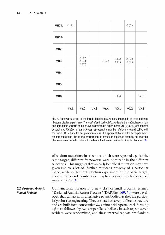
of random mutations; in selections which were repeated against the same target, different frameworks were dominant in the different selections. This suggests that an early benefi cial mutation may have given rise to a lot of (further mutated) progeny of a particular clone, while in the next selection experiment on the same target, another framework combination may have acquired such a benefi cial mutation (Fig. 3 ).
Combinatorial libraries of a new class of small proteins, termed “Designed Ankyrin Repeat Proteins” (DARPins) ( 69, 70 ) were devel-oped that can act as an alternative to antibodies, as they are particu-larly robust to engineering. They are based on a very different structure and are built from consecutive 33-amino acid repeats, each forming a b -turn followed by two antiparallel a -helices. In each repeat, seven residues were randomized, and these internal repeats are fl anked
6.2. Designed Ankyrin Repeat Proteins
Fig. 3. Framework usage of the insulin-binding HuCAL scFv fragments in three different ribosome display experiments. The vertical and horizontal axes denote the HuCAL heavy-chain and light-chain variable domains. ScFvs isolated in experiments ( A ), ( B ), or ( C ) are denoted accordingly. Numbers in parentheses represent the number of closely related scFvs with the same CDRs, but different point mutations. It is apparent that in different experiments random mutations lead to the proliferation of particular sequence families, but that this phenomenon occurred in different families in the three experiments. Adapted from ref. 38.
151 Ribosome Display: A Perspective
by constant capping repeats, to give one contiguous polypeptide chain with a randomized concave, groove-like binding surface, which is randomized in the library. The proteins contain no cysteine, can be expressed in soluble form in the cytoplasm of E. coli at very high levels, and are very stable and resistant to aggregation (refs. 71, 72 and references therein).
It may be these favorable biophysical properties, combined with the fact that high affi nity binders are obtained at high frequency, that cause the direct selection of binders from the diverse library to work very well with DARPins. Thus, binders against many targets, including diffi cult ones such as, e.g., detergent-solubilized GPCRs ( 73 ) or conformers of DNA (O. Scholz, unpublished), have been selected directly by ribosome display (e.g., see refs. 70, 74– 80 ) .
Binders based on the camelid VHH domains with micromolar affi nity have been isolated by ribosome display from a naive library ( 81 ) , and with nanomolar affi nity from an immunized llama ( 82 ) .
Ribosome display has been combined with other selection tech-nologies. It has been used as the affi nity maturation step of a phage display library (e.g., see ref. 68 ) , and thus used as the second stage in binder selection.
However, one can also use ribosome display as the fi rst step and follow it up by another technology to simplify the evaluation of individual clones. At the end of the ribosome display procedure, the fi nal selected pool is usually cloned in E. coli , and crude extracts of individual E. coli expression cultures are then analyzed by ELISA. Instead of going through enough rounds such that most of these clones will be positive, an earlier round can be cloned, and an in vivo selection can be applied to this selected pool. For this purpose, the pools of ribosome display were cloned after the fi rst, second, and third round in a protein fragment complementation assay (PCA) ( 2 ) , a split enzyme selection system using DHFR. This technology has a low discrimination power for affi nity, but essen-tially serves as a convenient qualitative screen of binding. It can be seen that even after one round of ribosome display, binders can be obtained, albeit with micromolar affi nity. These correspond to a random sampling of the library which has been enriched, perhaps 10 3 - to 10 4 -fold, and high affi nity binders are too rare to be expected to be found in this small sampling. However, already after the second round of ribosome display, binders with nano-molar affi nity are found ( 77 ) . This combination of ribosome display with PCA might become of interest in high-throughput applica-tions of ribosome display.
6.3. Other Scaffolds
7. Combining Ribosome Display with Other Selection Technologies
16 A. Plückthun
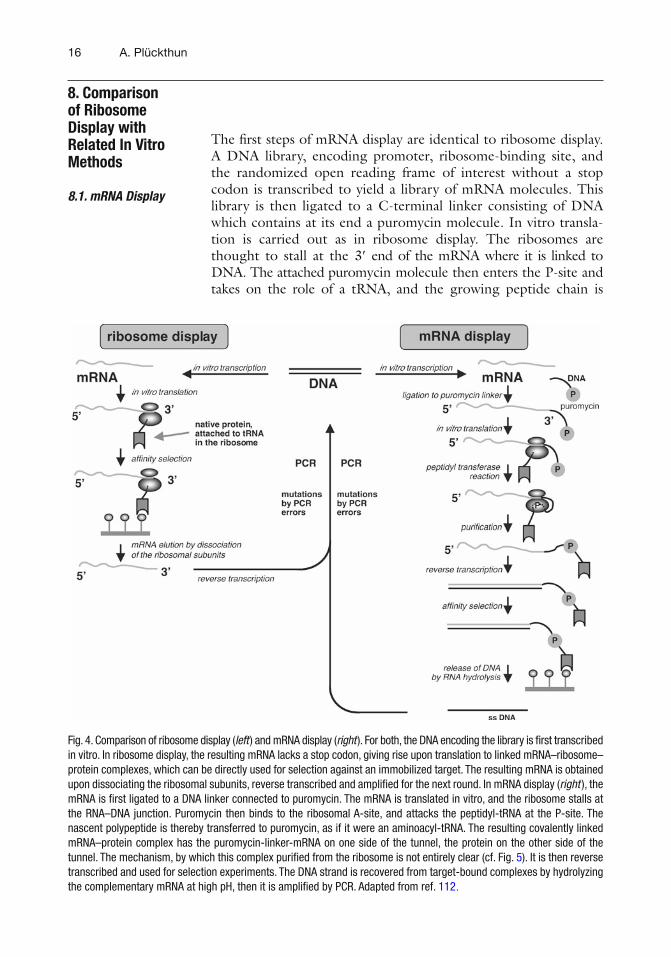
The fi rst steps of mRNA display are identical to ribosome display. A DNA library, encoding promoter, ribosome-binding site, and the randomized open reading frame of interest without a stop codon is transcribed to yield a library of mRN A molecules. This library is then ligated to a C-terminal linker consisting of DNA which contains at its end a puromycin molecule. In vitro transla-tion is carried out as in ribosome display. The ribosomes are thought to stall at the 3 ¢ end of the mRNA where it is linked to DNA. The attached puromycin molecule then enters the P-site and takes on the role of a tRNA, and the growing peptide chain is
8. Comparison of Ribosome Display with Related In Vitro Methods
8.1. mRNA Display
Fig. 4. Comparison of ribosome display ( left ) and mRNA display ( right ). For both, the DNA encoding the library is fi rst transcribed in vitro. In ribosome display, the resulting mRNA lacks a stop codon, giving rise upon translation to linked mRNA–ribosome–protein complexes, which can be directly used for selection against an immobilized target. The resulting mRNA is obtained upon dissociating the ribosomal subunits, reverse transcribed and amplifi ed for the next round. In mRNA display ( right ), the mRNA is fi rst ligated to a DNA linker connected to puromycin. The mRNA is translated in vitro, and the ribosome stalls at the RNA–DNA junction. Puromycin then binds to the ribosomal A-site, and attacks the peptidyl-tRNA at the P-site. The nascent polypeptide is thereby transferred to puromycin, as if it were an aminoacyl-tRNA. The resulting covalently linked mRNA–protein complex has the puromycin-linker-mRNA on one side of the tunnel, the protein on the other side of the tunnel. The mechanism, by which this complex purifi ed from the ribosome is not entirely clear (cf. Fig. 5 ). It is then reverse transcribed and used for selection experiments. The DNA strand is recovered from target-bound complexes by hydrolyzing the complementary mRNA at high pH, then it is amplifi ed by PCR. Adapted from ref. 112.
171 Ribosome Display: A Perspective
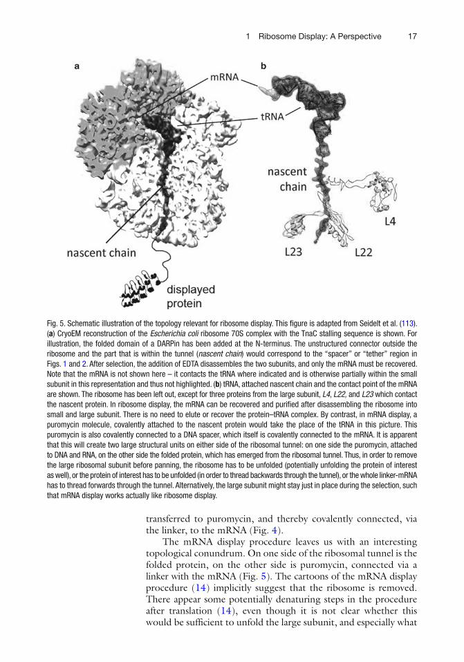
transferred to puromycin, and thereby covalently connected, via the linker, to the mRNA (Fig. 4 ).
The mRNA display procedure leaves us with an interesting topological conundrum. On one side of the ribosomal tunnel is the folded protein, on the other side is puromycin, connected via a linker with the mRNA (Fig. 5 ). The cartoons of the mRNA display procedure ( 14 ) implicitly suggest that the ribosome is removed. There appear some potentially denaturing steps in the procedure after translation ( 14 ) , even though it is not clear whether this would be suffi cient to unfold the large subunit, and especially what
Fig. 5. Schematic illustration of the topology relevant for ribosome display. This fi gure is adapted from Seidelt et al. ( 113 ) . ( a ) CryoEM reconstruction of the Escherichia coli ribosome 70S complex with the TnaC stalling sequence is shown. For illustration, the folded domain of a DARPin has been added at the N-terminus. The unstructured connector outside the ribosome and the part that is within the tunnel ( nascent chain ) would correspond to the “spacer” or “tether” region in Figs. 1 and 2 . After selection, the addition of EDTA disassembles the two subunits, and only the mRNA must be recovered. Note that the mRNA is not shown here – it contacts the tRNA where indicated and is otherwise partially within the small subunit in this representation and thus not highlighted. ( b ) tRNA, attached nascent chain and the contact point of the mRNA are shown. The ribosome has been left out, except for three proteins from the large subunit, L4 , L22, and L23 which contact the nascent protein. In ribosome display, the mRNA can be recovered and purifi ed after disassembling the ribosome into small and large subunit. There is no need to elute or recover the protein–tRNA complex. By contrast, in mRNA display, a puromycin molecule, covalently attached to the nascent protein would take the place of the tRNA in this picture. This puromycin is also covalently connected to a DNA spacer, which itself is covalently connected to the mRNA. It is apparent that this will create two large structural units on either side of the ribosomal tunnel: on one side the puromycin, attached to DNA and RNA, on the other side the folded protein, which has emerged from the ribosomal tunnel. Thus, in order to remove the large ribosomal subunit before panning, the ribosome has to be unfolded (potentially unfolding the protein of interest as well), or the protein of interest has to be unfolded (in order to thread backwards through the tunnel), or the whole linker-mRNA has to thread forwards through the tunnel. Alternatively, the large subunit might stay just in place during the selection, such that mRNA display works actually like ribosome display.
18 A. Plückthun
effect these conditions would have on the covalently bound protein of interest.
There are thus four possibilities: First, the large subunit of ribosome unfolds, thereby opening the exit tunnel, such that the protein can slide out sideways. It is unclear to what degree the protein of interest would unfold as well. Second, the DNA linker and the whole mRNA thread through the protein tunnel. Third, the protein of interest unfolds before the large subunit and threads backwards through the protein tunnel. Fourth, the large subunit of the ribosome is actually not completely removed, and is still present during panning, similar as in ribosome display. This question is not only of academic interest, as it may have some effect on the protein to be displayed.
A number of protein scaffolds have been used with mRNA display, e.g. some stabilized by metal ions ( 83– 85 ) , which would become unfolded by adding EDTA. However, selections have also been carried out using the fi bronectin scaffold ( 86– 88 ) which probably folds and unfolds reversibly, or scFv fragments ( 89 ) , as well as diverse other proteins ( 90 ) where it is not clear whether selection did require folded domains. The problem of topology in mRNA display has apparently not been solved.
The packaging of the translation extract into small droplets in the form of a water-in-oil emulsion combines the in vitro approach with the compartment concept of coupling genotype and pheno-type ( 91– 93 ) . If, on average, each droplet contains only one mRNA molecule, the protein content of each droplet is monoclonal. The most persuasive application of this technology is for enzymatic reactions ( 94, 95 ) . The basic challenge for evolving enzyme turn-over is that the phenotype, namely the production rate of the enzy-matic reaction product, cannot be easily linked to the enzyme molecule itself in a mixture of mutants in solution, at least not for reactions with multiple turnovers with high rates. The cellular con-fi nement solves this problem. Nonetheless, the full potential of this approach can be reached only if the reaction can be followed in the compartments directly, requiring optical detection and sorting.
On the other hand, the selection for a binding event in a com-partmentalized system is somewhat less compelling. Emulsions have to be broken and selections can be carried out in the bulk phase as in ribosome display and mRNA display. The generic detection of binding events within a droplet remains a challenge, and it is less clear how to achieve this in a semiquantitative way for binding strength , i.e., affi nity.
However, an interesting application of the emulsion technology for the selection of binding proteins was described by Sumida et al. ( 96 ) , in which they used the emulsion format to express both chains of a Fab fragment from a bicistronic operon within the same droplet. By fusing the heavy chain to streptavidin, and attaching biotin to
8.2. Water-in-Oil Emulsions
191 Ribosome Display: A Perspective
the DNA fragment via a photocleavable linker, both chains of the Fab fragment and the corresponding DNA stay together after breaking the emulsion. After panning, the DNA has to be recovered by photocleavage and can be amplifi ed. It will be interesting to see how well this system will perform with complex libraries.
The absolute functional library size in ribosome display is given by the number of different ternary complexes that are formed from mRNA and ribosomes and give rise to a nascent protein that can fold. This requires that the protein of interest is translated at least to the point that the relevant domain is outside the ribosomal tunnel.
From the amount of PCR fragment that is used as the input for transcription, we can calculate that under standard conditions ( 97 ) about 2–3 × 10 12 molecules input DNA are used. If the library template DNA to make this PCR is of good quality and highly diverse we can assume that these DNA molecules are all different. Also, further muta-tions will have been introduced while carrying out this very PCR.
The transcription of this linear PCR fragment will usually create multiple copies or mRNA per DNA molecule, and only an aliquot of the resulting mRNA is used for translation. In a standard ribo-some display reaction about 1–3 × 10 13 mRNA molecules are used. It is entirely possible that additional errors are introduced by the RNA polymerase, such that they will contain a greater diversity than the input DNA.
The other critical variable is the number of functional ribosomes. The number of assembled ribosomes in an E. coli cell depends on its growth rate and is between 18,000 and 72,000 ( 98 ) . From the amount of S30 extract used and the number of assembled ribosomes per cell, there should be about 1–4 × 10 14 assembled ribosomes in the standard ribosome display reaction. Thus, even if not all ribosomes are functional, there should be a suffi cient excess to translate most mRNA molecules. Also, while the number of ribosomes per mRNA will follow a binomial distribution, there should be a signifi cant proportion of monosomes (one ribosome per mRNA).
Using multi-ml quantities of S30 extract and more mRNA, almost arbitrarily large numbers for the library size can be stated. For example, in several review articles the library size of ribosome display and mRNA display have been “compared”, where the latter has usually referred to an experiment where extreme amounts of S30 extract have been used. This simple, direct relationship between the amount of extract used and the functional diversity of the library has not been recognized by all authors.
9. Comments on Library Size
20 A. Plückthun
More importantly, it remains to be seen whether the best use of the rather valuable S30 extract (and the even more valuable eukaryotic extract, or the truly precious purifi ed components) is to use it all at once in the fi rst round, as opposed to using it in smaller aliquots over a multi-step selection with built-in affi nity maturation.
Another important aspect is the functional fraction of the library, and whether the non-functional part is merely inactive, or instead “sticky” and thus becoming enriched during selections. This is a quantity that can almost not be objectively determined. This issue also sets a limit to the number of random mutations that is practically useful before the population becomes extinct.
The most attractive exploitation of the built-in possibility of generating mutations is to improve affi nity. Affi nity is usually quantifi ed by the equilibrium dissociation constant K D , which is the ratio of the dissociation rate constant k d (loosely referred to as off-rate) over the association rate constant k a (loosely referred to as on-rate). The association rate constant for protein–protein complexes falls in a remarkably small window, typically between 1 × 10 5 and 1 × 10 6 M −1 S −1 , as summarized from various experimental studies by Northrup et al. ( 99 ) and further computationally analyzed by these authors. The net association rate is often visualized as the collision rate times the fraction of “successful” collisions, in other words, where the two proteins have productive orientations. This means that affi nity is largely determined by off-rate, and that measures to improve affi nity should normally attempt to decrease the off-rate.
Of course, there are exceptions. A protein pair can be properly oriented upon approach by electrostatic forces, leading to a higher fraction of successful collisions ( 100 ) , and this can also be engineered ( 101 ) . However, this electrostatic steering will greatly diminish in importance in physiological buffers with high ionic strength and may thus not be so useful for practical applications. A second class of exceptions will be those interactions which are characterized by an unusually slow observed association rate, much slower than 10 5 M −1 S −1 . This can be due to two things: either one of the partners is not in a productive conformation, and only a small fraction of molecules are able to interact (conformational selection) ( 102, 103 ) , or a slow conformational change must occur in one of the partners before or upon binding (induced fi t) ( 104 ) . In sum-mary, if the on-rate of the protein to be improved is not unusually slow (say, only 10 3 –10 4 M −1 S −1 ), and if high affi nity should be achieved also under physiological conditions with considerable ionic strength, then an improvement of off-rate is the most likely route to success.
10. Selections for Higher Affi nity
211 Ribosome Display: A Perspective
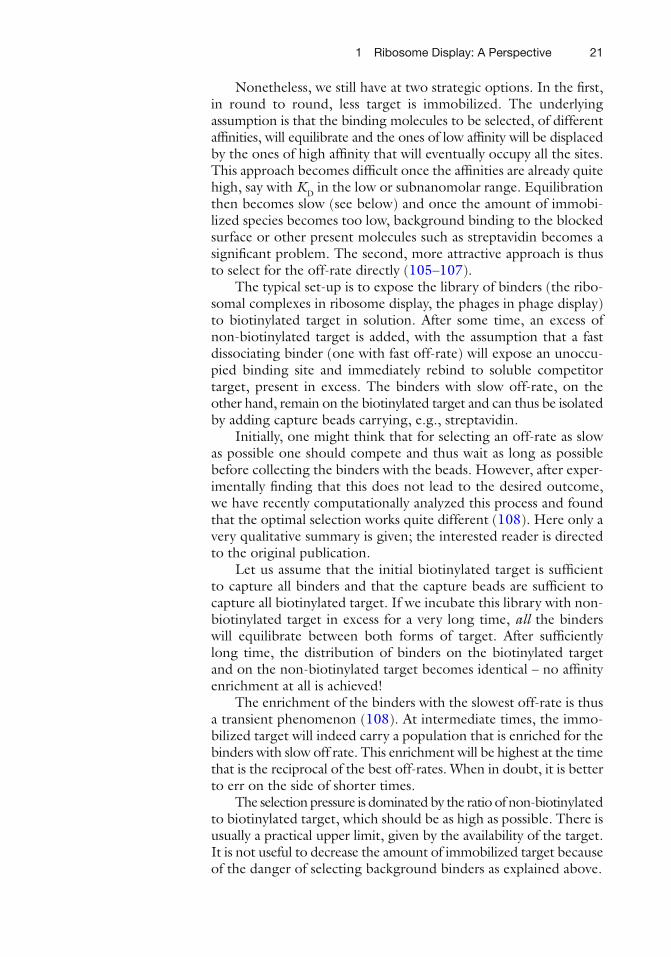
Nonetheless, we still have at two strategic options. In the fi rst, in round to round, less target is immobilized. The underlying assumption is that the binding molecules to be selected, of different affi nities, will equilibrate and the ones of low affi nity will be displaced by the ones of high affi nity that will eventually occupy all the sites. This approach becomes diffi cult once the affi nities are already quite high, say with K D in the low or subnanomolar range. Equilibration then becomes slow (see below) and once the amount of immobi-lized species becomes too low, background binding to the blocked surface or other present molecules such as streptavidin becomes a signifi cant problem. The second, more attractive approach is thus to select for the off-rate directly ( 105– 107 ) .
The typical set-up is to expose the library of binders (the ribo-somal complexes in ribosome display, the phages in phage display) to biotinylated target in solution. After some time, an excess of non-biotinylated target is added, with the assumption that a fast dissociating binder (one with fast off-rate) will expose an unoccu-pied binding site and immediately rebind to soluble competitor target, present in excess. The binders with slow off-rate, on the other hand, remain on the biotinylated target and can thus be isolated by adding capture beads carrying, e.g., streptavidin.
Initially, one might think that for selecting an off-rate as slow as possible one should compete and thus wait as long as possible before collecting the binders with the beads. However, after exper-imentally fi nding that this does not lead to the desired outcome, we have recently computationally analyzed this process and found that the optimal selection works quite different ( 108 ) . Here only a very qualitative summary is given; the interested reader is directed to the original publication.
Let us assume that the initial biotinylated target is suffi cient to capture all binders and that the capture beads are suffi cient to capture all biotinylated target. If we incubate this library with non-biotinylated target in excess for a very long time, all the binders will equilibrate between both forms of target. After suffi ciently long time, the distribution of binders on the biotinylated target and on the non-biotinylated target becomes identical – no affi nity enrichment at all is achieved!
The enrichment of the binders with the slowest off-rate is thus a transient phenomenon ( 108 ) . At intermediate times, the immo-bilized target will indeed carry a population that is enriched for the binders with slow off rate. This enrichment will be highest at the time that is the reciprocal of the best off-rates. When in doubt, it is better to err on the side of shorter times.
The selection pressure is dominated by the ratio of non-biotinylated to biotinylated target, which should be as high as possible. There is usually a practical upper limit, given by the availability of the target. It is not useful to decrease the amount of immobilized target because of the danger of selecting background binders as explained above.
22 A. Plückthun
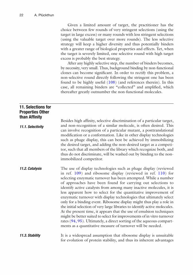
Given a limited amount of target, the practitioner has the choice between few rounds of very stringent selections (using the target in large excess) or many rounds with less stringent selections (using the valuable target over more rounds). The less selective strategy will keep a higher diversity and thus potentially binders with a greater range of biological properties and effects. Yet, when the target is severely limited, one selective round with high target excess is probably the best strategy.
After any highly selective step, the number of binders becomes, by necessity, very small. Thus, background binding by non-functional clones can become signifi cant. In order to rectify this problem, a non-selective round directly following the stringent one has been found to be highly useful ( 108 ) (and references therein). In this case, all remaining binders are “collected” and amplifi ed, which thereafter greatly outnumber the non-functional molecules.
Besides high affi nity, selective discrimination of a particular target, and non-recognition of a similar molecule, is often desired. This can involve recognition of a particular mutant, a posttranslational modifi cation or a conformation. Like in other display technologies such as phage display, this can best be achieved by immobilizing the desired target, and adding the non-desired target as a competi-tor, such that all members of the library which recognize both, and thus do not discriminate, will be washed out by binding to the non-immobilized competitor.
The use of display technologies such as phage display (reviewed in ref. 109 ) and ribosome display (reviewed in ref. 110 ) for selecting enzymatic turnover has been attempted. While a number of approaches have been found for carrying out selections to identify active catalysts from among many inactive molecules, it is less apparent how to select for the quantitative improvement of enzymatic turnover with display technologies that ultimately select only for a binding event. Ribosome display might thus play a role in the initial selection of very large libraries to identify active molecules. At the present time, it appears that the use of emulsion techniques might be better suited to select for improvements of in vitro turnover rates ( 94, 95 ) . Ultimately, a direct sorting of the aqueous compart-ments as a quantitative measure of turnover will be needed.
It is a widespread assumption that ribosome display is unsuitable for evolution of protein stability, and thus its inherent advantages
11. Selections for Properties Other than Affi nity
11.1. Selectivity
11.2. Catalysis
11.3. Stability
231 Ribosome Display: A Perspective
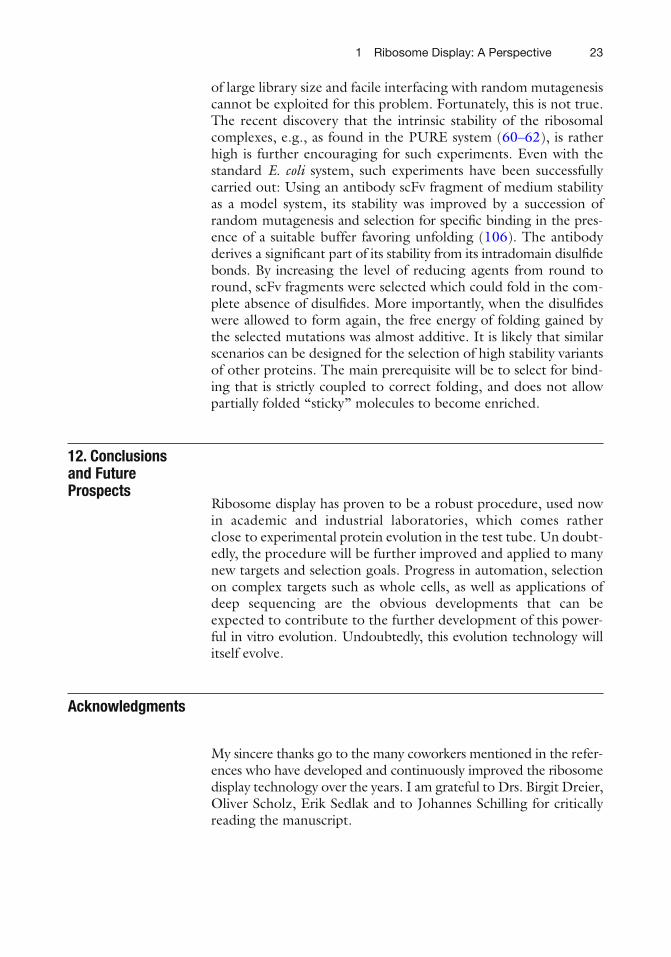
of large library size and facile interfacing with random mutagenesis cannot be exploited for this problem. Fortunately, this is not true. The recent discovery that the intrinsic stability of the ribosomal complexes, e.g., as found in the PURE system ( 60– 62 ) , is rather high is further encouraging for such experiments. Even with the standard E. coli system, such experiments have been successfully carried out: Using an antibody scFv fragment of medium stability as a model system, its stability was improved by a succession of random mutagenesis and selection for specifi c binding in the pres-ence of a suitable buffer favoring unfolding ( 106 ) . The antibody derives a signifi cant part of its stability from its intradomain disulfi de bonds. By increasing the level of reducing agents from round to round, scFv fragments were selected which could fold in the com-plete absence of disulfi des. More importantly, when the disulfi des were allowed to form again, the free energy of folding gained by the selected mutations was almost additive. It is likely that similar scenarios can be designed for the selection of high stability variants of other proteins. The main prerequisite will be to select for bind-ing that is strictly coupled to correct folding, and does not allow partially folded “sticky” molecules to become enriched.
Ribosome display has proven to be a robust procedure, used now in academic and industrial laboratories, which comes rather close to experimental protein evolution in the test tube. Un doubt-edly, the procedure will be further improved and applied to many new targets and selection goals. Progress in automation, selection on complex targets such as whole cells, as well as applications of deep sequencing are the obvious developments that can be expected to contribute to the further development of this power-ful in vitro evolution. Undoubtedly, this evolution technology will itself evolve.
Acknowledgments
My sincere thanks go to the many coworkers mentioned in the refer-ences who have developed and continuously improved the ribosome display technology over the years. I am grateful to Drs. Birgit Dreier, Oliver Scholz, Erik Sedlak and to Johannes Schilling for critically reading the manuscript.
12. Conclusions and Future Prospects
24 A. Plückthun
References
1. Fields, S. & Song, O. (1989) A novel genetic system to detect protein–protein interactions. Nature 340 , 245–246.
2. Pelletier, J. N., Arndt, K. M., Plückthun, A. & Michnick, S. W. (1999) An in vivo library-versus-library selection of optimized protein–protein interactions. Nat. Biotechnol. 17 , 683–690.
3. Wilson, C. G., Magliery, T. J. & Regan, L. (2004) Detecting protein–protein interac-tions with GFP-fragment reassembly. Nat. Methods 1 , 255–262.
4. Cabantous, S., Pedelacq, J. D., Mark, B. L., Naranjo, C., Terwilliger, T. C. & Waldo, G. S. (2005) Recent advances in GFP folding reporter and split-GFP solubility reporter technologies. Application to improving the folding and solubility of recalcitrant proteins from Mycobacterium tuberculosis. J. Struct. Funct. Genomics 6 , 113–119.
5. Rossi, F., Charlton, C. A. & Blau, H. M. (1997) Monitoring protein–protein interac-tions in intact eukaryotic cells by beta-galactosidase complementation. Proc. Natl. Acad. Sci. U. S. A. 94 , 8405–8410.
6. Francisco, J. A., Campbell, R., Iverson, B. L. & Georgiou, G. (1993) Production and fl uorescence-activated cell sorting of Escherichia coli expressing a functional anti-body fragment on the external surface. Proc. Natl. Acad. Sci. U. S. A. 90 , 10444–10448.
7. Boder, E. T. & Wittrup, K. D. (1997) Yeast surface display for screening combinatorial polypeptide libraries. Nat. Biotechnol. 15 , 553–557.
8. Smith, G. P. (1985) Filamentous fusion phage: novel expression vectors that display cloned antigens on the virion surface. Science 228 , 1315–1317.
9. Cwirla, S. E., Peters, E. A., Barrett, R. W. & Dower, W. J. (1990) Peptides on phage: a vast library of peptides for identifying ligands. Proc. Natl. Acad. Sci. U. S. A. 87 , 6378–6382.
10. Devlin, J. J., Panganiban, L. C. & Devlin, P. E. (1990) Random peptide libraries: a source of specifi c protein binding molecules. Science 249 , 404–406.
11. Scott, J. K. & Smith, G. P. (1990) Searching for peptide ligands with an epitope library. Science 249 , 386–390.
12. McCafferty, J., Griffi ths, A. D., Winter, G. & Chiswell, D. J. (1990) Phage antibodies: fi la-mentous phage displaying antibody variable domains. Nature 348 , 552–554.
13. Bass, S., Greene, R. & Wells, J. A. (1990) Hormone phage: an enrichment method for variant proteins with altered binding proper-ties. Proteins 8 , 309–314.
14. Takahashi, T. T. & Roberts, R. W. (2009) In vitro selection of protein and peptide libraries using mRNA display. Methods Mol. Biol. 535 , 293–314.
15. Levin, A. M. & Weiss, G. A. (2006) Optimizing the affinity and specificity of proteins with molecular display. Mol. Biosyst. 2 , 49–57.
16. Leemhuis, H., Stein, V., Griffi ths, A. D. & Hollfelder, F. (2005) New genotype-phenotype linkages for directed evolution of functional proteins. Curr. Opin. Struct. Biol. 15 , 472–478.
17. Daugherty, P. S. (2007) Protein engineering with bacterial display. Curr. Opin. Struct. Biol. 17 , 474–480.
18. Pepper, L. R., Cho, Y. K., Boder, E. T. & Shusta, E. V. (2008) A decade of yeast surface display technology: where are we now? Comb. Chem. High Throughput Screen. 11 , 127–134.
19. Mondon, P., Dubreuil, O., Bouayadi, K. & Kharrat, H. (2008) Human antibody librar-ies: a race to engineer and explore a larger diversity. Front. Biosci. 13 , 1117–1129.
20. Bratkovic, T. (2010) Progress in phage dis-play: evolution of the technique and its appli-cation. Cell. Mol. Life Sci. 67 , 749–767.
21. Labrou, N. E. (2010) Random mutagenesis methods for in vitro directed enzyme evolu-tion. Curr. Protein Pept. Sci. 11 , 91–100.
22. Horst, J. P., Wu, T. H. & Marinus, M. G. (1999) Escherichia coli mutator genes. Trends Microbiol. 7 , 29–36.
23. Wang, L., Jackson, W. C., Steinbach, P. A. & Tsien, R. Y. (2004) Evolution of new nonan-tibody proteins via iterative somatic hypermu-tation. Proc. Natl. Acad. Sci. U. S. A. 101 , 16745–16749.
24. Virnekäs, B., Ge, L., Plückthun, A., Schneider, K. C., Wellnhofer, G. & Moroney, S. E. (1994) Trinucleotide phosphoramidites: Ideal reagents for the synthesis of mixed oligonu-cleotides for random mutagenesis. Nucleic Acids Res. 22 , 5600–5607.
25. Zaccolo, M., Williams, D. M., Brown, D. M. & Gherardi, E. (1996) An approach to random mutagenesis of DNA using mixtures of triphosphate derivatives of nucleoside ana-logues. J. Mol. Biol. 255 , 589–603.
251 Ribosome Display: A Perspective
26. Zhao, H., Giver, L., Shao, Z., Affholter, J. A. & Arnold, F. H. (1998) Molecular evolu-tion by staggered extension process (StEP) in vitro recombination. Nat. Biotechnol. 16 , 258–261.
27. Stemmer, W. P. (1994) Rapid evolution of a protein in vitro by DNA shuffl ing. Nature 370 , 389–391.
28. Kraus, J. P. & Rosenberg, L. E. (1982) Purifi cation of low-abundance messenger RNAs from rat liver by polysome immunoad-sorption. Proc. Natl. Acad. Sci. U. S. A. 79 , 4015–4019.
29. Korman, A. J., Knudsen, P. J., Kaufman, J. F. & Strominger, J. L. (1982) cDNA clones for the heavy chain of HLA-DR antigens obtained after immunopurifi cation of polysomes by monoclonal antibody. Proc. Natl. Acad. Sci. U. S. A. 79 , 1844–1848.
30. Tuerk, C. & Gold, L. (1990) Systematic evo-lution of ligands by exponential enrichment: RNA ligands to bacteriophage T4 DNA poly-merase. Science 249 , 505–510.
31. Mattheakis, L. C., Dias, J. M. & Dower, W. J. (1996) Cell-free synthesis of peptide libraries displayed on polysomes. Methods Enzymol. 267 , 195–207.
32. Mattheakis, L. C., Bhatt, R. R. & Dower, W. J. (1994) An in vitro polysome display system for identifying ligands from very large peptide libraries. Proc. Natl. Acad. Sci. U. S. A. 91 , 9022–9026.
33. Kawasaki, G. H. (1991) Cell-free synthesis and isolation of novel genes and polypeptides. PCT Int. Appl. , WO 91/05058.
34. Gersuk, G. M., Corey, M. J., Corey, E., Stray, J. E., Kawasaki, G. H. & Vessella, R. L. (1997) High-affi nity peptide ligands to prostate-specifi c antigen identifi ed by polysome selection. Biochem. Biophys. Res. Commun. 232 , 578–582.
35. Hanes, J. & Plückthun, A. (1997) In vitro selection and evolution of functional proteins by using ribosome display. Proc. Natl. Acad. Sci. USA 94 , 4937–4942.
36. Ryabova, L. A., Desplancq, D., Spirin, A. S. & Plückthun, A. (1997) Functional antibody production using cell-free translation: Effects of protein disulfi de isomerase and chaperones. Nat. Biotechnol. 15 , 79–84.
37. Hanes, J., Jermutus, L., Weber-Bornhauser, S., Bosshard, H. R. & Plückthun, A. (1998) Ribosome display effi ciently selects and evolves high-affi nity antibodies in vitro from immune libraries. Proc. Natl. Acad. Sci. USA 95 , 14130–14135.
38. Hanes, J., Schaffi tzel, C., Knappik, A. & Plückthun, A. (2000) Picomolar affi nity anti-
bodies from a fully synthetic naive library selected and evolved by ribosome display. Nat. Biotechnol. 18 , 1287–1292.
39. He, M. & Taussig, M. J. (1997) Antibody-ribosome-mRNA (ARM) complexes as effi -cient selection particles for in vitro display and evolution of antibody combining sites. Nucleic Acids Res. 25 , 5132–5134.
40. He, M., Menges, M., Groves, M. A., Corps, E., Liu, H., Brüggemann, M. & Taussig, M. J. (1999) Selection of a human anti-progesterone antibody fragment from a transgenic mouse library by ARM ribosome display. J. Immunol. Methods 231 , 105–117.
41. Kisselev, L., Ehrenberg, M. & Frolova, L. (2003) Termination of translation: interplay of mRNA, rRNAs and release factors? EMBO J. 22 , 175–182.
42. Pavlov, M. Y., Antoun, A., Lovmar, M. & Ehrenberg, M. (2008) Complementary roles of initiation factor 1 and ribosome recycling factor in 70S ribosome splitting. EMBO J. 27 , 1706–1717.
43. Hauryliuk, V., Zavialov, A., Kisselev, L. & Ehrenberg, M. (2006) Class-1 release factor eRF1 promotes GTP binding by class-2 release factor eRF3. Biochimie 88 , 747–757.
44. Douthwaite, J. A., Groves, M. A., Dufner, P. & Jermutus, L. (2006) An improved method for an effi cient and easily accessible eukaryotic ribosome display technology. Protein Eng. Des. Sel. 19 , 85–90.
45. Cheng, K., Ivanova, N., Scheres, S. H., Pavlov, M. Y., Carazo, J. M., Hebert, H., Ehrenberg, M. & Lindahl, M. (2010) tmRNA·SmpB complex mimics native aminoacyl-tRNAs in the A site of stalled ribosomes. J. Struct. Biol. 169 , 342–348.
46. Moore, S. D. & Sauer, R. T. (2007) The tmRNA system for translational surveillance and ribosome rescue. Annu. Rev. Biochem. 76 , 101–124.
47. Shine, J. & Dalgarno, L. (1975) Terminal-sequence analysis of bacterial ribosomal RNA. Correlation between the 3 ¢ -terminal-polypyrimidine sequence of 16-S RNA and translational specifi city of the ribosome. Eur. J. Biochem. 57 , 221–230.
48. Salis, H. M., Mirsky, E. A. & Voigt, C. A. (2009) Automated design of synthetic ribo-some binding sites to control protein expres-sion. Nat. Biotechnol. 27 , 946–950.
49. Kozak, M. (1999) Initiation of translation in prokaryotes and eukaryotes. Gene 234 , 187–208.
50. Carpousis, A. J. (2007) The RNA degrado-some of Escherichia coli: an mRNA-degrading
26 A. Plückthun
machine assembled on RNase E. Annu. Rev. Microbiol. 61 , 71–87.
51. Carpousis, A. J., Luisi, B. F. & McDowall, K. J. (2009) Endonucleolytic initiation of mRNA decay in Escherichia coli. Prog. Mol. Biol. Transl. Sci. 85 , 91–135.
52. Regnier, P. & Hajnsdorf, E. (2009) Poly(A)-assisted RNA decay and modulators of RNA stability. Prog. Mol. Biol. Transl. Sci. 85 , 137–185.
53. Schatz, P. J. (1993) Use of peptide libraries to map the substrate specifi city of a peptide-modifying enzyme: a 13 residue consensus peptide specifi es biotinylation in Escherichia coli. Biotechnology (N. Y.) 11 , 1138–1143.
54. Hoffmann, A., Bukau, B. & Kramer, G. (2010) Structure and function of the molecu-lar chaperone Trigger Factor. Biochim. Biophys. Acta 1803 , 650–661.
55. Hoffmann, F. & Rinas, U. (2004) Roles of heat-shock chaperones in the production of recombinant proteins in Escherichia coli. Adv. Biochem. Eng. Biotechnol. 89 , 143–161.
56. Schimmele, B., Gräfe, N. & Plückthun, A. (2005) Ribosome display of mammalian receptor domains. Protein Eng. Des. Sel. 18 , 285–294.
57. Schimmele, B. & Plückthun, A. (2005) Identifi cation of a functional epitope of the Nogo receptor by a combinatorial approach using ribosome display. J. Mol. Biol. 352 , 229–241.
58. He, M. & Taussig, M. J. (2007) Eukaryotic ribosome display with in situ DNA recovery. Nat. Methods 4 , 281–288.
59. Villemagne, D., Jackson, R. & Douthwaite, J. A. (2006) Highly effi cient ribosome display selection by use of purifi ed components for in vitro translation. J. Immunol. Methods 313 , 140–148.
60. Matsuura, T., Yanagida, H., Ushioda, J., Urabe, I. & Yomo, T. (2007) Nascent chain, mRNA, and ribosome complexes generated by a pure translation system. Biochem. Biophys. Res. Commun. 352 , 372–377.
61. Ohashi, H., Shimizu, Y., Ying, B. W. & Ueda, T. (2007) Effi cient protein selection based on ribosome display system with purifi ed compo-nents. Biochem. Biophys. Res. Commun. 352 , 270–276.
62. Ueda, T., Kanamori, T. & Ohashi, H. (2010) Ribosome display with the PURE technol-ogy. Methods Mol. Biol. 607 , 219–225.
63. Hanes, J., Jermutus, L., Schaffi tzel, C. & Plückthun, A. (1999) Comparison of Escherichia coli and rabbit reticulocyte ribo-some display systems. FEBS Lett. 450 , 105–110.
64. Nakatogawa, H. & Ito, K. (2002) The ribo-somal exit tunnel functions as a discriminating gate. Cell 108 , 629–636.
65. Ogawa, A., Sando, S. & Aoyama, Y. (2005) In vitro read-through polysome/ribosome display of full-length protein ORF and its applica-tions. Nucleic Acids. Symp. Ser. (Oxf.) , 267–268.
66. Ogawa, A., Sando, S. & Aoyama, Y. (2006) Termination-free prokaryotic protein transla-tion by using anticodon-adjusted E. coli tRNA Ser as unifi ed suppressors of the UAA/UGA/UAG stop codons. Read-through ribo-some display of full-length DHFR with trans-lated UTR as a buried spacer arm. ChemBioChem 7 , 249–252.
67. Glockshuber, R., Malia, M., Pfi tzinger, I. & Plückthun, A. (1990) A comparison of strate-gies to stabilize immunoglobulin Fv-fragments. Biochemistry 29 , 1362–1367.
68. Groves, M., Lane, S., Douthwaite, J., Lowne, D., Rees, D. G., Edwards, B. & Jackson, R. H. (2006) Affi nity maturation of phage dis-play antibody populations using ribosome display. J. Immunol. Methods 313 , 129–139.
69. Binz, H. K., Stumpp, M. T., Forrer, P., Amstutz, P. & Plückthun, A. (2003) Designing repeat proteins: well-expressed, soluble and stable proteins from combinatorial libraries of consensus ankyrin repeat proteins. J. Mol. Biol. 332 , 489–503.
70. Binz, H. K., Amstutz, P., Kohl, A., Stumpp, M. T., Briand, C., Forrer, P., Grütter, M. G. & Plückthun, A. (2004) High-affi nity binders selected from designed ankyrin repeat protein libraries. Nat. Biotechnol. 22 , 575–582.
71. Wetzel, S. K., Ewald, C., Settanni, G., Jurt, S., Plückthun, A. & Zerbe, O. (2010) Residue-resolved stability of full-consensus ankyrin repeat proteins probed by NMR. J. Mol. Biol. 402 , 241–258.
72. Wetzel, S. K., Settanni, G., Kenig, M., Binz, H. K. & Plückthun, A. (2008) Folding and unfolding mechanism of highly stable full-consensus ankyrin repeat proteins. J. Mol. Biol. 376 , 241–257.
73. Milovnik, P., Ferrari, D., Sarkar, C. A. & Plückthun, A. (2009) Selection and charac-terization of DARPins specifi c for the neuro-tensin receptor 1. Protein Eng. Des. Sel. 22 , 357–366.
74. Zahnd, C., Wyler, E., Schwenk, J. M., Steiner, D., Lawrence, M. C., McKern, N. M., Pecorari, F., Ward, C. W., Joos, T. O. & Plückthun, A. (2007) A designed ankyrin repeat protein evolved to picomolar affi nity to Her2. J. Mol. Biol. 369 , 1015–1028.
271 Ribosome Display: A Perspective
75. Schweizer, A., Roschitzki-Voser, H., Amstutz, P., Briand, C., Gulotti-Georgieva, M., Prenosil, E., Binz, H. K., Capitani, G., Baici, A., Plückthun, A. & Grütter, M. G. (2007) Inhibition of caspase-2 by a designed ankyrin repeat protein: specifi city, structure, and inhibi-tion mechanism. Structure 15 , 625–636.
76. Zahnd, C., Pécorari, F., Straumann, N., Wyler, E. & Plückthun, A. (2006) Selection and characterization of Her2 binding-designed ankyrin repeat proteins. J. Biol. Chem. 281 , 35167–35175.
77. Amstutz, P., Koch, H., Binz, H. K., Deuber, S. A. & Plückthun, A. (2006) Rapid selection of specifi c MAP kinase-binders from designed ankyrin repeat protein libraries. Protein Eng. Des. Sel. 19 , 219–229.
78. Amstutz, P., Binz, H. K., Parizek, P., Stumpp, M. T., Kohl, A., Grütter, M. G., Forrer, P. & Plückthun, A. (2005) Intracellular kinase inhibitors selected from combinatorial librar-ies of designed ankyrin repeat proteins. J. Biol. Chem. 280 , 24715–24722.
79. Dreier, B., Mikheeva, G., Belousova, N., Parizek, P., Boczek, E., Jelesarov, I., Forrer, P., Plückthun, A. & Krasnykh, V. (2010) Her2-specifi c multivalent adapters confer designed tropism to adenovirus for gene tar-geting. J. Mol. Biol. 405 , 410–426.
80. Veesler, D., Dreier, B., Blangy, S., Lichière, J., Tremblay, D., Moineau, S., Spinelli, S., Tegoni, M., Plückthun, A., Campanacci, V. & Cambillau, C. (2009) Crystal structure of a DARPin neutralizing inhibitor of lactococcal phage TP901-1: comparison of DARPin and camelid VHH binding mode J. Biol. Chem. 384 , 30718–30726.
81. Yau, K. Y., Dubuc, G., Li, S., Hirama, T., Mackenzie, C. R., Jermutus, L., Hall, J. C. & Tanha, J. (2005) Affi nity maturation of a V(H)H by mutational hotspot randomization. J. Immunol. Methods 297 , 213–224.
82. Perruchini, C., Pecorari, F., Bourgeois, J. P., Duyckaerts, C., Rougeon, F. & Lafaye, P. (2009) Llama VHH antibody fragments against GFAP: better diffusion in fi xed tissues than classical monoclonal antibodies. Acta Neuropathol. 118 , 685–695.
83. Cho, G. S. & Szostak, J. W. (2006) Directed evolution of ATP binding proteins from a zinc fi nger domain by using mRNA display. Chem. Biol. 13 , 139–147.
84. Keefe, A. D. & Szostak, J. W. (2001) Functional proteins from a random-sequence library. Nature 410 , 715–718.
85. Seelig, B. & Szostak, J. W. (2007) Selection and evolution of enzymes from a partially
randomized non-catalytic scaffold. Nature 448 , 828–831.
86. Parker, M. H., Chen, Y., Danehy, F., Dufu, K., Ekstrom, J., Getmanova, E., Gokemeijer, J., Xu, L. & Lipovsek, D. (2005) Antibody mimics based on human fi bronectin type three domain engineered for thermostability and high-affi nity binding to vascular endothelial growth factor receptor two. Protein Eng. Des. Sel. 18 , 435–444.
87. Xu, L., Aha, P., Gu, K., Kuimelis, R. G., Kurz, M., Lam, T., Lim, A. C., Liu, H., Lohse, P. A., Sun, L., Weng, S., Wagner, R. W. & Lipovsek, D. (2002) Directed evolution of high-affi nity antibody mimics using mRNA display. Chem. Biol. 9 , 933–942.
88. Olson, C. A. & Roberts, R. W. (2007) Design, expression, and stability of a diverse protein library based on the human fi bronec-tin type III domain. Protein Sci. 16 , 476–484.
89. Fukuda, I., Kojoh, K., Tabata, N., Doi, N., Takashima, H., Miyamoto-Sato, E. & Yanagawa, H. (2006) In vitro evolution of single-chain antibodies using mRNA display. Nucleic Acids Res. 34 , e127.
90. Shen, X., Valencia, C. A., Szostak, J. W., Dong, B. & Liu, R. (2005) Scanning the human proteome for calmodulin-binding proteins. Proc. Natl. Acad. Sci. U. S. A. 102 , 5969–5974.
91. Doi, N. & Yanagawa, H. (1999) STABLE: protein-DNA fusion system for screening of combinatorial protein libraries in vitro. FEBS Lett. 457 , 227–230.
92. Yonezawa, M., Doi, N., Higashinakagawa, T. & Yanagawa, H. (2004) DNA display of bio-logically active proteins for in vitro protein selection. J. Biochem. 135 , 285–288.
93. Yonezawa, M., Doi, N., Kawahashi, Y., Higashinakagawa, T. & Yanagawa, H. (2003) DNA display for in vitro selection of diverse peptide libraries. Nucleic Acids Res. 31 , e118.
94. Griffi ths, A. D. & Tawfi k, D. S. (2003) Directed evolution of an extremely fast phos-photriesterase by in vitro compartmentaliza-tion. EMBO J. 22 , 24–35.
95. Miller, O. J., Bernath, K., Agresti, J. J., Amitai, G., Kelly, B. T., Mastrobattista, E., Taly, V., Magdassi, S., Tawfi k, D. S. & Griffi ths, A. D. (2006) Directed evolution by in vitro compartmentalization. Nat. Methods 3 , 561–570.
96. Sumida, T., Doi, N. & Yanagawa, H. (2009) Bicistronic DNA display for in vitro selection of Fab fragments. Nucleic Acids Res. 37 , e147.
28 A. Plückthun
97. Dreier, B. & Plückthun, A. (2012) Rapid selec-tion of high affi nity binders using ribosome display. Methods Mol. Biol. , 805, 261–286.
98. Bremer, H. & Dennis, P. P. (1996). Modulation of chemical composition and other parameters of the cell by growth rate. In Escherichia coli and Salmonella typhimurium: Cellular and Molecular Biology (Neidhard, F. C., Curtiss, R., Ingraham, J. L., Lin, E. C. C., Low, K. B., Magasanik, B., Reznikoff, W. S., Riley, M., Schaechter, M. & Umbarger, H. E., eds.), Vol. 2, pp. 1553–1569. American Society for Microbiology Press, Washington, DC.
99. Northrup, S. H. & Erickson, H. P. (1992) Kinetics of protein–protein association explained by Brownian dynamics computer simulation. Proc. Natl. Acad. Sci. U. S. A. 89 , 3338–3342.
100. Schreiber, G. & Fersht, A. R. (1996) Rapid, electrostatically assisted association of pro-teins. Nat. Struct. Biol. 3 , 427–431.
101. Selzer, T., Albeck, S. & Schreiber, G. (2000) Rational design of faster associating and tighter binding protein complexes. Nat. Struct. Biol. 7 , 537–541.
102. Berger, C., Weber-Bornhauser, S., Eggenberger, J., Hanes, J., Plückthun, A. & Bosshard, H. R. (1999) Antigen recognition by conformational selection. FEBS Lett. 450 , 149–153.
103. Foote, J. & Milstein, C. (1994) Conformational isomerism and the diversity of antibodies. Proc. Natl. Acad. Sci. U. S. A. 91 , 10370–10374.
104. Koshland, D. E. (1958) Application of a theory of enzyme specifi city to protein synthesis. Proc. Natl. Acad. Sci. U. S. A. 44 , 98–104.
105. Hawkins, R. E., Russell, S. J. & Winter, G. (1992) Selection of phage antibodies by bind-ing affi nity. Mimicking affi nity maturation. J. Mol. Biol. 226 , 889–896.
106. Jermutus, L., Honegger, A., Schwesinger, F., Hanes, J. & Plückthun, A. (2001) Tailoring in vitro evolution for protein affi nity or sta-bility. Proc. Natl. Acad. Sci. U.S.A. 98 , 75–80.
107. Zahnd, C., Spinelli, S., Luginbühl, B., Amstutz, P., Cambillau, C. & Plückthun, A. (2004) Directed in vitro evolution and crys-tallographic analysis of a peptide binding scFv antibody with low picomolar affi nity. J. Biol. Chem. 279 , 18870–18877.
108. Zahnd, C., Sarkar, C. A. & Plückthun, A. (2010) Computational analysis of off-rate selec-tion experiments to optimize affi nity matura-tion by directed evolution. Protein Eng. Des. Sel. 23 , 175–184.
109. Forrer, P., Jung, S. & Plückthun, A. (1999) Beyond binding: using phage display to select for structure, folding and enzymatic activity in proteins. Curr. Opin. Struct. Biol. 9 , 514–520.
110. Amstutz, P., Forrer, P., Zahnd, C. & Plückthun, A. (2001) In vitro display technologies: Novel developments and applications. Curr. Opin. Biotechnol. 12 , 400–405.
111. Zahnd, C., Amstutz, P. & Plückthun, A. (2007) Ribosome display: selecting and evolving proteins in vitro that specifically bind to a target. Nat. Methods 4 , 269–279.
112. Lipovsek, D. & Plückthun, A. (2004) In-vitro protein evolution by ribosome display and mRNA display. J. Immunol. Methods 290 , 51–67.
113. Seidelt, B., Innis, C. A., Wilson, D. N., Gartmann, M., Armache, J. P., Villa, E., Trabuco, L. G., Becker, T., Mielke, T., Schulten, K., Steitz, T. A. & Beckmann, R. (2009) Structural insight into nascent poly-peptide chain-mediated translational stalling. Science 326 , 1412–1415.