Embed Size (px)
Citation preview
How Cells Divide
Chapter 10

2
Bacterial Cell Division
• Bacteria divide by binary fission– No sexual life cycle
– Reproduction is clonal
• Single, circular bacterial chromosome is replicated
• Replication begins at the origin of replication and proceeds in two directions to site of termination
• New chromosomes are partitioned to opposite ends of the cell
• Septum forms to divide the cell into 2 cells

3
Prior to cell division,
the bacterial DNA
molecule replicates.
The replication of the
double-stranded,
Circular DNA mole-
cule that constitutes
the genome of a
bacterium begins at a
specific site, called
the origin of replica-
tion (green area).
The replication
enzymes move out
in both directions
from that site and
make copies of each
strand in the DNA
duplex. The enzymes
continue until they
meet at another
specific site, the
terminus of replication
(red area).
As the DNA is
replicated, the cell
elongates, and the
DNA is partitioned in
the cell such that the
origins are at the ¼
and ¾ positions in the
cell and the termini
are oriented toward
the middle of the cell.
1.
2.
3.
Bacterial cell
Bacterial chromosome:
Double-stranded DNA
Origin of
replication
Copyright © The McGraw-Hill Companies, Inc. Permission required for reproduction or display.

4
5.
Septation then begins, in which new membrane and cell wall material
begin to grow and form a septum at approximately the midpoint of the cell.
A protein molecule called FtsZ (orange dots) facilitates this process.
When the septum is complete, the cell pinches
in two, and two daughter cells are formed,
each containing a bacterial DNA molecule.
4.
Septum
Copyright © The McGraw-Hill Companies, Inc. Permission required for reproduction or display.

Eukaryotic Chromosomes
• Every species has a
different number of
chromosomes
• Humans have 46
chromosomes in 23
nearly identical pairs
– Additional/missing
chromosomes usually
fatal
5
950x
Copyright © The McGraw-Hill Companies, Inc. Permission required for reproduction or display.
© Biophoto Associates/Photo Researchers, Inc.
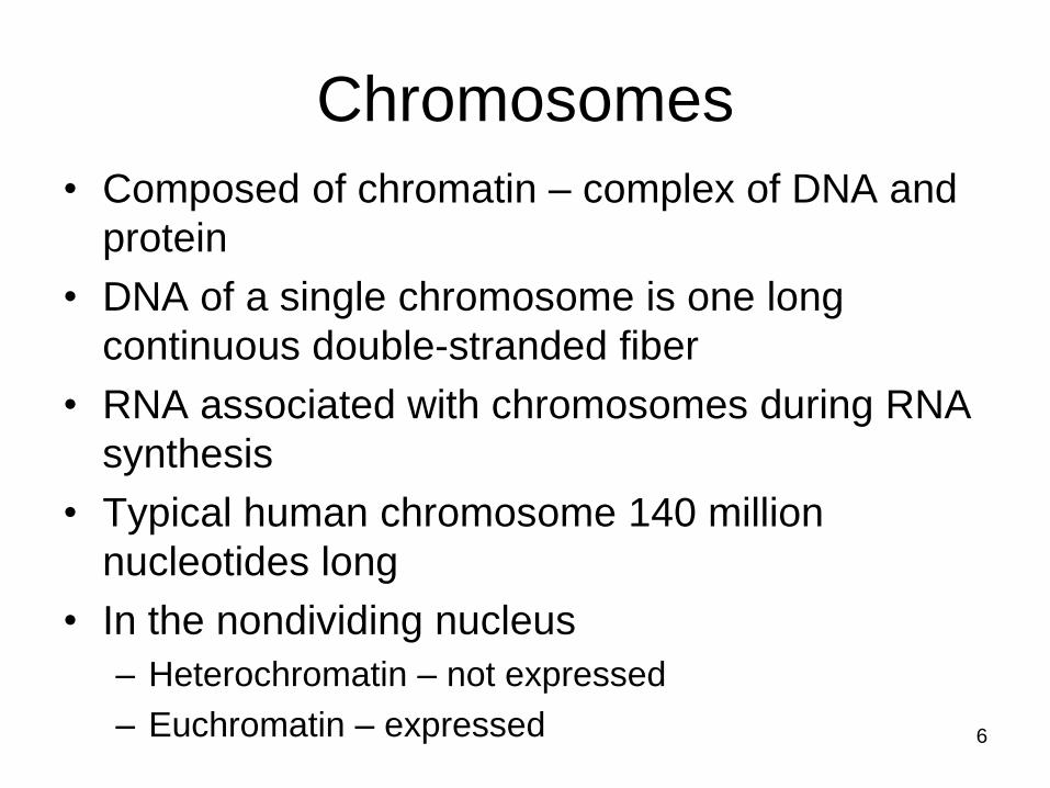
Chromosomes
• Composed of chromatin – complex of DNA and
protein
• DNA of a single chromosome is one long
continuous double-stranded fiber
• RNA associated with chromosomes during RNA
synthesis
• Typical human chromosome 140 million
nucleotides long
• In the nondividing nucleus
– Heterochromatin – not expressed
– Euchromatin – expressed 6

Chromosome Structure
• Nucleosome
– Complex of DNA and histone proteins
– Promote and guide coiling of DNA
– DNA duplex coiled around 8 histone proteins
every 200 nucleotides
– Histones are positively charged and strongly
attracted to negatively charged phosphate
groups of DNA
7

8
DNA Double Helix (duplex) Nucleosome
Histone coreDNA
Copyright © The McGraw-Hill Companies, Inc. Permission required for reproduction or display.

• Nucleosomes wrapped into higher order
coils called solenoids
– Leads to a fiber 30 nm in diameter
– Usual state of nondividing (interphase)
chromatin
• During mitosis, chromatin in solenoid
arranged around scaffold of protein to
achieve maximum compaction
– Radial looping aided by condensin proteins
9

10
Copyright © The McGraw-Hill Companies, Inc. Permission required for reproduction or display.
Mitotic Chromosome
Chromatin loop
Solenoid
Scaffold
proteinScaffold protein
Chromatin LoopRosettes of Chromatin Loops
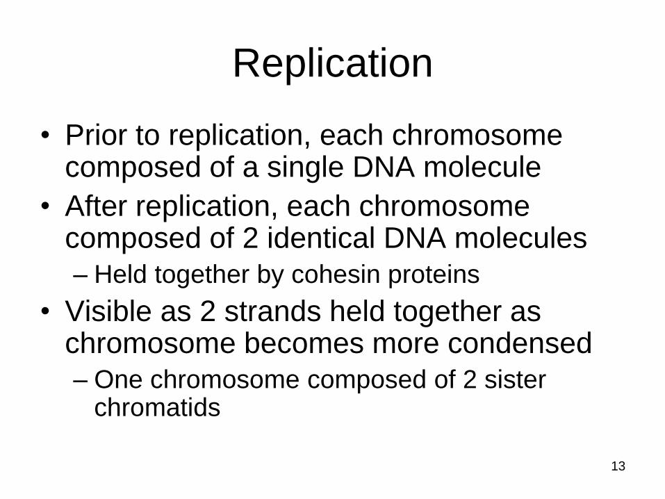
Replication
• Prior to replication, each chromosome composed of a single DNA molecule
• After replication, each chromosome composed of 2 identical DNA molecules
– Held together by cohesin proteins
• Visible as 2 strands held together as chromosome becomes more condensed
– One chromosome composed of 2 sister chromatids
13

14
Copyright © The McGraw-Hill Companies, Inc. Permission required for reproduction or display.
Homologous chromosomes Homologous chromosomes
Cohesin
proteins
Kinetochores
Sister chromatids
Sister chromatids
Kinetochore
Centromere
Replication
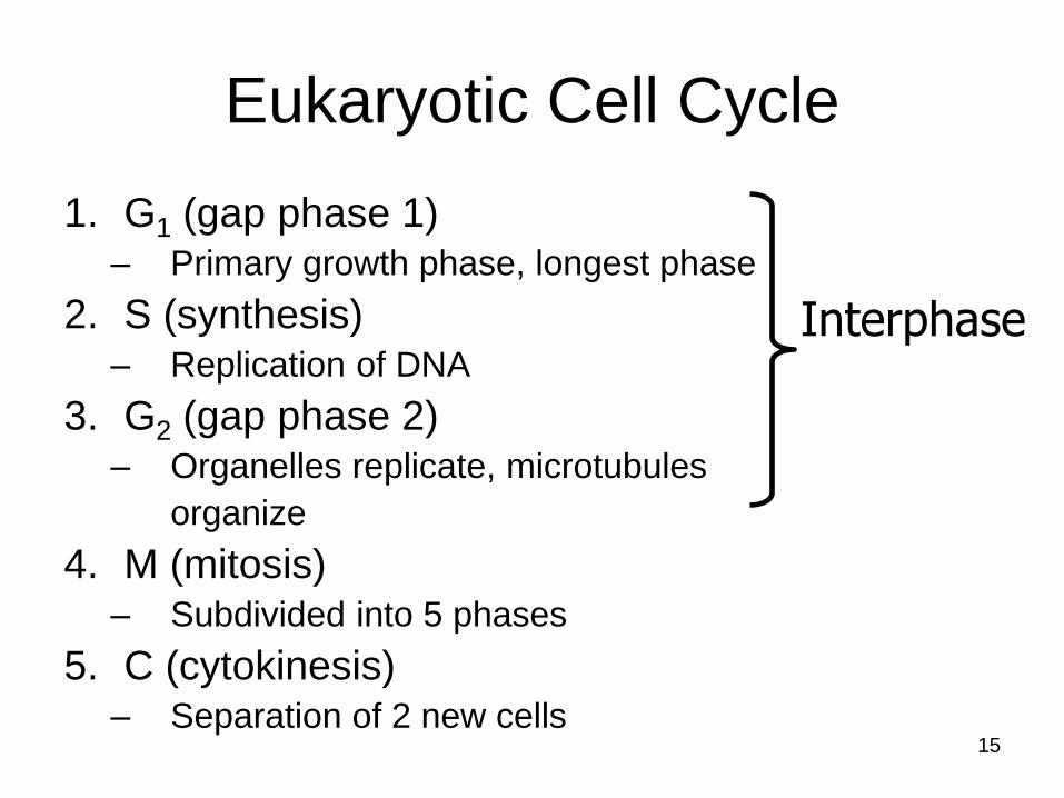
Eukaryotic Cell Cycle
1. G1 (gap phase 1)– Primary growth phase, longest phase
2. S (synthesis)– Replication of DNA
3. G2 (gap phase 2)– Organelles replicate, microtubules
organize
4. M (mitosis)– Subdivided into 5 phases
5. C (cytokinesis)– Separation of 2 new cells
15
Interphase

16
Duration
• Time it takes to complete a cell cycle varies greatly
• Fruit fly embryos = 8 minutes
• Mature cells take longer to grow– Typical mammalian cell takes 24 hours
– Liver cell takes more than a year
• Growth occurs during G1, G2, and S phases– M phase takes only about an hour
• Most variation in length of G1
– Resting phase G0 – cells spend more or less time here
17
Copyright © The McGraw-Hill Companies, Inc. Permission required for reproduction or display.
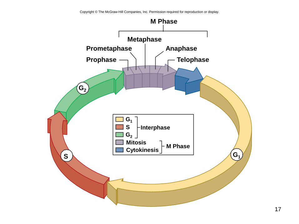
M Phase
Metaphase
Anaphase
Telophase
Prometaphase
Prophase
S
G2
G1
Interphase
M Phase
G1
Cytokinesis
Mitosis
S
G2
18
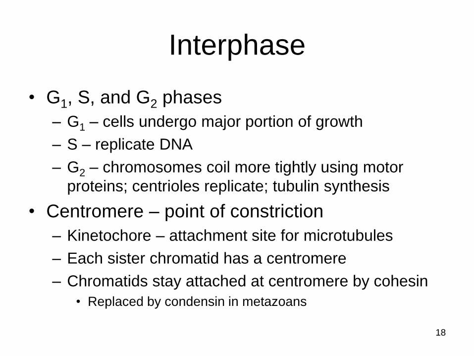
Interphase
• G1, S, and G2 phases
– G1 – cells undergo major portion of growth
– S – replicate DNA
– G2 – chromosomes coil more tightly using motor
proteins; centrioles replicate; tubulin synthesis
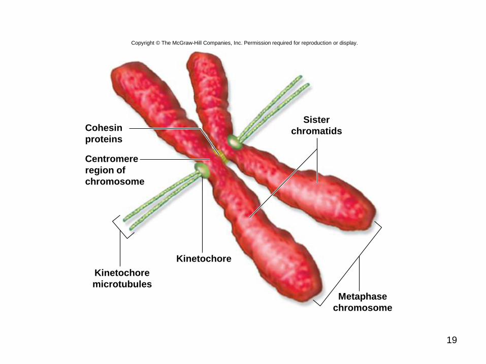
• Centromere – point of constriction
– Kinetochore – attachment site for microtubules
– Each sister chromatid has a centromere
– Chromatids stay attached at centromere by cohesin
• Replaced by condensin in metazoans
19
Copyright © The McGraw-Hill Companies, Inc. Permission required for reproduction or display.
Cohesin
proteins
Centromere
region of
chromosome
Kinetochore
microtubules
Kinetochore
Metaphase
chromosome
Sister
chromatids
20
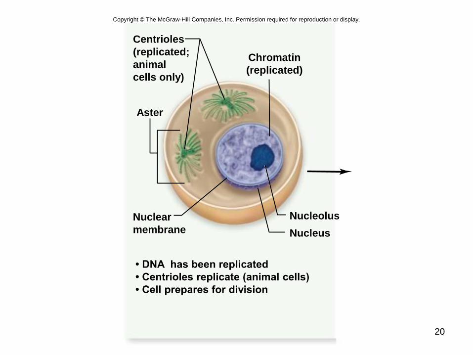
Nucleus
Nucleolus
Aster
Centrioles
(replicated;
animal
cells only)
Nuclear
membrane
• DNA has been replicated
• Centrioles replicate (animal cells)
• Cell prepares for division
Chromatin
(replicated)
Copyright © The McGraw-Hill Companies, Inc. Permission required for reproduction or display.

21
M phase
Mitosis is divided into 5 phases:
1. Prophase
2. Prometaphase
3. Metaphase
4. Anaphase
5. Telophase
22
Prophase
• Individual condensed chromosomes first
become visible with the light microscope
– Condensation continues throughout prophase
• Spindle apparatus assembles
– 2 centrioles move to opposite poles forming spindle
apparatus (no centrioles in plants)
– Asters – radial array of microtubules in animals (not
plants)
• Nuclear envelope breaks down
23
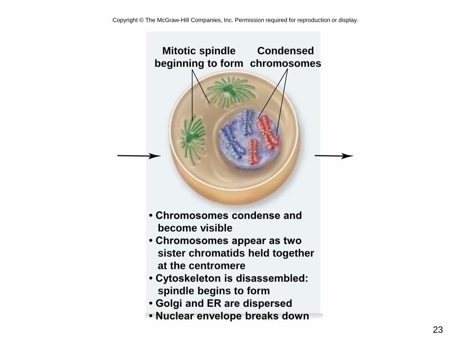
• Chromosomes condense and
become visible
• Chromosomes appear as two
sister chromatids held together
at the centromere
• Cytoskeleton is disassembled:
spindle begins to form
• Golgi and ER are dispersed
• Nuclear envelope breaks down
Mitotic spindle
beginning to form
Condensed
chromosomes
Copyright © The McGraw-Hill Companies, Inc. Permission required for reproduction or display.

24
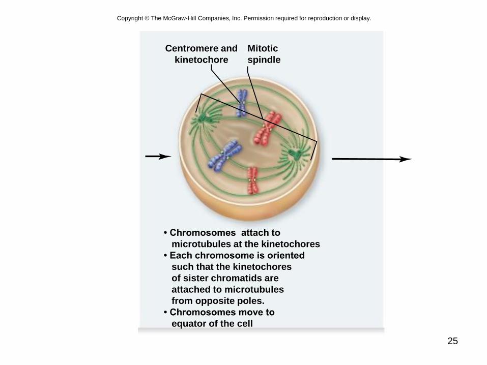
Prometaphase
• Transition occurs after disassembly of nuclear
envelope
• Microtubule attachment
– 2nd group grows from poles and attaches to
kinetochores
– Each sister chromatid connected to opposite poles
• Chromosomes begin to move to center of cell –
congression
– Assembly and disassembly of microtubules
– Motor proteins at kinetochores
25
• Chromosomes attach to
microtubules at the kinetochores
• Each chromosome is oriented
such that the kinetochores
of sister chromatids are
attached to microtubules
from opposite poles.
• Chromosomes move to
equator of the cell
Centromere and
kinetochore
Mitotic
spindle
Copyright © The McGraw-Hill Companies, Inc. Permission required for reproduction or display.

Metaphase
• Alignment of
chromosomes
along metaphase
plate
– Not an actual
structure
– Future axis of cell
division
26
Polar
microtubule
Centrioles
Metaphase
plate
Aster
Kinetochore
microtubule
Sister chromatids
57µm
Copyright © The McGraw-Hill Companies, Inc. Permission required for reproduction or display.
© Andrew S. Bajer, University of Oregon

27
• All chromosomes are aligned
at equator of the cell, called
the metaphase plate
• Chromosomes are attached
to opposite poles and are
under tension
Polar microtubule
Chromosomes
aligned on
metaphase plateKinetochore
microtubule
Copyright © The McGraw-Hill Companies, Inc. Permission required for reproduction or display.
28
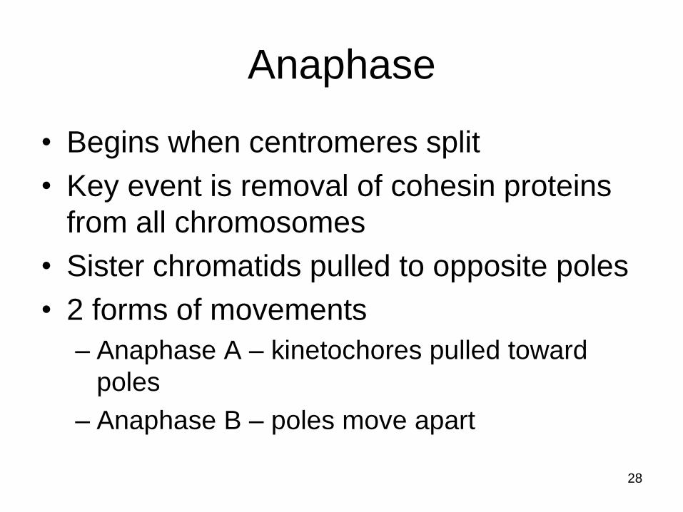
Anaphase
• Begins when centromeres split
• Key event is removal of cohesin proteins
from all chromosomes
• Sister chromatids pulled to opposite poles
• 2 forms of movements
– Anaphase A – kinetochores pulled toward
poles
– Anaphase B – poles move apart
29
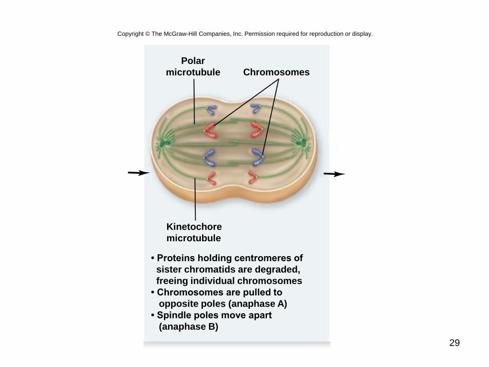
Chromosomes
• Proteins holding centromeres of
sister chromatids are degraded,
freeing individual chromosomes
• Chromosomes are pulled to
opposite poles (anaphase A)
• Spindle poles move apart
(anaphase B)
Kinetochore
microtubule
Polar
microtubule
Copyright © The McGraw-Hill Companies, Inc. Permission required for reproduction or display.

30
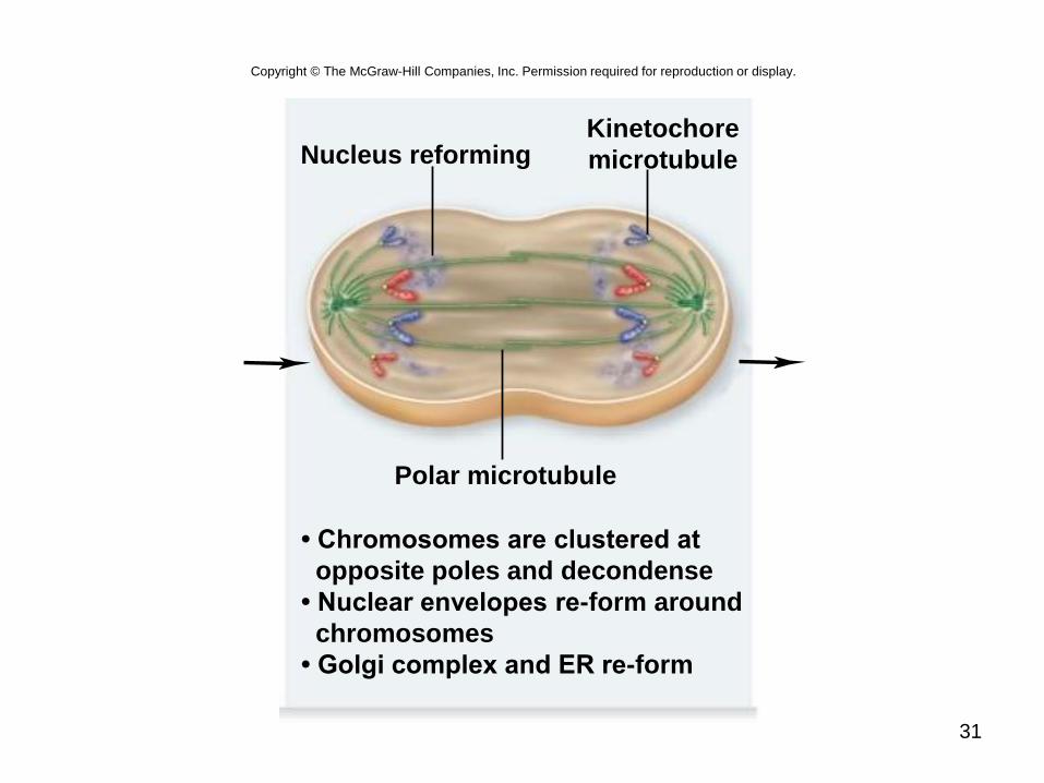
Telophase
• Spindle apparatus disassembles
• Nuclear envelope forms around each set
of sister chromatids
– Now called chromosomes
• Chromosomes begin to uncoil
• Nucleolus reappears in each new nucleus
31
Polar microtubule
Nucleus reforming
• Chromosomes are clustered at
opposite poles and decondense
• Nuclear envelopes re-form around
chromosomes
• Golgi complex and ER re-form
Kinetochore
microtubule
Copyright © The McGraw-Hill Companies, Inc. Permission required for reproduction or display.
32
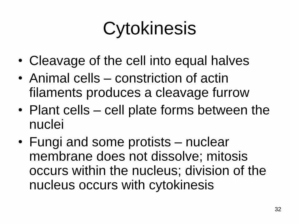
Cytokinesis
• Cleavage of the cell into equal halves
• Animal cells – constriction of actin filaments produces a cleavage furrow
• Plant cells – cell plate forms between the nuclei
• Fungi and some protists – nuclear membrane does not dissolve; mitosis occurs within the nucleus; division of the nucleus occurs with cytokinesis

33
Copyright © The McGraw-Hill Companies, Inc. Permission required for reproduction or display.
25 µmb.325 µma.a: © David M. Phillips/Visuals Unlimited; b: © Guenter Albrecht-Buehler, Northwestern University, Chicago

34
Copyright © The McGraw-Hill Companies, Inc. Permission required for reproduction or display.
Vesicles containing
membrane components
fusing to form cell plate
Cell wall
19,000×
(top): © E.H. Newcomb & W.P. Wergin/Biological Photo Service

35
Control of the Cell Cycle
Current view integrates 2 concepts
1.Cell cycle has two irreversible points
– Replication of genetic material
– Separation of the sister chromatids
2.Cell cycle can be put on hold at specific points
called checkpoints
– Process is checked for accuracy and can be halted if
there are errors
– Allows cell to respond to internal and external signals
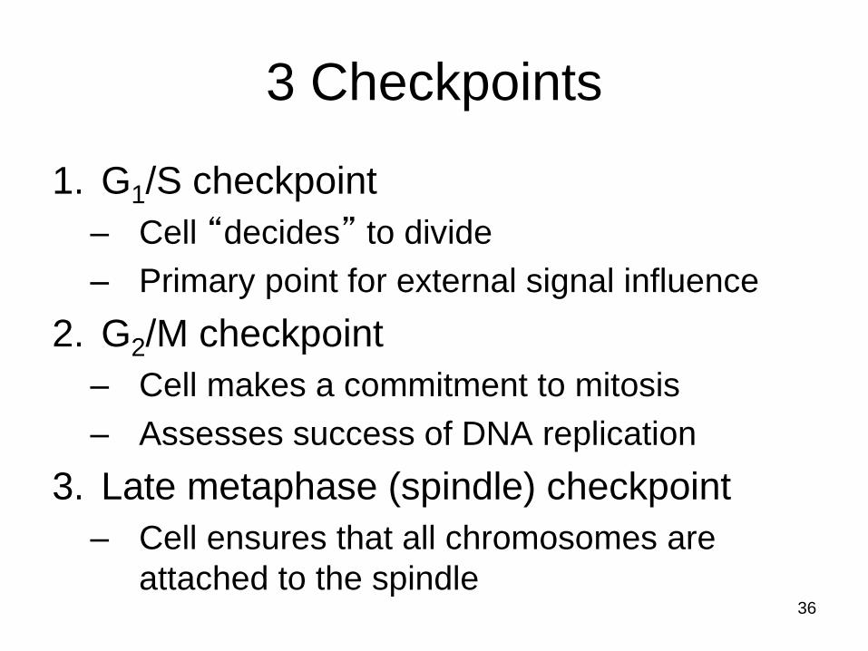
3 Checkpoints
1. G1/S checkpoint
– Cell “decides” to divide
– Primary point for external signal influence
2. G2/M checkpoint
– Cell makes a commitment to mitosis
– Assesses success of DNA replication
3. Late metaphase (spindle) checkpoint
– Cell ensures that all chromosomes are
attached to the spindle36
37
Copyright © The McGraw-Hill Companies, Inc. Permission required for reproduction or display.
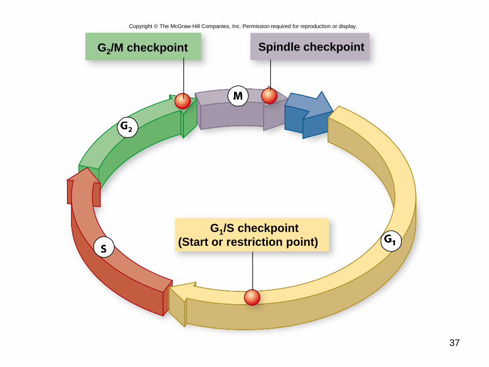
G2/M checkpoint Spindle checkpoint
G1/S checkpoint
(Start or restriction point)

38
Cyclin-dependent kinases (Cdks)
• Enzymes that phosphorylate proteins
• Primary mechanism of cell cycle control
• Cdks partner with different cyclins at different
points in the cell cycle
• For many years, a common view was that
cyclins drove the cell cycle – that is, the periodic
synthesis and destruction of cyclins acted as a
clock
• Now clear that Cdk itself is also controlled by
phosphorylation

39
Copyright © The McGraw-Hill Companies, Inc. Permission required for reproduction or display.
G2/M Checkpoint
Cdc2/Mitotic Cyclin
• DNA integrity
• Replication
completed
Spindle Checkpoint
APC
• Chromosomes
attached at
metaphase plate
M
G2
G1S
G1/S Checkpoint
• Size of cell
• Nutritional state
of cell
• Growth factors
Cdc2/G1 Cyclin
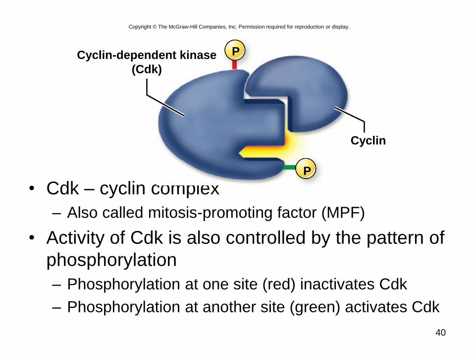
• Cdk – cyclin complex
– Also called mitosis-promoting factor (MPF)
• Activity of Cdk is also controlled by the pattern of
phosphorylation
– Phosphorylation at one site (red) inactivates Cdk
– Phosphorylation at another site (green) activates Cdk
40
Cyclin-dependent kinase
(Cdk)
Cyclin
P
P
Copyright © The McGraw-Hill Companies, Inc. Permission required for reproduction or display.
41
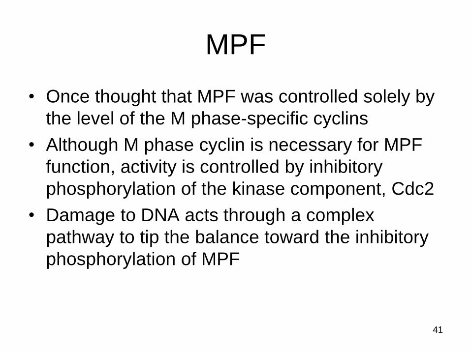
MPF
• Once thought that MPF was controlled solely by
the level of the M phase-specific cyclins
• Although M phase cyclin is necessary for MPF
function, activity is controlled by inhibitory
phosphorylation of the kinase component, Cdc2
• Damage to DNA acts through a complex
pathway to tip the balance toward the inhibitory
phosphorylation of MPF
42
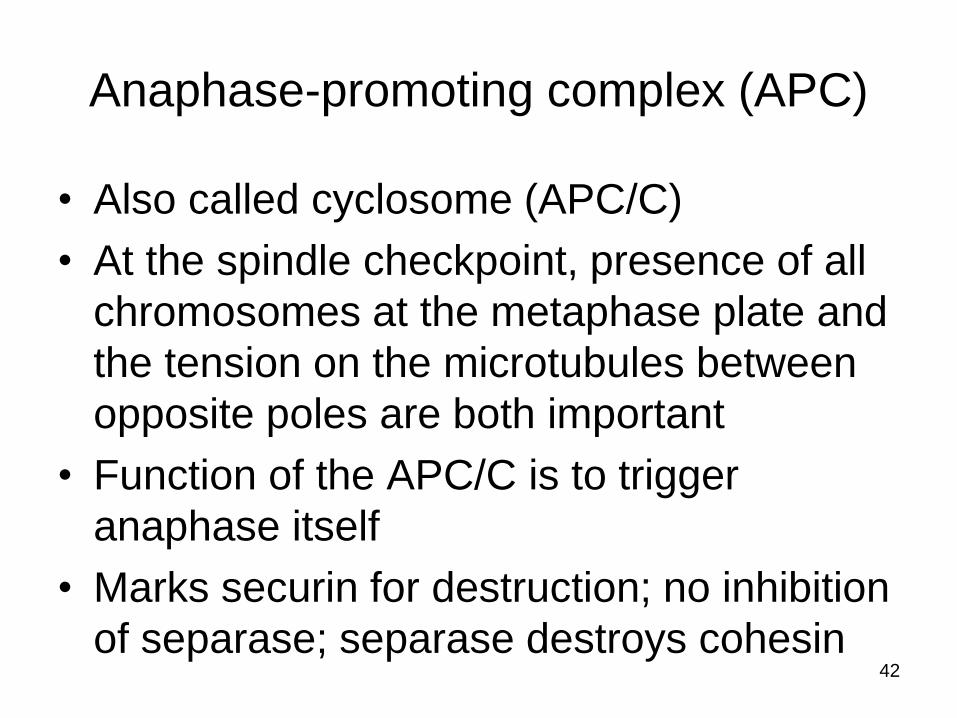
Anaphase-promoting complex (APC)
• Also called cyclosome (APC/C)
• At the spindle checkpoint, presence of all
chromosomes at the metaphase plate and
the tension on the microtubules between
opposite poles are both important
• Function of the APC/C is to trigger
anaphase itself
• Marks securin for destruction; no inhibition
of separase; separase destroys cohesin

43
Control in multicellular eukaryotes
• Multiple Cdks control the cycle as opposed
to the single Cdk in yeasts
• Animal cells respond to a greater variety of
external signals than do yeasts, which
primarily respond to signals necessary for
mating
• More complex controls allow the
integration of more input into control of the
cycle
44
Copyright © The McGraw-Hill Companies, Inc. Permission required for reproduction or display.
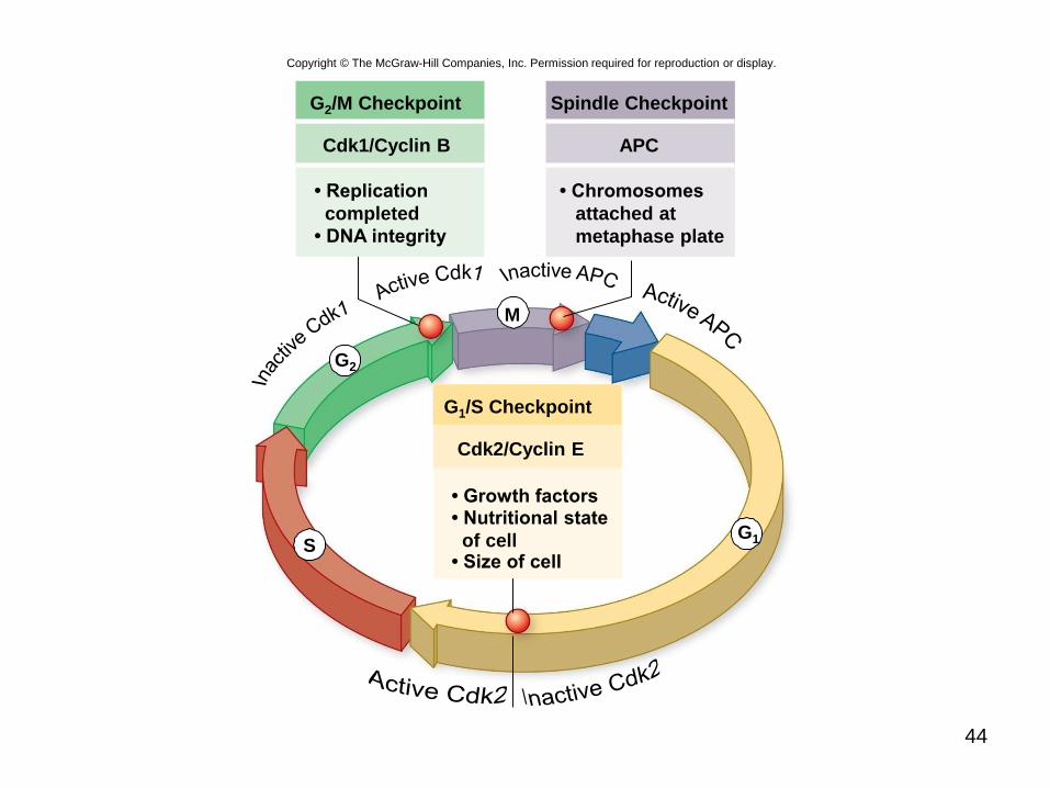
G2/M Checkpoint
Cdk1/Cyclin B
• DNA integrity
• Replication
completed
Spindle Checkpoint
APC
• Chromosomes
attached at
metaphase plate
M
G2
G1S
G1/S Checkpoint
• Size of cell
• Nutritional state
of cell
• Growth factors
Cdk2/Cyclin E

Growth factors
• Act by triggering intracellular signaling
systems
• Platelet-derived growth factor (PDGF) one
of the first growth factors to be identified
• PDGF receptor is a receptor tyrosine
kinase (RTK) that initiates a MAP kinase
cascade to stimulate cell division
• Growth factors can override cellular
controls that otherwise inhibit cell division45

46
GTP
5. MAP kinase (ERK)
activates proteins to
produce cellular
responses, including
transcription factors
that alter gene expression
4. MEK activates
MAP kinases (ERK)
1. Proteins bound to
receptor activate
Ras by exchanging
GDP for GTP.
2. Ras activates
the first
kinase (Raf)
3. Raf activates
the second
Kinase (MEK)
Growth factor
RAS
Cyclins/
proteins
for Sphase
ChromosomeP
Rb
Nucleus
E2F
Rb
ERK
MEK
P
PP
P
RAF
MEK
RAF
ERK
P
PE2F
PP
P
P
RAS
Copyright © The McGraw-Hill Companies, Inc. Permission required for reproduction or display.
GDP

47
Cancer
Unrestrained, uncontrolled growth of cells
• Failure of cell cycle control
• Two kinds of genes can disturb the cell
cycle when they are mutated
1. Tumor-suppressor genes
2. Proto-oncogenes
48
Tumor-suppressor genes
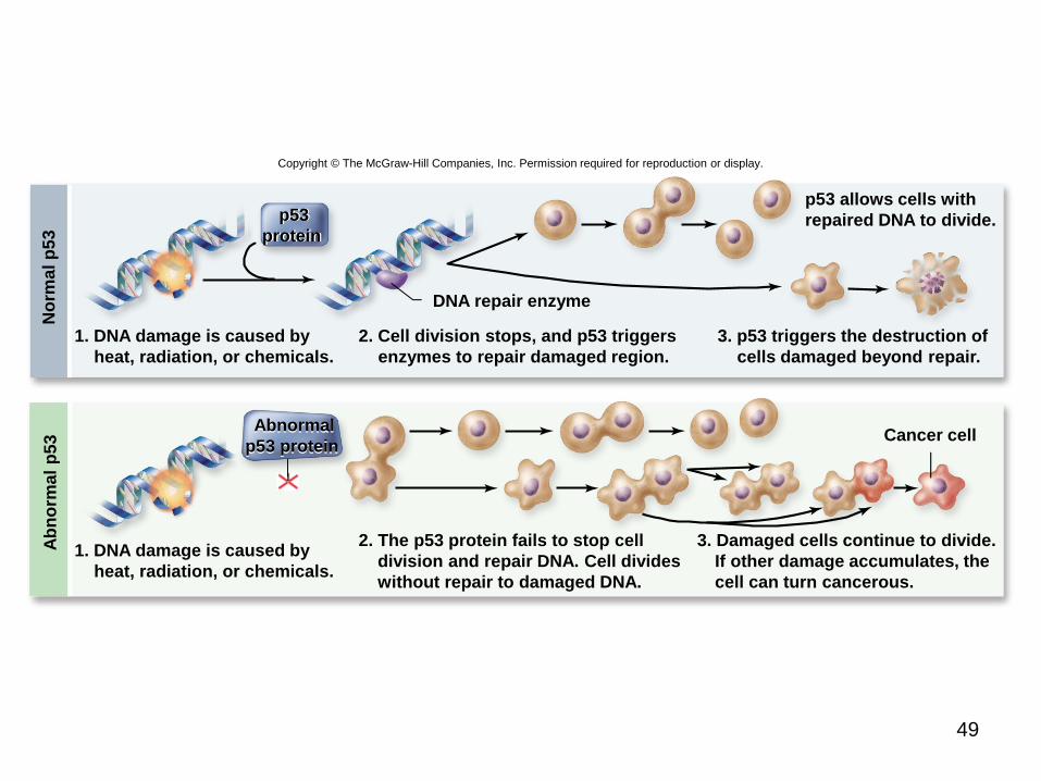
• p53 plays a key role in G1 checkpoint
• p53 protein monitors integrity of DNA
– If DNA damaged, cell division halted and repair
enzymes stimulated
– If DNA damage is irreparable, p53 directs cell to kill
itself
• Prevent the development of mutated cells
containing mutations
• p53 is absent or damaged in many cancerous
cells
49
Copyright © The McGraw-Hill Companies, Inc. Permission required for reproduction or display.
1. DNA damage is caused by
heat, radiation, or chemicals.
2. Cell division stops, and p53 triggers
enzymes to repair damaged region.
3. p53 triggers the destruction of
cells damaged beyond repair.
p53 allows cells with
repaired DNA to divide.
1. DNA damage is caused by
heat, radiation, or chemicals.
2. The p53 protein fails to stop cell
division and repair DNA. Cell divides
without repair to damaged DNA.
3. Damaged cells continue to divide.
If other damage accumulates, the
cell can turn cancerous.
DNA repair enzyme
Cancer cell
p53
protein
No
rma
l p
53
A
bn
orm
al
p5
3
Abnormal
p53 protein

50
Proto-oncogenes
• Normal cellular genes that become oncogenes
when mutated
– Oncogenes can cause cancer
• Some encode receptors for growth factors
– If receptor is mutated in “on,” cell no longer depends
on growth factors
• Some encode signal transduction proteins
• Only one copy of a proto-oncogene needs to
undergo this mutation for uncontrolled division to
take place

Tumor-suppressor genes
• p53 gene and many others
• Both copies of a tumor-suppressor gene
must lose function for the cancerous
phenotype to develop
• First tumor-suppressor identified was the
retinoblastoma susceptibility gene (Rb)
– Predisposes individuals for a rare form of
cancer that affects the retina of the eye
51
• Inheriting a single mutant copy of Rb means the
individual has only one “good” copy left
– During the hundreds of thousands of divisions that
occur to produce the retina, any error that damages
the remaining good copy leads to a cancerous cell
– Single cancerous cell in the retina then leads to the
formation of a retinoblastoma tumor
• Rb protein integrates signals from growth factors
– Role to bind important regulatory proteins and prevent
stimulation of cyclin or Cdk production
52
53
Copyright © The McGraw-Hill Companies, Inc. Permission required for reproduction or display.
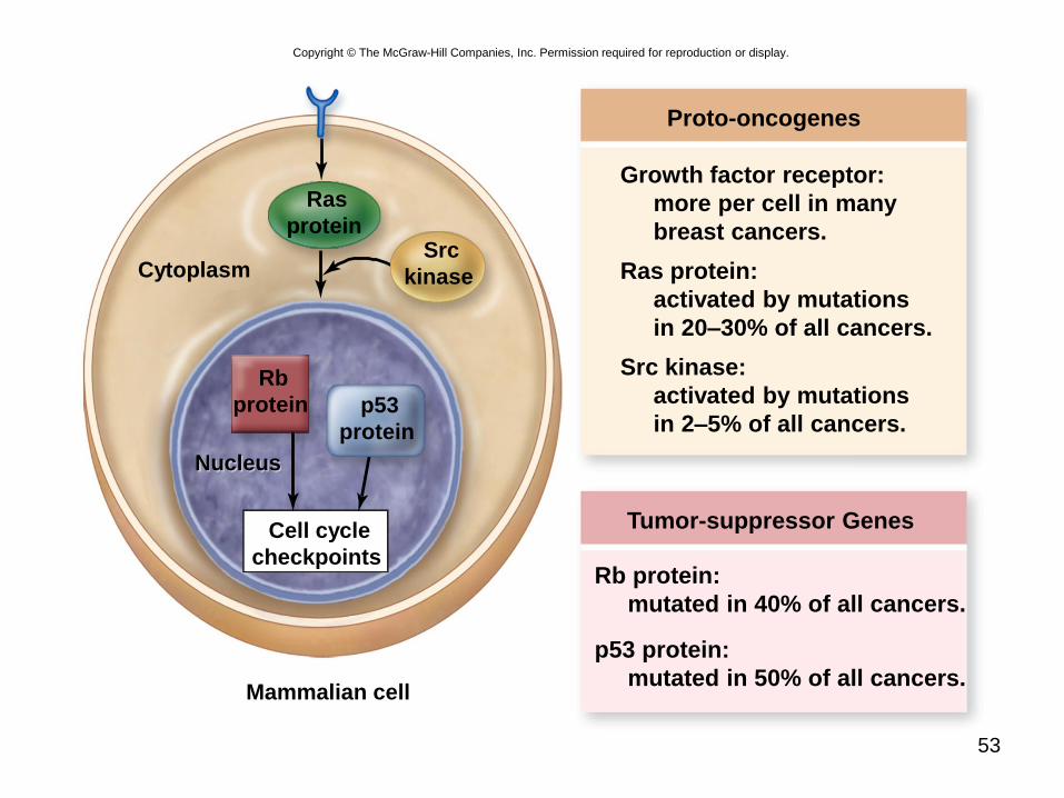
Proto-oncogenes
Growth factor receptor:
more per cell in many
breast cancers.
Ras protein:
activated by mutations
in 20–30% of all cancers.
Src kinase:
activated by mutations
in 2–5% of all cancers.
Tumor-suppressor Genes
Rb protein:
mutated in 40% of all cancers.
p53 protein:
mutated in 50% of all cancers.
Cell cycle
checkpoints
Ras
protein
Rb
protein p53
protein
Src
kinase
Mammalian cell
Nucleus
Cytoplasm