Embed Size (px)
Citation preview
Sherlock’s Diseases of the Liver and Biliary System, Twelfth Edition. Edited by James S. Dooley, Anna S.F. Lok, Andrew K. Burroughs, E. Jenny Heathcote.© 2011 by Blackwell Publishing Ltd. Published 2011 by Blackwell Publishing Ltd.
257
CHAPTER 12
Gallstones and Benign Biliary Diseases
James S. Dooley University College London Medical School and the Royal Free Hampstead NHS Trust, London, UK
The spectrum of benign biliary disease is wide (Table 12.1 ), although the majority of clinical events are due to gallstones, either in gallbladder or bile duct. Symptoms and laboratory tests are likely to be suggestive but non - specifi c, but this does not obviate the careful assessment of history, examination fi ndings and blood tests. An orderly work - up usually starts with ultrasonography. For the hepatologist, there will be the added complica-tion of benign biliary disease coexisting or developing in a patient with primary hepatic disease, which may complicate normal approaches to management. Less diffi cult to recognize are the biliary complications of cholecystectomy and liver transplantation, although their management may be complex.
Gallbladder disease is in the differential diagnosis of right upper quadrant pain, some features of which par-ticular suggest this origin. However, there are many other sources of similar pain or discomfort, emph-asizing the need for collateral evidence from scanning.
Learning points
• Presentation of benign biliary disease is often non - specifi c; imaging is central in management, ultrasonog-raphy being the fi rst approach used.
• Gallstone formation is multifactorial, though lifestyle has an important infl uence. Genetic markers of increased risk are now recognized in humans.
• Laparoscopic cholecystectomy is the standard surgical approach for patients with cholecystitis. Open cholecys-tectomy is still needed in some cases, and because of the comorbidities in this selected patient group has a higher complication rate and mortality.
• Bile duct damage at cholecystectomy occurs in around 1 in 200 patients; management requires a multidisciplinary team approach (radiologist, endoscopist, surgeon).
• Biliary tract intervention has a greater risks in patients with cirrhosis.
Cholestatic liver function tests with or without jaundice, itching, pain or fever focus attention on possible bile duct disease, although again these features are not spe-cifi c for bile duct obstruction.
Examination may be useful in showing characteristic pain or tenderness in the right upper quadrant, or a large nodular liver suspicious of malignancy. Jaundice and scratch marks suggest cholestasis. Splenomegaly raises the question of chronic liver disease, although haemato-logical and other causes need to be remembered.
Liver function tests (bilirubin, transaminases, alkaline phosphatase, γ - glutamyl transpeptidase) will generally be normal in gallbladder disease, although there may be mild abnormalities with sepsis. However, abnormalities have to raise the possibility of bile duct disease. Characteristically, serum alkaline phosphatase and γ - glutamyl transpeptidase, with or without bilirubin, are high when bile drainage is impaired. However a sudden rise (and usually fall) of transaminases may be seen when acute obstruction occurs due to a stone, leading to an initial search for a hepatitis. Polymorph leucocy-tosis will relate to underlying infection.
In these situations, as in most liver - related algorithms, ultrasonography is the fi rst imaging approach of choice. It is effective in showing gallbladder disease, and bile duct dilatation. It may show all that is required; if not, other modalities are used. A single fl ow chart for all clinical scenarios is not appropriate. Direct cholangiog-raphy (percutaneous cholangiography (PTC), endo-scopic retrograde cholangiopancreatography (ERCP)) are now done with a specifi c purpose, therapeutic or to obtain tissue, such is the effectiveness of modern scanning. These techniques compliment management approaches where surgery is an alternative, and this emphasizes the need for a multidisciplinary team.
Cholestatic jaundice without or with pain may be due to malignant disease of the biliary system, described in Chapter 13 .
258 Chapter 12
Table 12.1. Benign biliary diseases
Gallbladder
Stones
Cholecystitis
calculous
— acute
— chronic
acalculous
— acute
— chronic
— gallbladder dyskinesia
empyema
Polyps
infl ammatory
neoplastic
Miscellaneous
adenomyomatosis
cholesterolosis
porcelain
xanthogranulomatous
congenital anomalies
associated with infections (e.g. HIV related, Salmonella)
Bile duct
Stones
Common duct
— asymptomatic
— without cholangitis
— with cholangitis
Intrahepatic
Strictures
Postoperative
— cholecystectomy
— transplantation (Chapter 36 )
— anastomotic
Primary sclerosing cholangitis (Chapter 16 )
Chronic pancreatitis
Others
Sphincter of Oddi dysfunction
Autoimmune pancreatitis
Haemobilia
Mirizzi syndrome
Parasites
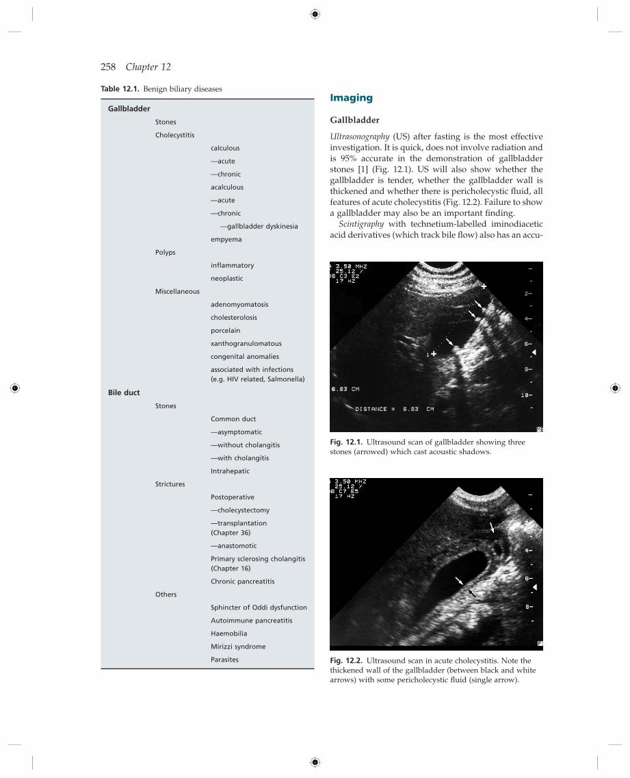
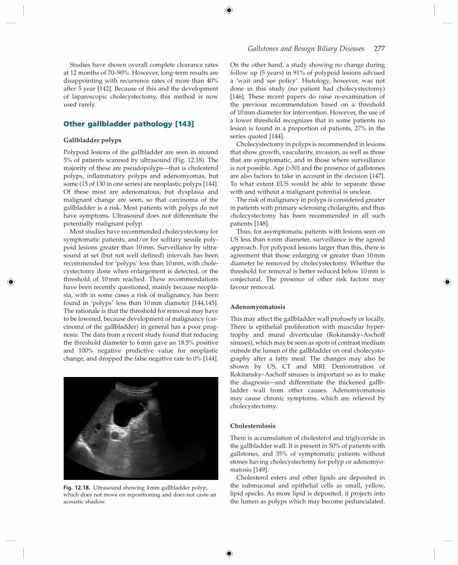
Fig. 12.1. Ultrasound scan of gallbladder showing three stones (arrowed) which cast acoustic shadows.
Fig. 12.2. Ultrasound scan in acute cholecystitis. Note the thickened wall of the gallbladder (between black and white arrows) with some pericholecystic fl uid (single arrow).
Imaging
Gallbladder
Ultrasonography (US) after fasting is the most effective investigation. It is quick, does not involve radiation and is 95% accurate in the demonstration of gallbladder stones [1] (Fig. 12.1 ). US will also show whether the gallbladder is tender, whether the gallbladder wall is thickened and whether there is pericholecystic fl uid, all features of acute cholecystitis (Fig. 12.2 ). Failure to show a gallbladder may also be an important fi nding.
Scintigraphy with technetium - labelled iminodiacetic acid derivatives (which track bile fl ow) also has an accu-
Gallstones and Benign Biliary Diseases 259
racy of 95% for acute cholecystitis (non - fi lling of gall-bladder) (Fig. 12.3 ), but is may be more diffi cult to arrange quickly, takes longer and involves radioisotope. US takes precedence as the diagnostic approach.
CT and MRI scanning can show stones, but are most complementary in showing gallbladder size, wall thick-ness and evidence of infl ammation as in acute cholecys-titis [1] . They are second - line approaches after US.
Bile d uct
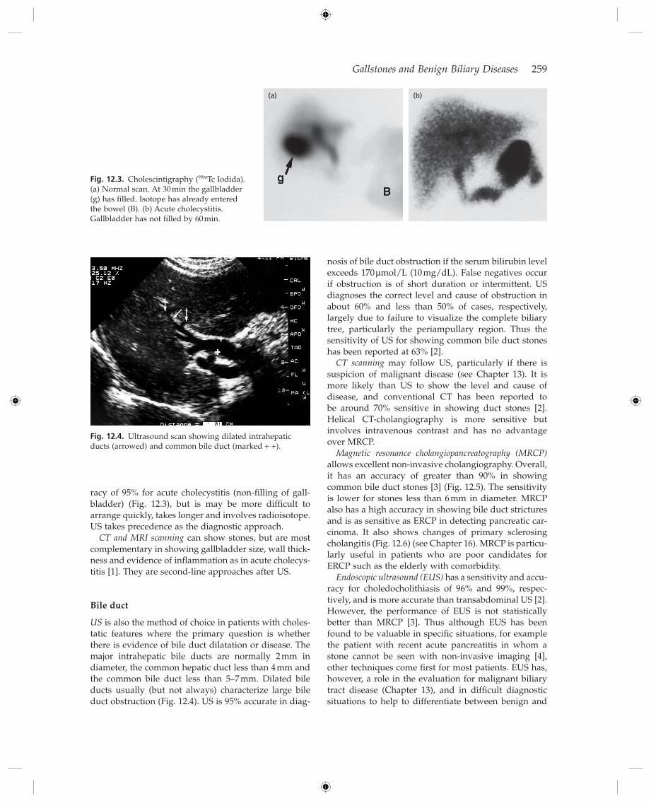
US is also the method of choice in patients with choles-tatic features where the primary question is whether there is evidence of bile duct dilatation or disease. The major intrahepatic bile ducts are normally 2 mm in diameter, the common hepatic duct less than 4 mm and the common bile duct less than 5 – 7 mm. Dilated bile ducts usually (but not always) characterize large bile duct obstruction (Fig. 12.4 ). US is 95% accurate in diag-
Fig. 12.3. Cholescintigraphy ( 99m Tc Iodida). (a) Normal scan. At 30 min the gallbladder (g) has fi lled. Isotope has already entered the bowel (B). (b) Acute cholecystitis. Gallbladder has not fi lled by 60 min.
(a) (b)
Fig. 12.4. Ultrasound scan showing dilated intrahepatic ducts (arrowed) and common bile duct (marked + + ).
nosis of bile duct obstruction if the serum bilirubin level exceeds 170 μ mol/L (10 mg/dL). False negatives occur if obstruction is of short duration or intermittent. US diagnoses the correct level and cause of obstruction in about 60% and less than 50% of cases, respectively, largely due to failure to visualize the complete biliary tree, particularly the periampullary region. Thus the sensitivity of US for showing common bile duct stones has been reported at 63% [2] .
CT scanning may follow US, particularly if there is suspicion of malignant disease (see Chapter 13 ). It is more likely than US to show the level and cause of disease, and conventional CT has been reported to be around 70% sensitive in showing duct stones [2] . Helical CT - cholangiography is more sensitive but involves intravenous contrast and has no advantage over MRCP.
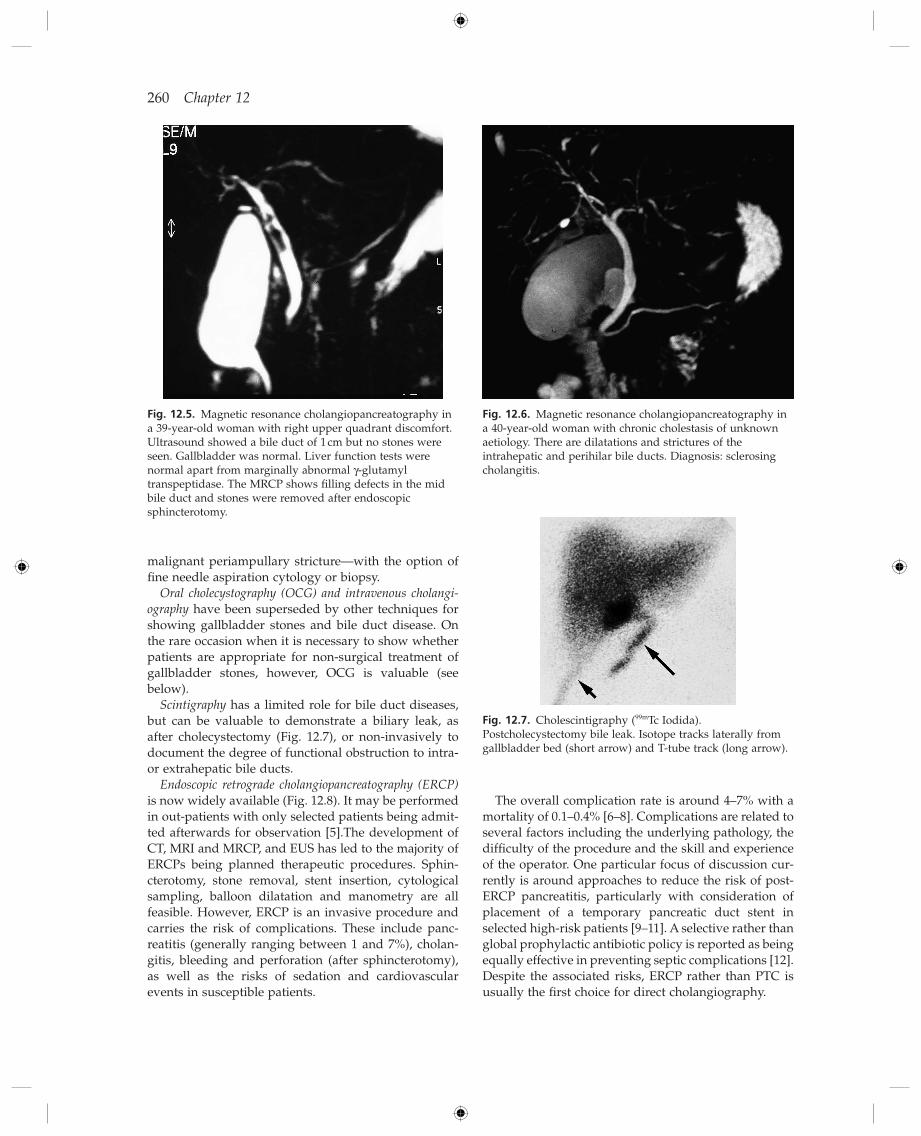
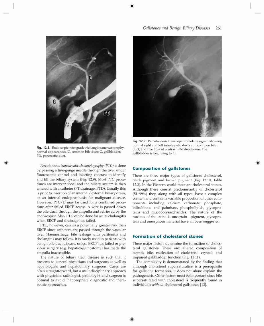
Magnetic resonance cholangiopancreatography (MRCP) allows excellent non - invasive cholangiography. Overall, it has an accuracy of greater than 90% in showing common bile duct stones [3] (Fig. 12.5 ). The sensitivity is lower for stones less than 6 mm in diameter. MRCP also has a high accuracy in showing bile duct strictures and is as sensitive as ERCP in detecting pancreatic car-cinoma. It also shows changes of primary sclerosing cholangitis (Fig. 12.6 ) (see Chapter 16 ). MRCP is particu-larly useful in patients who are poor candidates for ERCP such as the elderly with comorbidity.
Endoscopic ultrasound (EUS) has a sensitivity and accu-racy for choledocholithiasis of 96% and 99%, respec-tively, and is more accurate than transabdominal US [2] . However, the performance of EUS is not statistically better than MRCP [3] . Thus although EUS has been found to be valuable in specifi c situations, for example the patient with recent acute pancreatitis in whom a stone cannot be seen with non - invasive imaging [4] , other techniques come fi rst for most patients. EUS has, however, a role in the evaluation for malignant biliary tract disease (Chapter 13 ), and in diffi cult diagnostic situations to help to differentiate between benign and
260 Chapter 12
Fig. 12.5. Magnetic resonance cholangiopancreatography in a 39 - year - old woman with right upper quadrant discomfort. Ultrasound showed a bile duct of 1 cm but no stones were seen. Gallbladder was normal. Liver function tests were normal apart from marginally abnormal γ - glutamyl transpeptidase. The MRCP shows fi lling defects in the mid bile duct and stones were removed after endoscopic sphincterotomy.
Fig. 12.6. Magnetic resonance cholangiopancreatography in a 40 - year - old woman with chronic cholestasis of unknown aetiology. There are dilatations and strictures of the intrahepatic and perihilar bile ducts. Diagnosis: sclerosing cholangitis.
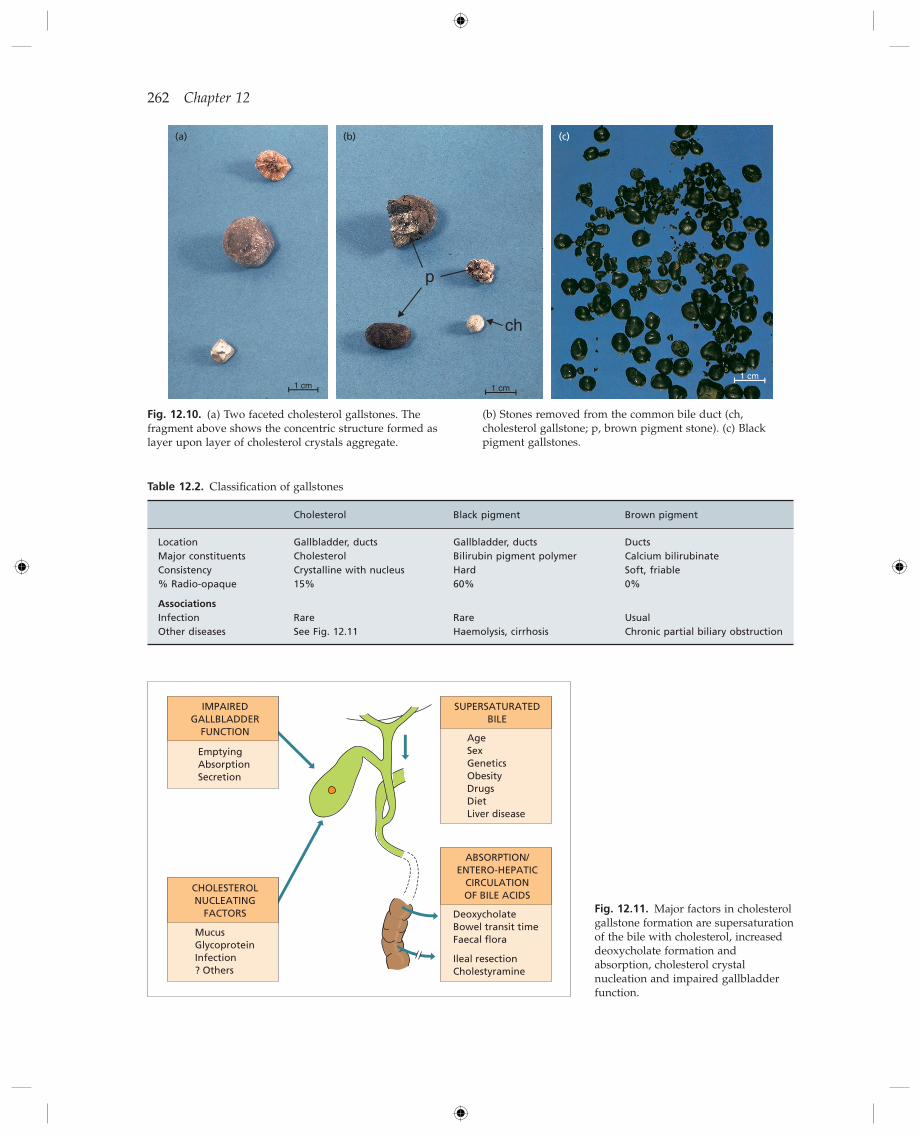
Fig. 12.7. Cholescintigraphy ( 99m Tc Iodida). Postcholecystectomy bile leak. Isotope tracks laterally from gallbladder bed (short arrow) and T - tube track (long arrow).
malignant periampullary stricture — with the option of fi ne needle aspiration cytology or biopsy.
Oral cholecystography (OCG) and intravenous cholangi-ography have been superseded by other techniques for showing gallbladder stones and bile duct disease. On the rare occasion when it is necessary to show whether patients are appropriate for non - surgical treatment of gallbladder stones, however, OCG is valuable (see below).
Scintigraphy has a limited role for bile duct diseases, but can be valuable to demonstrate a biliary leak, as after cholecystectomy (Fig. 12.7 ), or non - invasively to document the degree of functional obstruction to intra - or extrahepatic bile ducts.
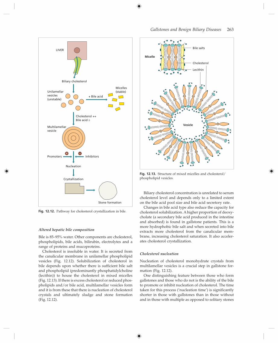
Endoscopic retrograde cholangiopancreatography (ERCP) is now widely available (Fig. 12.8 ). It may be performed in out - patients with only selected patients being admit-ted afterwards for observation [5] .The development of CT, MRI and MRCP, and EUS has led to the majority of ERCPs being planned therapeutic procedures. Sphin-cterotomy, stone removal, stent insertion, cytological sampling, balloon dilatation and manometry are all feasible. However, ERCP is an invasive procedure and carries the risk of complications. These include panc-reatitis (generally ranging between 1 and 7%), cholan-gitis, bleeding and perforation (after sphincterotomy), as well as the risks of sedation and cardiovascular events in susceptible patients.
The overall complication rate is around 4 – 7% with a mortality of 0.1 – 0.4% [6 – 8] . Complications are related to several factors including the underlying pathology, the diffi culty of the procedure and the skill and experience of the operator. One particular focus of discussion cur-rently is around approaches to reduce the risk of post - ERCP pancreatitis, particularly with consideration of placement of a temporary pancreatic duct stent in selected high - risk patients [9 – 11] . A selective rather than global prophylactic antibiotic policy is reported as being equally effective in preventing septic complications [12] . Despite the associated risks, ERCP rather than PTC is usually the fi rst choice for direct cholangiography.
Gallstones and Benign Biliary Diseases 261
Composition of g allstones
There are three major types of gallstone: cholesterol, black pigment and brown pigment (Fig. 12.10 , Table 12.2 ). In the Western world most are cholesterol stones. Although these consist predominantly of cholesterol (51 – 99%) they, along with all types, have a complex content and contain a variable proportion of other com-ponents including calcium carbonate, phosphate, bilirubinate and palmitate, phospholipids, glycopro-teins and mucopolysaccharides. The nature of the nucleus of the stone is uncertain — pigment, glycopro-tein and amorphous material have all been suggested.
Formation of c holesterol s tones
Three major factors determine the formation of choles-terol gallstones. These are: altered composition of hepatic bile, nucleation of cholesterol crystals and impaired gallbladder function (Fig. 12.11 ).
The complexity is demonstrated by the fi nding that although cholesterol supersaturation is a prerequisite for gallstone formation, it does not alone explain the pathogenesis. Other factors must be important since bile supersaturated with cholesterol is frequently found in individuals without cholesterol gallstones [13] .
Fig. 12.8. Endoscopic retrograde cholangiopancreatography, normal appearances. C, common bile duct; G, gallbladder; PD, pancreatic duct.
Percutaneous transhepatic cholangiography (PTC) is done by passing a fi ne - gauge needle through the liver under fl uoroscopic control and injecting contrast to identify and fi ll the biliary system (Fig. 12.9 ). Most PTC proce-dures are interventional and the biliary system is then entered with a catheter (PT drainage, PTD). Usually this is prior to insertion of an internal/ external biliary drain, or an internal endoprosthesis for malignant disease. However, PTC/D may be used for a combined proce-dure after failed ERCP access. A wire is passed down the bile duct, through the ampulla and retrieved by the endoscopist. Also, PTD can be done for acute cholangitis when ERCP and drainage has failed.
PTC, however, carries a potentially greater risk than ERCP since catheters are passed through the vascular liver. Haemorrhage, bile leakage with peritonitis and cholangitis may follow. It is rarely used in patients with benign bile duct disease, unless ERCP has failed or pre-vious surgery (e.g. hepaticojejunostomy) has made the ampulla inaccessible.
The nature of biliary tract disease is such that it presents to general physicians and surgeons as well as hepatologists and hepatobiliary surgeons. Cases are often straightforward, but a multidisciplinary approach with physician, radiologist, pathologist and surgeon is optimal to avoid inappropriate diagnostic and thera-peutic approaches.
Fig. 12.9. Percutaneous transhepatic cholangiogram showing normal right and left intrahepatic ducts and common bile duct, and free fl ow of contrast into duodenum. The gallbladder is beginning to fi ll.
262 Chapter 12
Fig. 12.10. (a) Two faceted cholesterol gallstones. The fragment above shows the concentric structure formed as layer upon layer of cholesterol crystals aggregate.
(b) Stones removed from the common bile duct (ch, cholesterol gallstone; p, brown pigment stone). (c) Black pigment gallstones.
1 cm 1 cm
p
ch
1 cm
(a) (b) (c)
Table 12.2. Classifi cation of gallstones
Cholesterol Black pigment Brown pigment
Location Gallbladder, ducts Gallbladder, ducts Ducts Major constituents Cholesterol Bilirubin pigment polymer Calcium bilirubinate Consistency Crystalline with nucleus Hard Soft, friable % Radio - opaque 15% 60% 0%
Associations
Infection Rare Rare Usual Other diseases See Fig. 12.11 Haemolysis, cirrhosis Chronic partial biliary obstruction
Fig. 12.11. Major factors in cholesterol gallstone formation are supersaturation of the bile with cholesterol, increased deoxycholate formation and absorption, cholesterol crystal nucleation and impaired gallbladder function.
SUPERSATURATEDBILE
AgeSexGeneticsObesityDrugsDietLiver disease
ABSORPTION/ENTERO-HEPATIC
CIRCULATIONOF BILE ACIDS
DeoxycholateBowel transit timeFaecal flora
Ileal resectionCholestyramine
CHOLESTEROLNUCLEATING
FACTORS
MucusGlycoproteinInfection? Others
IMPAIREDGALLBLADDER
FUNCTION
EmptyingAbsorptionSecretion
Gallstones and Benign Biliary Diseases 263
Biliary cholesterol concentration is unrelated to serum cholesterol level and depends only to a limited extent on the bile acid pool size and bile acid secretory rate.
Changes in bile acid type also reduce the capacity for cholesterol solubilization. A higher proportion of deoxy-cholate (a secondary bile acid produced in the intestine and absorbed) is found in gallstone patients. This is a more hydrophobic bile salt and when secreted into bile extracts more cholesterol from the canalicular mem-brane, increasing cholesterol saturation. It also acceler-ates cholesterol crystallization.
Cholesterol n ucleation
Nucleation of cholesterol monohydrate crystals from multilamellar vesicles is a crucial step in gallstone for-mation (Fig. 12.12 ).
One distinguishing feature between those who form gallstones and those who do not is the ability of the bile to promote or inhibit nucleation of cholesterol. The time taken for this process ( ‘ nucleation time ’ ) is signifi cantly shorter in those with gallstones than in those without and in those with multiple as opposed to solitary stones
Fig. 12.12. Pathway for cholesterol crystallization in bile.
Nucleation
LIVER
Biliary cholesterol
Unilamellarvesicles(unstable)
Multilamellarvesicle
Promoters+ –
Inhibitors
Crystallization
Stone formation
Micelles(stable)
+ Bile acid
Cholesterol ++Bile acid ±
Altered h epatic b ile c omposition
Bile is 85 – 95% water. Other components are cholesterol, phospholipids, bile acids, bilirubin, electrolytes and a range of proteins and mucoproteins.
Cholesterol is insoluble in water. It is secreted from the canalicular membrane in unilamellar phospholipid vesicles (Fig. 12.12 ). Solubilization of cholesterol in bile depends upon whether there is suffi cient bile salt and phospholipid (predominantly phosphatidylcholine (lecithin)) to house the cholesterol in mixed micelles (Fig. 12.13 ). If there is excess cholesterol or reduced phos-pholipids and/or bile acid, multilamellar vesicles form and it is from these that there is nucleation of cholesterol crystals and ultimately sludge and stone formation (Fig. 12.12 ).
Fig. 12.13. Structure of mixed micelles and cholesterol/ phospholipid vesicles.
Bile salts
Micelle
Vesicle
Cholesterol
Lecithin
264 Chapter 12
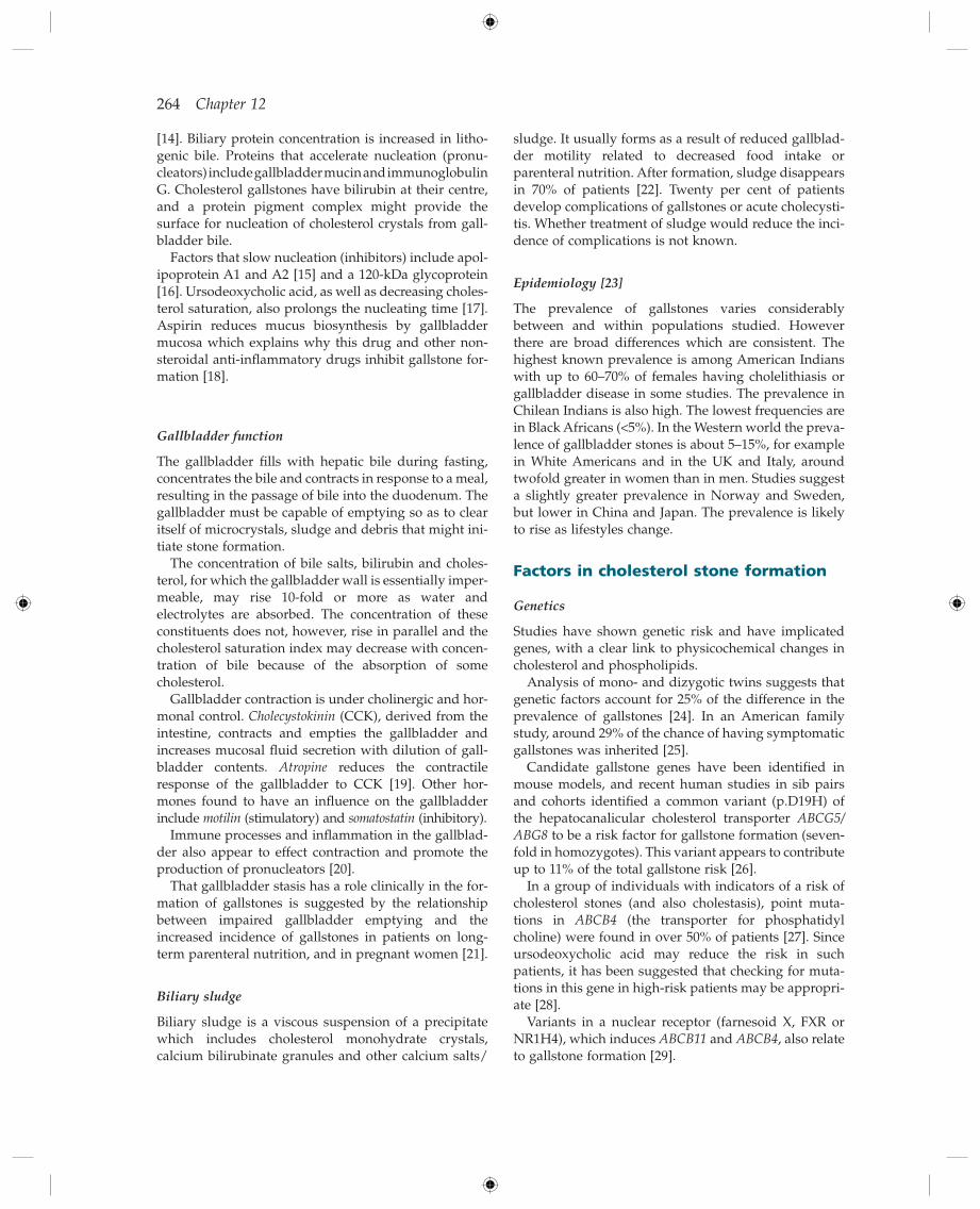
sludge. It usually forms as a result of reduced gallblad-der motility related to decreased food intake or parenteral nutrition. After formation, sludge disappears in 70% of patients [22] . Twenty per cent of patients develop complications of gallstones or acute cholecysti-tis. Whether treatment of sludge would reduce the inci-dence of complications is not known.
Epidemiology [23]
The prevalence of gallstones varies considerably between and within populations studied. However there are broad differences which are consistent. The highest known prevalence is among American Indians with up to 60 – 70% of females having cholelithiasis or gallbladder disease in some studies. The prevalence in Chilean Indians is also high. The lowest frequencies are in Black Africans ( < 5%). In the Western world the preva-lence of gallbladder stones is about 5 – 15%, for example in White Americans and in the UK and Italy, around twofold greater in women than in men. Studies suggest a slightly greater prevalence in Norway and Sweden, but lower in China and Japan. The prevalence is likely to rise as lifestyles change.
Factors in c holesterol s tone f ormation
Genetics
Studies have shown genetic risk and have implicated genes, with a clear link to physicochemical changes in cholesterol and phospholipids.
Analysis of mono - and dizygotic twins suggests that genetic factors account for 25% of the difference in the prevalence of gallstones [24] . In an American family study, around 29% of the chance of having symptomatic gallstones was inherited [25] .
Candidate gallstone genes have been identifi ed in mouse models, and recent human studies in sib pairs and cohorts identifi ed a common variant (p.D19H) of the hepatocanalicular cholesterol transporter ABCG5/ABG8 to be a risk factor for gallstone formation (seven-fold in homozygotes). This variant appears to contribute up to 11% of the total gallstone risk [26] .
In a group of individuals with indicators of a risk of cholesterol stones (and also cholestasis), point muta-tions in ABCB4 (the transporter for phosphatidyl choline) were found in over 50% of patients [27] . Since ursodeoxycholic acid may reduce the risk in such patients, it has been suggested that checking for muta-tions in this gene in high - risk patients may be appropri-ate [28] .
Variants in a nuclear receptor (farnesoid X, FXR or NR1H4), which induces ABCB11 and ABCB4 , also relate to gallstone formation [29] .
[14] . Biliary protein concentration is increased in litho-genic bile. Proteins that accelerate nucleation (pronu-cleators) include gallbladder mucin and immunoglobulin G. Cholesterol gallstones have bilirubin at their centre, and a protein pigment complex might provide the surface for nucleation of cholesterol crystals from gall-bladder bile.
Factors that slow nucleation (inhibitors) include apol-ipoprotein A1 and A2 [15] and a 120 - kDa glycoprotein [16] . Ursodeoxycholic acid, as well as decreasing choles-terol saturation, also prolongs the nucleating time [17] . Aspirin reduces mucus biosynthesis by gallbladder mucosa which explains why this drug and other non - steroidal anti - infl ammatory drugs inhibit gallstone for-mation [18] .
Gallbladder f unction
The gallbladder fi lls with hepatic bile during fasting, concentrates the bile and contracts in response to a meal, resulting in the passage of bile into the duodenum. The gallbladder must be capable of emptying so as to clear itself of microcrystals, sludge and debris that might ini-tiate stone formation.
The concentration of bile salts, bilirubin and choles-terol, for which the gallbladder wall is essentially imper-meable, may rise 10 - fold or more as water and electrolytes are absorbed. The concentration of these constituents does not, however, rise in parallel and the cholesterol saturation index may decrease with concen-tration of bile because of the absorption of some cholesterol.
Gallbladder contraction is under cholinergic and hor-monal control. Cholecystokinin (CCK), derived from the intestine, contracts and empties the gallbladder and increases mucosal fl uid secretion with dilution of gall-bladder contents. Atropine reduces the contractile response of the gallbladder to CCK [19] . Other hor-mones found to have an infl uence on the gallbladder include motilin (stimulatory) and somatostatin (inhibitory).
Immune processes and infl ammation in the gallblad-der also appear to effect contraction and promote the production of pronucleators [20] .
That gallbladder stasis has a role clinically in the for-mation of gallstones is suggested by the relationship between impaired gallbladder emptying and the increased incidence of gallstones in patients on long - term parenteral nutrition, and in pregnant women [21] .
Biliary s ludge
Biliary sludge is a viscous suspension of a precipitate which includes cholesterol monohydrate crystals, calcium bilirubinate granules and other calcium salts/
Gallstones and Benign Biliary Diseases 265
gallstones. Clinical problems present most frequently between the ages of 50 and 70.
Sex and o estrogens
Gallstones are twice as common in women as in men, and this is particularly so before the age of 50.
The incidence is higher in multiparous than in nul-liparous women. Incomplete emptying of the gallblad-der in late pregnancy leaves a large residual volume and thus retention of cholesterol crystals. Biliary sludge occurs frequently but is generally asymptomatic and disappears spontaneously after delivery in two - thirds [42] . In the postpartum period gallstones are present in 8 – 12% of women (nine times that in a matched group) [43] . One - third of those with a functional gallbladder are symptomatic. Small stones disappear spontaneously in 30%.
The bile becomes more lithogenic when women are placed on birth control pills. Women on long - term oral contraceptives have a twofold increased incidence of gallbladder disease over controls [44] . Postmenopausal women taking oestrogen - containing drugs have a sig-nifi cant increase frequency (around 1.8 times) of gall-bladder disease [45] . In men given oestrogen for prostatic carcinoma the bile becomes saturated with cholesterol and gallstones may form [46] .
Serum f actors
The highest risk of gallstones (both cholesterol and pigment) is associated with low HDL levels and high triglyceride levels, which may be more important than body mass [47] . High serum cholesterol is not a deter-minant of gallstone risk.
Cirrhosis
About 30% of patients with cirrhosis have gallstones. The risk of developing stones is most strongly associ-ated with Child ’ s grade C and alcoholic cirrhosis with a yearly incidence of about 5% [48] . The mechanisms are uncertain. All patients with hepatocellular disease show a variable degree of haemolysis. Although bile acid secretion is reduced, the stones are usually of the black pigment type. Phospholipid and cholesterol secretion are also lowered so that the bile is not supersaturated.
Cholecystectomy in patients with cirrhosis carries an increased morbidity and mortality [49,50] . In Child ’ s group A and B the laparoscopic approach is preferred to open cholecystectomy because of lower morbidity and mortality. In Child C patients and those with a higher MELD score the risk of cholecystectomy is particularly
Lifestyle
Lack of physical activity [30] is also an association. There also is an association with the metabolic syn-drome, and related conditions of obesity, type 2 diabetes and dyslipidaemia [31] . At the molecular level this appears to relate to insulin resistance leading to biliary cholesterol hypersecretion and impaired synthesis of bile acids [32] .
Obesity
This seems to be more common among gallstone suffer-ers than in the general population [33] and is a particu-lar risk factor in women less than 50 years old. Obesity is associated with increased cholesterol synthesis. There are no consistent changes in postprandial gallbladder volume. Fifty per cent of markedly obese patients have gallstones at surgery.
Dieting (2100 kJ/day) can result in biliary sludge and the formation of symptomatic gallstones in obese indi-viduals [34] .
Gallstone formation during weight loss following gastric bypass surgery for obesity is prevented by giving ursodeoxycholic acid [35] .
Dietary f actors
Epidemiological studies show that chronic over - nutrition with refi ned carbohydrates and triglycerides increases the risk [36] .
Increasing dietary cholesterol increases biliary choles-terol but there is no epidemiological or dietary data to link cholesterol intake with gallstones.
In Western countries, gallstones have been linked to dietary fi bre defi ciency and a longer intestinal transit time [37] . This increases deoxycholic acid in bile, and renders bile more lithogenic. Deoxycholate is derived from dehydroxylation of cholic acid in the colon by faecal bacteria. There is an enterohepatic circulation. Gallstone patients have signifi cantly prolonged small bowel transit times [38] and increased bacterial dehy-droxylating activity in faeces [39] .
A diet low in carbohydrate and a shorter overnight fasting period protects against gallstones, as does a moderate alcohol intake in males [40] . Vegetarians get fewer gallstones irrespective of their tendency to be slim [41] .
Age
There is a steady increase in gallstone prevalence with advancing years, probably due to the increased choles-terol content in bile. By age 75, around 20% of men and 35% of women in some Western countries have
266 Chapter 12
Black pigment stones are largely composed of an insolu-ble bilirubin pigment polymer mixed with calcium phosphate and carbonate. There is no cholesterol. The mechanism of formation is not well understood, but supersaturation of bile with unconjugated bilirubin, changes in pH and calcium, and overproduction of an organic matrix (glycoprotein) play a role [59] . Overall, 20 – 30% of gallbladder stones are black. The incidence rises with age. They may pass into the bile duct. Black stones accompany chronic haemolysis, usually heredi-tary spherocytosis or sickle cell disease, and mechanical prostheses, for example heart valves, in the circulation. They show an increased prevalence with all forms of cirrhosis, particularly alcoholic [48] . Patients with ileal Crohn ’ s disease may form pigment stones because of increased colonic absorption of bilirubin due to failure of ileal absorption of bile acid [60] .
Brown pigment stones contain calcium bilirubinate, calcium palmitate, and stearate, as well as cholesterol. The bilirubinate is polymerized to a lesser extent than in black stones.
Brown stones are rare in the gallbladder. They form in the bile duct and are related to bile stasis and infected bile. They are usually radiolucent. Bacteria are present in more than 90%. Stone formation is related to the deconjugation of bilirubin diglucuronide by bacterial β - glucuronidase [59] . Insoluble unconjugated bili-rubinate precipitates.
Brown pigment stones form above biliary strictures in sclerosing cholangitis and in the dilated segments of Caroli ’ s disease. There is an association with juxtapapil-lary duodenal diverticula [61] . In Oriental countries, these stones are associated with parasitic infestations of the biliary tract such as Clonorchis sinensis and Ascaris lumbricoides . These stones are frequently intrahepatic.
Natural h istory of g allbladder s tones (Fig. 12.14 )
Gallstones can be dated from the atmospheric radiocar-bon produced by nuclear bomb explosions. This sug-gests a time lag of about 12 years between initial stone formation and symptoms culminating in cholecystec-tomy [62] .
However, gallbladder stones are usually asympto-matic and diagnosed by chance by imaging or during investigation for some other condition. A small propor-tion develop symptoms. Around 8 – 10% of patients with asymptomatic gallstones developed symptoms within 5 years and only 5% required surgery [63,64] . Only about half the patients with symptomatic gallstones come to cholecystectomy within 6 years of diagnosis. Patients with gallstones seem to tolerate their symptoms for long periods of time, preferring this to cholecystectomy. If
high, making management decisions more diffi cult [49,50] . In such patients with symptomatic gallbladder and bile duct stones, non - surgical techniques have to be considered as alternatives to surgery.
Infection
Although infection is thought to be of little importance in cholesterol stone formation, bacterial DNA is found in these stones [51] . Conceivably, bacteria might decon-jugate bile salts, allowing their absorption and reducing cholesterol solubility.
Diabetes m ellitus
Diabetics have a higher prevalence of gallstones (or a history of cholecystectomy) than non - diabetics, particu-larly females (42 versus 23%) [52] . The older diabetic tends to be obese, and this may be the important factor in gallstone formation.
Patients with diabetes may have large, poorly con-tracting and poorly fi lling gallbladders [53] . A ‘ diabetic neurogenic gallbladder ’ syndrome has been postulated.
Patients with diabetes mellitus undergoing cholecys-tectomy, whether emergency or elective, have an increased risk of complications. These are probably related to associated cardiovascular or renal disease and to more advanced age.
Other f actors
Hepatitis C is associated with a higher incidence of gall-bladder stones than patients with hepatitis B, or those without hepatitis B or C (11.7 vs. 5.4 vs. 6.0% respec-tively [54] ), but the reason for the link is not known.
Ileal resection breaks the enterohepatic circulation of bile salts, reduces the total bile salt pool and is followed by gallstone formation. The same is found in subtotal or total colectomy [55] .
Gastrectomy increases the incidence of gallstones [56] . Long - term cholestyramine therapy increases bile salt loss
with a reduced bile acid pool size and gallstone formation.
Parenteral nutrition leads to a dilated, sluggish gall-bladder containing stones.
Endoscopic sphincterotomy improves gallbladder emptying and decreases the lithogenicity of bile in patients with gallstone disease [57] . Patients with gall-bladder stones have signifi cantly higher sphincter of Oddi tone [58] .
Pigment g allstones
This term is used for stones containing less than 30% cholesterol. There are two types: black and brown (see Table 12.2 ).

Gallstones and Benign Biliary Diseases 267
Acute c alculous c holecystitis
Aetiology
In 95% of patients the cystic duct is obstructed by a gallstone. The imprisoned bile salts have a toxic action on the gallbladder wall. Lipids may penetrate the Rokitansky – Aschoff sinuses and exert an irritant reac-tion. The rise in pressure compresses blood vessels in the gallbladder wall; infarction and gangrene may follow.
Pancreatic enzymes may also cause acute cholecysti-tis, presumably by regurgitation into the biliary system when there is a common biliary and pancreatic channel.
Bacterial infl ammation is an integral part of acute cholecystitis. Bacterial deconjugation of bile salts may produce toxic bile acids which can injure the mucosa.
Pathology
The gallbladder is usually distended, but after previous infl ammation the wall becomes thickened and con-tracted. There may be vascular adhesions to adjacent structures.
Histology shows haemorrhage and moderate oedema reaching a peak by about the fourth day and diminish-ing by the seventh day. As the acute reaction subsides it is replaced by fi brosis.
Bacteriology. Cultures of both gallbladder wall and bile usually show organisms of intestinal type, including anaerobes. Common infecting organisms are Escherichia coli , Streptococcus faecalis and Klebsiella , often in combina-tion. Anaerobes are present, if sought, and are usually found with aerobes. They include Bacteroides and Clostridium sp .
Clinical f eatures
These vary according to whether there is only mild infl ammation or more severe disease such as
symptoms develop, they are unlikely to present as an emergency.
Data suggest that elective cholecystectomy is an appropriate choice for patients with biliary colic [65] .
Prophylactic cholecystectomy should not be per-formed for asymptomatic gallbladder stones [66] . It should not be done to prevent gallbladder cancer since the risk is small and less than that of cholecystectomy [67] .
Migration of a stone to the neck of the gallbladder causes obstruction of the cystic duct and a rise in gallblad-der pressure. There is chemical irritation of the gallblad-der mucosa by the retained bile, followed by bacterial invasion. According to the severity of the changes, acute or chronic cholecystitis results. Empyema may follow; perforation, fi stula formation and emphysematous cholecystitis are rare.
Migration of a stone into the bile duct may present with pain, jaundice, cholangitis or pancreatitis.
Possible a ssociation of s tones with g allbladder and b ile d uct c ancer
Population surveys have investigated the link between stones and malignancy of the gallbladder and extrahe-patic bile ducts.
The relative risk of gallbladder carcinoma is 2.4 if gallbladder stones are 2.0 – 2.9 cm in diameter, and greater with larger stones [68] . The issue is whether this is causative or an association, meaning that there is a common factor(s) that predisposes to both stones and carcinoma. This remains a possibility [69] and diffi cult to rule in or out.
There appears to be a lower risk of extrahepatic bile duct carcinoma during follow up of patients who had cholecystectomy, raising the question of a link between gallbladder stones or gallbladders containing stones with extrahepatic bile duct malignant changes [70] .
Fig. 12.14. The natural history of gallstones.
1–2% per yearof symptomatic20–50%
recurrencein 1st year
~30%recurrence
in 1st month
2–3% per year ~0.2% per year
ACUTECHOLECYSTITIS
BILE DUCT STONE– pancreatitis– cholangitis– jaundice
CHRONICCHOLECYSTITIS
Gallbladder carcinoma(~0.08% per year ofsymptomatic patients>50 year old)
BILIARYCOLIC
ASYMPTOMATIC
268 Chapter 12
Acute calculous cholecystitis is suggested by the fi nding of stones with: • a thickened gallbladder wall ( > 5 mm) (see Fig. 12.2 ); • a positive sonographic Murphy sign — the presence of maximum tenderness, elicited by direct pressure of the transducer, over a sonographically localized gallbladder; • gallbladder distension; • pericholecystic fl uid; • subserosal oedema (without ascites); • intramural gas; • a sloughed mucosal membrane.
Cholescintigraphy. Technetium - labelled iminodiacetic acid derivatives (IDA) are cleared from the plasma by hepatocellular organic anion transport and excreted in the bile (see Fig. 12.3 a).
Hepatic IDA scanning may be used to determine patency of the cystic duct in suspected acute cholecys-titis. If the gallbladder fails to visualize, despite common bile duct patency and intestinal visualization (Fig. 12.3 b), the probability of acute cholecystitis is 80 – 90%. False - negative results are more common the later the gallbladder fi lls [73] .
CT and MRI scanning are not indicated as the initial assessment of a patient with suspected acute cholecystitis.
Differential d iagnosis
Acute cholecystitis is liable to be confused with other causes of sudden pain and tenderness in the right hypo-chondrium. Below the diaphragm, acute retrocaecal appendicitis, intestinal obstruction, a perforated peptic ulcer or acute pancreatitis may produce similar clinical features.
Myocardial infarction should always be considered. Referred pain from muscular and spinal root lesions
may cause similar pain.
Prognosis
Spontaneous recovery follows disimpaction of the stone in 85% of patients. Recurrent acute cholecystitis may follow — approximately a 30% chance over the next 3 months [74] .
Rarely, acute cholecystitis proceeds rapidly to gan-grene or empyema of the gallbladder, fi stula formation, hepatic abscesses or even generalized peritonitis. The acute fulminating disease is becoming less common because of earlier antibiotic therapy and more frequent cholecystectomy for recurrent gallbladder symptoms.
fulminating gangrene of the gallbladder wall. The acute attack is often an exacerbation of underlying chronic cholecystitis.
The sufferers are often obese, female and over 40, but no type, age or sex is immune.
Pain often occurs late at night or in the early morning, usually in the right upper abdomen or epigastrium and is referred to the angle of the right scapula, to the right shoulder [71] , or rarely to the left side. It may simulate angina pectoris.
The pain usually rises to a plateau and can last 30 – 60 min without relief, unlike the short spasm of biliary colic. Attacks may be precipitated by late - night, heavy meals or fatty food.
Distension pain coming in waves is due to the gallblad-der contracting to overcome the blocked cystic duct.
Peritoneal pain is superfi cial with skin tenderness, hyperaesthesia and muscular rigidity. The fundus of the gallbladder is in apposition to the diaphragmatic peri-toneum, which is supplied by the phrenic and last six intercostal nerves. Stimulation of the anterior branches produces right upper quadrant pain and of the posterior cutaneous branch leads to the characteristic right infra-scapular pain.
Examination
The patient appears ill. The temperature rises with bac-terial invasion. Jaundice usually indicates associated stones in the common bile duct.
The gallbladder is usually impalpable; occasionally a tender mass of gallbladder and adherent omentum may be felt. Murphy ’ s sign is positive.
The leucocyte count may be raised with a moderate increase in polymorphs. In the febrile patient blood cul-tures may be positive.
For the patient with acute abdominal pain of uncer-tain cause, a plain X - ray will be taken during the work - up, but if acute cholecystitis is suspected scanning is indicated. Only about 10% of gallstones are radio - opaque, compared with 90% of renal calculi.
Imaging
The diagnosis of gallbladder disease depends upon scanning because of the overall lack of power of any specifi c symptom [72] . As described earlier, ultrasound is the test of choice. Scintigraphy is also accurate but second line.
Gallstones cast intense echoes with obvious posterior acoustic shadows (see Fig. 12.1 ). They change in posi-tion with turning of the patient. Stones 3 mm or more in size are usually seen. Diagnostic accuracy is 96% but less experienced operators may not achieve this success.
Gallstones and Benign Biliary Diseases 269
Treatment is with antibiotics and surgery. There is a high postoperative rate of septic complications [79] . Percutaneous cholecystostomy is considered if the patient is unfi t for surgery.
Emphysematous c holecystitis
The term is used to denote infection of the gallbladder with gas - producing organisms ( Escherichia coli , Clostridium welchii ) or anaerobic streptococci. The primary lesion is occlusion of the cystic duct or cystic artery. Infection is secondary [80] . The condition classi-cally affects male diabetics who develop features of severe, toxic, acute cholecystitis. An abdominal mass may be palpable.
On a plain abdominal Xray the gallbladder may be seen as a sharply outlined pear - shaped gas shadow. Occasionally air may be seen infi ltrating the wall and surrounding tissue. Gas is not apparent in the cystic duct, which is blocked by a gallstone. In the erect posi-tion, a fl uid level is seen in the gallbladder. However, plain abdominal X - ray may not show the characteristic changes. Ultrasound is diagnostic in around 50% of cases. CT may also show characteristic features.
Standard treatment is with antibiotics and emergency cholecystectomy. In the severely ill patient percutaneous cholecystostomy is an alternative [81] .
Chronic c alculous c holecystitis
This is the commonest type of clinical gallbladder disease. The association of chronic cholecystitis with stones is almost constant. Aetiological factors therefore include all those related to gallstones. The chronic infl ammation may follow acute cholecystitis, but usually develops insidiously.
Pathology
The gallbladder is usually contracted with a thickened, sometimes calcifi ed, wall. Stones are seen lying loosely embedded in the wall or in meshes of an organizing fi brotic network. One stone is usually lodged in the neck. Histologically, the wall is thickened and congested with lymphocytic infi ltration and occasionally complete destruction of the mucosa.
Clinical f eatures
Chronic cholecystitis is diffi cult to diagnose because of the ill - defi ned symptoms. Episodes of acute cholecysti-tis punctuate the course.
Treatment
Medical. This depends upon the clinical severity for which a grading has been described [75,76] . This is based on the white cell count, clinical fi ndings, duration and features of systemic/ multisystem signs or compli-cations. General measures during the acute phase include intravenous fl uids, nothing given orally, analge-sia and antibiotics. Tokyo guidelines recommend man-agement depending upon the severity [75,76] .
Antibiotic(s) are given if there is clinical evidence of sepsis, and should have a spectrum to cover the likely micro - organisms. Choice is according to hospital policy, but a second or third - generation cephalosporin or com-bination of a quinolone with metronidazole are usually adequate for the stable patient with pain and mild fever. Patients with features of severe sepsis require broader - spectrum antibiotics such as piperacillin/ tazobactam, combined if necessary with an aminoglycoside. The elderly, and those with diabetes or immunodefi ciency, are at particular risk of severe sepsis.
Cholecystectomy ( s ee b elow). For those with mild acute cholecystitis, early cholecystectomy is recommended [75,76] . Meta - analysis of randomized controlled trials shows that this approach (within 1 week) is superior to delayed cholecystectomy (2 to 3 months later) because of avoidance of gallstone - related complications during the waiting period [65,77] . These could lead to emer-gency surgery which is known to carry a higher risk than elective operation, particularly in elderly patients over 75 years old and in the diabetic patient where early elective cholecystectomy is preferred once symptoms have developed [78] .
For moderate acute cholecystitis there may be early or delayed cholecystectomy, but if early laparoscopic surgery is done, it should be by a highly experienced surgeon so that decisions to alter the approach can be made and carried out if the operation becomes compli-cated [75,76] .
For severe acute cholecystitis, based on the Tokyo guidelines, initial intensive medical treatment with anti-biotics is recommended with, if needed, percutaneous cholecystostomy [75,76] ; surgical decisions are then cus-tomized for individual patients according to the clinical course and degree of surgical risk.
Empyema of the g allbladder
If the cystic duct remains blocked by a stone and infec-tion sets in, empyema may develop. Symptoms may be of an intra - abdominal abscess (fever, rigors, pain), although the elderly patient may appear relatively well.

270 Chapter 12
is especially so when indefi nite symptoms are associ-ated with a well - functioning gallbladder. The general condition of the patient may contraindicate surgery. The infrequent place of medical dissolution and shock - wave lithotripsy of radiolucent stones is dis-cussed later.
Obesity should be addressed. A low - fat diet is advisable.
If the patient is symptomatic, particularly with repeated episodes of pain, cholecystectomy (see below) is indicated.
Acalculous c holecystitis
Acute
About 5 – 10% of acute cholecystitis in adults and about 30% in children occurs in the absence of stones. The most frequent predisposing cause is an associated criti-cal condition such as after major non - biliary surgery, multiple injuries, major burns, recent childbirth, severe sepsis, mechanical ventilation and parenteral nutrition.
The pathogenesis is unclear and probably multifacto-rial, but bile stasis (lack of gallbladder contraction), increased bile viscosity and lithogenicity, and gallblad-der ischaemia are thought to play a role. Administration of opiates, which increase sphincter of Oddi tone, may also reduce gallbladder emptying.
Clinical features should be those of acute calcul-ous cholecystitis with fever, leucocytosis and right upper quadrant pain but diagnosis is often diffi cult because of the overall clinical state of the patient who may be intubated, ventilated and receiving narcotic analgesics.
There may be laboratory features of cholestasis with a raised bilirubin and alkaline phosphatase. Ultrasound and CT are complementary and useful in showing a thickened gallbladder wall ( > 5mm), pericholecystic fl uid or subserosal oedema (without ascites), intramural gas or a sloughed mucosal membrane. The sensitivity of ultrasound varies widely between studies (30 – 100%), but prospective studies have suggested that this is a useful technique [83,84] .Cholescintigraphy is reported to have a sensitivity of 60 – 90% for acalculous cholecys-titis [85,86] , but moving patients to the imaging unit for the time required for scanning may not be practical.
Because of the diffi culties of diagnosis a high index of suspicion is needed, particularly in patients at risk. Gangrene and perforation of the gallbladder are common. The mortality is high, 41% in one series [85] , often due to delayed diagnosis.
Treatment is emergency cholecystectomy. In the critically ill patient percutaneous cholecystostomy under ultrasound guidance may be life saving (see below).
Abdominal distension or epigastric discomfort, espe-cially after a fatty meal, may be temporarily relieved by belching. Nausea is common, but vomiting is unusual unless there are stones in the common bile duct. Apart from a constant dull ache in the right hypochondrium and epigastrium, pain may be experienced in the right scapular region, substernally or at the right shoulder. Postprandial pain may be relieved by alkalis.
Local tenderness over the gallbladder and a positive Murphy sign are very suggestive.
Investigations
The temperature, leucocyte count, haemoglobin and erythrocyte sedimentation rate are within normal limits. A plain abdominal X - ray may show calcifi ed gallstones. However, the imaging technique of fi rst choice is ultra-sound, which may show gallstones within a fi brosed gallbladder with a thickened wall. Non - visualization of the gallbladder is also a signifi cant fi nding. CT scan may show gallstones but this technique is not usually appro-priate in the diagnostic work - up. Endoscopy may be necessary to rule out gastric or duodenal infl ammation or ulceration.
Differential d iagnosis
Fat intolerance, fl atulence and postprandial discomfort are common symptoms. Even if associated with imaging evidence of gallstones, the calculi are not necessarily responsible since stones are frequently present in the symptom - free.
Other disorders producing a similar clinical picture must be excluded before cholecystectomy is advised, otherwise symptoms persist postoperatively. These include peptic ulceration or infl ammation, hiatus hernia, irritable bowel syndrome and functional dyspepsias.
Since approximately 10% of young to middle - aged adults have gallstones, symptomatic gallbladder disease may be over - diagnosed. Conversely, ultrasound is only about 95% accurate and symptomatic gallbladder disease may therefore sometimes be unrecognized.
Prognosis
This chronic disease is compatible with good life expect-ancy. However, once symptoms, particularly biliary colic, are experienced, the patients tend to remain symp-tomatic with about a 40% chance of recurrence within 2 years [82] . Gallbladder cancer is a rare, later develop-ment (see above).
Treatment
Medical measures may be tried if the diagnosis is uncertain and a period of observation is desirable. This
Gallstones and Benign Biliary Diseases 271
bladder are carefully identifi ed and clipped. Haemostasis is achieved by electrocautery or laser. The gallbladder is dissected from the gallbladder bed on the liver and removed whole. When necessary large stones are frag-mented while they are still within the gallbladder to allow its delivery through the anterior abdominal wall.
Results
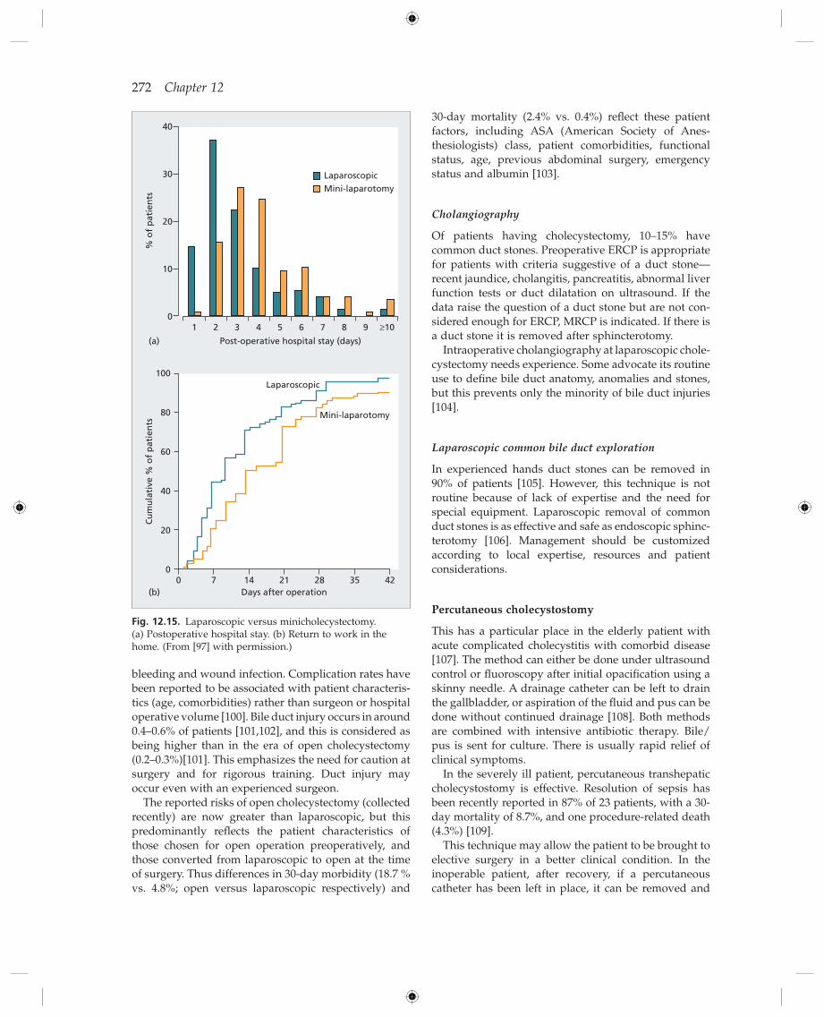
Systemic reviews and meta - analyses show that there is no overall difference in outcome measures of mortality and complications between open, small - incision and laparoscopic cholecystectomy [93,96] . However, the minimally invasive methods (laparoscopic and small incision) were associated with a signifi cantly shorter postoperative hospital stay (around 3 days) compared with open cholecystectomy, and convalescence was shortened (around 22 days). The results from laparo-scopic and small incision cholecystectomy were similar. The smaller incision approach interestingly had a shorter operative time and possible lower cost than laparo-scopic cholecystectomy.
The Cochrane review raises the question of why laparoscopic rather than mini - incision cholecystectomy has become the standard approach for patients with symptomatic disease, and suggests that to address this other outcomes need more concerted analysis, such as symptom relief and complications. One study involving minilaparotomy and laparoscopic cholecystectomy evaluated pain scores, physical function and psycho-logical health 1 week after operation and found that the laparoscopic approach gave a signifi cantly better outcome [97] (Fig. 12.15 ). The wide use of laparoscopic cholecystectomy also appears to refl ect patient prefer-ence and the overall pattern of practice available. Thus practitioners of mini - incision surgery are in the minor-ity, because of its technical diffi culty and the fewer opportunities for training.
Laparoscopic cholecystectomy is successful in about 95% of patients. In the remainder, the operation has to be converted to open cholecystectomy. This is more likely if there is acute cholecystitis, particularly with empyema [98] . In these cases, initial laparoscopic assess-ment is appropriate and conversion to open operation made if indicated. In experienced hands laparoscopic cholecystectomy for acute and gangrenous cholecystitis is as safe and effective as open cholecystectomy although there is a moderately high conversion rate (16%) to the open procedure [99] .
Complications
The perioperative mortality lies between 0 and 0.3% [65,96] . The complication rate is around 5% [96] , and includes bile duct injury, biliary leak, postoperative
Chronic ( i ncluding g allbladder d yskinesia)
This is a diffi cult diagnosis as the clinical condition resembles others, particularly irritable bowel syndrome and functional dyspepsias. A description of biliary - like pain has been endorsed by the Rome committee on functional biliary and pancreatic disorders [87] : an epi-sodic, severe, constant pain, in epigastrium or right upper quadrant, lasting at least 30 min, severe enough to interrupt daily activities or lead to consultation with a physician. Laboratory investigations (liver enzymes, conjugated bilirubin, amylase, lipase) are normal; routine transabdominal ultrasound scan shows a normal gallbladder.
Cholescintigraphy with measurement of the gallblad-der ejection fraction 15 min after CCK infusion has been used to try and identify patients who have putative gallbladder pathology and would benefi t from cholecys-tectomy. Normal individuals have an ejection fraction of around 70%. In those with a low ejection fraction (usually regarded as less than 35 – 40%) or who develop pain during the infusion, symptom relief after cholecys-tectomy is reported in between 70 and 90% of patients [88 – 90] . However, decisions on management based on the results of a single isotope scan alone may not appear appropriate. There are many issues regarding the actual technique used (e.g. dose and rate of CCK infusion) and that a low ejection fraction is not specifi c for functional gallbladder disease [91] . Results of scanning should be taken in the context of the other clinical features of the patient. Of note is that EUS may detect small gallblad-der stones missed by transabdominal US and in these patients cholecystectomy resulted in loss of pain [92] .
In patients with acalculous gallbladder disease under-going cholecystectomy, chronic cholecystitis, cholestero-losis, muscle hypertrophy and/or a narrowed cystic duct have been shown in patients in whom symptoms were relieved [89,90] .
Cholecystectomy
Laparoscopic cholecystectomy , introduced in the late 1980s, is the current standard treatment for symptomatic gall-bladder stones, and mild and moderate acute cholecys-titis, based on superior outcomes compared with open cholecystectomy [93 – 95] . Open cholecystectomy is still required where the laparoscopic approach fails, or is not possible. Thus expertise is still needed for the open operation.
Operative a pproach for l aparoscopic c holecystectomy
Under general anaesthesia the abdominal cavity is insuffl ated with CO 2 and the laparoscope and operating channels inserted. Cystic duct and vessels to the gall-
272 Chapter 12
30 - day mortality (2.4% vs. 0.4%) refl ect these patient factors, including ASA (American Society of Anes-thesiologists) class, patient comorbidities, functional status, age, previous abdominal surgery, emergency status and albumin [103] .
Cholangiography
Of patients having cholecystectomy, 10 – 15% have common duct stones. Preoperative ERCP is appropriate for patients with criteria suggestive of a duct stone — recent jaundice, cholangitis, pancreatitis, abnormal liver function tests or duct dilatation on ultrasound. If the data raise the question of a duct stone but are not con-sidered enough for ERCP, MRCP is indicated. If there is a duct stone it is removed after sphincterotomy.
Intraoperative cholangiography at laparoscopic chole-cystectomy needs experience. Some advocate its routine use to defi ne bile duct anatomy, anomalies and stones, but this prevents only the minority of bile duct injuries [104] .
Laparoscopic c ommon b ile d uct e xploration
In experienced hands duct stones can be removed in 90% of patients [105] . However, this technique is not routine because of lack of expertise and the need for special equipment. Laparoscopic removal of common duct stones is as effective and safe as endoscopic sphinc-terotomy [106] . Management should be customized according to local expertise, resources and patient considerations.
Percutaneous c holecystostomy
This has a particular place in the elderly patient with acute complicated cholecystitis with comorbid disease [107] . The method can either be done under ultrasound control or fl uoroscopy after initial opacifi cation using a skinny needle. A drainage catheter can be left to drain the gallbladder, or aspiration of the fl uid and pus can be done without continued drainage [108] . Both methods are combined with intensive antibiotic therapy. Bile/ pus is sent for culture. There is usually rapid relief of clinical symptoms.
In the severely ill patient, percutaneous transhepatic cholecystostomy is effective. Resolution of sepsis has been recently reported in 87% of 23 patients, with a 30 - day mortality of 8.7%, and one procedure - related death (4.3%) [109] .
This technique may allow the patient to be brought to elective surgery in a better clinical condition. In the inoperable patient, after recovery, if a percutaneous catheter has been left in place, it can be removed and
bleeding and wound infection. Complication rates have been reported to be associated with patient characteris-tics (age, comorbidities) rather than surgeon or hospital operative volume [100] . Bile duct injury occurs in around 0.4 – 0.6% of patients [101,102] , and this is considered as being higher than in the era of open cholecystectomy (0.2 – 0.3%) [101] . This emphasizes the need for caution at surgery and for rigorous training. Duct injury may occur even with an experienced surgeon.
The reported risks of open cholecystectomy (collected recently) are now greater than laparoscopic, but this predominantly refl ects the patient characteristics of those chosen for open operation preoperatively, and those converted from laparoscopic to open at the time of surgery. Thus differences in 30 - day morbidity (18.7 % vs. 4.8%; open versus laparoscopic respectively) and
Fig. 12.15. Laparoscopic versus minicholecystectomy. (a) Postoperative hospital stay. (b) Return to work in the home. (From [97] with permission.)
% o
f p
atie
nts
0
10
20
30
40
1
(a)
2 3 4 5 6 7 8 9
LaparoscopicMini-laparotomy
≥10
Post-operative hospital stay (days)
Cu
mu
lati
ve %
of
pat
ien
ts
0
20
40
60
100
80 Mini-laparotomy
Laparoscopic
0 7 14(b)
21 28 35 42Days after operation

Gallstones and Benign Biliary Diseases 273
ence and a long procedure are also associated with damage [112,113] . The threshold at which the decision is made to convert from laparoscopic to open surgery is also important.
Clinical f eatures
Complete ligation, clipping or transection will become clear clinically in the immediate perioperative period. With partial injury, the occlusion develops slowly. About 60% of patients with bile duct injury present within 3 months of operation, and 80% within 1 year [114] .
If unrecognized at the time of cholecystectomy, presentation depends upon the degree of damage. Postoperative anorexia, nausea, vomiting, pain, abdom-inal distension, ileus and delayed recovery should raise the possibility of damage [112] , although the presenta-tion is usually more obvious.
The appearance of bile - stained fl uid in the surgical drain raises the possibility of duct damage. Complete transection of the main bile duct usually gives pain (bile peritonitis), fever and cholestatic jaundice 3 – 7 days postoperatively. Alternatively, an external biliary fi stula develops. The fi stula may drain intermittently with epi-sodes of jaundice when it is closed. Subhepatic abscesses may develop.
Ligation or clipping the main duct, or a later stricture, gives escalating cholestatic jaundice with or without cholangitis.
With current awareness of the complications of lapar-oscopic cholecystectomy, and the availability of ERCP and other imaging techniques, patients should not develop the chronic complications of biliary obstruction. Biliary cirrhosis with portal hypertension and splenom-egaly will develop with time if the obstruction is not recognized and relieved effectively.
Patients unfortunate enough to suffer bile duct damage at cholecystectomy may become increasingly introspective as the months pass. Some keep the most detailed notes of their symptoms and, understandably, become querulous and suspicious of their medical advi-sors. They need considerable support.
Investigations
The history of recent cholecystectomy, the postoperative features and the biochemical and imaging data should lead to cholangiography and the correct diagnosis.
Liver function tests may show cholestasis, but may be normal.
Radiology. The fi rst step is scanning with ultrasound or CT. Where duct damage has led to a bile leak, ultra-sound or CT will show an intra - abdominal collection which may be drained under scanning control. Bile
the patient treated conservatively, often without recur-rence [107] .
In the situation of the patient not being a surgical candidate, and/or having a coagulopathy or ascites that precludes cholecystostomy, ERCP with selective cannu-lation of the cystic duct and nasobiliary tube or stent placement in the gallbladder is possible [110] , although technically diffi cult. A systematic review reported over 90% technical and 80 – 90% clinical success, with compli-cations in 1 – 4% [111] .
Postcholecystectomy b ile d uct d amage
Bile duct damage occurs in around 0.4 – 0.6% of patients [101,102] . Injuries include bile leak from cystic duct or gallbladder bed, complete transection of the duct and complete or partial stricture due to clips or damage during dissection.
Several factors contribute to duct injury. There may be mistaken interpretation of the anatomy due to oedema or haemorrhage around an infl amed gallblad-der, anomalies of the cystic duct or right hepatic duct (Fig. 12.16 ), or lack of operator experience.
Risk factors for laparoscopic bile duct injury include obesity, bleeding, acute cholecystitis and scarring in Calot ’ s triangle (the area between the cystic duct and common hepatic duct). Uncertain anatomy, inexperi-
Fig. 12.16. Benign bile duct stricture following laparoscopic cholecystectomy (arrow). Note anomalous right - sided bile duct (a).
274 Chapter 12
injuries into four major categories and this more often used by biliary endoscopists: • Type A: cystic duct leak, or leakage from aberrant or peripheral hepatic radicals; • Type B: major bile duct leak with or without a con-comitant bile duct stricture; • Type C: bile duct stricture without bile leakage; • Type D: complete transection of the duct, with or without excision of some portion of the biliary tree.
Other classifi cations are more detailed, such as that by Strasberg et al . [118] , and are used for planning of surgical repair.
Treatment
Prevention. The majority of strictures would be pre-vented if: (1) cholecystectomy was only performed by experienced surgeons; (2) the top – down approach was used with thorough dissection at the junction of the gallbladder infundibulum and cystic duct; and (3) there was an appropriate threshold for conversion from lapar-oscopic to open surgery. This is particularly so in the presence of acute cholecystitis.
Medical. Fluid and electrolyte balance must be main-tained, particularly in the jaundiced septic patient and those with a biliary fi stula. Antibiotic therapy, based if possible on blood and bile culture, will improve septi-caemia but, if there is bile duct obstruction or a leak, bile duct catheterization and drainage by the endoscopic or percutaneous route is essential to treat sepsis. Bile col-lections may need percutaneous drainage under scan-ning control.
The overriding principle is the importance of early referral to a specialist hepatobiliary centre where there will be a multidisciplinary approach by surgeon, radi-ologist and endoscopist [119] .
Interventional e ndoscopy and r adiology; s urgical r epair. Bile leakage from a cystic duct stump or tiny ducts in the gallbladder bed can usually be managed endoscopi-cally by stent insertion [116] . This is the fi rst - choice procedure.
For the incomplete stricture, endoscopic balloon dila-tation and stenting, using repeat procedures with grad-ated increase in balloon diameter and number of stents, over a year gives a successful outcome in 80 – 90% of postcholecystectomy strictures [120 – 122] . Success with strictures at the hilar confl uence is lower.
An analysis of mesh metal stents in this scenario sug-gests that they should not be used unless life expectancy is less than 2 years, because of occlusion [123] . Whether removable covered mesh metal stents have any role awaits outcome analysis.
ducts may not be dilated. Biliary scintigraphy detects around 50% of leaks (see Fig. 12.7 ) [115] . When there is a stricture without a leak, dilated intrahepatic bile ducts are seen.
The route of cholangiography depends upon the clini-cal data. If bile leakage from a cystic duct or a partial low duct stricture are suspected then ERCP is the fi rst choice (see Fig. 12.16 ). MRCP may be valuable depend-ing upon the type of injury. For duct transection and discontinuity or a high stricture, percutaneous cholangi-ography and drainage are appropriate as part of the preoperative work - up and management. CT or MR angi-ography is indicated if vascular injury is suspected, and may be done as a road map for patients with high duct damage.
Classifi cations
There are many classifi cations of types of injury, in an attempt to defi ne different patterns and facilitate man-agement decisions [116] . Bergman et al . [117] separate
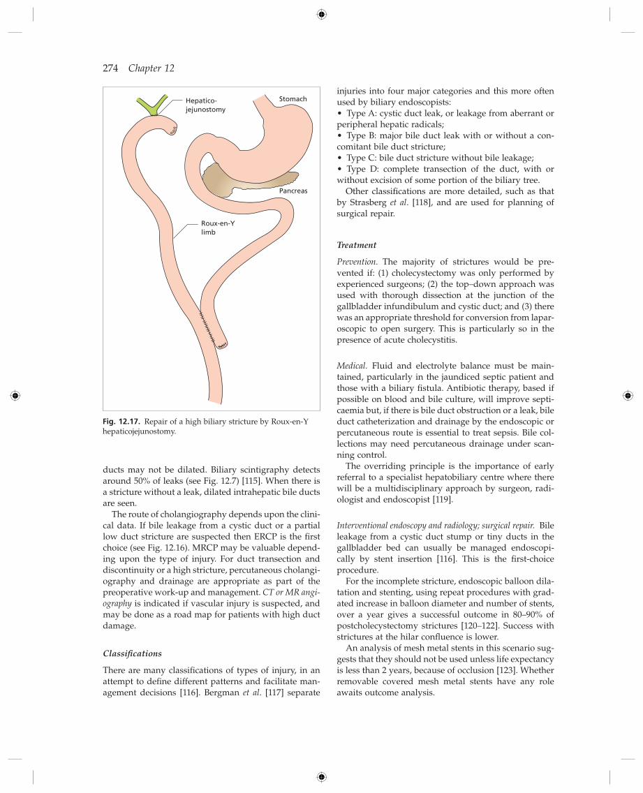
Fig. 12.17. Repair of a high biliary stricture by Roux - en - Y hepaticojejunostomy.
Hepatico-jejunostomy
Roux-en-Ylimb
Stomach
Pancreas
Gallstones and Benign Biliary Diseases 275
if stones are found at cholecystectomy and if a period of relief follows the operation. The colon and pancreas are common alternative culprits.
Postoperative symptoms may be related to technical diffi culties at the time of surgery. These include trau-matic biliary stricture and residual calculi (see below).
Amputation neuromas can be demonstrated in some patients but removal offers no relief and this seems unlikely to be the cause of the symptoms.
Chronic pancreatitis , a common association of choledo-cholithiasis , may persist postoperatively.
US is the fi rst test to image the bile duct. Depending on the result and the clinical features MRCP may be indicated. Despite all these, ERCP is usually necessary. Residual calculi, stricture, ampullary stenosis or normal appearances are signifi cant fi ndings.
Sphincter of Oddi d ysfunction [129,130]
This has been an area of controversy but now appears to be a cause of postcholecystectomy pain in some patients. Two forms exist.
Papillary stenosis is defi ned as narrowing of all or part of the sphincter of Oddi. There is fi brosis. It may follow injury due to stones [131] , operative instrumentation, biliary infection or pancreatitis. There may be episodes of pain associated with abnormal liver function tests. On ERCP the bile duct is dilated and drains slowly. The basal sphincter tone is raised on manometry and is not reduced by smooth muscle relaxants. Endoscopic sphincterotomy is helpful [132] .
Sphincter of Oddi (biliary) dyskinesia is a more diffi cult area. Biliary manometry shows a range of abnorma-lities including sphincter spasm, increased phasic contrac-tion frequency (tachyoddia), paradoxical contraction response to CCK and abnormal propagation of phasic waves.
Clinical features (Table 12.3 ) are valuable in manage-ment decisions. Group I benefi t from sphincterotomy in around 90% of cases. In group II manometry is impor-tant. Patients with an elevated basal sphincter pressure have had greater benefi t from sphincterotomy than those with a normal pressure (91 vs. 42%) [133] , although a recent expert review suggests a lower success rate (50 – 70% vs. 30%) based on manometry result [130] . Studies continue in group III. Duodenal distension reproduces the symptoms in most patients [134] . Sphincterotomy in those with abnormal manometry may be benefi cial in only 20 – 30% of patients [130] . This is a diffi cult group of patients, and it has been suggested that such patients should fi rst have a trial of medical therapy (including proton pump inhibitors and/or calcium channel block-ers (nifedipine)) before considering ERCP and manom-etry, with the attendant risks, in those without a response [135] .
For the completely obstructed or transected bile duct, surgery is necessary after investigation and preparatory percutaneous bile drainage, as appropriate to the indi-vidual patient. The endoscopic route is likely to be of no value. These are complex patients requiring a specialist multidisciplinary team.
Preoperative percutaneous transhepatic biliary drain-age is performed. Intra - abdominal collections are drained. Other preoperative investigations are done, including angiography to detect vascular damage. Surgery may then be performed electively in the sub-sequent weeks under optimal conditions.
The operation chosen will depend mainly on two factors — the site and length of the stricture and the amount of duct available for repair. Any operation must provide excision of the stricture with mucosal apposi-tion between the duct lining and the intestinal mucosa. The anastomosis must be as large as possible and not under tension.
Even if suffi cient duct is available proximally, excision of the stricture and end - to - end anastomosis of the duct is rarely performed. Differences between the calibre of the duct above and below the stricture are too great for a satisfactory anastomosis. Recurrent stricture occurs in 60% of cases.
The usual operation is between the bile duct and a Roux - en - Y segment of jejunum ( choledochojejunostomy ). In the case of high stricture, the hepatic duct is used ( hepaticojejunostomy ) (Fig. 12.17 ).
Successful long - term results of hepaticojejunostomy (mean 5 years) have been reported in 50 of 54 patients in one series [124] . Predictors of poor outcome were peritonitis at the time of reconstruction, combined vas-cular and bile duct injuries and injury at or above the level of the biliary bifurcation. Another series also analysed associations with a poor outcome which included three or more attempts at operative repair before referral, hypoalbuminaemia, high serum bilirubin, the presence of liver disease and portal hyper-tension [125] .
Stenosis of a hepaticojejunostomy done either as the primary repair, or after failed other approaches, may be managed by interventional radiological approaches, or by surgical revision (see below).
Postcholecystectomy s yndromes
About 90 – 95% of those with gallstones are freed of symp-toms or improved postoperatively. The absence of stones questions the original diagnosis. These patients may have been suffering from a psychosomatic or some other disorder including non - visceral pain [126] . Results of surgery are poor when done for vague symptoms such as abdominal bloating or dyspepsia, or in patients using psychiatric medication [127,128] . A biliary cause is likely
276 Chapter 12
edly obese. The overall success rate for oral bile acid therapy is approximately 40%, rising to 60% with careful patient selection [138] . Stones of 5 mm or less in diam-eter that fl oat dissolve more quickly (80 – 90% complete dissolution by 12 months). Larger non - fl oating stones take longer or never disappear.
The effect of bile acid therapy on symptoms is varia-ble. Biliary pain is less frequent in those patients on long - term ursodeoxycholic acid therapy [139] . Stone recurrence develops in 25 – 50% of patients at a rate of 10% per year. They are most likely in the fi rst 2 years and unlikely after the fi rst 3 years. Recurrence is higher in those with multiple rather than solitary stones.
Side effects are absent. During treatment the stones may undergo surface calcifi cation [140] , but this is prob-ably of little signifi cance.
Extracorporeal s hock - w ave t herapy [141]
Gallbladder stones can be fragmented by shock waves generated extracorporeally using the same principle as that developed for kidney stones. Ultrasound is usually used to target stones. Oral bile acid therapy is given to dissolve those fragments remaining in the gallbladder, although when pulverization is achieved bile acid therapy is not necessary [141] . The gallbladder shows bruising and oedema after the shock waves but these are reversible.
Results
These vary from one machine, centre and protocol to another. Only 20 – 25% of patients referred satisfy the treatment criteria which included: three or fewer radi-olucent gallbladder stones with a total diameter of less than 30 mm, in a functioning gallbladder (on chole-cystography), in a symptomatic patient who is other-wise healthy.
Non - s urgical t reatment of g allstones in the g allbladder
The widespread availability and acceptance of laparo-scopic cholecystectomy has markedly reduced the use of non - surgical treatments for gallbladder stones. However, a small group of patients remain where these approaches need to be considered, including those unfi t for or refusing surgery.
Dissolution t herapy with u rsodeoxycholic a cid [136]
Ursodeoxycholic acid decreases biliary cholesterol secretion as well as cholesterol absorption and increases solubility of cholesterol by the formation of liquid crys-tals [28] . Ursodeoxycholic acid also prolongs nucleation time.
Indications
The patient must be compliant and prepared for at least 2 years of treatment. Symptoms must be mild to moder-ate and silent stones should not be treated. On oral cholecystography the cystic duct must be patent ( ‘ func-tioning gallbladder ’ ) and stones radiolucent, preferably fl oating. They should be less than 15 mm in diameter. Best results are for stones less than 5 mm diameter.
Unfortunately, no imaging technique accurately determines the composition of gallstones and therefore solubility. Ultrasound is of little value. CT can be useful and, because of the expense of bile acid therapy, cost - effective in assessing stones. Stones with an attenuation value of less than 100 Hounsfi eld units (refl ecting low calcium content) are more likely to dissolve [137] .
Results
The dose of ursodeoxycholic acid is at least 10 mg/kg per day with more being needed if the patient is mark-
Table 12.3. Sphincter of Oddi dysfunction: classifi cation (modifi ed according to [130] )
Group I (defi nite)
Biliary - type pain
Abnormal liver function tests (serum transaminases or alkaline phosphatase > 1.5 × normal) documented on two or more occasions, with normalization between attacks
Dilated common bile duct > 8mm Manometry unnecessary
Group II (presumptive)
Biliary - type pain and one of other group I criteria Manometry essential
Group III (possible)
Biliary - type pain only. No other abnormalities Manometry essential if intervention contemplated
Gallstones and Benign Biliary Diseases 277
On the other hand, a study showing no change during follow up (5 years) in 91% of polypoid lesions advised a ‘ wait and see policy ’ . Histology, however, was not done in this study (no patient had cholecystectomy) [146] . These recent papers do raise re - examination of the previous recommendation based on a threshold of 10 mm diameter for intervention. However, the use of a lower threshold recognizes that in some patients no lesion is found in a proportion of patients, 27% in the series quoted [144] .
Cholecystectomy in polyps is recommended in lesions that show growth, vascularity, invasion, as well as those that are symptomatic, and in those where surveillance is not possible. Age ( > 50) and the presence of gallstones are also factors to take in account in the decision [147] . To what extent EUS would be able to separate those with and without a malignant potential is unclear.
The risk of malignancy in polyps is considered greater in patients with primary sclerosing cholangitis, and thus cholecystectomy has been recommended in all such patients [148] .
Thus, for asymptomatic patients with lesions seen on US less than 6 mm diameter, surveillance is the agreed approach. For polypoid lesions larger than this, there is agreement that those enlarging or greater than 10 mm diameter be removed by cholecystectomy. Whether the threshold for removal is better reduced below 10 mm is conjectural. The presence of other risk factors may favour removal.
Adenomyomatosis
This may affect the gallbladder wall profusely or locally. There is epithelial proliferation with muscular hyper-trophy and mural diverticulae (Rokitansky – Aschoff sinuses), which may be seen as spots of contrast medium outside the lumen of the gallbladder on oral cholecysto-graphy after a fatty meal. The changes may also be shown by US, CT and MRI. Demonstration of Rokitansky – Aschoff sinuses is important so as to make the diagnosis — and differentiate the thickened gallb-ladder wall from other causes. Adenomyomatosis may cause chronic symptoms, which are relieved by cholecystectomy.
Cholesterolosis
There is accumulation of cholesterol and triglyceride in the gallbladder wall. It is present in 50% of patients with gallstones, and 35% of symptomatic patients without stones having cholecystectomy for polyp or adenomyo-matosis [149] .
Cholesterol esters and other lipids are deposited in the submucosal and epithelial cells as small, yellow, lipid specks. As more lipid is deposited, it projects into the lumen as polyps which may become pedunculated.
Studies have shown overall complete clearance rates at 12 months of 70 – 90%. However, long - term results are disappointing with recurrence rates of more than 40% after 5 year [142] . Because of this and the development of laparoscopic cholecystectomy, this method is now used rarely.
Other g allbladder p athology [143]
Gallbladder p olyps
Polypoid lesions of the gallbladder are seen in around 5% of patients scanned by ultrasound (Fig. 12.18 ). The majority of these are pseudopolyps — that is cholesterol polyps, infl ammatory polyps and adenomyomas, but some (15 of 130 in one series) are neoplastic polyps [144] . Of these most are adenomatous, but dysplasia and malignant change are seen, so that carcinoma of the gallbladder is a risk. Most patients with polyps do not have symptoms. Ultrasound does not differentiate the potentially malignant polyp.
Most studies have recommended cholecystectomy for symptomatic patients, and/or for solitary sessile poly-poid lesions greater than 10 mm. Surveillance by ultra-sound at set (but not well defi ned) intervals has been recommended for ‘ polyps ’ less than 10 mm, with chole-cystectomy done when enlargement is detected, or the threshold of 10 mm reached. These recommendations have been recently questioned, mainly because neopla-sia, with in some cases a risk of malignancy, has been found in ‘ polyps ’ less than 10 mm diameter [144,145] . The rationale is that the threshold for removal may have to be lowered, because development of malignancy (car-cinoma of the gallbladder) in general has a poor prog-nosis. The data from a recent study found that reducing the threshold diameter to 6 mm gave an 18.5% positive and 100% negative predictive value for neoplastic change, and dropped the false negative rate to 0% [144] .
Fig. 12.18. Ultrasound showing 4 mm gallbladder polyp, which does not move on repositioning and does not caste an acoustic shadow.

278 Chapter 12
in only about 0.2% of patients with typhoid fever. Colonization may be facilitated by biofi lm formation on cholesterol gallstones [152] .
Acute typhoid cholecystitis is becoming very rare. Signs of acute cholecystitis appear at the end of the second week or even during convalescence, and are sometimes followed by perforation of the gallbladder.
Chronic typhoid fever cholecystitis and the typhoid carrier state. The typhoid carrier passes organisms in the faeces derived from a focus of infection in the gallbladder or biliary tract. Chronic typhoid cholecystitis is symp-tomless. The carrier state is not cured by antibiotic therapy. Cholecystectomy is successful if there is no associated infection of the biliary ducts. Chronic typhoid cholecystitis is not an important cause of gallstones, but carries an increased risk of gallbladder carcinoma [153] .
Biliary carriers of other salmonellae have been repor-ted and treated with ampicillin and cholecystectomy.
Acute c holecystitis in AIDS
This is thought to occur because of gallbladder stasis and increased bile lithogenicity in the critically ill patient, opportunistic pathogens such as cytomegalovi-rus (CMV) and cryptosporidium, or vascular insuffi -ciency due to oedema or infection. Calculous and acalculous cholecystitis are seen.
Patients present with fever, right upper quadrant pain and tenderness. The white cell count is often normal but with a left shift of neutrophils. Ultrasound shows fea-tures of acute cholecystitis.
Treatment is by cholecystectomy. In the late1980s, this carried a mortality of around 30% due to sepsis [154] . However, a mortality of 2% was reported in a series of 53 patients with acute cholecystitis in 1999 from the same group, thought due to improvement in medical treatment [155] . Highly active antiretroviral therapy (HAART) is associated with a reduction in post-operative complications after cholecystectomy [156] .
Acute cholecystitis is one part of the spectrum of hepatobiliary disease seen in patients with HIV. There may be parenchymal liver disease (see Chapter 22 ). Pancreatitis may be seen from HIV - related medications or less often opportunistic infections. AIDS - related dis-eases include not only acalculous cholecystitis, but also Kaposi ’ s sarcoma, gallstones and AIDS cholangiopathy [157,158] .
AIDS cholangiopathy has several patterns: papillary stenosis with bile duct dilatation above; a sclerosing cholangitis - like appearance of intra - and extrahepatic ducts; a combination of the two; or segmental extrahe-patic strictures. Patients may be asymptomatic, or present with malaise, fever or right upper quadrant pain. US and CT may be useful, but the combination of
The change is confi ned to the gallbladder and never extends to the ducts.
The cholesterolosis is related to the biliary, not blood, cholesterol concentration.
The aetiology is uncertain. The gallbladder mucosa may simply be taking up excess cholesterol from bile. Other possibilities are a defect in submucosal macro-phages, impaired transport of cholesterol out of the mucosa [149] or increased cholesterol ester synthesis by the gallbladder mucosa [150] .
There is controversy concerning the relation of cho-lesterolosis to symptoms. However, cholesterolosis may sometimes cause right upper quadrant pain and fea-tures causing confusion with the irritable bowel syn-drome. Diagnosis is from histology since the changes are generally not shown radiologically.
Xanthogranulomatous c holecystitis
This is an uncommon infl ammatory disease of the gall-bladder characterized by a focal or diffuse destructive infl ammatory process with lipid - laden macrophages. Macroscopically, areas of xanthogranulomatous chole-cystitis appear as yellow masses within the wall of the gallbladder. The gallbladder wall is invariably thick-ened and cholesterol or mixed gallstones are usually present.
The pathogenesis is uncertain, but an infl ammatory response to extravasated bile, possibly from ruptured Rokitansky – Aschoff sinuses, is likely.
Symptoms often begin with an episode of acute chole-cystitis which persist. There is extension of yellow tissue into adjacent organs. US and/or CT may show hypoe-choic or low attenuation areas or bands in the gallblad-der wall [143] . Because of the diffi culty in pre - and perioperative differentiation from carcinoma of the gall-bladder in some cases, extended resection as for malig-nant disease has been reported [151] .
Porcelain g allbladder
This rare condition (0.4 – 0.8% at cholecystectomy) is due to extensive calcifi cation of the gallbladder wall. Circum-ferential calcifi cation is seen on abdominal X - ray or CT [143] . Ultrasound is helpful in showing the extent of involvement of the gallbladder wall. It was thought that porcelain gallbladder has a relationship to gallblad-der carcinoma, although this is conjectural. Particular patterns of calcifi cation may be relate to risk (see Chapter 13 ).
Typhoid c holecystitis
Circulating typhoid bacilli are fi ltered by the liver and excreted in the bile. The biliary tract, however, is infected

Gallstones and Benign Biliary Diseases 279
vascular insuffi ciency or immunodefi ciency such as atherosclerosis, diabetes mellitus, collagen diseases, cor-ticosteroid use or decompensated cirrhosis. The diagno-sis should be suspected in any immunocompromised patient, such as a patient with AIDS with an acute abdomen. Prognosis is poor with a mortality of about 30%. Treatment is by massive antibiotics and restoration of the fl uid balance. The gangrenous gallbladder wall is removed or drained percutaneously or surgically. Any abscess must also be drained.
2 Subacute with pericholecystic abscess. These patients have chronic gallstone disease and the picture is inter-mediate between the acute and chronic types.
3 Chronic with cholecystenteric fi stula formation, such as between the gallbladder and colon (see below and Fig. 12.19 ).
Biliary fi stulae
External
These follow procedures such as cholecystotomy, trans-hepatic biliary drainage or T - tube choledochotomy, after the tube or catheter has been removed.
Because of the sodium and bicarbonate content of bile, patients with large leaks run a risk of hyponatrae-mic acidosis and impaired renal function.
MRI and MRCP is valuable [159] . ERCP allows biopsies and bile to be taken, and if appropriate sphincterotomy to be done. Since the introduction of HAART, the inci-dence of AIDS cholangiograpthy has declined (as with other AIDS - related diseases), and survival with this entity has improved signifi cantly [157] . A worse outcome is associated with opportunistic infections (especially Cryptosporidia ) and an alkaline phosphatase of greater than eight times normal.
Other a ssociations i ncluding i nfection
Diseases involving the cystic artery , such as polyarteritis nodosa, may lead to cholecystitis [160] .
The gallbladder may be involved in Crohn ’ s disease . Actinomycosis can very rarely involve the gallbladder,
as may Vibrio cholerae [161] and Leptospirosis [162] , which have been associated with acalculous cholecystitis. The pathological signifi cance of Helicobacter spp. in the biliary tree still seems uncertain.
Congenital g allbladder a nomalies (see Chapter 14)
Possible gallbladder anomalies include absence, double, left - sided, intrahepatic and folded. Accessory bile ducts and anomalous cystic duct are seen. All are rare, as is fl oating gallbladder with torsion [163] which may present with pain.
Altrhough these congenital defects seem rarely related to symptoms the range of case reports show that aware-ness of them is of importance to the radiologist and to the biliary and hepatic transplant surgeon.
Perforation of the g allbladder
Acute calculous cholecystitis may lead to complete necrosis of the gallbladder wall and perforation. The gallstone may erode the necrotic wall; alternatively, dilated infected Rokitansky – Aschoff sinuses may provide a weak point for rupture.
Rupture usually takes place at the fundus which is the least well - vascularized part of the gallbladder. Discharge into the free peritoneal cavity is rare and, more usually, adhesions form between adjacent organs with local abscess formation. Rupture into adjacent viscera leads to internal biliary fi stula.
The patient presents with nausea, right upper quad-rant pain and vomiting. A right upper quadrant mass is palpable in 50%, and a similar number are febrile. The diagnosis is often overlooked. CT and ultrasound are of value in showing peritoneal fl uid, abscess and gallstones.
There are three clinical types:
1 Acute with bile peritonitis. A history of gallbladder disease is rare. Associated systemic conditions include
Fig. 12.19. Endoscopic retrograde cholangiopancreatography showing a fi stula between the gallbladder and colon (large arrow).

280 Chapter 12
or colon [165] . The impacted gallstone may excite an infl ammatory reaction in the intestinal wall, or cause intussusception. Gallstone ileus is very rare but is a cause of non - strangulated intestinal obstruction.
The patient is usually an elderly, afebrile female, pos-sibly with a preceding history suggestive of chronic cholecystitis. The onset is insidious, with nausea, occa-sional vomiting, colicky abdominal pain and a some-what distended but fl accid abdomen. There is obstruction to the bowel, and ‘ ileus ’ is a misnomer.
A plain X - ray of the abdomen may show loops of distended bowel with fl uid levels and possibly the obstructing stone. Gas may be seen in the biliary tract and gallbladder, indicating a biliary fi stula.
The plain fi lm on admission is diagnostic in about 50% of patients. Ultrasound, barium studies and CT provide diagnostic information in a further 25%.
Preoperative diagnosis is made in about 70% of cases [165] .
Treatment
The intestinal obstruction should be relieved surgically. Whether fi stula repair and cholecystectomy are done at the time of the fi rst operation to relieve intestinal obstruction is debated [166] . The decision may be based on the clinical state of the patient and the operative feasibility [165] . If not done at the initial operation, sub-sequent cholecystectomy is not mandatory. Recurrent gallstone ileus has been reported at 17% with such treat-ment [165] . Surgical mortality in series is reported from 10 to 30%.
Bile p eritonitis
Aetiology
Postcholecystectomy. Bile may leak from small bile chan-nels between the gallbladder and liver or from an imper-fectly ligated cystic duct (see also pg 274). If the biliary pressure is raised, perhaps by a residual common duct stone or papillary stenosis, leakage is facilitated and the subsequent paraductal bile accumulation favours the development of biliary stricture.
Post - t ransplantation. Leakage of bile from the bile duct anastomosis is a recognized complication of liver trans-plantation (see Chapter 36 ).
Rupture of the g allbladder. Empyema or gangrene of the gallbladder may lead to rupture and the formation of an abscess; this is localized by previous infl ammatory adhesions.
Distal bile duct obstruction may contribute to the failure of a fi stula to heal and the placement of an endo-scopic stent is likely to be benefi cial.
Internal
In 80% these are due to long - standing calculous chole-cystitis. The infl amed gallbladder, containing stones, adheres and ruptures into a segment of the intestine, usually the duodenum and less often the colon (Fig. 12.19 ). The ejected gallstones may be passed or cause intestinal obstruction ( gallstone ileus ), usually in the ter-minal ileum.
Biliary fi stulae may also follow rupture of a chronic duodenal ulcer into the gallbladder or common bile duct. Fistulae may also develop between the colon and biliary tract in ulcerative colitis or Crohn ’ s disease.
Clinical f eatures
The fi stula may be symptomless and, when the gall-stones have discharged into the intestine successfully, the fi stula closes. Such instances are often diagnosed only at the time of a later cholecystectomy.
About one - third give a history of jaundice or are jaun-diced on admission. Pain may be absent or as severe as biliary colic. The features of cholangitis may be present. In cholecystocolic fi stula the common bile duct may be fi lled with calculi, putrefying matter and faeces, which cause the severe cholangitis. Bile salts entering the colon produce severe diarrhoea.
Radiological f eatures
These include gas in the biliary tract and the presence of a gallstone in an unusual position. The biliary tree may be fi lled from barium meal in the case of a chole-cystoduodenal fi stula, or at barium enema, in the case of a cholecystocolic fi stula. ERCP should be diagnostic (Fig. 12.19 ).
Treatment
Fistulae due to gallbladder disease are treated surgically. Endoscopic treatment of common duct stones can
result in closure of cholecystocolic and bronchobiliary fi stulae [164] .
Gallstone i leus
A gallstone over 2.5 cm in diameter entering the intes-tine causes obstruction, usually of the ileum, less often of the duodenojejunal junction, duodenal bulb, pylorus
Gallstones and Benign Biliary Diseases 281
Association b etween c holecystectomy and c olorectal c ancer
Both cohort studies and meta - analyses have suggested a possible increase in carcinoma of the proximal colon after cholecystectomy [168 – 170] . This is not seen in all populations studied, and some have suggested that some bias may be present in the studies. The data relat-ing cholecystectomy (and cholelithiasis) to colonic adenoma are even less secure and prospective data in larger populations have been suggested. The putative mechanism for any risk relates to possible changes in faecal bile acids and cholesterol metabolites, which may promote colorectal oncogenesis. Cholecystectomy may allow greater exposure of conjugated primary bile acids to anaerobic intestinal bacteria and so increase produc-tion of carcinogens. Since cholecystectomy is done for clinical reasons this possible association is of question-able clinical relevance — particularly with the develop-ment of surveillance programmes for colonic tumour.
Common d uct s tones
The majority of stones in the common bile duct have passed from the gallbladder. Migration is related to the size of the stone relative to the cystic and common bile duct. Stones may pass uneventfully into the duodenum, cause acute pancreatitis, acute cholangitis, isolated abdominal pain or remain clinically silent in the duct. They may result in partial obstruction to the common bile duct with intermittent obstructive jaundice. Infection behind the obstruction, cholangitis , is common and is one cause of liver abscess.
After cholecystectomy stones may be left unintention-ally in the common bile duct, or slip there from the cystic duct stump. These present in a similar way.
Stones not of gallbladder origin but forming in the duct usually follow partial biliary obstruction due to other residual calculi, traumatic stricture, sclerosing cholangitis or congenital biliary abnormalities. Infection may be the initial event. Stones are brown (see Fig. 12.10b ), single or multiple, often oval and conforming to the long axis of the duct.
Effects of c ommon b ile d uct s tones
Bile duct obstruction is usually partial and intermittent since the calculus exerts a ball - valve effect at the lower end of the common bile duct.
Asymptomatic. Duct stones are sometimes discovered incidentally on scanning after investigation of mildly cholestatic liver function tests, or when another system is being scanned (e.g. virtual colonoscopy). It is often surprising that the patient has had no symptoms. In the
Trauma. Crushing or gunshot wounds may involve the biliary tree. Needle biopsy of the liver or percutaneous cholangiography may rarely be complicated by punc-ture of the gallbladder or of a dilated intrahepatic bile duct in a patient with deep cholestasis. Oozing of bile rarely follows operative liver biopsy.
Spontaneous. Biliary peritonitis may develop in patients with prolonged, deep obstructive jaundice without demonstrable breach of the biliary tree. This is presum-ably due to bursting of minute superfi cial intrahepatic bile ducts.
Common bile duct perforation is exceedingly rare. The factors concerned are similar to those for perforated gallbladder. They include increases of intraductal pres-sure, calculous erosion and necrosis of the duct wall secondary to thrombosis [167] .
Spontaneous perforation of the extrahepatic bile ducts is a rare cause of jaundice in infancy, the most common site being at the confl uence of the cystic and common hepatic duct. The pathogenesis is unknown (see Chapter 14 ).
Clinical p icture
This depends on whether the bile is localized or free in the peritoneal cavity, sterile or infected. Free rupture of bile into the peritoneal cavity causes severe shock. Due to the irritant effect of bile salts, large quantities of plasma are poured into the ascitic fl uid. The onset is with excruciating, generalized, abdominal pain. Examination shows a shocked patient, with low blood pressure and persistent tachycardia. There is board - like rigidity of the diffusely tender abdomen. Paralytic ileus is a frequent complication. Bile peritonitis should always be considered in any patient with unexplained intestinal obstruction. In a matter of hours secondary infection follows and the temperature rises while abdominal pain and tenderness persist.
Laboratory fi ndings are non - contributory. Abdominal paracentesis reveals bile, usually infected. Serum bilirubin rises and this is followed by an increase in alkaline phosphatase levels. Cholescintigraphy or cholan giography will show the leakage of bile.
Treatment
Rupture of the gallbladder is treated by cholecystec-tomy. Biliary leakage from the common bile duct can be treated by endoscopic stenting (with or without sphinc-terotomy) or nasobiliary drainage. If the leak does not seal over in 7 – 10 days, surgery may be necessary. Scanning - guided drainage of collections may be necessary.
282 Chapter 12
Imaging
As already discussed, where there is suspect biliary tract disease, ultrasonography is the investigation of choice. Dilated ducts and the stone may be seen, but the absence of these should not be regarded as defi nitive. If the symptoms are strongly suggestive of bile duct stone and there are gallbladder stones, then investigation should be taken further. MRCP is valuable (see Fig. 12.5 ). Alternatives are EUS or CT. The aim is to detect the duct stone before ERCP is done, usually with sphincterotomy.
Management of duct stones
This depends on the clinical situation — emergency or elective — on the age and general condition of the patient and on the facilities and clinical expertise available. Antibiotics will be given for their systemic effect to treat or prevent septicaemia, and this is probably more rele-vant than their entry into bile. They are only temporarily effective in controlling the septicaemia if the bile duct is completely obstructed. Decompression is needed. Other measures include control of fl uid and electrolyte balance and intravenous vitamin K, if the prothrombin time is prolonged.
Common d uct s tones without c holangitis
These are usually treated by elective ERCP, sphincter-otomy and stone removal. Antibiotics are given to cover the procedure. Stone removal without sphincterotomy is possible, in most cases after balloon dilatation of the sphincter [175] . However the risk of pancreatitis appears to higher using this technique than sphincterotomy [176] .
Patients with g allbladder in s itu
Endoscopic sphincterotomy is defi nitive for residual postcholecystectomy stones with only 10% having further biliary problems [177] — a similar outcome to surgical treatment.
If the gallbladder is still in situ and contains stones, subsequent management depends upon the age and clinical state of the patient. In the elderly, several studies have shown that, after endoscopic sphincterotomy, 5 – 10% need cholecystectomy for gallbladder disease during 1 – 9 years ’ follow - up [178] . However, a rand-omized trial of sphincterotomy alone versus open chole-cystectomy with surgical removal of duct stones found that 15% of patients treated by sphincterotomy alone subsequently required cholecystectomy during a mean follow - up of 17 months [179] . This compared with 4%
elderly, they may present simply as general malaise, or mental and physical debility [171] .
Pain with a bnormal l iver t ests. Pain occurs in about three - quarters of patients, is usually severe, colicky and inter-mittent and needs analgesics for its relief. Sometimes it is a constant, sharp, severe pain. The site may be right upper quadrant or epigastric. It radiates to the back and to the right scapula. It is associated with vomiting. Palpation of the epigastrium is painful. The serum has the changes of cholestasis with one or more of raised alkaline phosphatase, γ - glutamyl transpeptidase and conjugated bilirubin. In acute obstruction the transami-nase levels may be briefl y very high. Full blood count may show neutrophilia.
Cholangitis. The classical picture is of jaundice, abdomi-nal pain, chills and fever (Charcot ’ s triad). This triad also is not specifi c to duct stones, and can be seen occa-sionally in viral hepatitis.
It is important to make a judgement on the severity of cholangitis, since this has a major infl uence on man-agement. Most patients have mild or moderate cholan-gitis, based on clinical and laboratory data and the response to antibiotic therapy [172] . In those who do not respond to antibiotic therapy, or have features of septic shock, resuscitation and urgent biliary decompression is necessary. Confusion and hypotension added to Charcot ’ s triad has been termed Reynold ’ s pentad, which was associated with acute obstructive suppurative cholangitis , and the need to perform urgent bile duct drainage [173] . Grades of severity of cholangitis have been defi ned recently in the Tokyo Guidelines [174] with the severe type being that with associated organ dys-function (one only of hypotension, confusion, respira-tory or renal dysfunction (creatinine > 170 μ mol/L), or raised INR > 1.5 or platelets < 100 × 10 9 /L). Mild and mod-erate cholangitis (by defi nition without organ dysfunc-tion) separate according to the response to antibiotics. The message is clear – severe cholangitis must be recog-nized and managed urgently.
The bile is infected, probably from the duodenum. However the presence of bacteria alone is insuffi cient to give signs of systemic infection. The biliary pressure must rise due to bile duct blockage. Sepsis may be intermittent or constant. Blood culture should be per-formed during the febrile episode. Escherichia coli is the commonest infecting organism. Others include Klebsiella , Streptococcus , Pseudomonas , Bacteroides and Clostridium sp..
Acute pancreatitis is due to a stone wedging at, or passing through, the ampulla. The patient presents with an acute abdomen with or without vomiting, and sys-temic collapse if severe. Serum amylase is raised.
Gallstones and Benign Biliary Diseases 283
Antibiotics should cover Gram - negative colonic bac-teria, and/or any organism cultured. There are several alternatives but piperacillin/ tazobactam is a good choice, with an aminoglycoside (e.g. gentamicin) and metronidazole if the clinical picture is life threatening. Aminoglycoside should only be used for as short a period as possible because of the risk of nephro - and ototoxicity. Most cases are caused by common duct stones. ERCP is done with sphincterotomy and stone removal, if coagulation and anatomy permit. If not, then a stent or nasobiliary tube is inserted.
The aim of any procedure is to guarantee decompression of the biliary system . The endoscopic approach is now accepted as the fi rst choice, although there is still a mor-tality of around 5 – 10% [181,182] . If this method fails, percutaneous transhepatic external bile drainage is the second choice. Every attempt should be made not to raise the biliary pressure more than necessary by con-trast injection, simply to place a drain (either by endo-scopic or percutaneous approach), to avoid worsening septicaemia. A full diagnostic cholangiogram is not usually necessary. Surgical operation carries a greater mortality than non - surgical techniques, being between 16 and 40% [182] .
After biliary decompression there is usually rapid resolution of septicaemia and toxaemia. If not, drainage of the biliary system should be checked, or another source of sepsis sought, such as empyema of the gall-bladder or liver abscess.
Acute g allstone p ancreatitis
Gallstones travelling down the bile duct may produce acute pancreatitis as they pass through the ampulla. The stones are usually small and pass into the faeces. The infl ammation then subsides. Sometimes the stone does not pass out of the ampulla and pancreatitis persists and may be severe. Abnormal liver function tests, particu-larly transaminases, and ultrasound are the most useful tests to identify the patient with pancreatitis due to gall-stones [183] . Early (within 72 h) ERCP and sphincterot-omy to remove the stone(s) has been shown to reduce complications and cholangitis in patients with severe, but not mild, biliary pancreatitis, and in those with coin-cident jaundice or cholangitis [184,185] . The optimal timing and selection of patients awaits further study.
Biliary sludge may also cause attacks of acute pan-creatitis [186] . Biliary microscopy or EUS may be useful to defi ne this.
Large c ommon d uct s tones
Stones greater than 15 mm in diameter are sometimes diffi cult or impossible to remove with a standard basket
of the surgical group needing sphincterotomy after the cholecystectomy for a retained duct stone.
In an otherwise fi t patient with duct and gallbladder stones, endoscopic sphincterotomy with subsequent cholecystectomy is the favoured approach unless there are medical or other factors rendering the patient unfi t for surgery [172] .
Acute c holangitis
The strategy is treatment with antibiotics, with appro-priate fl uid management followed by ERCP and sphinc-terotomy. The urgency of endoscopic interventional is governed by the clinical state of the patient, the response to antibiotic and the availability of ERCP with an appro-priate team.
The choice of antibiotic depends upon the state of the patient and local policy. A cephalosporin usually suf-fi ces. Quinolones (e.g. ciprofl oxacin) are an alternative. Cholangiography is timed according to the state of the patient and the response to antibiotics. Stones are removed after endoscopic sphincterotomy. If the stones cannot be extracted, bile drainage is provided by inser-tion of an endoprosthesis or nasobiliary tube.
This management is necessary independent of whether the gallbladder is in situ or not. Subsequent decisions on cholecystectomy have been discussed above.
Acute cholangitis is also the frequent presentation of patients with a blocking or blocked stent placed for a biliary stricture. Management is stent replacement, with antibiotics beforehand. Antibiotic choice may be more complicated if patients have received several different agents previously, and have resistant biliary micro - organisms. Previous bile culture should be checked to direct the choice.
Multivariate analysis has identifi ed seven features associated with a poor outcome in a mixed group of patients with cholangitis treated surgically and by non - surgical techniques. These were acute renal failure, cholangitis associated with liver abscess or liver cirrho-sis, cholangitis secondary to high malignant biliary strictures or after percutaneous transhepatic cholangi-ography, female gender and age over 50 years [180] .
Acute o bstructive s uppurative c holangitis
As described above, clinical features that identify this syndrome are fever, jaundice, pain, and, more impor-tantly, features of single or multiple organ dysfunction. Confusion and hypotension may be followed by renal failure and thrombocytopenia, as part of a disseminated intravascular coagulopathy. This situation indicates the need for biliary decompression by the least invasive approach, as soon as the patient ’ s state allows.
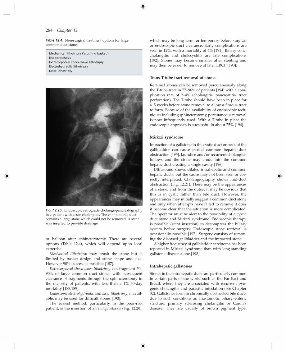
284 Chapter 12
or balloon after sphincterotomy. There are several options (Table 12.4 ), which will depend upon local expertise.
Mechanical lithotripsy may crush the stone but is limited by basket design and stone shape and size. However 90% success is possible [187] .
Extracorporeal shock - wave lithotripsy can fragment 70 – 90% of large common duct stones with subsequent clearance of fragments through the sphincterotomy in the majority of patients, with less than a 1% 30 - day mortality [188,189] .
Endoscopic electrohydraulic and laser lithotripsy , if avail-able, may be used for diffi cult stones [190] .
The easiest method, particularly in the poor - risk patient, is the insertion of an endoprosthesis (Fig. 12.20 ),
Table 12.4. Non - surgical treatment options for large common duct stones
Mechanical lithotripsy ( ‘ crushing basket ’ ) Endoprosthesis Extracorporeal shock - wave lithotripsy Electrohydraulic lithotripsy Laser lithotripsy
Fig. 12.20. Endoscopic retrograde cholangiopancreatography in a patient with acute cholangitis. The common bile duct contains a large stone which could not be removed. A stent was inserted to provide drainage.
which may be long term, or temporary before surgical or endoscopic duct clearance. Early complications are seen in 12%, with a mortality of 4% [191] . Biliary colic, cholangitis and cholecystitis are late complications [192] . Stones may become smaller after stenting and may then be easier to remove at later ERCP [193] .
Trans T - t ube t ract r emoval of s tones
Retained stones can be removed percutaneously along the T - tube tract in 77 – 96% of patients [194] with a com-plication rate of 2 – 4% (cholangitis, pancreatitis, tract perforation). The T - tube should have been in place for 4 – 5 weeks before stone removal to allow a fi brous tract to form. Because of the availability of endoscopic tech-niques including sphincterotomy, percutaneous removal is now infrequently used. With a T - tube in place the endoscopic approach is successful in about 75% [194] .
Mirizzi s yndrome
Impaction of a gallstone in the cystic duct or neck of the gallbladder can cause partial common hepatic duct obstruction [195] . Jaundice and/or recurrent cholangitis follows and the stone may erode into the common hepatic duct creating a single cavity [196] .
Ultrasound shows dilated intrahepatic and common hepatic ducts, but the cause may not been seen or cor-rectly interpreted. Cholangiography shows mid - duct obstruction (Fig. 12.21 ). There may be the appearances of a stone, and from the outset it may be obvious that this is in cystic rather than bile duct. However, the appearances may initially suggest a common duct stone and only when attempts have failed to remove it does it become clear that the situation is more complicated. The operator must be alert to the possibility of a cystic duct stone and Mirizzi syndrome. Endoscopic therapy is possible (stent insertion) to decompress the biliary system before surgery. Endoscopic stone retrieval is occasionally possible [197] . Surgery consists of remov-ing the diseased gallbladder and the impacted stone.
A higher frequency of gallbladder carcinoma has been reported in Mirizzi syndrome than with long - standing gallstone disease alone [198] .
Intrahepatic g allstones
Stones in the intrahepatic ducts are particularly common in certain parts of the world such as the Far East and Brazil, where they are associated with recurrent pyo-genic cholangitis and parasitic infestation (see Chapter 32 ). Gallstones form in chronically obstructed bile ducts due to such conditions as anastomotic biliary – enteric stricture, primary sclerosing cholangitis or Caroli ’ s disease. They are usually of brown pigment type.
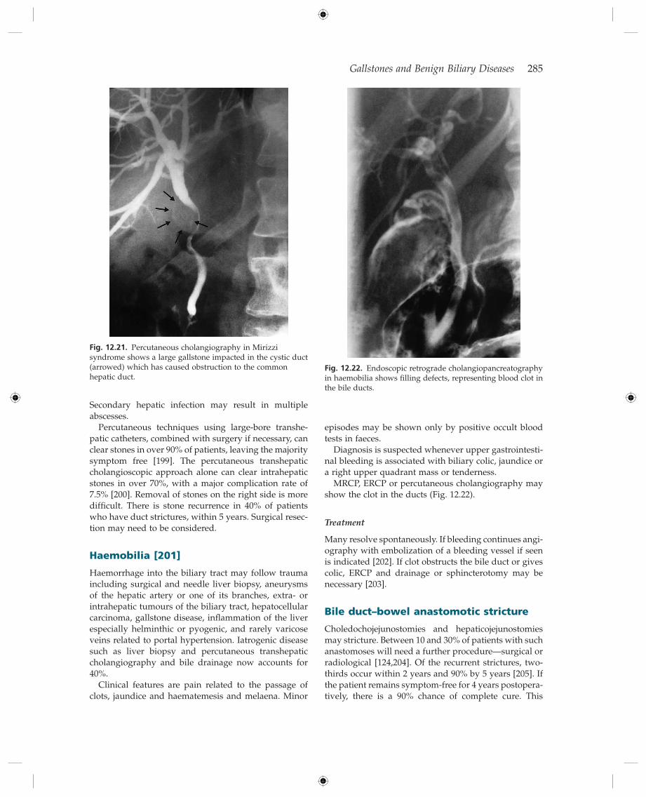
Gallstones and Benign Biliary Diseases 285
Fig. 12.21. Percutaneous cholangiography in Mirizzi syndrome shows a large gallstone impacted in the cystic duct (arrowed) which has caused obstruction to the common hepatic duct.
episodes may be shown only by positive occult blood tests in faeces.
Diagnosis is suspected whenever upper gastrointesti-nal bleeding is associated with biliary colic, jaundice or a right upper quadrant mass or tenderness.
MRCP, ERCP or percutaneous cholangiography may show the clot in the ducts (Fig. 12.22 ).
Treatment
Many resolve spontaneously. If bleeding continues angi-ography with embolization of a bleeding vessel if seen is indicated [202] . If clot obstructs the bile duct or gives colic, ERCP and drainage or sphincterotomy may be necessary [203] .
Bile d uct – b owel a nastomotic s tricture
Choledochojejunostomies and hepaticojejunostomies may stricture. Between 10 and 30% of patients with such anastomoses will need a further procedure — surgical or radiological [124,204] . Of the recurrent strictures, two - thirds occur within 2 years and 90% by 5 years [205] . If the patient remains symptom - free for 4 years postopera-tively, there is a 90% chance of complete cure. This
Secondary hepatic infection may result in multiple abscesses.
Percutaneous techniques using large - bore transhe-patic catheters, combined with surgery if necessary, can clear stones in over 90% of patients, leaving the majority symptom free [199] . The percutaneous transhepatic cholangioscopic approach alone can clear intrahepatic stones in over 70%, with a major complication rate of 7.5% [200] . Removal of stones on the right side is more diffi cult. There is stone recurrence in 40% of patients who have duct strictures, within 5 years. Surgical resec-tion may need to be considered.
Haemobilia [201]
Haemorrhage into the biliary tract may follow trauma including surgical and needle liver biopsy, aneurysms of the hepatic artery or one of its branches, extra - or intrahepatic tumours of the biliary tract, hepatocellular carcinoma, gallstone disease, infl ammation of the liver especially helminthic or pyogenic, and rarely varicose veins related to portal hypertension. Iatrogenic disease such as liver biopsy and percutaneous transhepatic cholangiography and bile drainage now accounts for 40%.
Clinical features are pain related to the passage of clots, jaundice and haematemesis and melaena. Minor
Fig. 12.22. Endoscopic retrograde cholangiopancreatography in haemobilia shows fi lling defects, representing blood clot in the bile ducts.
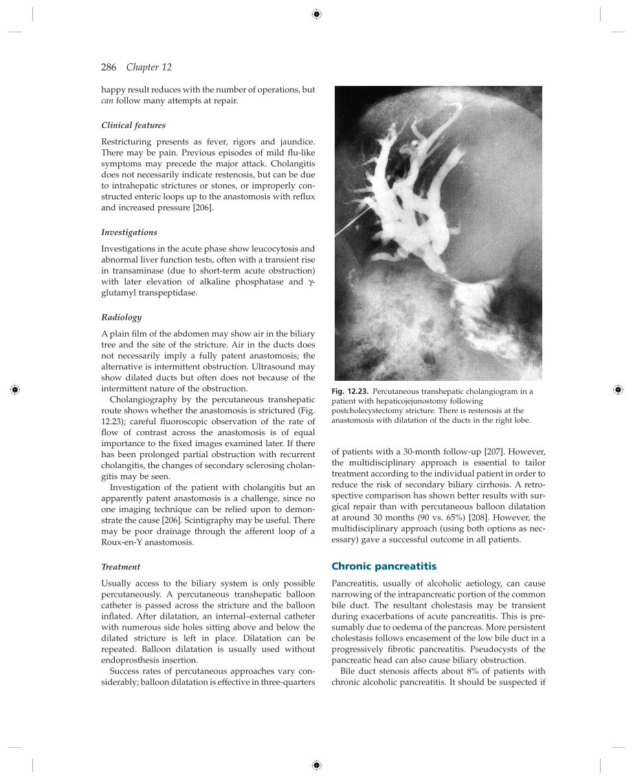
286 Chapter 12
of patients with a 30 - month follow - up [207] . However, the multidisciplinary approach is essential to tailor treatment according to the individual patient in order to reduce the risk of secondary biliary cirrhosis. A retro-spective comparison has shown better results with sur-gical repair than with percutaneous balloon dilatation at around 30 months (90 vs. 65%) [208] . However, the multidisciplinary approach (using both options as nec-essary) gave a successful outcome in all patients.
Chronic p ancreatitis
Pancreatitis, usually of alcoholic aetiology, can cause narrowing of the intrapancreatic portion of the common bile duct. The resultant cholestasis may be transient during exacerbations of acute pancreatitis. This is pre-sumably due to oedema of the pancreas. More persistent cholestasis follows encasement of the low bile duct in a progressively fi brotic pancreatitis. Pseudocysts of the pancreatic head can also cause biliary obstruction.
Bile duct stenosis affects about 8% of patients with chronic alcoholic pancreatitis. It should be suspected if
happy result reduces with the number of operations, but can follow many attempts at repair.
Clinical f eatures
Restricturing presents as fever, rigors and jaundice. There may be pain. Previous episodes of mild fl u - like symptoms may precede the major attack. Cholangitis does not necessarily indicate restenosis, but can be due to intrahepatic strictures or stones, or improperly con-structed enteric loops up to the anastomosis with refl ux and increased pressure [206] .
Investigations
Investigations in the acute phase show leucocytosis and abnormal liver function tests, often with a transient rise in transaminase (due to short - term acute obstruction) with later elevation of alkaline phosphatase and γ - glutamyl transpeptidase.
Radiology
A plain fi lm of the abdomen may show air in the biliary tree and the site of the stricture. Air in the ducts does not necessarily imply a fully patent anastomosis; the alternative is intermittent obstruction. Ultrasound may show dilated ducts but often does not because of the intermittent nature of the obstruction.
Cholangiography by the percutaneous transhepatic route shows whether the anastomosis is strictured (Fig. 12.23 ); careful fl uoroscopic observation of the rate of fl ow of contrast across the anastomosis is of equal importance to the fi xed images examined later. If there has been prolonged partial obstruction with recurrent cholangitis, the changes of secondary sclerosing cholan-gitis may be seen.
Investigation of the patient with cholangitis but an apparently patent anastomosis is a challenge, since no one imaging technique can be relied upon to demon-strate the cause [206] . Scintigraphy may be useful. There may be poor drainage through the afferent loop of a Roux - en - Y anastomosis.
Treatment
Usually access to the biliary system is only possible percutaneously. A percutaneous transhepatic balloon catheter is passed across the stricture and the balloon infl ated. After dilatation, an internal – external catheter with numerous side holes sitting above and below the dilated stricture is left in place. Dilatation can be repeated. Balloon dilatation is usually used without endoprosthesis insertion.
Success rates of percutaneous approaches vary con-siderably; balloon dilatation is effective in three - quarters
Fig. 12.23. Percutaneous transhepatic cholangiogram in a patient with hepaticojejunostomy following postcholecystectomy stricture. There is restenosis at the anastomosis with dilatation of the ducts in the right lobe.

Gallstones and Benign Biliary Diseases 287
llic stents may have a place but are under study [211] . Acute cholangitis, biliary cirrhosis and protracted jaundice are strong indicators for surgery [212] . Choledochoenterostomy is the usual procedure.
Primary s clerosing c holangitis and a utoimmune p ancreatitis (see Chapter 16)
Extra - and intrahepatic bile ducts are diffusely involved in approximately 80% of patients with primary sclero-sing cholangitis. If the patient develops persistent jaundice or recurrent sepsis, investigations are neces-sary to show whether there is a dominant stricture, that is one which appears to be causing signifi cant obstruction compared with the diffuse changes else-where. Ultrasound may show duct dilatation; MRCP or ERCP will show a dominant stricture if present. Brush cytology is necessary. Differentiation of benign strictur-ing from cholangiocarcinoma is diffi cult and often impossible.
In autoimmune pancreatitis the biliary tree may be involved with sclerosing cholangitis - like changes, with or without the characteristic pancreatic changes. Steroids typically reverse the bile duct changes, differentiating this syndrome from classic PSC [213] .
Bile d uct p athology f ollowing l iver t ransplantation
See Chapter 36 .
References
1 Gore RM , Thakrar KH , Newmark GM et al . Gallbladder imaging . Gastroenterol. Clin. N. Am. 2010 ; 39 : 265 – 287 .
2 Sugiyama M , Atomi Y . Endoscopic ultrasonography for diagnosing choledocholithiasis; a prospective compara-tive study with ultrasonography and computed tomogra-phy . Gastrointest. Endosc. 1997 ; 45 : 143 – 146 .
3 Verma D , Kapadia A , Eisen GM et al . EUS v MRCP for detection of choledocholithiasis . Gastrointest. Endosc. 2006 ; 64 : 248 – 254 .
4 Liu CL , Lo CM , Chan JK et al . EUS for detection of occult cholelithiasis in patients with idiopathic pancreatitis . Gastrointest. Endosc. 2000 ; 51 : 28 – 32 .
5 Jeurnick SM , Poley JW , Steyerberg EW et al . ERCP as an outpatient treatment: a review . Gastrointest. Endosc. 2008 ; 68 : 118 – 123 .
6 Andriulli A , Loperfi do S , Napolitano G et al . Incidence rates of post - ERCP complications: a systematic survey of prospective studies . Am. J. Gastroenterol. 2007 ; 102 : 1781 – 1788 .
7 Williams EJ , Taylor S , Fairclough P et al . Are we meeting the standards set for endoscopy? Results of a large - scale prospective survey of Endoscopic retrograde cholangio - pancreatograph practice . Gut 2007 ; 56 : 821 – 829 .
8 Cotton PB , Garrow DA , Gallagher J et al . Risk factors for complications after ERCP: a multivariate analysis of 11,497
the serum alkaline phosphatase is more than twice ele-vated for longer than 1 month. ERCP shows a smooth narrowing of the lower end of the bile duct, sometimes adopting a rat tail confi guration (Fig. 12.24 ). The main pancreatic duct may be tortuous, irregular and dilated. Pancreatic calcifi cation may be present.
Liver biopsy shows portal fi brosis, features of biliary obstruction and sometimes biliary cirrhosis. Features of alcoholic liver disease are unusual. Hepatic fi brosis regresses after biliary decompression [209] .
Management
Early diagnosis is essential as biliary cirrhosis and acute cholangitis can develop in the absence of clinical jaundice.
If alcohol is responsible for the pancreatitis the patient must abstain completely.
The place of surgery is controversial. Clinical, labora-tory and imaging data do not necessarily distinguish those patients with signifi cant bile duct obstruction from those with alcoholic liver disease or normal liver histology. Liver biopsy is valuable in deciding whether surgical decompression of the bile duct is necessary.
Plastic stents successfully relieve bile duct obstruction due to chronic pancreatitis. Placement of multiple plastic stents, to preserve wider dilatation of the stricture, for 12 months has led to an effective outcome after 4 years ’ follow up, though patients with calcifi c do worse than those with non - calcifi c pancreatitis [210] Covered meta-
Fig. 12.24. Endoscopic retrograde cholangiopancreatography in a patient with alcoholic chronic pancreatitis. Note the ‘ rat tail ’ narrowing of the distal common bile duct (arrow).

288 Chapter 12
27 Rosmorduc O , Hermelin B , Boelle PY et al . ABCB4 gene mutation - associated cholelithiasis in adults . Gastro-enterology 2003 ; 125 : 452 – 459 .
28 Lammert F , Miquel J - F . Gallstone disease: from genes to evidence - based therapy . J. Hepatol. 2008 ; 48 : S124 – 135 .
29 Kovacs P , Kress R , Rocha J et al . Variation of the gene encoding the nuclear bile salt receptor FXR and gallstone susceptibility in mice and humans . J. Hepatol. 2008 ; 48 : 116 – 124 .
30 Leitzmann MF , Rimm EB , Willett WC et al . Recreational physical activity and the risk of cholecystectomy in women . N. Engl. J. Med. 1999 ; 341 : 777 – 784 .
31 Portincasa P , Moschetta A , Palasciano G . Cholesterol gall-stone disease . Lancet 2006 ; 368 : 230 – 239 .
32 Biddinger SB , Haas JT , Yu BB et al . Hepatic insulin resist-ance directly promotes formation of cholesterol gallstones . Nat. Med. 2008 ; 14 : 778 – 782 .
33 Maclure KM , Hayes KC , Colditz GA et al . Weight, diet and the risk of symptomatic gallstones in middle - aged women . N. Engl. J. Med. 1989 ; 321 : 563 – 9 .
34 Liddle RA , Goldstein RB , Saxton J . Gallstone formation during weight - reduction dieting . Arch. Intern. Med. 1989 ; 149 : 1750 – 1753 .
35 Sugerman HJ , Brewer WH , Schiffman ML et al . A multi-center, placebo - controlled, randomised, double - blind, prospective trial of prophylactic ursodiol for the preven-tion of gallstone formation following gastric - bypass - induced rapid weight loss . Am. J. Surg. 1995 ; 169 : 91 – 96 .
36 Tsai CJ , Leitzmann MF , Willett WC et al . Dietary carbo-hydrates and glycaemic load and the incidence of sym-ptomatic gall stone disease in men . Gut 2005 ; 54 : 823 – 828 .
37 Heaton KW , Emmett PM , Symes CL et al . An explanation for gallstones in normal - weight women: slow intestinal transit . Lancet 1993 ; 341 : 8 – 10 .
38 Azzaroli F , Mazzella G , Mazelo P et al . Sluggish small bowel motility is involved in determining increased biliary deoxycholic acid in cholesterol gallstone patients . Am. J. Gastroenterol. 1999 ; 94 : 2453 – 2459 .
39 Wells JE , Berr F , Thomas LA et al . Isolation and characteri-zation of cholic acid 7 alpha - dehydroxylating fecal bacte-ria from cholesterol gallstone patients . J. Hepatol. 2000 ; 32 : 4 – 10 .
40 Attili AF , Scafato E , Marchiolo R et al . Diet and gallstones in Italy: the cross - sectional MICOL results . Hepatology 1998 ; 27 : 1492 – 1498 .
41 Pixley F , Wilson D , McPherson K et al . Effect of vegetarian-ism on development of gallstones in women . Br. Med. J. 1985 ; 291 : 11 – 12 .
42 Maringhini A , Ciambra M , Baccelliere P et al . Biliary sludge and gallstones in pregnancy: incidence, risk factors, and natural history . Ann. Intern. Med. 1993 ; 119 : 116 – 120 .
43 Valdivieso V , Covarrubias C , Siegel F et al . Pregnancy and cholelithiasis: pathogenesis and natural course of gall-stones diagnosed in early puerperium . Hepatology 1993 ; 17 : 1 – 4 .
44 Boston Collaborative Drug Surveillance Program . Oral contraceptives and venous thromboembolic disease: sur-gically confi rmed gall - bladder disease and breast tumours . Lancet 1973 ; i : 1399 – 1404 .
procedures over 12 years . Gastrointest. Endosc. 2009 ; 70 : 80 – 88 .
9 Freeman ML . Pancreatic stents for prevention of post - ERCP pancreatitis: for everyday practice or for experts only? Gastrointest. Endosc. 2010 ; 71 : 940 – 944 .
10 Dumonceau J - M , Andriulli A , Deviere J et al . European Society of Gastrointestinal Endoscopy (ESGE) Guideline: Prophylaxis of post - ERCP pancreatitis . Endoscopy 2010 ; 42 : 503 – 515 .
11 Kennedy PTF , Russo E , Kumar N et al . The safety and utility of prophylactic pancreatic duct stents in the preven-tion of post - ERCP pancreatitis: an analysis of practice in a single UK tertiary referral center . Surg. Endosc. 2010 ; 24 : 1923 – 1928 .
12 Cotton PB , Connor P , Rawls E et al. Infection after ERCP and antibiotic prophylaxis: a sequential quality - improvement approach over 11 years . Gastrointest. Endosc. 2008 ; 67 : 471 – 475 .
13 Holzbach RT , Marsh M , Olszewski M et al . Chol-esterol solubility in bile: evidence that supersaturated bile is frequent in healthy man . J. Clin. Invest. 1973 ; 52 : 1467 – 1479 .
14 J ü ngst D , Lang T , von Ritter C et al . Cholesterol nucleation time in gallbladder bile of patients with solitary or multi-ple cholesterol gallstones . Hepatology 1992 ; 15 : 804 – 808 .
15 Kibe A , Holzbach RT , LaRusso NF et al . Inhibition of cho-lesterol crystal formation by apolipoproteins in supersatu-rated model bile . Science 1984 ; 225 : 514 – 516 .
16 Ohya T , Schwarzendrube J , Busch N et al . Isolation of a human biliary glycoprotein inhibitor of cholesterol crys-tallization . Gastroenterology 1993 ; 104 : 527 – 538 .
17 Portincasa P , van Erpecum KJ , Vanberge - Henegouwen GP . Cholesterol crystallization in bile . Gut 1997 ; 41 : 138 – 141 .
18 Hood K , Gleeson D , Ruppin DC et al . Prevention of gall-stone recurrence by nonsteroidal anti - infl ammatory drugs . Lancet 1988 ; ii : 1223 – 1225 .
19 Hopman WP , Jansen JB , Rosenbusch G et al . Role of chole-cystokinin and the cholinergic system in intestinal stimu-lation of gallbladder contraction in man . J. Hepatol. 1990 ; 11 : 261 – 265 .
20 Maurer KJ , Carey MC , Fox JG . Roles of infection, infl am-mation, and the immune system in cholesterol gallstone formation . Gastroenterology 2009 ; 136 : 425 – 440 .
21 Van Bodegraven AA , B ö hmer CJM , Manoliu RA et al . Gallbladder contents and fasting gallbladder volumes during and after pregnancy . Scand. J. Gastroenterol. 1998 ; 33 : 993 – 997 .
22 Janowitz J , Kratzer W , Zemmler T et al . Gallbladder sludge: spontaneous course and incidence of complications in patients without stones . Hepatology 1994 ; 20 : 291 – 294 .
23 Shaffer EA . Epidemiology of gallbladder stone disease . Best Pract. Res. Clin. Gastroenterol. 2006 ; 20 : 981 – 996 .
24 Katsika D , Grjibovski A , Einarsson C et al . Genetic and environmental infl uences on symptomatic gallstone disease: a Swedish study of 43,141 twin pairs . Hepatology 2005 ; 41 : 1138 – 1143 .
25 Nakeeb A , Comuzzie AG , Martin L et al . Gallstones: genet-ics versus environment . Ann. Surg. 2002 ; 235 : 842 – 849 .
26 Gr ü nhage F , Acalovschi M , Tirziu S et al . Increased gall-stone risk in humans conferred by common variant of hepatic ATP - binding cassette transporter for cholesterol . Hepatology 2007 ; 46 : 793 – 801 .
Gallstones and Benign Biliary Diseases 289
matic and asymptomatic patients . Ann. Surg. 1985 ; 202 : 59 – 63 .
64 Halldestam I , Enell E - L , Kullman E et al . Development of symptoms and complications in individuals with asymp-tomatic gallstones . Br. J. Surg. 2004 ; 91 : 734 – 738 .
65 Gurusamy KS , Davidson BR . Surgical treatment of gall-stones . Gastroenterol. Clin. N. Am. 2010 ; 39 : 229 – 244 .
66 Ransohoff DF , Gracie WA , Wolfenson LB et al . Prophylactic cholecystectomy or expectant management for silent gall-stones . Ann. Intern. Med. 1983 ; 99 : 199 – 204 .
67 Diehl AK , Beral V . Cholecystectomy and changing mortal-ity from gallbladder cancer . Lancet 1981 ; i : 187 – 189 .
68 Diehl AK . Gallstone size and the risk of gallbladder cancer . JAMA 1983 ; 250 : 2323 – 6 .
69 Misra S , Chaturvedi A , Misra NC et al . Carcinoma of the gallbladder . Lancet Oncol. 2003 ; 4 : 167 – 176 .
70 Ekbom A , Hsieh CC , Yuen J et al . Risk of extrahepatic bile duct cancer after cholecystectomy . Lancet 1993 ; 342 : 1262 – 1265 .
71 Festi D , Sottili S , Colecchia A et al . Clinical manifestations of gallstone disease: evidence from the multicentre Italian study on cholelithiasis (MICOL) . Hepatology 1999 ; 30 : 839 – 846 .
72 Berger MY , van der Velden JJIM , Lijmer JG et al . Abdominal symptoms: do they predict gallstones? Scand. J. Gastroenterol. 2000 ; 35 : 70 – 76 .
73 Hicks RJ , Kelly MJ , Kalff V . Association between false negative hepatobiliary scans and initial gallbladder visualization after 30 min . Eur. J. Nucl. Med. 1990 ; 16 : 747 – 753 .
74 Ransohoff DF , Gracie WA . Treatment of gallstones . Ann. Intern. Med. 1993 ; 119 : 606 – 619 .
75 Hirota M , Takada T , Kawarada Y et al . Diagnostic criteria and severity assessment of acute cholecystitis: Tokyo guidelines . J. Hepatobiliary. Pancreat. Surg. 2007 ; 14 : 78 – 82 .
76 Strasberg SM . Acute calculous cholecystitis . N. Engl. J. Med. 2008 ; 358 : 2804 – 2811 .
77 Papi C , Catarci M , D ’ Ambrosio L et al . Timing of cholecys-tectomy for acute calculous cholecystitis: a meta - analysis . Am. J. Gastroenterol. 2004 ; 99 : 147 – 155 .
78 Hickman MS , Schwesinger WH , Page CP . Acute cholecys-titis in the diabetic. A case control study of outcome . Arch. Surg. 1988 ; 123 : 409 – 411 .
79 Chow WC , Ong CL , Png JC et al . Gall bladder empyema — another good reason for early cholecystectomy . J. R. Coll. Surg. Edin. 1993 ; 38 : 213 – 215 .
80 Garcia - SanchoTellez L , Rodriquez - Montes JA , Fernandez de Lis S et al . Acute emphysematous cholecystitis. Report of twenty cases . Hepatogastroenterology 1999 ; 46 : 2144 – 2148 .
81 Zeebregts CJ , Wijffels RT , de Jong KP et al . Percutaneous drainage of emphysematous cholecystitis associated with pneumoperitoneum . Hepatogastroenterology 1999 ; 46 : 771 – 774 .
82 Thistle JL , Cleary PA , Lachin JM et al . The natural history of cholelithiasis: the National Cooperative Gallstone Study . Ann. Intern. Med. 1984 ; 101 : 171 – 175 .
83 Owen CC , Jain R . Acute acalculous cholecystitis . Curr. Treat. Options Gastroenterol. 2005 ; 8 : 99 – 104 .
84 Huffman JL , Schenker S . Acute acalculous cholecystitis: a review . Clin. Gastroenterol. Hepatol. 2010 ; 8 : 15 – 22 .
45 Cirillo DJ , Wallace RB , Rodabough RJ et al . Effect of estro-gen therapy on gallbladder disease . JAMA 2005 ; 293 : 330 – 339 .
46 Henriksson P , Einarsson K , Eriksson A et al . Estrogen - induced gallstone formation in males . J. Clin. Invest. 1989 ; 84 : 811 – 816 .
47 Thijs C , Knipschild P , Brombacher P . Serum lipids and gallstones: a case – control study . Gastroenterology 1990 ; 99 : 843 – 849 .
48 Fornari F , Imberti D , Squillante MM et al . Incidence of gallstones in a population of patients with cirrhosis . J. Hepatol. 1994 ; 20 : 797 – 801 .
49 Lucidi V , Buggenhout A , Donckier V . Cholecystectomy in cirrhotic patients: pitfalls and reasonable recommenda-tions . Acta Chir. Belg. 2009 ; 109 : 477 – 480 .
50 Frey JW , Peri RE . Perioperative risk assessment for patients with cirrhosis and liver disease . Expert Rev. Gastroenterol. Hepatol. 2009 ; 3 : 65 – 75 .
51 Swidsinski A , Khilkin M , Pahlig H et al . Time dependent changes in the concentration and type of bacterial sequences found in cholesterol gallstones . Hepatology 1998 ; 27 : 662 – 665 .
52 Chapman BA , Wilson IR , Frampton CM et al . Prevalence of gallbladder disease in diabetes mellitus . Dig. Dis. Sci. 1996 ; 41 : 2222 – 2228 .
53 Keshavarzian A , Dunne M , Iber FL . Gallbladder volume and emptying in insulin requiring male diabetics . Dig. Dis. Sci. 1987 ; 32 : 824 – 828 .
54 Chang TE - S , Lo SK , Shyr H - Y et al . Hepatitis C virus infec-tion facilitates gallstone formation . J. Gastroenterol. Hepatol. 2005 ; 20 : 1416 – 1421 .
55 Makino I , Chijiiwa K , Higashijima H et al . Rapid choles-terol nucleation time and cholesterol gall stone formation after subtotal or total colectomy in humans . Gut 1994 ; 35 : 1760 – 1764 .
56 Inoue K , Fuchigami A , Higashide S et al . Gallbladder sludge and stone formation in relation to contractile func-tion after gastrectomy . Ann. Surg. 1992 ; 215 : 19 – 26 .
57 Dhiman RK , Phanish MK , Chawla YK et al . Gallbladder motility and lithogenicity of bile in patients with choledo-cholithiasis after endoscopic sphincterotomy . J. Hepatol. 1997 ; 26 : 1300 – 1305 .
58 Cicala M , Habib FI , Fiocca F et al . Increased sphincter of Oddi basal pressure in patients affected by gall stone disease: a role for biliary stasis and colicy pain? Gut 2001 ; 48 : 414 – 417 .
59 Leuschner U , G ü ld ü tuna S , Hellstern A . Pathogenesis of pigment stones and medical treatment . J. Gastroenterol. Hepatol. 1994 ; 9 : 87 – 98 .
60 Brink MA , Slors JFM , Keulemans YCA et al . Enterohepatic cycling of bilirubin: a putative mechanism for pigment gallstone formation in ileal Crohn ’ s disease . Gastroenterology 1999 ; 116 : 1420 – 1427 .
61 Sandstad O , Osnes T , Skar V et al . Common bile duct stones are mainly brown and associated with duodenal diverticula . Gut 1994 ; 35 : 1464 – 1467 .
62 Mok HYI , Druffel ERM , Rampone WH . Chronology of cholelithiasis. Dating gallstones from atmospheric radio-carbon produced by nuclear bomb explosions . N. Engl. J. Med. 1986 ; 344 : 1075 – 1077 .
63 McSherry CK , Ferstenberg H , Calhoun WF et al . The natural history of diagnosed gallstone disease in sympto-
290 Chapter 12
cholecystectomy in Veterans Health Administration hos-pitals . Am. J. Surg. 2010 ; 200 : 32 – 40 .
104 Barkun JS , Fried GM , Barkun AN et al . Cholecystectomy without operative cholangiography: implications for common bile duct injury and retained common bile duct stones . Ann. Surg. 1993 ; 218 : 371 – 377 .
105 Millat B , Fingerhut A , Deleuze A et al . Prospective evalu-ation in 121 consecutive unselected patients undergoing laparoscopic treatment of choledocholithiasis . Br. J. Surg. 1995 ; 82 : 1266 – 1269 .
106 Clayton ES , Connor S , Alexakis N et al . Meta - analysis of endoscopy and surgery versus surgery alone for common bile duct stones with the gallbladder in situ . Br. J. Surg. 2006 ; 93 : 1185 – 1191
107 Van Steenbergen W , Ponette E , Marchal G et al . Percutaneous transhepatic cholecystostomy for acute complicated cholecystitis in elderly patients . Am. J. Gastroenterol. 1990 ; 85 : 1363 – 1369 .
108 Verbanck JJ , Demol JW , Ghillebert GL et al . Ultrasound - guided puncture of the gallbladder for acute cholecystitis . Lancet 1993 ; 341 : 1132 – 1133 .
109 Chok KSH , Chu FSK , Cheung TT et al . Results of percuta-neous transhepatic cholecystostomy for high surgical risk patients with acute cholecystitis . ANZ J. Surg. 2010 ; 80 : 280 – 283 .
110 Johlin FC , Neil GA . Drainage of the gallbladder in patients with acute acalculous cholecystitis by transpapillary endoscopic cholecystotomy . Gastrointest. Endosc. 1993 ; 39 : 645 – 651 .
111 Itoi T , Coehlo - Prabhu N , Baron TH . Endoscopic gallblad-der drainage for management of acute cholecystitis . Gastrointest. Endosc. 2010 ; 71 : 1038 – 1045 .
112 Rossi RL , Schirmer WJ , Braasch JW et al . Laparoscopic bile duct injuries: risk factors, recognition, and repair . Arch. Surg. 1992 ; 127 : 596 – 601 .
113 Schol FPG , Go PM , Gouma DJ . Risk factors for bile duct injury in laparoscopic cholecystectomy: analysis of 49 cases . Br. J. Surg. 1994 ; 81 : 1786 – 1788 .
114 Lillemoe KD . Benign postoperative bile duct strictures . Bailli è re ’ s Clin. Gastroenterol. 1997 ; 11 : 749 – 79 .
115 Kurzawinski TR , Selves L , Farouk M et al . Prospective study of hepatobiliary scintigraphy and endoscopic cholangiography for the detection of early biliary compli-cations after orthotopic liver transplantation . Br. J. Surg. 1997 ; 84 : 620 – 623 .
116 Lau WY , Lai ECH , Lau SHY . Management of bile duct injury after laparoscopic cholecystectomy: a review . ANZ J. Surg. 2010 ; 80 : 75 – 81 .
117 Bergman JJGHM , van der Brink GR , Rauws EAJ et al . Treatment of bile duct lesions after laparoscopic cholecys-tectomy . Gut 1996 ; 38 : 141 – 147 .
118 Strasberg SM , Herd M , Soper NJ . An analysis of the problem of bile duct injury during laparoscopic cholecys-tectomy . J. Am. Coll. Surg. 1995 ; 180 : 101 – 125 .
119 Nuzzo G , Giuliante F , Giovannini I et al . Advantages of multidisciplinary management of bile duct injuries occur-ring during cholecystectomy . Am. J. Surg. 2008 ; 195 : 763 – 769 .
120 Vitale GC , Tran TC , Davis BR et al . Endoscopic manage-ment of post cholecystectomy bile duct strictures . J. Am. Coll. Surg. 2008 ; 206 : 918 – 923 .
121 Costamagna G , Pandolfi M , Mutignani M et al . Long term results of endoscopic management of postoperative bile
85 Kalliafas S , Ziegler DW , Flancbaum L et al . Acute acalcu-lous cholecystitis: incidence, risk factors, diagnosis, and outcome . Am. Surg. 1998 ; 64 : 471 – 475 .
86 Mariat G , Mahul P , Pr é v TN et al . Contribution of ultra-sonography and cholescintigraphy to the diagnosis of acute acalculous cholecystitis in intensive care unit patients . Intensive Care Med. 2000 ; 26 : 1658 – 1663 .
87 Drossman DA , Dumitrascu DL . Rome III: new standard for functional gastrointestinal disorders . J. Gastrointest. Liver Dis. 2006 ; 15 : 307 – 312 .
88 Skipper K , Sligh S , Dunn E et al . Laparoscopic cholecys-tectomy for abnormal hepato - iminodiacetic acid scan: a worthwhile procedure . Am. Surg. 2000 ; 66 : 30 – 32 .
89 Yap L , Wycherley AG , Morphett AD et al . Acalculous biliary pain: cholecystectomy alleviates symptoms in patients with abnormal cholescintigraphy . Gastroenterology 1991 ; 101 : 786 – 793 .
90 Sabbaghian MS , Rich BS , Rothberger GD et al . Evaluation of surgical outcomes and gallbladder characteristics in patients with biliary dyskinesia . J. Gastrointest. Surg. 2008 ; 12 : 1324 – 1330 .
91 Hansel SL , DiBaise JK . Functional gallbladder disorder: gallbladder dyskinesia . Gastroenterol. Clin. N. Am. 2010 ; 39 : 369 – 379 .
92 Thorboli J , Vilmann P , Hassan H . Endoscopic ultrasonog-raphy in detection of cholelithiasis in patients with biliary pain and negative transabdominal ultrasonogrpahy . Scand. J. Gastroenterol. 2004 ; 39 : 267 – 269 .
93 Keus F , Gooszen HG , van Laarhoven CJHM . Open, small - incision, or laparoscopic cholecystectomy for patients with symptomatic cholecystolithiasis. An overview of Cochrane Hepato - Biliary Group reviews . Cochrane Database Syst. Rev. 2006 ; CD008318.
94 Gurusamy KS , Samraj K . Early versus delayed laparo-scopic cholecystectomy for acute cholecystitis . Cochrane Database Syst. Rev. 2010 ; CD005440.
95 Yamashita Y , Takada T , Kawarada Y et al . Surgical treat-ment of patients with acute cholecystitis. Tokyo guide-lines . J. Hepatobiliary Pancreat. Surg. 2007 ; 14 : 91 – 97 .
96 Keus F , Gooszen HG , Van Laarhoven CJHM . Systematic review: open, small - incision or laparoscopic cholecystec-tomy for symptomatic cholecystolithiasis . Aliment Pharmacol. Ther. 2009 ; 29 : 359 – 378 .
97 McMahon AJ , Russell IT , Baxter JN et al . Laparoscopic vs. minilaparotomy cholecystectomy: a randomised trial . Lancet 1994 ; 343 : 135 – 138 .
98 Cox MR , Wilson TG , Luck AJ et al . Laparoscopic cholecys-tectomy for acute infl ammation of the gall bladder . Ann. Surg. 1993 ; 218 : 630 – 634 .
99 Kiviluoto T , Sir é n J , Luukkonen P et al . Randomised trial of laparoscopic vs. open cholecystectomy for acute and gangrenous cholecystitis . Lancet 1998 ; 351 : 321 – 325 .
100 Murphy MM , Ng SC , Simons JP et al . Predictors of major complications after laparoscopic cholecystectomy: surgeon, hospital or patient? J. Am. Coll. Surg. 2010 ; 211 : 73 – 80 .
101 Lillemoe KD , Melton GB , Cameron JL et al . Postoperative bile duct strictures: management and outcome in the 1990s . Ann. Surg. 2000 ; 232 : 430 – 441 .
102 Flum DR , Cheadle A , Prela C et al . Bile duct injury during cholecystectomy and survival in Medicare benefi ciaries . JAMA 2003 ; 290 : 2168 – 2173 .
103 Kaafarani HMA , Smith TS , Neumayer L et al . Trends, out-comes, and predictors of open and conversion to open
Gallstones and Benign Biliary Diseases 291
140 Bateson MC , Bouchier IAD , Trash DB et al . Calcifi cation of radiolucent gallstones during treatment with ursodeoxy-cholic acid . Br. Med. J. 1981 ; 283 : 645 – 646 .
141 Mulagha E , Fromm H . Extracorporeal shock wave lithotripsy of gallstones revisited: current status and future promises . J. Gastroenterol. Hepatol. 2000 ; 15 ; 239 – 243 .
142 Rabenstein T , Radespiel - Tr ö ger M , H ö pfner L et al . Ten year experience with piezoelectric extracorporeal shock-wave lithotripsy of gallbladder stones . Eur. J. Gastroenterol. Hepatol. 2005 ; 17 : 629 – 639 .
143 Ash - Miles J , Roach H , Virjee J et al . More than just stones: a pictorial review of common and less common gallblad-der pathologies . Curr. Probl. Diagn. Radiol. 2008 ; 37 : 189 – 202 .
144 Zielinski MD , Atwell TD , Davis PW et al . Comparison of surgically resected polypoid lesions of the gallbladder to their pre - operative ultrasound characteristics . J. Gast-roenterol. Surg. 2009 ; 13 : 19 – 25 .
145 Park JY , Hong SP , Kim YJ et al . Long - term follow up of gall bladder polyps . J. Gastroenterol. Hepatol. 2009 ; 24 : 219 – 222 .
146 Colecchia A , Larocca A , Scaioli E et al . Natural history of small gallbladder polyps is benign: evidence from a clini-cal and pathogenetic study . Am. J. Gastroentrol. 2009 ; 104 : 624 – 629 .
147 Gallahan WC , Conway JD . Diagnosis and management of gallbladder polyps . Gastroenterol. Clin. N. Am. 2010 ; 39 : 359 – 367 .
148 Leung UC , Wong PY , Roberts RH et al . Gall bladder polyps in sclerosing cholangitis: does the 1 - cm rule apply? ANZ J. Surg. 2007 ; 77 : 355 – 357
149 Sahlin S , St å hlberg D , Einarsson K . Cholesterol metabo-lism in liver and gallbladder mucosa of patients with cho-lesterolosis . Hepatology 1995 ; 21 : 1269 – 1275 .
150 Watanabe F , Hanai H , Kaneko E . Increased acylCoA - cholesterol ester acyltransferase activity in gallbladder mucosa in patients with gallbladder cholesterolosis . Am. J. Gastroenterol. 1998 ; 93 : 1518 – 1523 .
151 Spinelli A , Schumacher G , Pascher A et al . Extended surgi-cal resection for xanthogranulomatous cholecystitis mim-icking advanced gallbladder carcinoma: a case report and review of literature . World J. Gastroenterol. 2006 ; 12 : 2293 – 2296 .
152 Crawford RW , Rosales - Reyes R , Ramirez - Aguilar M de la L et al . Gallstones play a signifi cant role in Salmonella spp. gallbladder colonization and carriage . PNAS 2010 ; 107 : 4353 – 4358 .
153 Caygill CPJ , Hill MJ , Braddick M et al . Cancer mortality in chronic typhoid and paratyphoid carriers . Lancet 1994 ; 343 : 83 – 84 .
154 LaRaja RD , Rothenberg RE , Odom JW et al . The incidence of intra - abdominal surgery in acquired immunodefi ciency syndrome: a statistical review of 904 patients . Surgery 1989 ; 105 : 175 – 179 .
155 Ricci M , Puente AO , Rothenberg RE et al . Open and lapar-oscopic cholecystectomy in acquired immunodefi ciency syndrome: indications and results in fi fty - three patients . Surgery 1999 ; 125 : 172 – 177 .
156 Foschi D , Cellerino P , Corsi F et al . Impact of highly active antiretroviral therapy on outcome of cholecystectomy in patients with human immunodefi ciency virus infection . Br. J. Surg. 2006 ; 93 : 1383 – 1389 .
duct strictures with increasing number of stents . Gastrointest. Endosc. 2001 ; 54 : 162 – 168 .
122 Kuzela L , Oltman M , Sutka J et al . Prospective follow up of patients with bile duct strictures due to laparoscopic cholecystectomy, treated endoscopically with multiple stents . Hepatogastroenterology 2005 ; 52 : 1357 – 1361 .
123 Siriwardana HP , Siriwardana AK . Systemic appraisal of the role of metallic endobiliary stents in the treatment of benign biliary stricture . Ann. Surg. 2005 ; 242 : 10 – 19 .
124 Schmidt SC , Langrehr JM , Hintze RE et al . Long - term results and risk factors infl uencing outcomes of major bile duct injuries following cholecystectomy . Br. J. Surg. 2005 ; 92 : 76 – 82 .
125 Chapman WC , Halevy A , Blumgart LH et al . Postcholecystectomy bile duct strictures: management and outcomes in 130 patients . Arch. Surg. 1995 ; 130 : 597 – 602 .
126 Sharpstone D , Colin - Jones DG . Chronic, non - visceral abdominal pain . Gut 1994 ; 35 : 833 – 836 .
127 Luman W , Adams WH , Nixon SN et al . Incidence of per-sistent symptoms after laparoscopic cholecystectomy: a prospective study . Gut 1996 : 39 : 863 – 866 .
128 Vander Velpen GC , Shimi SM , Cuschieri A . Outcome after cholecystectomy for symptomatic gall stone disease and effect of surgical access: laparoscopic vs. open approach . Gut 1993 ; 34 : 1448 – 1451 .
129 Menees S , Elta GH . Sphincter of Oddi dysfunction . Curr. Treat. Options Gastrenterol. 2005 ; 8 : 109 – 115 .
130 Baillie J . Sphincter of Oddi dysfunction . Curr. Gastroenterol. Rep. 2010 ; 12 : 130 – 134 .
131 Hernandez CA , Lerch MM . Sphincter stenosis and gall-stone migration through the biliary tract . Lancet 1993 ; 341 : 1371 – 1373 .
132 Toouli J , Roberts - Thomson IC , Kellow J et al . Manometry based randomised trial of endoscopic sphincterotomy for sphincter of Oddi dysfunction . Gut 2000 ; 46 : 98 – 102 .
133 Geenen JE , Hogan WJ , Dodds WJ et al . The effi cacy of endoscopic sphincterotomy after cholecystectomy in patients with sphincter of Oddi dysfunction . N. Engl. J. Med. 1989 ; 320 : 82 – 87 .
134 Desautels SG , Slivka A , Hutson WR et al . Postchole-cystectomy pain syndrome: pathophysiology of abdomi-nal pain in sphincter of Oddi type III . Gastroenterology 1999 ; 116 : 900 – 905 .
135 Behar J , Corazziari E , Guelrud M et al . Functional gallblad-der and sphincter of Oddi disorders . Gastroenterology 2006 ; 130 : 1498 – 1509 .
136 Howard DE , Fromm H . Nonsurgical management of gallstone disease . Gastroenterol. Clin. North Am. 1999 ; 28 : 133 – 144 .
137 Walters JRF , Hood KA , Gleeson D et al . Combination therapy with oral ursodeoxycholic and chenodeoxycholic acids: pretreatment computed tomography of the gall bladder improves gall stone dissolution effi cacy . Gut 1992 ; 33 : 375 – 380 .
138 Gleeson D , Ruppin DC , Saunders A et al . Final outcome of ursodeoxycholic acid treatment in 126 patients with radi-olucent gallstones . Q. J. Med. 1990 ; 279 : 711 – 729 .
139 Tomida S , Abei M , Yamaguchi T et al . Long - term ursode-oxycholic acid therapy is associated with reduced risk of biliary pain and acute cholecystitis in patients with gall-bladder stones: a cohort analysis . Hepatology 1999 ; 30 : 6 – 13 .
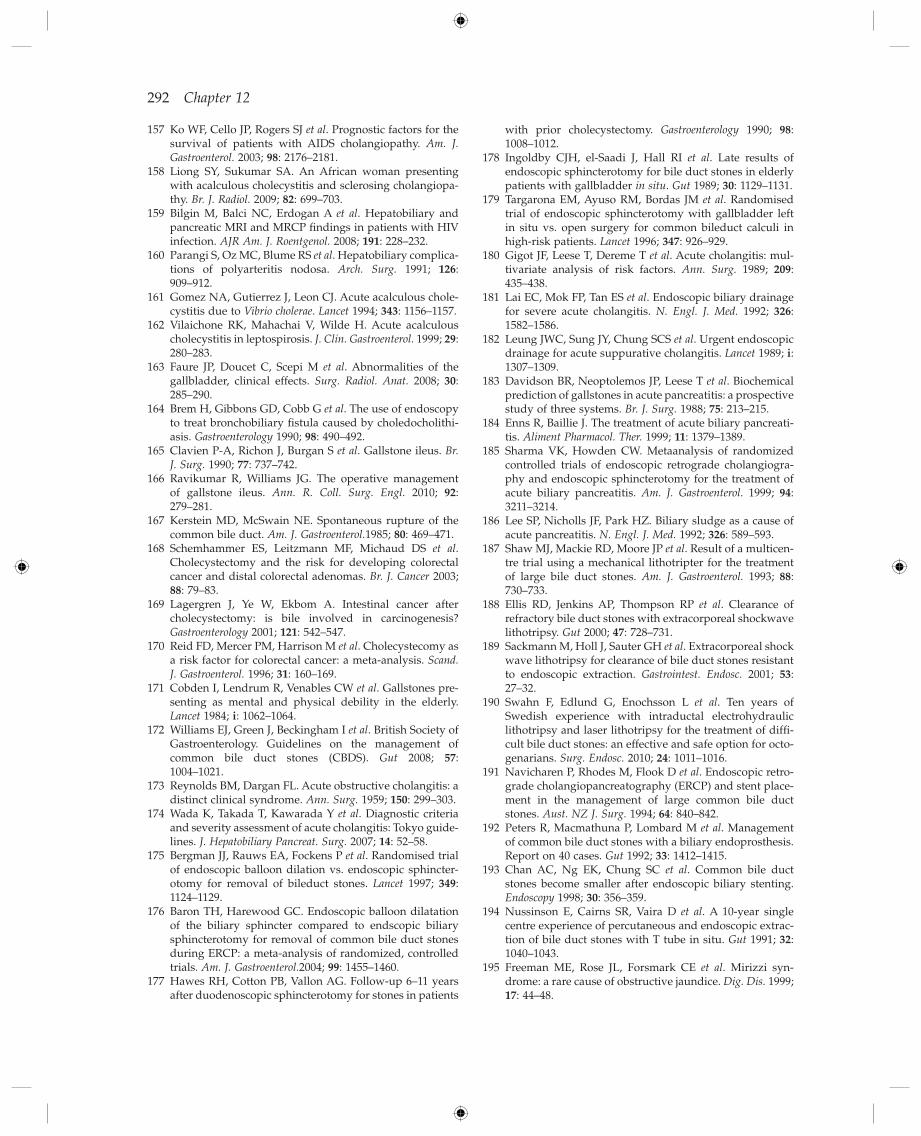
292 Chapter 12
with prior cholecystectomy . Gastroenterology 1990 ; 98 : 1008 – 1012 .
178 Ingoldby CJH , el - Saadi J , Hall RI et al . Late results of endoscopic sphincterotomy for bile duct stones in elderly patients with gallbladder in situ . Gut 1989 ; 30 : 1129 – 1131 .
179 Targarona EM , Ayuso RM , Bordas JM et al . Randomised trial of endoscopic sphincterotomy with gallbladder left in situ vs. open surgery for common bileduct calculi in high - risk patients . Lancet 1996 ; 347 : 926 – 929 .
180 Gigot JF , Leese T , Dereme T et al . Acute cholangitis: mul-tivariate analysis of risk factors . Ann. Surg. 1989 ; 209 : 435 – 438 .
181 Lai EC , Mok FP , Tan ES et al . Endoscopic biliary drainage for severe acute cholangitis . N. Engl. J. Med. 1992 ; 326 : 1582 – 1586 .
182 Leung JWC , Sung JY , Chung SCS et al . Urgent endoscopic drainage for acute suppurative cholangitis . Lancet 1989 ; i : 1307 – 1309 .
183 Davidson BR , Neoptolemos JP , Leese T et al . Biochemical prediction of gallstones in acute pancreatitis: a prospective study of three systems . Br. J. Surg. 1988 ; 75 : 213 – 215 .
184 Enns R , Baillie J . The treatment of acute biliary pancreati-tis . Aliment Pharmacol. Ther. 1999 ; 11 : 1379 – 1389 .
185 Sharma VK , Howden CW . Metaanalysis of randomized controlled trials of endoscopic retrograde cholangiogra-phy and endoscopic sphincterotomy for the treatment of acute biliary pancreatitis . Am. J. Gastroenterol. 1999 ; 94 : 3211 – 3214 .
186 Lee SP , Nicholls JF , Park HZ . Biliary sludge as a cause of acute pancreatitis . N. Engl. J. Med. 1992 ; 326 : 589 – 593 .
187 Shaw MJ , Mackie RD , Moore JP et al . Result of a multicen-tre trial using a mechanical lithotripter for the treatment of large bile duct stones . Am. J. Gastroenterol. 1993 ; 88 : 730 – 733 .
188 Ellis RD , Jenkins AP , Thompson RP et al . Clearance of refractory bile duct stones with extracorporeal shockwave lithotripsy . Gut 2000 ; 47 : 728 – 731 .
189 Sackmann M , Holl J , Sauter GH et al . Extracorporeal shock wave lithotripsy for clearance of bile duct stones resistant to endoscopic extraction . Gastrointest. Endosc. 2001 ; 53 : 27 – 32 .
190 Swahn F , Edlund G , Enochsson L et al . Ten years of Swedish experience with intraductal electrohydraulic lithotripsy and laser lithotripsy for the treatment of diffi -cult bile duct stones: an effective and safe option for octo-genarians . Surg. Endosc. 2010 ; 24 : 1011 – 1016 .
191 Navicharen P , Rhodes M , Flook D et al . Endoscopic retro-grade cholangiopancreatography (ERCP) and stent place-ment in the management of large common bile duct stones . Aust. NZ J. Surg. 1994 ; 64 : 840 – 842 .
192 Peters R , Macmathuna P , Lombard M et al . Management of common bile duct stones with a biliary endoprosthesis. Report on 40 cases . Gut 1992 ; 33 : 1412 – 1415 .
193 Chan AC , Ng EK , Chung SC et al . Common bile duct stones become smaller after endoscopic biliary stenting . Endoscopy 1998 ; 30 : 356 – 359 .
194 Nussinson E , Cairns SR , Vaira D et al . A 10 - year single centre experience of percutaneous and endoscopic extrac-tion of bile duct stones with T tube in situ . Gut 1991 ; 32 : 1040 – 1043 .
195 Freeman ME , Rose JL , Forsmark CE et al . Mirizzi syn-drome: a rare cause of obstructive jaundice . Dig. Dis. 1999 ; 17 : 44 – 48 .
157 Ko WF , Cello JP , Rogers SJ et al . Prognostic factors for the survival of patients with AIDS cholangiopathy . Am. J. Gastroenterol. 2003 ; 98 : 2176 – 2181 .
158 Liong SY , Sukumar SA . An African woman presenting with acalculous cholecystitis and sclerosing cholangiopa-thy . Br. J. Radiol. 2009 ; 82 : 699 – 703 .
159 Bilgin M , Balci NC , Erdogan A et al . Hepatobiliary and pancreatic MRI and MRCP fi ndings in patients with HIV infection . AJR Am. J. Roentgenol. 2008 ; 191 : 228 – 232 .
160 Parangi S , Oz MC , Blume RS et al . Hepatobiliary complica-tions of polyarteritis nodosa . Arch. Surg. 1991 ; 126 : 909 – 912 .
161 Gomez NA , Gutierrez J , Leon CJ . Acute acalculous chole-cystitis due to Vibrio cholerae . Lancet 1994 ; 343 : 1156 – 1157 .
162 Vilaichone RK , Mahachai V , Wilde H . Acute acalculous cholecystitis in leptospirosis . J. Clin. Gastroenterol. 1999 ; 29 : 280 – 283 .
163 Faure JP , Doucet C , Scepi M et al . Abnormalities of the gallbladder, clinical effects . Surg. Radiol. Anat. 2008 ; 30 : 285 – 290 .
164 Brem H , Gibbons GD , Cobb G et al . The use of endoscopy to treat bronchobiliary fi stula caused by choledocholithi-asis . Gastroenterology 1990 ; 98 : 490 – 492 .
165 Clavien P - A , Richon J , Burgan S et al . Gallstone ileus . Br. J. Surg. 1990 ; 77 : 737 – 742 .
166 Ravikumar R , Williams JG . The operative management of gallstone ileus . Ann. R. Coll. Surg. Engl. 2010 ; 92 : 279 – 281 .
167 Kerstein MD , McSwain NE . Spontaneous rupture of the common bile duct . Am. J. Gastroenterol. 1985 ; 80 : 469 – 471 .
168 Schemhammer ES , Leitzmann MF , Michaud DS et al . Cholecystectomy and the risk for developing colorectal cancer and distal colorectal adenomas . Br. J. Cancer 2003 ; 88 : 79 – 83 .
169 Lagergren J , Ye W , Ekbom A . Intestinal cancer after cholecystectomy: is bile involved in carcinogenesis? Gastroenterology 2001 ; 121 : 542 – 547 .
170 Reid FD , Mercer PM , Harrison M et al . Cholecystecomy as a risk factor for colorectal cancer: a meta - analysis . Scand. J. Gastroenterol. 1996 ; 31 : 160 – 169 .
171 Cobden I , Lendrum R , Venables CW et al . Gallstones pre-senting as mental and physical debility in the elderly . Lancet 1984 ; i : 1062 – 1064 .
172 Williams EJ , Green J , Beckingham I et al . British Society of Gastroenterology. Guidelines on the management of common bile duct stones (CBDS) . Gut 2008 ; 57 : 1004 – 1021 .
173 Reynolds BM , Dargan FL . Acute obstructive cholangitis: a distinct clinical syndrome . Ann. Surg. 1959 ; 150 : 299 – 303 .
174 Wada K , Takada T , Kawarada Y et al . Diagnostic criteria and severity assessment of acute cholangitis: Tokyo guide-lines . J. Hepatobiliary Pancreat. Surg. 2007 ; 14 : 52 – 58 .
175 Bergman JJ , Rauws EA , Fockens P et al . Randomised trial of endoscopic balloon dilation vs. endoscopic sphincter-otomy for removal of bileduct stones . Lancet 1997 ; 349 : 1124 – 1129 .
176 Baron TH , Harewood GC . Endoscopic balloon dilatation of the biliary sphincter compared to endscopic biliary sphincterotomy for removal of common bile duct stones during ERCP: a meta - analysis of randomized, controlled trials . Am. J. Gastroenterol. 2004 ; 99 : 1455 – 1460 .
177 Hawes RH , Cotton PB , Vallon AG . Follow - up 6 – 11 years after duodenoscopic sphincterotomy for stones in patients
Gallstones and Benign Biliary Diseases 293
after biliary - enteric bypass . Arch. Surg. 1993 ; 128 : 269 – 272 .
207 Vos PM , van Beek EJR , Smits NJ et al . Percutaneous balloon dilatation for benign hepaticojejunostomy strictures . Abdom. Imaging 2000 ; 25 : 134 – 138.
208 Lillemoe KD , Martin SA , Cameron JA et al . Major bile duct injuries during laparoscopic cholecystectomy. Follow - up after combined surgical and radiological management . Ann. Surg. 1997 ; 225 : 459 – 468 .
209 Hammel P , Couvelard A , O ’ Toole D et al . Regression of liver fi brosis after biliary drainage in patients with chronic pancreatitis and stenosis of the common bile duct . N. Engl. J. Med. 2001 ; 333 : 418 – 423 .
210 Dragonov P , Hoffmann B , Marsh W et al . Long - term outcome in patients with benign biliary strictures treated endoscopically with multiple stents . Gastrointest. Endosc. 2002 ; 55 : 680 – 686 .
211 Isayama H , Nakai Y , Togawa O et al . Covered metallic stents in the management of malignant and benign pan-creatobiliary strictures . J. Hepatobiliary Pancreat Surg. 2009 ; 16 : 624 – 627 .
212 Stahl TJ , Allen MO ’ C , Ansel HJ et al . Partial biliary obstruc-tion caused by chronic pancreatitis: an appraisal of indica-tions for surgical biliary drainage . Ann. Surg. 1988 ; 207 : 26 – 32 .
213 Webster GJM , Pereira SP , Chapman RW . Autoimmune pancreatitis/IgG4 - associated cholangitis and primary sclerosing cholangitis – overlapping or separate diseases . J. Hepatol. 2009 ; 51 : 398 – 402 .
196 Csendes A , Carlos Diaz J , Burdiles P et al . Mirizzi syn-drome and cholecystobiliary fi stula: a unifying classifi ca-tion . Br. J. Surg. 1989 ; 76 : 1139 – 1143 .
197 England RE , Martin DF . Endoscopic management of Mirizzi ’ s syndrome . Gut 1997 ; 40 : 272 – 276 .
198 Redaelli CA , B ü chler MW , Schilling MK et al . High coin-cidence of Mirizzi syndrome and gallbladder carcinoma . Surgery 1997 ; 121 : 58 – 63 .
199 Pitt HA , Venbrux AC , Coleman J et al . Intrahepatic stones. The transhepatic team approach . Ann. Surg. 1994 ; 219 : 527 – 535 .
200 Cheung M - T , Wai S - H , Kwok PC - H . Percutaneous tran-shepatic choledochoscopic removal of intrahepatic stones . Br. J. Surg. 2003 ; 90 : 1409 – 1415 .
201 Bloechle C , Izbicki JR , Rashed MY et al . Hemobilia: pres-entation, diagnosis and management . Am. J. Gastroenterol. 1994 ; 89 : 1537 – 1540 .
202 Czerniak A , Thompson JN , Hemingway AP et al . Hemobilia: a disease in evolution . Arch. Surg. 1988 ; 123 : 718 – 721 .
203 Kroser J , Rothstein RD , Kochman ML . Endoscopic man-agement of obstructive jaundice caused by haemobilia . Gastrointest. Endosc. 1996 ; 44 : 618 – 619 .
204 Murr MM , Gigot JF , Nagorney DM et al . Long - term results of biliary reconstruction after laparoscopic bile duct injury . Arch. Surg. 1999 ; 134 : 604 – 609 .
205 Pellegrini CA , Thomas MJ , Way LW . Recurrent biliary stricture. Patterns of recurrence and outcome of surgical therapy . Am. J. Surg. 1984 ; 147 : 175 – 180.
206 Matthews JB , Baer HU , Schweizer WP et al . Recurrent cholangitis with and without anastomotic stricture