Embed Size (px)
Citation preview
Chapter 13 Disordered Seeing with Normal Eyes
Chapter 13 Disordered Seeing with Normal Eyes
A neurological approach to vision
Kate Denker Welch
A neurological approach to vision
Kate Denker Welch
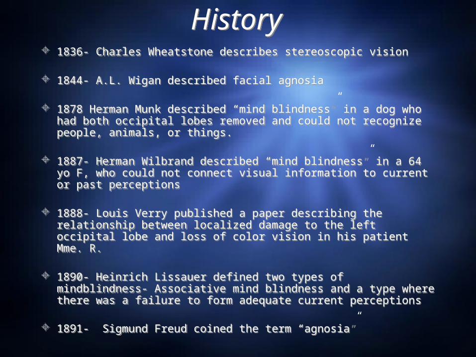
HistoryHistory 1836- Charles Wheatstone describes stereoscopic vision
1844- A.L. Wigan described facial agnosia
1878 Herman Munk described “mind blindness” in a dog who had both occipital lobes removed and could not recognize people, animals, or things.
1887- Herman Wilbrand described “mind blindness” in a 64 yo F, who could not connect visual information to current or past perceptions
1888- Louis Verry published a paper describing the relationship between localized damage to the left occipital lobe and loss of color vision in his patient Mme. R.
1890- Heinrich Lissauer defined two types of mindblindness- Associative mind blindness and a type where there was a failure to form adequate current perceptions
1891- Sigmund Freud coined the term “agnosia”
1836- Charles Wheatstone describes stereoscopic vision
1844- A.L. Wigan described facial agnosia
1878 Herman Munk described “mind blindness” in a dog who had both occipital lobes removed and could not recognize people, animals, or things.
1887- Herman Wilbrand described “mind blindness” in a 64 yo F, who could not connect visual information to current or past perceptions
1888- Louis Verry published a paper describing the relationship between localized damage to the left occipital lobe and loss of color vision in his patient Mme. R.
1890- Heinrich Lissauer defined two types of mindblindness- Associative mind blindness and a type where there was a failure to form adequate current perceptions
1891- Sigmund Freud coined the term “agnosia”
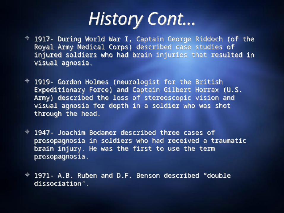
History Cont…History Cont… 1917- During World War I, Captain George Riddoch (of the
Royal Army Medical Corps) described case studies of injured soldiers who had brain injuries that resulted in visual agnosia.
1919- Gordon Holmes (neurologist for the British Expeditionary Force) and Captain Gilbert Horrax (U.S. Army) described the loss of stereoscopic vision and visual agnosia for depth in a soldier who was shot through the head.
1947- Joachim Bodamer described three cases of prosopagnosia in soldiers who had received a traumatic brain injury. He was the first to use the term prosopagnosia.
1971- A.B. Ruben and D.F. Benson described “double dissociation”.
1917- During World War I, Captain George Riddoch (of the Royal Army Medical Corps) described case studies of injured soldiers who had brain injuries that resulted in visual agnosia.
1919- Gordon Holmes (neurologist for the British Expeditionary Force) and Captain Gilbert Horrax (U.S. Army) described the loss of stereoscopic vision and visual agnosia for depth in a soldier who was shot through the head.
1947- Joachim Bodamer described three cases of prosopagnosia in soldiers who had received a traumatic brain injury. He was the first to use the term prosopagnosia.
1971- A.B. Ruben and D.F. Benson described “double dissociation”.
What Causes Visual Agnosia?What Causes Visual Agnosia?
Damage to temporal, occipital, or the parietal lobe.
Different types of agnosia relate to specific damaged regions of the brain.
Overall, visual agnosia means that there is an inability to recognize stimuli.
Damage to temporal, occipital, or the parietal lobe.
Different types of agnosia relate to specific damaged regions of the brain.
Overall, visual agnosia means that there is an inability to recognize stimuli.
Visual AgnosiaVisual Agnosia
Types:Associative Visual Agnosia Apperceptive Visual AgnosiaColor AgnosiaObject AgnosiaProsopagnosiaMovement agnosiaDepth Agnosia
Types:Associative Visual Agnosia Apperceptive Visual AgnosiaColor AgnosiaObject AgnosiaProsopagnosiaMovement agnosiaDepth Agnosia
Associative Visual AgnosiaAssociative Visual Agnosia
Inability to connect current perception of what is presented with past experience.
Can not recognize something even though the shape itself can be perceived. Perception exists but comprehension of perception does not.
Can draw things that they do not recognize. Damage to an area among the ventral stream. Most common cause of associative visual agnosia is
carbon monoxide poisoning.
MRI of CO victim with Visual AgnosiaMRI of CO victim with Visual Agnosia
QuickTime™ and aTIFF (LZW) decompressor
are needed to see this picture.
Apperceptive AgnosiaApperceptive Agnosia
Typically related to damage in the ventral stream occipital region
Inability to recognize and discriminate between overall shapes and visual space
Can not copy or draw objects without great difficulty.
Typically related to damage in the ventral stream occipital region
Inability to recognize and discriminate between overall shapes and visual space
Can not copy or draw objects without great difficulty.
Visual Agnosia for FormVisual Agnosia for Form
Form Agnosia is the inability to perceive shape
Goodale and Miller described a case study of a 34 yo woman called DF.
Form Agnosia is the inability to perceive shape
Goodale and Miller described a case study of a 34 yo woman called DF.
The Case of DFThe Case of DF 34 yo F, lost consciousness from carbon monoxide poisoning.
Awoke from coma blind. Her vision returned after 10 days but with poor perception of shape.She could identify things like the color or size of an object (dorsal stream must be intact) but not the object itself (ventral stream is damaged).
Tested by Melvyn Goodale and David Milner Orientation test- asked to put a card through a slot
She failed that task but succeeded when asked to put it through an imaginary slot
Efron square test- asked to discriminate between rectangle plaques and square plaques. Next asked to estimate width of plaques with her fingers.
She failed at the two tasks. She succeeded at a task where she could pick up the plaque with appropriate finger width.
Drawing- She poorly copied drawings but was much better at drawing objects from memory (despite not recognizing what she had drawn).
Diagnosed with visual agnosia for form, the damage to her brain was primarily in the ventral stream on visual areas V2, V3, and V4.
34 yo F, lost consciousness from carbon monoxide poisoning. Awoke from coma blind. Her vision returned after 10 days but with poor perception of shape.She could identify things like the color or size of an object (dorsal stream must be intact) but not the object itself (ventral stream is damaged).
Tested by Melvyn Goodale and David Milner Orientation test- asked to put a card through a slot
She failed that task but succeeded when asked to put it through an imaginary slot
Efron square test- asked to discriminate between rectangle plaques and square plaques. Next asked to estimate width of plaques with her fingers.
She failed at the two tasks. She succeeded at a task where she could pick up the plaque with appropriate finger width.
Drawing- She poorly copied drawings but was much better at drawing objects from memory (despite not recognizing what she had drawn).
Diagnosed with visual agnosia for form, the damage to her brain was primarily in the ventral stream on visual areas V2, V3, and V4.
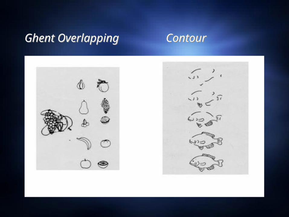
Ghent Overlapping ContourGhent Overlapping Contour
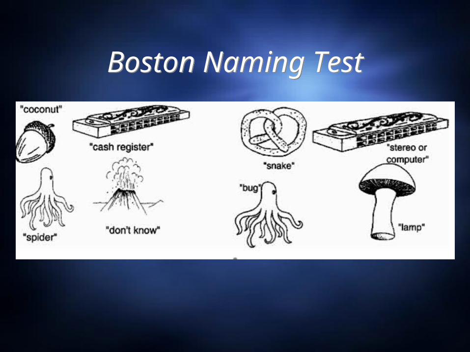
Boston Naming TestBoston Naming Test
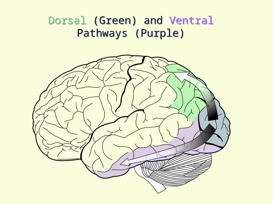
Dorsal (Green) and Ventral Pathways (Purple)
Dorsal (Green) and Ventral Pathways (Purple)
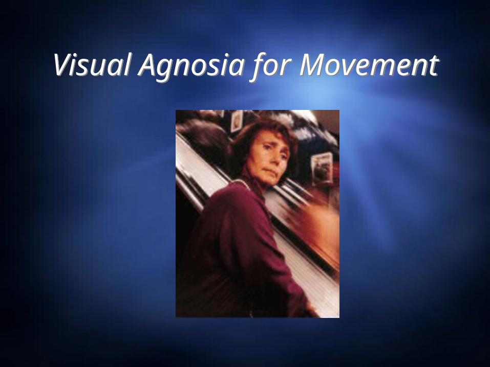
Visual Agnosia for Movement
Visual Agnosia for Movement
Visual Agnosia for Movement
Visual Agnosia for Movement
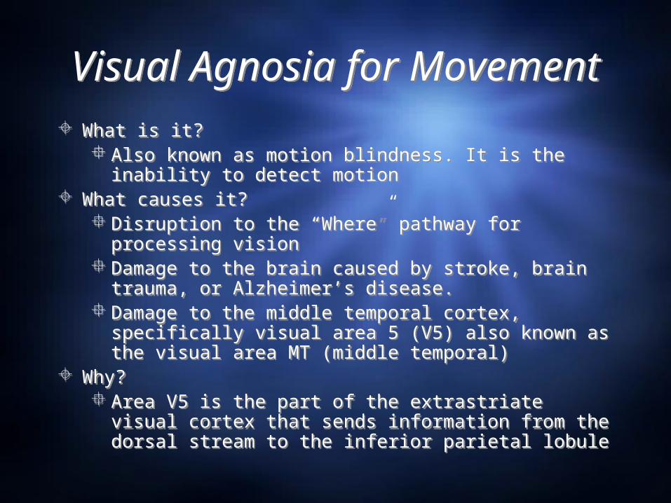
What is it? Also known as motion blindness. It is the inability to
detect motion What causes it?
Disruption to the “Where” pathway for processing vision
Damage to the brain caused by stroke, brain trauma, or Alzheimer’s disease.
Damage to the middle temporal cortex, specifically visual area 5 (V5) also known as the visual area MT (middle temporal)
Why? Area V5 is the part of the extrastriate visual cortex
that sends information from the dorsal stream to the inferior parietal lobule
Zilhl’s PatientZilhl’s Patient “‘I see the world in snapshots – like the frames of a
movie, but most of the frames are missing.” Patient was a 45 yo F who was admitted to the hospital
with headache, nausea, vertigo, and an inability to detect movement. She had normal vision for color, shape, and facial recognition.
Zilhl’s patient had experienced a stroke that effected the V5 area of her brain and caused her to have motion blindness.
She was unable to see the movement of people, cars, or objects. However, she could discriminate between movement and stationary objects in her peripheral vision.
Evolutionarily, her intact peripheral vision for movement speaks to the importance of peripheral vision in predator/prey tracking and self-preservation.
“‘I see the world in snapshots – like the frames of a movie, but most of the frames are missing.”
Patient was a 45 yo F who was admitted to the hospital with headache, nausea, vertigo, and an inability to detect movement. She had normal vision for color, shape, and facial recognition.
Zilhl’s patient had experienced a stroke that effected the V5 area of her brain and caused her to have motion blindness.
She was unable to see the movement of people, cars, or objects. However, she could discriminate between movement and stationary objects in her peripheral vision.
Evolutionarily, her intact peripheral vision for movement speaks to the importance of peripheral vision in predator/prey tracking and self-preservation.

Case of Colonel T.Case of Colonel T.
Described by Captain George Riddoch of the Royal Army Medical Corps during WWI
Colonel T. was shot in the head. His right occipital lobe was damaged which resulted in his inability to see still objects in the left visual field.
“Moving things” could be detected in the left visual field but they looked grey blobs.
Visual Agnosia for DepthVisual Agnosia for Depth
What is it? Lack of stereoscopic vision
What causes it?Damage to right posterior
hemisphereCan also be caused by poor
development
What is it? Lack of stereoscopic vision
What causes it?Damage to right posterior
hemisphereCan also be caused by poor
development
Case Studies of Depth AgnosiaCase Studies of Depth Agnosia
Captain de W. Described by George Riddoch Cpn. W had shrapnel removed from his brain that dmaged
the right occipital lobe and left frontal lobe. He lost all ability to understand depth and everything
looked flat. Riddoch determined that stereoscopic vision relies on more
than just binocular vision to produce 3 dimensional images.
George Holmes and Captain Gilbert Horax They treated a soldier who had been shot through the back
of the head. The patient reported that everything looked flat to him.
Color AgnosiaColor Agnosia
What causes it?Damage to the extrastriate visual cortex,
specifically the V4 area.The visual area 4 (V4) is cut off from
sending information to the inferior temporal lobe. Without the ability to send that information to the “What” pathway of the temporal lobe, the color cannot be recognized.
What causes it?Damage to the extrastriate visual cortex,
specifically the V4 area.The visual area 4 (V4) is cut off from
sending information to the inferior temporal lobe. Without the ability to send that information to the “What” pathway of the temporal lobe, the color cannot be recognized.
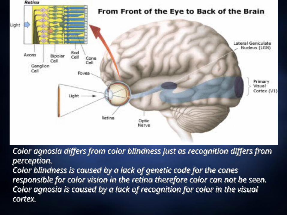
Color agnosia differs from color blindness just as recognition differs from perception. Color blindness is caused by a lack of genetic code for the cones responsible for color vision in the retina therefore color can not be seen.Color agnosia is caused by a lack of recognition for color in the visual cortex.
Color agnosia differs from color blindness just as recognition differs from perception. Color blindness is caused by a lack of genetic code for the cones responsible for color vision in the retina therefore color can not be seen.Color agnosia is caused by a lack of recognition for color in the visual cortex.
Deficit a short film by Calum MacAulayDeficit a short film by Calum MacAulay
QuickTime™ and a decompressor
are needed to see this picture.
ProsopagnosiaProsopagnosia
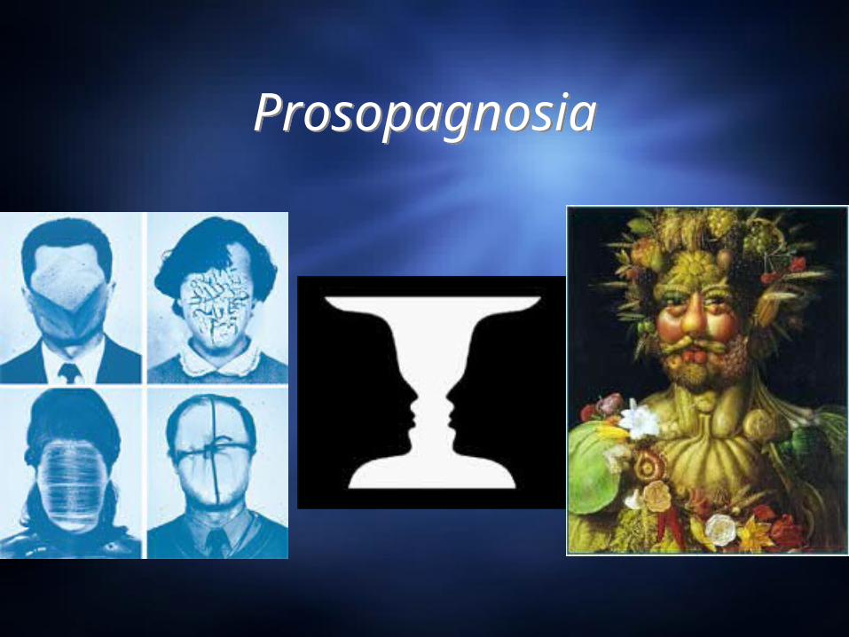
ProsopagnosiaProsopagnosia What is prosopagnosia?
Inability to recognize faces. Also known as face blindness Types of prosopagnosia:
Associative prosopagnosiaMore common version Less severe because faces are not recognized but facial positioning is
understood (they know where the mouth and nose should be placed)May be able to draw faces but not recognize the face they drew
Apperceptive prosopagnosiaFaces are not recognized and facial position is not understoodDrawing faces may be very difficultMay have covert recognition
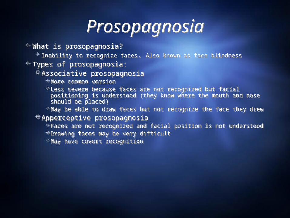
What is prosopagnosia? Inability to recognize faces. Also known as face blindness
Types of prosopagnosia:Associative prosopagnosia
More common version Less severe because faces are not recognized but facial positioning is
understood (they know where the mouth and nose should be placed)May be able to draw faces but not recognize the face they drew
Apperceptive prosopagnosiaFaces are not recognized and facial position is not understoodDrawing faces may be very difficultMay have covert recognition
ProsopagnosiaProsopagnosia
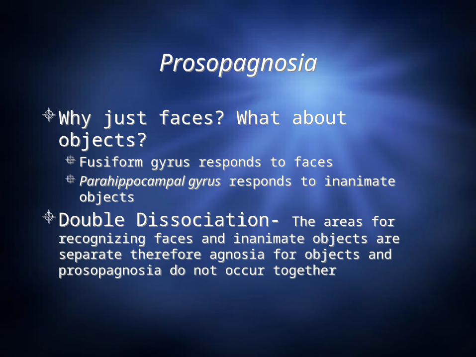
Why just faces? What about objects? Fusiform gyrus responds to faces Parahippocampal gyrus responds to inanimate objects
Double Dissociation- The areas for recognizing faces and inanimate objects are separate therefore agnosia for objects and prosopagnosia do not occur together
Why just faces? What about objects? Fusiform gyrus responds to faces Parahippocampal gyrus responds to inanimate objects
Double Dissociation- The areas for recognizing faces and inanimate objects are separate therefore agnosia for objects and prosopagnosia do not occur together
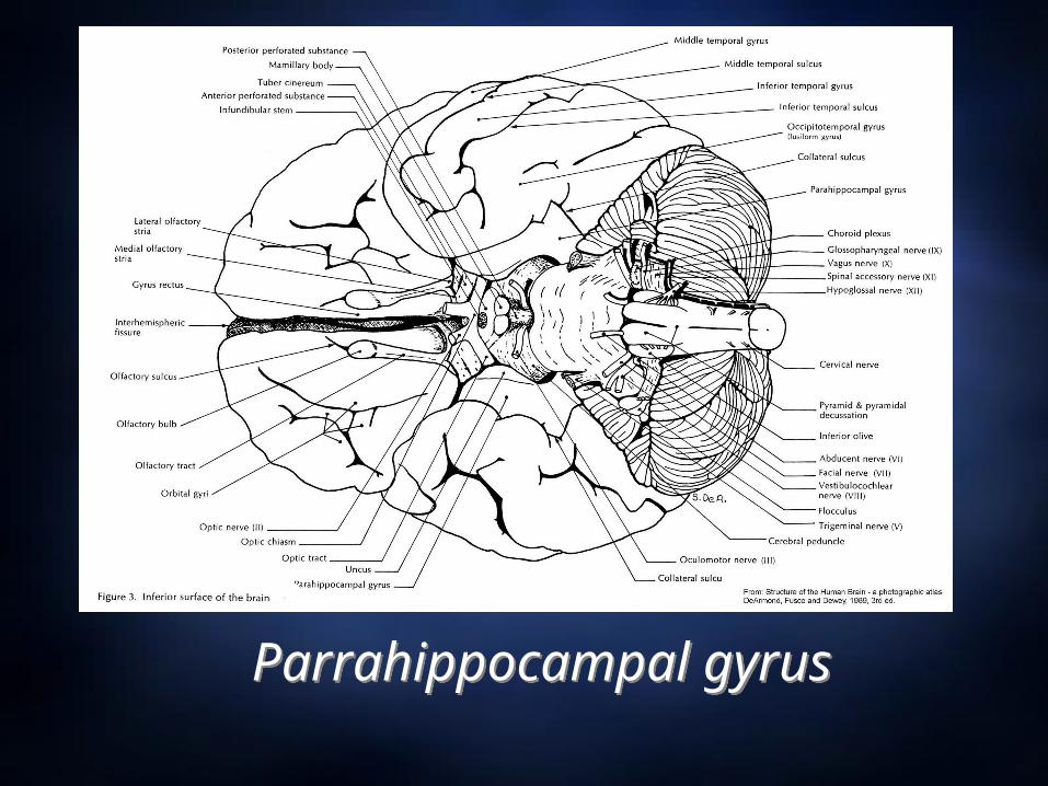
Parrahippocampal gyrusParrahippocampal gyrus
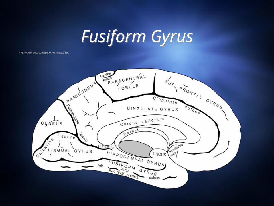
Fusiform GyrusFusiform GyrusThe Fusiform gyrus is located in the temporal lobeThe Fusiform gyrus is located in the temporal lobe
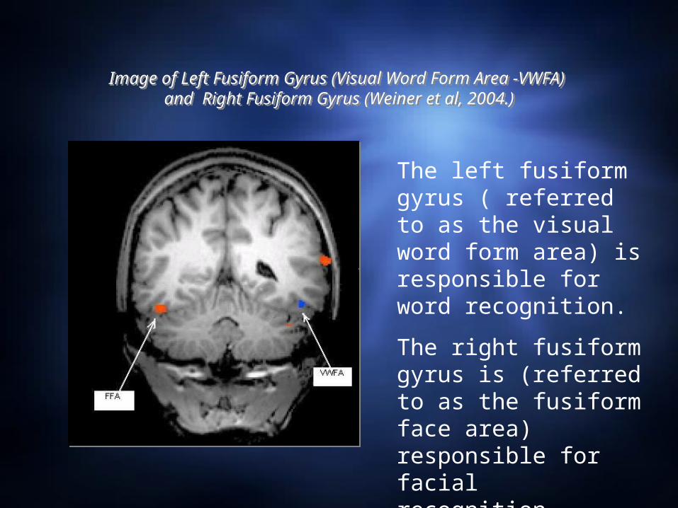
Image of Left Fusiform Gyrus (Visual Word Form Area -VWFA) and Right Fusiform Gyrus (Weiner et al, 2004.)
Image of Left Fusiform Gyrus (Visual Word Form Area -VWFA) and Right Fusiform Gyrus (Weiner et al, 2004.)
The left fusiform gyrus ( referred to as the visual word form area) is responsible for word recognition.
The right fusiform gyrus is (referred to as the fusiform face area) responsible for facial recognition.
What Causes Prosopagnosia?What Causes Prosopagnosia?
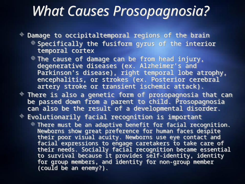
Damage to occipitaltemporal regions of the brain Specifically the fusiform gyrus of the interior temporal
cortex The cause of damage can be from head injury,
degenerative diseases (ex. Alzheimer’s and Parkinson’s disease), right temporal lobe atrophy, encephalitis, or strokes (ex. Posterior cerebral artery stroke or transient ischemic attack).
There is also a genetic form of prosopagnosia that can be passed down from a parent to child. Prosopagnosia can also be the result of a developmental disorder.
Evolutionarily facial recognition is important There must be an adaptive benefit for facial recognition.
Newborns show great preference for human faces despite their poor visual acuity. Newborns use eye contact and facial expressions to engage caretakers to take care of their needs. Socially facial recognition became essential to survival because it provides self-identity, identity for group members, and identity for non-group member (could be an enemy?).
Early DescriptionsEarly Descriptions
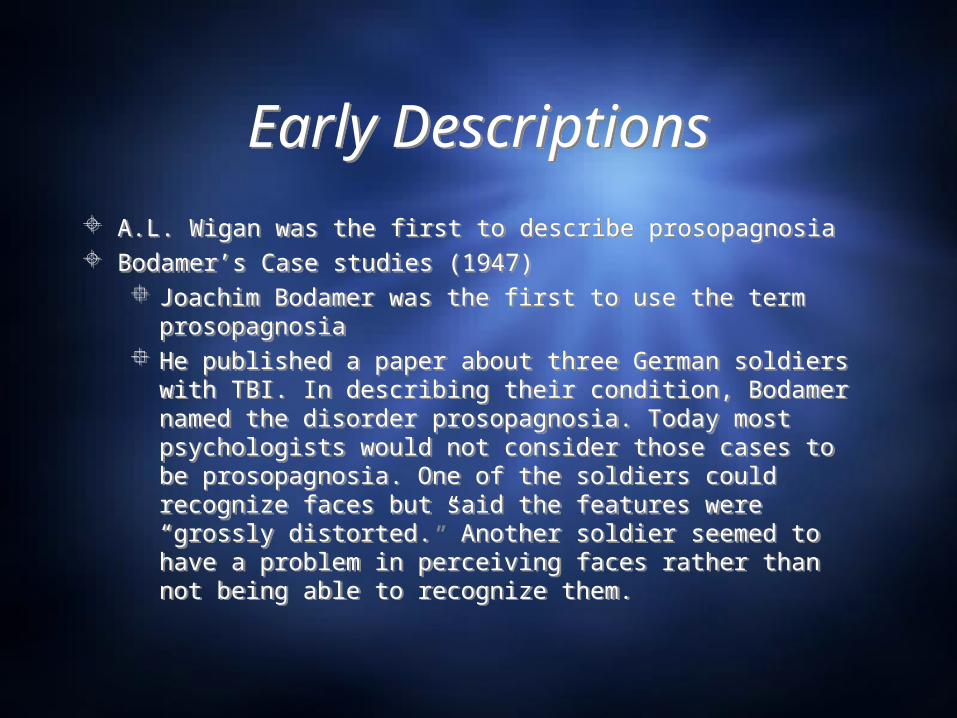
A.L. Wigan was the first to describe prosopagnosia Bodamer’s Case studies (1947)
Joachim Bodamer was the first to use the term prosopagnosia
He published a paper about three German soldiers with TBI. In describing their condition, Bodamer named the disorder prosopagnosia. Today most psychologists would not consider those cases to be prosopagnosia. One of the soldiers could recognize faces but said the features were “grossly distorted.” Another soldier seemed to have a problem in perceiving faces rather than not being able to recognize them.
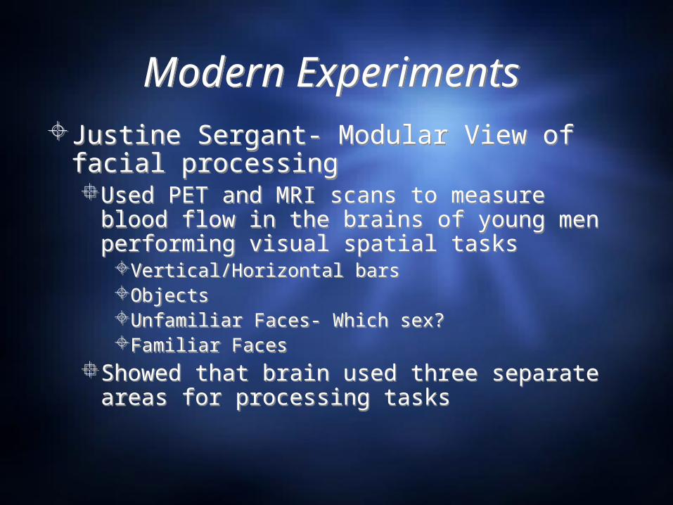
Modern Experiments Modern Experiments Justine Sergant- Modular View of facial
processingUsed PET and MRI scans to measure blood
flow in the brains of young men performing visual spatial tasksVertical/Horizontal barsObjectsUnfamiliar Faces- Which sex?Familiar Faces
Showed that brain used three separate areas for processing tasks
Justine Sergant- Modular View of facial processingUsed PET and MRI scans to measure blood
flow in the brains of young men performing visual spatial tasksVertical/Horizontal barsObjectsUnfamiliar Faces- Which sex?Familiar Faces
Showed that brain used three separate areas for processing tasks
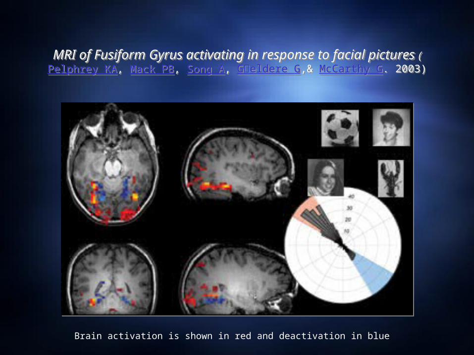
MRI of Fusiform Gyrus activating in response to facial pictures (Pelphrey KA, Mack PB, Song A, G毟eldere G,& McCarthy G.
2003)
MRI of Fusiform Gyrus activating in response to facial pictures (Pelphrey KA, Mack PB, Song A, G毟eldere G,& McCarthy G.
2003)
Brain activation is shown in red and deactivation in blue
Covert Recognition in Facial Agnosia
Covert Recognition in Facial Agnosia
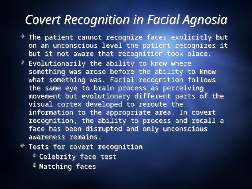
The patient cannot recognize faces explicitly but on an unconscious level the patient recognizes it but it not aware that recognition took place.
Evolutionarily the ability to know where something was arose before the ability to know what something was. Facial recognition follows the same eye to brain process as perceiving movement but evolutionary different parts of the visual cortex developed to reroute the information to the appropriate area. In covert recognition, the ability to process and recall a face has been disrupted and only unconscious awareness remains.
Tests for covert recognition Celebrity face test Matching faces
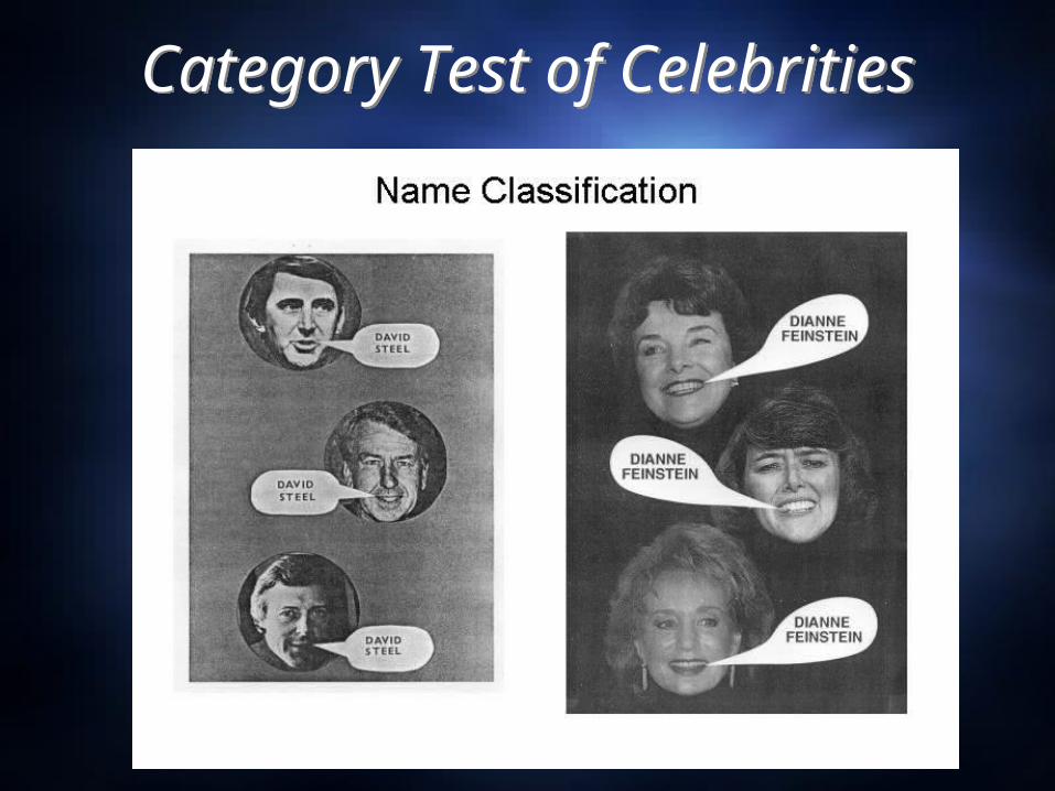
Category Test of Celebrities
Category Test of Celebrities
The Case of PHThe Case of PH
19 yo M, head injury from motorcycle accident Awoke from a coma after 12 days and discovered he
could no longer recognize faces He had no overt ability to recognize faces On covert recognition tasks, he was slow in replying and
recognition occurred even though he was unaware of it.
19 yo M, head injury from motorcycle accident Awoke from a coma after 12 days and discovered he
could no longer recognize faces He had no overt ability to recognize faces On covert recognition tasks, he was slow in replying and
recognition occurred even though he was unaware of it.
Blind SightBlind Sight
Blind sight is also known as unconscious vision. Discovered by E. Poppel at MIT
Noticed that brain damaged patients who reported some visual field blindness were able to follow the movement of a light shone in the “blind spot” of their visual field. Although the patients denied moving their eyes in the correct direction, nevertheless their eyes followed the light even without their knowledge.
Larry Weiskrantz named the disorder blindsight after studying a patient named DB.
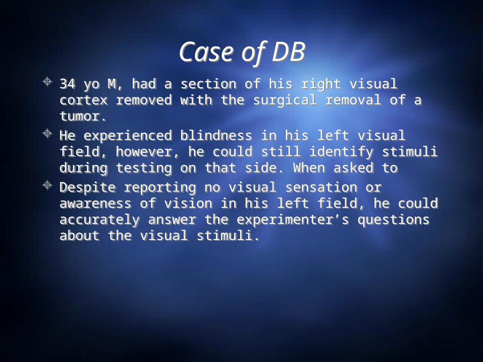
Case of DBCase of DB 34 yo M, had a section of his right visual cortex
removed with the surgical removal of a tumor. He experienced blindness in his left visual field,
however, he could still identify stimuli during testing on that side. When asked to
Despite reporting no visual sensation or awareness of vision in his left field, he could accurately answer the experimenter’s questions about the visual stimuli.
34 yo M, had a section of his right visual cortex removed with the surgical removal of a tumor.
He experienced blindness in his left visual field, however, he could still identify stimuli during testing on that side. When asked to
Despite reporting no visual sensation or awareness of vision in his left field, he could accurately answer the experimenter’s questions about the visual stimuli.
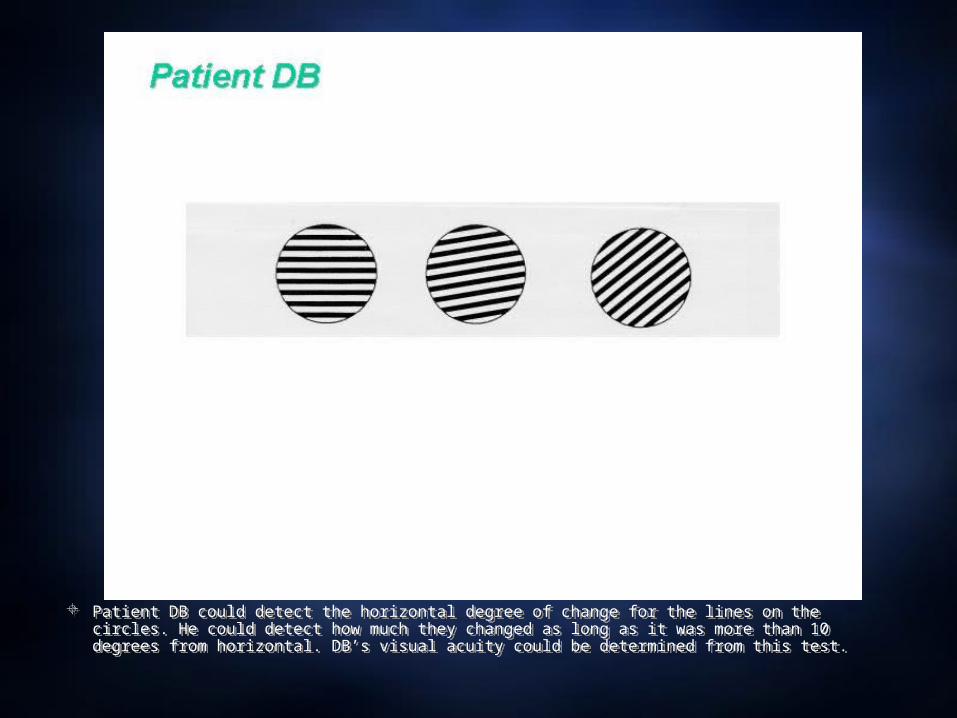
Patient DB could detect the horizontal degree of change for the lines on the circles. He could detect how much they changed as long as it was more than 10 degrees from horizontal. DB’s visual acuity could be determined from this test.
Patient DB could detect the horizontal degree of change for the lines on the circles. He could detect how much they changed as long as it was more than 10 degrees from horizontal. DB’s visual acuity could be determined from this test.
Non-Invasive Methods for Scanning the Living BrainNon-Invasive Methods for Scanning the Living Brain MRI- Magnetic Resonance Imaging
Pros-Functional MRI can detect brain activityCan detect tumors with good accuracyCan detect differences between normal and
diseased similar tissue Cons
ExpensiveMagnetic force could be a problem for implantable
devices PET- Positron Emission Tomography
ProsCan detect brain activity during different tasksCan see how glucose is metabolized in brain
ConsRadioactive injection
MRI- Magnetic Resonance Imaging Pros-
Functional MRI can detect brain activityCan detect tumors with good accuracyCan detect differences between normal and
diseased similar tissue Cons
ExpensiveMagnetic force could be a problem for implantable
devices PET- Positron Emission Tomography
ProsCan detect brain activity during different tasksCan see how glucose is metabolized in brain
ConsRadioactive injection
Non-Invasive Methods for Scanning the Living Brain
Non-Invasive Methods for Scanning the Living Brain
CAT- Computed Axial Tomography Pros-
can help detect intercranial hemorrhages, strokes, and some tumors
Help plan surgerySlides can be rendered into a 3D model
Cons- Cannot detect artery blockage before a stroke,
intercranial pressure, as well as not the best tool for detecting tumors
Contrast can not pick up differences between similar tissues
Ionizing radiation exposureAllergic reaction to iodine contrast agent
fMRI Visual Spatial StudyfMRI Visual Spatial Study
QuickTime™ and aSorenson Video 3 decompressorare needed to see this picture.
QuestionsQuestions 1. Describe some of the early cases of visual agnosia (or mind-blindness). What
is the central deficit of the disorder? 2. Describe the case of DF, what sort of tests reveal the nature of her visual
deficit? 3. What is meant by 'double dissociation' between face and object recognition?
What does it indicate about the neural basis of these abilities? 4. What part of the brain was implicated in Bodamer's early studies of
prosopagnosia? Why wouldn't these cases be consider prosopagnosia today? 5. What is covert recognition? How is it measured and what does it indicate
about the evolutionary origins of processing facial information? 6. What dissociations of movement perception have been revealed by different
types of brain damage? 7. What is blindsight and what part(s) of the brain is (are) implicated in it? 8. Describe the three major non-invasive methods for monitoring on-going brain
activity and their strengths and weakness: CAT; MRI; PET.
1. Describe some of the early cases of visual agnosia (or mind-blindness). What is the central deficit of the disorder?
2. Describe the case of DF, what sort of tests reveal the nature of her visual deficit?
3. What is meant by 'double dissociation' between face and object recognition? What does it indicate about the neural basis of these abilities?
4. What part of the brain was implicated in Bodamer's early studies of prosopagnosia? Why wouldn't these cases be consider prosopagnosia today?
5. What is covert recognition? How is it measured and what does it indicate about the evolutionary origins of processing facial information?
6. What dissociations of movement perception have been revealed by different types of brain damage?
7. What is blindsight and what part(s) of the brain is (are) implicated in it? 8. Describe the three major non-invasive methods for monitoring on-going brain
activity and their strengths and weakness: CAT; MRI; PET.


![Die Herz-Denker. [Leseprobe]](https://img.pdfslide.net/doc/110x75/568c381f1a28ab02359de2eb/die-herz-denker-leseprobe.jpg)
