Embed Size (px)
Citation preview

CHAPTER 13
Principal Components Analysis: A Reviewof its Application on Molecular Dynamics
Data
Sarah A. Mueller Stein,1 Anne E. Loccisano,1 Steven M. Firestine2
and Jeffrey D. Evanseck1
1Contribution from the Center for Computational Sciences and the Department ofChemistry and Biochemistry Duquesne University, 600 Forbes Avenue,
Pittsburgh, PA 15282, USA2Eugene Applebaum College of Pharmacy and Health Science, 3134 EugeneApplebaum Building, Wayne State University, 259 Mack Avenue, Detroit, MI
48201, USA
Contents1. Introduction 233
2. Multivariate methods 235
3. Principal components analysis 236
3.1. Background 236
3.2. Principal components 237
3.3. Covariance matrix 238
3.4. Index of selectivity 239
3.5. Eigenanalysis 239
3.6. Scree test dimensionality determination 240
3.7. Visualization 241
4. Essential dynamics 242
4.1. Background 242
4.2. Applications to protein systems 244
4.3. Applications to nucleic acids 244
5. Related methods 245
5.1. Independent component analysis 245
5.2. Singular value decomposition 245
6. Limitations and common errors 246
7. Conclusion 247
Acknowledgments 247
References 248
1. INTRODUCTION
Molecular dynamics is a proven and powerful tool in the exploration and
study of the structure and dynamics that define biomolecular energy
ANNUAL REPORTS IN COMPUTATIONAL CHEMISTRY, VOLUME 2ISSN: 1574 1400 DOI 10.1016/S1574 1400(06)02013 5
r 2006 Elsevier B.V.All rights reserved

landscapes [1–7]. Technological advances in simulation methodology
[8–14] and computer architecture [15,16] have significantly extended both
the time scale and length (size) scale of molecular dynamics trajectories
[17–22]. Reports in the literature show that the time scale of contemporary
molecular dynamics trajectories, carried out with modest computer re-
sources, have increased by roughly four orders of magnitude since the
inception of biomolecular simulation. A microsecond simulation has been
reported in 2000 [23]; however, after 5 years, this still remains the ex-
ception rather than the rule in computational studies. The length scale of
practical molecular dynamics simulations has not witnessed such a dra-
matic increase, since the urgency for larger systems is not as great as for
longer time. System sizes have increased by nearly 25 times, where sim-
ulations of 25,000 particles are not uncommon.
To put the growth of molecular dynamics simulations into perspective, a
rough analogy to Moore’s Law [24] can be created. Since the first reported
biomolecular simulation over three decades ago, the time scale of re-
ported protein simulations is found to double roughly every 2 years. In
terms of length scale, simulations have nearly doubled in size every 6
years. For example, the first molecular dynamics simulation involving bo-
vine pancreatic trypsin inhibitor (BPTI) was carried out for 9.2 picoseconds
involving approximately 1082 atoms using a united-atom force field in
vacuum [25]. In contrast, it is now fairly routine to simulate models incor-
porating explicit solvent, periodic boundary conditions, and extended
electrostatics with second generation all-atom molecular force fields for
10–100nanoseconds. As an illustration, lysozyme, a small protein of
comparable size to BPTI, has been simulated using a solvated model of
explicit waters for 28 nanoseconds involving over 13,000 atoms [26]. Ex-
amples including nucleic acid simulation show even greater growth, where
a total of 0.6microseconds of simulation for unique tetranucleotide se-
quences of DNA containing �24,000 atoms has been reported [27,28].
It is obvious that the escalation of computing power, resources, and
software development has made it easier to create significantly larger and
more complex sets of data stemming from molecular dynamics simula-
tions. However, analysis of molecular dynamics trajectories has never
been and is currently not trivial. Extracting meaningful information from
even the shortest time simulations is an artform requiring solid chemical
intuition, physical insight, and technical expertise [29,30]. The increased
complexity and size of molecular dynamics trajectories further amplifies an
already difficult situation. As such, computational chemists have been
searching for new computational tools to mine molecular dynamics data for
meaningful information connecting biological function to structure and dy-
namics. The goal of this review is to demonstrate the need for multivariate
S. A. M. Stein et al.234

analysis in biophysical studies, present how principal components analysis
(PCA) can be implemented in the analysis of molecular dynamics data, and
provide insights into the pitfalls and common errors associated with mul-
tivariate techniques.
2. MULTIVARIATE METHODS
Systematic variation of a single variable is usually desired in scientific
study; however, researchers in the biological, chemical, physical, and so-
cial sciences frequently collect measurements on several variables si-
multaneously. This is especially true for molecular dynamics simulations,
where the coordinates and momenta of all atoms are typically sampled
every few femtoseconds over millions of time steps. Within the context of
molecular dynamics simulations, the challenge is to discover the molec-
ular motion(s) responsible for the phenomena of biochemical interest
within the vast range of dynamics ‘‘noise’’ [17,18,29,30]. Some progress
has been made, where localized molecular motion has been linked to
biochemical function as a gateway in acetylcholinesterase [31–33], a
hinged-lid in triose phosphate isomerase [34–38], and combined levers
and gates in carbonmonoxy myoglobin [39]. A database of more than
120molecular motions has been reported [40].
A molecular dynamics trajectory is by definition multivariate data, where
a large number of variables (atomic positions) are typically found to be
interrelated, correlated, or dependent on each other. To decipher these
large data sets, multivariate statistical analysis is one approach that is
gaining popularity. References that present an organized overview of
multivariate methods highlighting their statistical utility and connection
between each of the techniques are available [41–43]. There are also
excellent sources on individual multivariate methods giving an in-depth
mathematical review coupled with illustrative examples and scientific
problems suited for such applications [44,45]. For our purposes, multi-
variate analysis has been applied to molecular dynamics trajectories in
two general ways:
(1) Data reduction or structural simplification. The goal is to reduce the original
large number of dependent variables (atomic coordinates) to a smaller and
independent set to explain the phenomena of interest. Data reduction
through PCA is unique when applied to molecular dynamics trajectories,
since three or less principal components, composed of linear combinations
of the original Cartesian coordinates, are typically identified to clarify im-
portant biomolecular motions.
Principal Components Analysis 235

(2) Sorting, classification, and grouping. The goal is to group or classify objects
based upon measured characteristics. In this specific application of mul-
tivariate analysis, the dimensionality of the data set remains the same. The
data set is simply partitioned into different groups to gain a sense of order
or classification. For instance, cluster analysis has been used to identify
similar geometric or conformational features from molecular dynamics
simulations to further understand complex energy landscapes or design
new drugs in pharmaceutical drug design studies.
The method of analysis depends heavily on whether one is interested in
interrelationships or in comparisons, and on whether variables are qual-
itative or quantitative. In many situations, there will not be a single best
method of analysis. When applied to molecular dynamics trajectories, the
major classifications of multivariate analysis involve PCA [39,46–104],
factor analysis [105,106], discriminant function analysis [107], cluster
analysis [50,107–122], canonical correlation analysis [123–125], and mul-
tidimensional scaling [53,112,113,115,126–130].
A full description for each of these methods is beyond the scope of this
review and may be found in other sources [41–45]. There is some overlap
between a few of the methods where each technique is generally unique
in carrying out either reduction or grouping of multivariate data. However,
one of the most commonly applied techniques to molecular dynamics data
sets is PCA, which will be the focus of this review.
3. PRINCIPAL COMPONENTS ANALYSIS
3.1. Background
Principal components analysis (PCA) is the simplest of multivariate tech-
niques that is used to reduce or simplify large and complicated sets of
data. The PCA procedure was first introduced for only a few variables in
1901 by Karl Pearson [131]. With the advent of computers, PCA was
extended as a practical computing method by Hotelling in 1933 for a
greater number of variables [132]. Since this time, many variations have
been proposed and implemented, such as the essential dynamics method,
which has been extensively used and reported in the recent literature.
However, the underlying mathematical procedure for essential dynamics
remains the same as PCA.
The commonly stated goal of PCA is to reduce the dimensionality of a
multivariate data set by taking p interrelated variables, x1, x2,y, xp, andfinding combinations of these based upon variances to produce a
S. A. M. Stein et al.236

transformed set of variables, z1, z2,y, zp, that are uncorrelated. The in-
dices zi are called the principal components (PCs). Statistically, the point
of PCA is straightforward, but this type of explanation is far from a physical
interpretation that would be meaningful to scientists employing such a
technique.
It is important first to realize that PCA is predicated on the assumption
that the phenomena of interest can be explained by the variances and
covariances between the p variables in the original data set. Unless the
number of variables p is small, it is not possible to examine all of the
variances or the covariances between the variables manually. PCA over-
comes this limitation and transforms the data such that the uncorrelated
variables or principal components are ranked by the variance of the data
set in a single analysis. In terms of molecular dynamics simulations, PCA
ultimately gives a view of the atoms that move anisotropically to maximize
the variance.
3.2. Principal components
Before understanding the mathematical process on how PCA is carried
out, it is instructive to define the principal components. The first principal
component, z1, is simply a linear combination (dot product) of the original
variables x1, x2,y, xp,with a. Note that the mathematical dot product op-
erator takes two vectors and gives a scalar, or a new variable (principal
component) to describe the data.
z1 ¼ aT1x ¼ a11x1 þ a12x2 þ � � � þ a1pxp ¼Xp
j¼1
a1jxj ð1Þ
The weights (a11, a12,y, a1p) are mathematically determined to maximize
the variation of the original data in x, subject to the normalization con-
straint that
a211 þ a212 þ � � � þ a21p ¼ 1 ð2Þ
The constraint is necessary; otherwise, the maximum can simply be in-
creased by increasing any component of aj. To be discussed later, the
weights for a particular component are used to interpret and account for
the variability in the data. Next, the second principal component, z2 ¼ a2Tx,
is determined having a maximum variance that is uncorrelated with z1subject to the same normalization constraint on a2p, and so on, so that the
kth principal component, zk ¼ akTx, has maximum variance subject to
being uncorrelated with z1, z2,y, zk 1. The computed number of principal
Principal Components Analysis 237
components will be same as the number of p original variables. However,
in highly correlated data sets, most of the variation from x will be ac-
counted for in a few principal components. In uncorrelated data sets, PCA
provides no statistical advantage in treating the data. Obviously, it is de-
sirable to have the value of m much less than the value of p to attain a
significant reduction in dimensionality of the data set, where m is the
number of principal components necessary to account for the majority of
the variation in the data set. The lack of correlation between the principal
components is a useful property, since the indices can be interpreted as
different ‘‘dimensions’’ describing the variation in the data set.
3.3. Covariance matrix
The process of determining the principal components starts with the con-
struction of the p� p covariance or correlation matrix from a collection of nsnapshot structures from molecular dynamics trajectories. The structure
matrix x is composed of the Cartesian coordinates for each time stamp
that defines each of the rows. The p columns of x are given by the 3NCartesian coordinates for each atom. It is necessary to transform x to re-
move rotation and translation contamination that does not contribute to the
real dynamics of the system. This is accomplished by aligning the structures
to a common structure. The reference structure for the alignment process
can be an averaged structure, any structure from the trajectory, or an ex-
perimental structure. Many techniques have been reported for comparing
and overlaying proteins for applications other than for PCA [133–149]. The
underlying procedure is essentially the same, where a subset of atoms for
the alignment process is selected, and then alignment is carried out using a
standard root-mean-square-deviation (RMSD) fit on the selected atoms
[150]. In studies involving PCA, it is most common to use the alpha carbons
or all of the non-hydrogen atoms in the alignment process.
Once x has been aligned, it is possible to compute the covariance matrix
elements. The average position oxi4 of the i th atom is computed along
the entire trajectory. The convariance between the i th and j th atoms over
the collection of n structures can be calculated as shown in equation (3).
Each covariance matrix element is determined, as shown in equation (3).
cij ¼1
n
Xn
k¼1
ðxik � xih iÞðxjk � xj� �
Þ i ¼ 1; 2; . . . ;p j ¼ 1;2; . . . ;p ð3Þ
The diagonal of the matrix is simply the variance of each coordinate. The
covariance is the difference between a variable and its mean multiplied by
S. A. M. Stein et al.238

the difference of another variable and its mean. Thus, if variable xi varieslargely from its mean, and variable xj varies largely in the same direction,
then the covariance matrix element, cij, will be large and positive. However,
the covariance matrix element for xi and xj will be small, if either or both
values are close to their corresponding means. With respect to a molecular
system, the covariance matrix element between two atoms will be large
and positive, if each of those atoms deviate largely from their equilibrium
positions and the deviations are in the same direction. Mathematically, the
covariance matrix summarizes the covariance between all variable com-
binations. This matrix is symmetric, so each row and column represents
coordinates from the same structures in the same order, i.e. the kth row
contains the same data points as the kth column.
3.4. Index of selectivity
The index of selectivity is simply the set of atoms identified for analysis.
The index of selectivity is a modification of the possible values of i and j inequation (3). It is often assumed in the vast majority of studies utilizing
PCA that all atoms should be included in the covariance matrix construc-
tion. It is important to realize that selection of all atoms, all non-hydrogen
atoms, or all alpha carbons biases PCA to extract information involving
large-scale global motion. Thus, if localized events are important, and all
atoms are selected in the analysis, then the principal components method
will likely fail to discover the localized motions, forcing an analysis on
motion involving all of the atoms. This problem has been shown for the
understanding of the dynamics of carbonmonoxy myoglobin [92]. When all
of the non-hydrogen atoms were selected for the PCA, isotropic motion
was found, where over 15 dimensions were required to understand the
dynamics. However, when smaller and smaller volumes centered about
the carbon monoxide ligand were used to select a subset of atoms, the
amount of variance was found to be a maximum in two dimensions. Thus,
two amino acids, histidine 64 and arginine 45, were found to be respon-
sible for a majority of the anisotropic motion. The dynamics of the two
residues were found to explain the spectroscopic A-states of carbon-
monoxy myoglobin consistent with available kinetic and mutation data
[151–155]. Consequently, the index of selectivity is an important step in
the proper use of PCA.
3.5. Eigenanalysis
Analysis using PCA simply involves finding the eigenvectors and eigen-
values of the covariance matrix. The computed eigenvalues from the
Principal Components Analysis 239

covariance matrix are the principal component variances. The eigenval-
ues are ordered from the largest to the smallest, so that l1 X l2 XyX lpX 0. Specifically, the kth eigenvalue, lk, indicates the magnitude of the
variance of the data in the direction of the corresponding kth eigenvector.
The resulting eigenvectors provide the coefficients (weights) for the linear
combination of observed structures. These eigenvectors are often re-
ferred to as the ‘‘loadings’’ for the principal components, and referred to as
aj in our previous discussion above. Thus, the linear combination of ob-
served structures, zi ¼ aiTx, is known as the ith principal component.
zi ¼ ai1x1 þ ai2x2 þ � � � þ aipxp ð4Þ
In protein and nucleic acids, the important data variance can be accounted
for by a much smaller number of derived variables (principal components)
than the p variables from which the analysis begins. For example in the
case of nucleic acids, three principal components may account for 85% of
the variance in the data [156]. The first two or three principal components
often account for enough of the variance that important motions of the
protein or nucleic acid can be extracted.
3.6. Scree test dimensionality determination
A key step in PCA is the determination of the number of dimensions to which
the data is reduced. This is most easily accomplished by performing the
scree test, or by creating a scree plot [157,158]. This type of plot involves the
eigenvalues that are determined in the diagonalization of the covariance
matrix. In a scree plot, the x-axis is an index of the number of eigenvalues
determined. The eigenvalues are ordered from the strongest to weakest.
The y-axis gives the magnitude of the eigenvalues from the covariance
matrix diagonalization. It is customary to scale the eigenvalues such that
they sum to unity in order to determine more easily the percent of the
variance of the data accounted for the associated eigenvector. To accom-
plish this, each eigenvalue is divided by the sum of all of the eigenvalues.
To determine the appropriate dimensionality from the resulting analysis, it
is necessary to locate the kink in the scree plot, where the variance rapidly
falls to a relatively stable value. If the data is highly correlated initially, then
the first few dimensions will have large eigenvalues, which indicates that a
great amount of variance is described in those dimensions. The variance
should drop rapidly and form a relatively flat plateau. The correct dimen-
sionality is typically the dimension prior to the eigenvalue reaching the pla-
teau. The interpretation is such that adding the extra dimension does not
S. A. M. Stein et al.240
result in any appreciable gain in information (variance) on the system, as
compared to the complexity of adding an additional dimension.
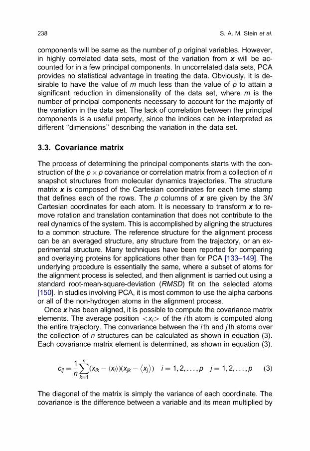
Two example scree plots from our earlier work on carbonmonoxy my-
oglobin are given below [92]. From both plots, it is possible to identify the
associated problems with the improper use of the index of selectivity, as
commonly assumed (Fig. 1).
When all atoms are used in the PCA, a well-defined kink is never re-
alized. The relative inertia monotonically decreases as the dimension in-
creases. A scree plot with this type of signature indicates that the variance
of the molecular system cannot be consolidated under a few dimensions.
In using such an index of selectivity, it would be impossible to determine
the correct dimensionality for further analysis. However, in Fig. 1b, where
a subset of atoms is used to define the index of selectivity, it is clear that a
significant portion of the system’s variance is captured in the first two
dimensions. In fact, approximately 70% of the information is found in the
first two principal components. In this specific case, the third dimension
delivers additional 10% of information; however, no useful data was found
upon examination. The two plots illustrate the problem associated with
assuming that all of the atoms should be used in the index of selectivity.
3.7. Visualization
The next step is visualization of the data using the dimensionality deter-
mined from the previous step. Projections of the original structures onto
the weights (eigenvectors) of the associated principal components are
plotted against each other as the scree plot dictates. Consequently, if Mstructures are collected and used in the covariance matrix construction,
Fig. 1. Scree plots using indices of selectivity that include (a) all heavyatoms and (b) His64, the CO ligand and the heme.
Principal Components Analysis 241

then M data points will be realized on the plots. The principal components
plots give information on the similarity of the M structures used to form the
covariance matrix. As an example, the two-dimensional plot correspond-
ing to 1045 structures and scree plot in Fig. 1b is given below.
Each point on the plot corresponds to a structure and its relation to all
other structures in the dimension(s) plotted. If two points are close to each
other on the plot, then those structures are similar. If two points on such a
plot are far from each other, then they are dissimilar in some fashion. It is
clear that this specific plot yields three general basins of structural sim-
ilarity. This behavior is consistent with the current ideas of energy land-
scapes, where multiple minima are clustered into regions that are
separated by higher energy barriers [159]. It is at this juncture that the
origin of dissimilarity between the three energy basins cannot be derived
from the principal components plots alone. All that is known is that the
structures are different given the index of selectivity utilized. In this specific
instance, the difference was determined to be a result of the structural
change of histidine 64, since that was the primary constituent of the index
of selectivity. More traditional methods such as constructing the RMSDbetween the two structures from the different energy basins as a function
of its sequence can usually pinpoint the molecular reasons of conformat-
ional differences. It is through the pair wise comparison of structures from
the different basins that a molecular interpretation may be formulated in
describing the different conformations sampled by the molecular system.
A major result of the Schulze and Evanseck study was that histidine 64
moved in from the solvent to the ligand by �10 A on a timescale consistent
with experiment [151], as shown in Fig. 2b.
When more than three dimensions are indicated by the scree plot, it is
possible to examine the variance at the higher dimensions. As an exam-
ple, when the first and second principal components are determined (us-
ing the scree test) to describe large amounts of variance, and when the
third and fourth are very close together in magnitude, two separate plots
may be created to help characterize molecular motion. The first plot may
have the first, second, and third principal components as the axes, and the
second plot would use first, second, and fourth as the axes.
4. ESSENTIAL DYNAMICS
4.1. Background
Certain types of internal motions allow proteins to perform their biological
functions. These motions may enable the binding of substrates, adaptation
S. A. M. Stein et al.242

to various environments, or conformational changes that allow binding of a
substrate at another site on the protein (allosteric effects). The internal
motions may be subtle and can involve complicated correlations between
atomic motions, and thus the challenge presented is to identify these mo-
tions, determine how they relate to protein function, and to separate the
complicated dynamics from the essential degrees of freedom
[102,160,161]. Amadei and coworkers first developed the method known
as essential dynamics in order to separate the concerted structural rear-
rangement from irrelevant motions [161]. Their method is based on the
hypothesis that by using PCA, atomic positional fluctuations can be used to
separate a protein’s conformational space into two subspaces: an ‘‘essen-
tial’’ subspace which contains only a few degrees of freedom that describe
the motions relevant for protein function (e.g., opening and closing and
hinge bending motions) and the remaining subspace (‘‘constrained sub-
space’’) that describes the irrelevant local fluctuations of the protein. This
group used lysozyme as their test case of the essential dynamics method,
and they concluded that the essential dynamics of most proteins can be
described in a subspace of only a few degrees of freedom, while all other
degrees of freedom represent much less important and mostly independent
fluctuations of the molecule.
The essential dynamics method involves the use of a covariance matrix
constructed from structures sampled throughout a molecular dynamics
simulation. By diagonalization of a covariance matrix of the atomic
Fig. 2. (a) The two dimensional principal component plot of carbon-monoxy myoglobin using the coordinates of His64, the ligand and heme.The different symbols indicate different starting conditions of the multipleshort time trajectories used to form the ensemble of structures. (b) Vectorsshowing the conformational change extracted from Fig. 2a.
Principal Components Analysis 243
coordinates of the system, the motions of a structure that are responsible
for the most variance in atomic position are targeted. The essential sub-
space is determined by ranking the eigenvalues elucidated by PCA, of the
covariance matrix from the molecular dynamics trajectory. The mathe-
matical equivalence between PCA and essential dynamics has been
noted before [88,162–168] and has described within this document.
4.2. Applications to protein systems
Many authors of protein simulation studies have used the essential dy-
namics method in order to identify important molecular vibrations to un-
derstand more about large correlated protein motions and how they are
critical to biological function. PCA has been used in a wide array of ap-
plications ranging from crystallographic and NMR structure ensembles
[63,64,73,80,169–177], protein and peptide folding/unfolding
[47,54,66,67,99,167,178–184], structural determinants of transmembrane
proteins and channels [49,51,55,69,90,101,185–191], large-scale domain
motion [58,77,98,104,192–199], locally accessible conformational sub-
states [52,60,92,96,97,103,200], correlated and functional motion
[39,56,57,61,71,84,163,166,168,201–215], dynamic effects from muta-
tions and domain swapping [216–219], mutation impact upon binding
[220–223], connection between structural similarity and dynamic behavior
[87,89,164,224], ligand binding and migration [53,74,82,225–233], con-
formations of small molecules [72,79,91], protonation effects on dynamics
[234], liquid behavior and spectroscopy [48,75,76], testing and develop-
ment of methodology [46,59,62,65,78,83,85,86,235], protein docking al-
gorithms [68,70,236–238], homology modeling [100], and atomic and
molecular properties [50,95].
4.3. Applications to nucleic acids
PCA has been shown to be a powerful tool in evaluation of DNA flexibility
in molecular dynamics simulations [162,239–241]. This technique has
been employed to examine nucleic acid flexibility [239,240,242], flexibility
of hybrid nucleic acids [243], flexibility of DNA in the crystal environment
[240], behavior of A-tract DNA [93], electrostatic interactions of nucleic
acids [83,156], sequence effects [27,28], DNA containing chemical mod-
ifications [81,244–246], broken strand DNA [247], base flipping [248], and
nucleic acid mispairs [242]. The potential energy surface of nucleic acid
conformational changes have also been investigated using PCA [156]. As
new techniques in molecular dynamics simulations emerge, PCA has
been used to evaluate the quality of simulations [249–251].
S. A. M. Stein et al.244

5. RELATED METHODS
5.1. Independent component analysis
Independent component analysis (ICA) is a multivariate technique that is
used to separate independent variables in a data set [252,253]. Unlike
PCA, ICA is not typically used as a dimensionality reduction technique. In
ICA, the data must be fit to a model (not necessarily a linear model) [254]
in which the derived variables are as statistically independent as possible.
In chemical applications, it is generally favorable if the number of derived
variables (principal components, latent variables) is much smaller than the
number of original observations collected. Therefore, a data reduction
technique such as PCA may be performed before standard ICA is carried
out [252,255]. Thus, the focus of ICA is on the subspace accounting for
the most variance in the data set when the analysis begins [252]. Westad
and Kermit investigated validation methods for ICA and found that cross-
validation was a valuable tool for determining the number of principal
components to use from the preliminary reduction step and the number of
ICs to extract in the actual ICA [256]. Yadava and Chaudhary applied ICA
to determine analyte solvation parameters on polymer-coated surface
acoustic wave vapor sensors [255]. ICA was employed because PCA did
not yield derived variables that were interpretable for this particular type of
experiment. ICA has been used in analysis of spectroscopic data
[257,258]. Medical image processing is also an area in which ICA has
been used [259]. Other uses of ICA include analysis of natural systems
such as seismological and atmospheric data [260] and atmospheric aer-
osol content [261].
5.2. Singular value decomposition
The singular value decomposition (SVD) technique was established by
several mathematicians who worked independently to develop the theory
leading to the efficient diagonalization of a matrix [262]. Although SVD has
many uses, it is commonly used to extract eigenvalues from a symmetric
matrix [263]. As such, the technique has been used in PCA [94,264,265].
SVD may be used as a tool to execute PCA on a variety of systems
including NMR spectroscopy [194,266] and X-ray photoelectron spectro-
microscopy [265]. Andrews and coworkers used the SVD algorithm to
perform PCA on myoglobin. In their work, SVD was chosen because it is
computationally efficient. SVD was carried out on the internal coordinates
of the myoglobin, which gave similar results as the SVD of the Cartesian
coordinates [94]. Tomfohr and coworkers used SVD to diagonalize a ma-
Principal Components Analysis 245

trix for dimensionality reduction of gene expression data [264]. SVD may
also be used to carry out Gaussian network model analysis [267].
6. LIMITATIONS AND COMMON ERRORS
Multivariate techniques can be very powerful in data analysis. However,
there are only a few papers that critically examine the possible weak-
nesses of multivariate analyses [102,268]. When using these statistical
tools on molecular dynamics simulations, one should realize that there
exist potential sources of error that could bias the analysis and provide
misleading or wrong interpretations of the data.
The first and most important source of error deals with the well-
documented sampling issues with molecular dynamics simulations
[8,9,11,13,39]. The goal of applying PCA to molecular dynamics trajec-
tories is to extract and understand the dynamics of the system. Conse-
quently, if the trajectory samples only a portion of available structures from
the true ensemble, then PCA will extract and provide information on the
incomplete representation of phase space. Multivariate analysis will
not create data to correct problems with the generation of the original
data set.
Secondly, the index of selectivity is crucial to a successful PCA, which is
often overlooked in a majority of studies utilizing dimensionality reduction
and molecular dynamics. Care needs to be exercised in atom selection,
where all atoms, all nonhydrogen atoms, or all alpha carbons of proteins
are typically used for analysis. Selection based upon all atoms is correct,
as long as low-frequency, large-scale motions are desired. However, it
should be clear that molecular motion need not be large scale. As men-
tioned before, many well-understood examples show that local-motion is
connected with function, as gateways [31–33], hinged-lids [34–38], and
combined levers and gates [39]. Therefore, important motions could be
localized and only a subset of the atoms is needed within the range of
molecular motions. In carbonmonoxy myoglobin, it was necessary to
modify the index of selectivity, based upon previous knowledge of the
binding site, in order to discover the local motion responsible for the
spectroscopic A-states [39]. Indices of selectivity can bias multivariate
analysis, where it is necessary to have a course idea of the type of dy-
namics of interest, i.e., local or global motion, in molecular dynamics
simulations.
Lastly, when working with PCA, it is essential to bear in mind that the
major assumption is that the sources of largest variance are of importance
to the problem being addressed. However, caution needs to be exercised
S. A. M. Stein et al.246

in mixed data sets that involve more than the atomic coordinates from
molecular dynamics trajectories. For example, differences in the units
could be involved, where the original data may be composed of differently
measured characteristics. For example, the variation in angstroms in
atomic position is obviously different than the variation of pH or temper-
ature. Even when the same units are used, it is plausible that one meas-
ured quantity may have a completely different range of behavior
compared to another. Consider the variation in covalent bond length ver-
sus the variation in intermolecular hydrogen bonding. When variables with
large variance are compared with variables of small variance, those with
larger associated variance will be weighted more heavily in construction of
the principal components. This weighting is simply due to the fact that the
goal in constructing the principal components is to maximize variance. In
cases with variables with widely ranging variance, using a covariance
matrix of standardized variables, or correlation matrix to determine the
principal components may help to alleviate this issue [45].
7. CONCLUSION
The continued advances in readily available computer power coupled with
the desire to explore dynamics at longer time scales means that the
magnitude and complexity of accessible dynamics data will keep growing.
By necessity, methods to reduce the size of this data will continue to be
valued by computational chemists. In this review, we have sought to
highlight the utility of PCA to reduce the complexity of variables describing
the dynamics data. PCA and the mathematically identical essential dy-
namics, have proved useful in the detection of important motions in bio-
molecules ranging from proteins to nucleic acids. Provided that
appropriate care is taken with the use of these methods, computational
chemists should find PCA useful in managing large, complex data sets
and discovering molecular motions that are biochemically relevant.
ACKNOWLEDGMENTS
This work was funded in part by the National Science Foundation (CHE-
0321147, CHE-0354052, AAB/PSC CHE-030008P), Department of Edu-
cation (P116Z040100 and P116Z050331), SGI and Clarix Corporations,
the National Institutes of Health (GM069549-01), and the Center for
Computational Sciences at Duquesne University.
Principal Components Analysis 247
REFERENCES
[1] M. Karplus and J. Kuriyan, Molecular dynamics and protein function, Proc. Natl.Acad. Sci., 2005, 102, 6679–6685.
[2] T. Hansson, C. Oostenbrink and W. F. van Gunsteren, Molecular dynamics sim-ulations, Curr. Opin. Struc. Biol., 2002, 12, 190–196.
[3] M. Karplus, Molecular dynamics simulations of biomolecules, Acc. Chem. Res.,2002, 35, 321–323.
[4] M. Karplus and J. A. McCammon, Molecular dynamics simulations of biomolecules,Nat. Struct. Biol., 2002, 9, 646–652.
[5] W. Wang, O. Donini, C. M. Reyes and P. A. Kollman, Biomolecular simulations:Recent developments in force fields, simulations of enzyme catalysis, protein-ligand,protein-protein, and protein-nucleic acid noncovalent interactions. Annu. Rev.Biophys. Biomol. Struct., 2001, 30, 211–243.
[6] M. Karplus and G. A. Petsko, Molecular dynamics simulations in biology, Nature,1990, 347, 631–639.
[7] M. Karplus, Molecular dynamics simulations of proteins, Phys. Today, 1987, 40,68–70.
[8] X. Cheng, G. Cui, V. Hornak and C. Simmerling, Modified replica exchange sim-ulation methods for local structure refinement, J. Phys. Chem. B, 2005, 109,8220–8230.
[9] A. E. Loccisano, O. Acevedo, J. DeChancie, B. G. Schulze and J. D. Evanseck,Enhanced sampling by multiple molecular dynamics trajectories: carbonmonoxymyoglobin 10 microsecond A0 4 A1-3 transition from ten 400 picosecond simu-lations, J. Mol. Graph. Model, 2004, 22, 369–376.
[10] P. Minary, M. E. Tuckerman and G. T. Martyna, Long time molecular dynamics forenhanced conformational sampling in biomolecular systems, Phys. Rev. Lett., 2004,93, 1520201/1–1520201/4.
[11] I. Andricioaei, A. R. Dinner and M. Karplus, Self-guided enhanced sampling meth-ods for thermodynamic averages, J. Chem. Phys., 2003, 118, 1074–1084.
[12] T. Schlick, Molecular Modeling and Simulation, Springer, New York, 2002.[13] Z. Zhu, M. E. Tuckerman, S. O. Samuelson and G. T. Martyna, Using novel variable
transformations to enhance conformational sampling in molecular dynamics, Phys.Rev. Lett., 2002, 88, 100201/1–100201/4.
[14] H. Grubmuller, Predicting slow structural transitions in macromolecular systems:Conformational Flooding, Phys. Rev. E, 1995, 52, 2893–2906.
[15] G. Bhanota, D. Chen, A. Gara and P. Vranas, The BlueGene/L supercomputer,Nucl. Phys. B (Proc. Suppl.), 2003, 119, 114–121.
[16] F. Bodin, P. Boucaud, N. Cabibbo, G. Cascino, F. Calvayrac, M. Della Morte, A. DelRe, R. De Pietri, P. Deriso and F. Di Carlo, APE computers – past, present andfuture, Comput. Phys. Commun., 2002, 147, 402–409.
[17] A. H. Zewail, Femtochemistry, Atomic-scale dynamics of the chemical bond usingultrafast lasers Nobel lecture. In Les Prix Nobel (ed. T. Frangsmyr), Almqvist andWiksell International, Stockholm, 2000, pp. 110–203.
[18] R. M. Hochstrasser, Ultrafast spectroscopy of protein dynamics, J. Chem. Educ.,1998, 75, 559–564.
[19] V. Reat, H. Patzelt, M. Ferrand, C. Pfister, D. Oesterhelt and G. Zaccai, Dynamics ofdifferent functional parts of bacteriorhodopsin: H-2 H labeling and neutron scatter-ing, Proc. Natl. Acad. Sci., 1998, 95, 4970–4975.
[20] M. Ben-Nun, J. Cao and K. R. Wilson, Ultrafast X-ray and electron diffraction: The-oretical considerations, J. Phys. Chem. A, 1997, 101, 8743–8761.
[21] E. Chen, R. A. Goldbeck and D. S. Kliger, Nanosecond time-resolved spectroscopyof biomolecular processes, Annu. Rev. Biophys. Biomol. Struct., 1997, 26, 327–355.
S. A. M. Stein et al.248

[22] T. Schlick, E. Barth and M. Mandziuk, Biomolecular dynamics at long timesteps:Bridging the timescale gap between simulation and experimentation, Annu. Rev.Biophys. Biomol. Struct., 1997, 26, 181–222.
[23] Y. Duan and P. A. Kollman, Pathways to a protein folding intermediate observed in a1-microsecond simulation in aqueous solution, Science, 1998, 282, 740–744.
[24] G. E. Moore, Cramming more components onto integrated circuits, Electronics,1965, 38, 114–117.
[25] J. A. McCammon, B. R. Gelin and M. Karplus, Dynamics of folded proteins, Nature,1977, 267, 585–590.
[26] M. Marchi, F. Sterpone and M. Ceccarelli, Water rotational relaxation and diffusion inhydrated lysozyme, J. Am. Chem. Soc., 2002, 124, 6787–6791.
[27] S. B. Dixit, D. L. Beveridge, D. A. Case, T. E. Cheatham III, E. Giudice, F. Lankas, R.Lavery, J. H. Maddocks, R. Osman, H. Sklenar, K. M. Thayer and P. Varnai, Mo-lecular dynamics simulations of the 136 unique tetranucleotide sequences of DNAoligonucleotides. II: Sequence context effects on the dynamical structures of the 10unique dinucleotide steps, Biophys. J., 2005, 89, 3721–3740.
[28] D. L. Beveridge, G. Barreiro, K. S. Byun, D. A. Case, T. E. Cheatham III, S. B. Dixit,E. Giudice, F. Lankas, R. Lavery, J. H. Maddocks, R. Osman, E. Seibert, H. Sklenar,G. Stoll, K. M. Thayer, P. Varnai and M. A. Young, Molecular dynamics simulationsof the 136 unique tetranucleotide sequences of DNA oligonucleotides, I. Researchdesign and results on d(CpG) steps, Biophys. J., 2004, 87, 3799–3813.
[29] C. L. Brooks, III, M. Karplus and B. M. Pettitt, Proteins: A Theoretical Perspective ofDynamics, Structure, and Thermodynamics, Wiley, New York, 1988.
[30] J. A. McCammon and S. C. Harvey, Dynamics of Proteins and Nucleic Acids,Cambridge University Press, Cambridge, 1988.
[31] T. Y. Shen, T. Kaihsu and J. A. McCammon, Statistical analysis of the fractal gatingmotions of the enzyme acetylcholinesterase, Phys. Rev. E, 2001, 63, 041902/1–041902/6.
[32] N. A. Baker and J. A. McCammon, Non-Boltzmann rate distributions in stochasticallygated reactions, J. Phys. Chem. B, 1999, 103, 615–617.
[33] H.-X. Zhou, S. T. Wlodek and J. A. McCammon, Conformation gating as a mech-anism for enzyme specificity, Proc. Natl. Acad. Sci., 1998, 95, 9280–9283.
[34] J. Sun and N. S. Sampson, Understanding protein lids: Kinetic analysis of activehinge mutants in triosephosphate isomerase, Biochemistry, 1999, 38, 11474–11481.
[35] P. Derreumaux and T. Schlick, The loop opening/closing motion of the enzymetriosephosphate isomerase, Biophys. J., 1998, 74, 72–81.
[36] K. Yuksel, A. Sun, R. Gracy and K. Schnackerz, The hinged lid of yeast triose-phosphate isomerase. Determination of the energy barrier between the two con-formations, J. Biol. Chem., 1994, 269, 5005–5008.
[37] N. S. Sampson and J. R. Knowles, Segmental motion in catalysis: Investigation of ahydrogen bond critical for loop closure in the reaction of triosephosphate isomerase,Biochemistry, 1992, 31, 8488–8494.
[38] D. Joseph, G. A. Petsko and M. Karplus, Anatomy of a conformational change:Hinged ‘‘lid’’ motion of the triosephosphate isomerase loop, Science, 1990, 249,1425–1428.
[39] B. G. Schulze, H. Grubmuller and J. D. Evanseck, Functional significance of hier-archical tiers in carbonmonoxy myoglobin: Conformational substates and transitionsstudied by conformational flooding simulations, J. Am. Chem. Soc., 2000, 122,8700–8711.
[40] M. Gerstein and W. Krebs, A database of macromolecular motions, Nucleic AcidsRes, 1998, 26, 4280–4290.
[41] L. G. Grimm and P. R. Yarnold, Reading and Understanding Multivariate Statistics,American Psychological Association, Washington, DC, 1998.
Principal Components Analysis 249

[42] B. F. J. Manly, Multivariate Statistical Methods: A Primer, Chapman and Hall, Lon-don, 1994.
[43] R. A. Johnson and D. W. Wichern, Applied Multivariate Statistical Analysis, PrenticeHall, Upper Saddle River, 1992.
[44] I. T. Jolliffe, In Principal Component Analysis, 2nd ed, Springer, New York, 2002.[45] G. H. Dunteman, In: M.S. Lewis-Beck (Ed.), Principal Components Analysis, 1st Ed.,
vol. 69, Sage, Newbury Park, 1989, p. 96–97.[46] C. P. Barrett and M. E. M. Noble, Dynamite extended: Two new services to simplify
protein dynamic analysis, Bioinformatics, 2005, 21, 3174–3175.[47] C. Chen, Y. Xiao and L. Zhang, A directed essential dynamics simulation of peptide
folding, Biophys. J., 2005, 88, 3276–3285.[48] M. D’Abramo, M. D’Alessandro, A. Di Nola, D. Roccatano and A. Amadei, Char-
acterization of liquid behavior by means of local density fluctuations, J. Mol. Liq.,2005, 117, 17–21.
[49] S. Haider, A. Grottesi, B. A. Hall, F. M. Ashcroft and M. S. P. Sansom, Confor-mational dynamics of the ligand-binding domain of inward rectifier K channels asrevealed by molecular dynamics simulations: Toward an understanding of Kir chan-nel gating, Biophys. J., 2005, 88, 3310–3320.
[50] O. Horovitz and C. Sarbu, Characterization and classification of lanthanides bymultivariate analysis methods, J. Chem. Ed., 2005, 82, 473–483.
[51] A. Hung, K. Tai and M. S. P. Sansom, Molecular dynamics simulation of the M2helices within the nicotinic acetylcholine receptor transmembrane domain: Structureand collective motions, Biophys. J., 2005, 88, 3321–3333.
[52] A. Leo-Macias, P. Lopez-Romero, D. Lupyan, D. Zerbino and A. R. Ortiz, An anal-ysis of core deformations in protein superfamilies, Biophys. J., 2005, 88, 1291–1299.
[53] Y. Li, Z. Zhou and C. B. Post, Dissociation of an antiviral compound from the internalpocket of human rhinovirus 14 capsid, Proc. Natl. Acad. Sci., 2005, 102, 7529–7534.
[54] J. T. MacDonald, A. G. Purkiss, M. A. Smith, P. Evans, J. M. Goodfellow and C.Slingsby, Unfolding crystallins: The destabilizing role of a b-hairpin cysteine in bB2-crystallin by simulation and experiment, Protein Sci, 2005, 14, 1282–1292.
[55] S. Oyama, Jr., P. Pristovsek, L. Franzoni, A. Pertinhez Thelma, E. Schinina, C.Lucke, H. Ruterjans, C. Arantes Eliane and A. Spisni, Probing the pH-dependentstructural features of a-KTx12.1, a potassium channel blocker from the scorpionTityus serrulatus, Protein Sci, 2005, 14, 1025–1038.
[56] P. W. Pan, R. J. Dickson, H. L. Gordon, S. M. Rothstein and S. Tanaka, Functionallyrelevant protein motions: Extracting basin-specific collective coordinates from mo-lecular dynamics trajectories, J. Chem. Phys., 2005, 122, 034904.
[57] G. R. Smith, M. J. Sternberg and P. A. Bates, The relationship between the flexibilityof proteins and their conformational states on forming protein-protein complexeswith an application to protein-protein docking, J. Mol. Biol., 2005, 347, 1077–1101.
[58] Z. Zhou, M. Madrid, J. D. Evanseck and J. D. Madura, Effect of a bound non-nucleoside RT inhibitor on the dynamics of wild-type and mutant HIV-1 reversetranscriptase, J. Am. Chem. Soc., 2005, 127, 17253–17260.
[59] L. Afzelius, F. Raubacher, A. Karlen, F. S. Jorgensen, T. B. Andersson, C. M.Masimirembwa and I. Zamora, Structural analysis of CYP2C9 and CYP2C5 and anevaluation of commonly used molecular modeling techniques, Drug Metab. Dispos.,2004, 32, 1218–1229.
[60] B. Alakent, P. Doruker and M. C. Camurdan, Application of time series analysis onmolecular dynamics simulations of proteins: A study of different conformationalspaces by principal component analysis, J. Chem. Phys., 2004, 121, 4759–4769.
[61] B. Alakent, P. Doruker and M. C. Camurdan, Time series analysis of collectivemotions in proteins, J. Chem. Phys., 2004, 120, 1072–1088.
[62] C. P. Barrett, B. A. Hall and M. E. M. Noble, Dynamite: A simple way to gain insightinto protein motions, Acta Cryst. D., 2004, 60, 2280–2287.
S. A. M. Stein et al.250

[63] F. Corzana, S. Motawia Mohammed, C. Herve du Penhoat, F. van den Berg, A.Blennow, S. Perez and B. Engelsen Soren, Hydration of the amylopectin branchpoint. Evidence of restricted conformational diversity of the a-(1 46) linkage, J.Am. Chem. Soc., 2004, 126, 13144–13155.
[64] E. G. Emberly, R. Mukhopadhyay, C. Tang and N. S. Wingreen, Flexibility of b-sheets: Principal component analysis of database protein structures, Proteins, 2004,55, 91–98.
[65] D. Flock, I. Daidone and A. Di Nola, A molecular dynamics study of acylphosphatasein aggregation-promoting conditions: The influence of trifluoroethanol/water solvent,Biopolymers, 2004, 75, 491–496.
[66] N. J. Marianayagam and S. E. Jackson, The folding pathway of ubiquitin from all-atom molecular dynamics simulations, Biophys. Chem., 2004, 111, 159–171.
[67] A. Palazoglu, A. Gursoy, Y. Arkun and B. Erman, Folding dynamics of proteins fromdenatured to native state: Principal component analysis, J. Comp. Biol., 2004, 11,1149–1168.
[68] R. Tatsumi, Y. Fukunishi and H. K. Nakamura, A hybrid method of molecular dy-namics and harmonic dynamics for docking of flexible ligand to flexible receptor, J.Comp. Chem., 2004, 25, 1995–2005.
[69] Y. S. Watanabe, Y. Fukunishi and H. K. Nakamura, Modelling of third cytoplasmicloop of bovine rhodopsin by multicanonical molecular dynamics, J. Mol. Graph.Model, 2004, 23, 59–68.
[70] M. Zacharias, Rapid protein–ligand docking using soft modes from molecular dy-namics simulations to account for protein deformability: Binding of FK506 to FKBP,Proteins, 2004, 54, 759–767.
[71] G. Chillemi, P. Fiorani, P. Benedetti and A. Desideri, Protein concerted motions inthe DNA-human topoisomerase I complex, Nucleic Acids Res., 2003, 31,1525–1535.
[72] X. Fradera, M. Marquez, B. D. Smith, M. Orozco and F. J. Luque, Molecular dy-namics study of [2]rotaxanes: Influence of solvation and cation on co-conformation,J. Org. Chem., 2003, 68, 4663–4673.
[73] J.-C. Hus, W. Peti, C. Griesinger and R. Bruschweiler, Self-consistency analysis ofdipolar couplings in multiple alignments of ubiquitin, J. Am. Chem. Soc., 2003, 125,5596–5597.
[74] A. Nijnik, R. Mott, D. P. Kwiatkowski and I. R. Udalova, Comparing the finespecificity of DNA binding by NF-kB p50 and p52 using principal coordinates anal-ysis, Nucleic Acids Res., 2003, 31, 1497–1501.
[75] R. A. Wheeler and H. Dong, Optimal spectrum estimation in statistical mechanics,ChemPhysChem, 2003, 4, 1227–1230.
[76] R. A. Wheeler, H. Dong and S. E. Boesch, Quasiharmonic vibrations of water, waterdimer, and liquid water from principal component analysis of quantum and QM/MMtrajectories, ChemPhysChem, 2003, 4, 382–384.
[77] N. P. Barton, C. S. Verma and L. S. D. Caves, Inherent flexibility of calmodulindomains: A normal-mode analysis study, J. Phys. Chem. B., 2002, 106,11036–11040.
[78] L. S. D. Caves and C. S. Verma, Congruent qualitative behavior of complete andreconstructed phase space trajectories from biomolecular dynamics simulation,Proteins Struct. Funct. Genet., 2002, 47, 25–30.
[79] M. D’Alessandro, A. Tenenbaum and A. Amadei, Coherent dynamics in a butanemolecule, Phys. Rev. E, 2002, 66, 020901/1–020901/4.
[80] R. Dvorsky, V. Hornak, J. Sevcik, G. P. Tyrrell, L. S. D. Caves and C. S. Verma,Dynamics of RNase Sa: A simulation perspective complementary to NMR/X-ray, J.Phys. Chem. B, 2002, 106, 6038–6048.
[81] H. Ishida, Molecular dynamics simulation of 7,8-dihydro-8-oxoguanine DNA, J.Biomol. Struct. Dyn., 2002, 19, 839–851.
Principal Components Analysis 251
[82] M. J. Millan, L. Maiofiss, D. Cussac, V. Audinot, J. A. Boutin and A. Newman-Tancredi, Differential actions of anti-Parkinson agents at multiple classes of mon-oaminergic receptor. 1. A multivariate analysis of the binding profiles of 14 drugs at21 native and cloned human receptor subtypes, J. Pharm. Exp. Ther., 2002, 303,791–804.
[83] M. Nina and T. Simonson, Molecular dynamics of the tRNA Ala acceptor stem:Comparison between continuum reaction field and Particle-Mesh Ewald electrostatictreatments, J. Phys. Chem. B., 2002, 106, 3696–3705.
[84] J. T. A. Saarala, K. Tuppurainen, M. Perakyla, H. Santa and Laatikainen, Correlativemotions and memory effects in molecular dynamics simulations of molecules: Prin-cipal components and rescaled range analysis suggest that the motions of nativeBPTI are more correlated than those of its mutants, Biophys. Chem., 2002, 95,49–57.
[85] N. Ota and D. A. Agard, Enzyme specificity under dynamic control II. Principalcomponents analysis of a-lytic protease using global and local solvent boundaryconditions,, Protein Sci., 2001, 10, 1403–1414.
[86] R. Dvorsky, J. Sevcik, L. S. D. Caves, R. E. Hubbard and C. S. Verma, Temperatureeffects on protein motions: A molecular dynamics study of RNase-Sa, J. Phys.Chem. B, 2000, 104, 10387–10397.
[87] A. Giuliani, R. Benigni, P. Sirabella, J. P. Zbilut and A. Colosimo, Nonlinear methodsin the analysis of protein sequences: A case study in rubredoxins, Biophys. J., 2000,78, 136–148.
[88] B. Hess, Similarities between principal components of protein dynamics and randomdiffusion, Phys. Rev. E, 2000, 62, 8438–8448.
[89] M. A. Ceruso, A. Amadei and A. Di Nola, Mechanics and dynamics of B1 domain ofProtein G: Role of packing and surface hydrophobic residues, Protein Sci., 1999, 8,147–160.
[90] J. M. Koshi and W. J. Bruno, Major structural determinants of transmembrane pro-teins identified by principal components analysis, Proteins, 1999, 34, 333–340.
[91] H. Lanig, M. Gottschalk, S. Schneider and T. W. Clark, Conformational analysis oftetracycline using molecular mechanical and semiempirical MO-calculations, J. Mol.Mod., 1999, 5, 46–62.
[92] B. G. Schulze and J. D. Evanseck, Cooperative role of Arg45 and His64 in thespectroscopic A3 state of carbonmonoxy myoglobin: Molecular dynamics simula-tions, multivariate anlaysis and quantum mechanical computations, J. Am. Chem.Soc., 1999, 121, 6444–6454.
[93] E. C. Sherer, S. A. Harris, R. Soliva, M. Orozco and C. A. Laughton, Moleculardynamics studies of DNA A-tract structure and flexibility, J. Am. Chem. Soc., 1999,121, 5981–5991.
[94] B. K. Andrews, T. Romo, J. B. Clarage, B. M. Pettitt and G. N. Phillips, Jr., Char-acterizing global substates of myoglobin, Structure, 1998, 6, 587–594.
[95] E. Bolzacchini, V. Consonni, R. Lucini, M. Orlandi and B. Rindone, High-perform-ance size-exclusion chromatographic behavior of substituted benzoylpoly L-lysinesby principal component analysis and molecular dynamics simulations, J. Chroma-togr. A, 1998, 813, 255–265.
[96] L. S. D. Caves, J. D. Evanseck and M. Karplus, Locally accessible conformations ofproteins: Multiple molecular dynamics simulations of crambin, Protein Sci., 1998, 7,649–666.
[97] R. Laatikainen, J. T. A. Saarala, K. Tuppurainen and T. Hassinen, Internal motionsof native lysozyme are more organized than those of mutants: A principal compo-nent analysis of molecular dynamics data, Biophys. Chem., 1998, 73, 1–5.
[98] S. Hayward, A. Kitao and H. J. C. Berendsen, Model-free methods of analyzingdomain motions in proteins from simulation: A comparison of normal mode analysisand molecular dynamics simulation of lysozyme, Proteins, 1997, 27, 425–437.
S. A. M. Stein et al.252

[99] T. Lazaridis, I. Lee and M. Karplus, Dynamics and unfolding pathways of a hype-rthermophilic and a mesophilic rubredoxin, Protein Sci, 1997, 6, 2589–2605.
[100] K. Ogata and H. Umeyama, Prediction of protein side-chain conformations by prin-cipal component analysis for fixed main-chain atoms, Protein Eng., 1997, 10,353–359.
[101] S. T. Wlodek, T. W. Clark, L. R. Scott and J. A. McCammon, Molecular dynamics ofacetylcholinase dimer complexed wtih tacrine, J. Am. Chem. Soc., 1997, 119,9513–9522.
[102] M. A. Balsera, W. Wriggers, Y. Oono and K. Schulten, Principal component analysisand long time protein dynamics, J. Phys. Chem., 1996, 100, 2567–2572.
[103] S. Hayward, A. Kitao and N. Go, Harmonic and anharmonic aspects in the dynamicsof BPTI: A normal mode analysis and principal component analysis, Protein Sci.,1994, 3, 936–943.
[104] A. E. Garcia, Large-amplitude nonlinear motions in proteins, Phys. Rev. Lett., 1992,68, 2696–2699.
[105] M. Kronen, H. Gorls, H.-H. Nguyen, S. Reissman, M. Bohl, J. Suhnel and U. Grafe,Crystal structure and conformational analysis of ampullosporin A, J. Pept. Sci.,2003, 9, 729–744.
[106] J. Hanus, I. Barvik, K. Ruszovz-Chmelova, J. Stepanek, P.-Y. Turpin and J. Bok, I.Rosenberg and M. Petrova-Endova, -CH2-lengthening of the internucleotide linkagein the ApA dimer can improve its conformational compatibility with its natural po-lynucleotide counterpart, Nucleic Acids Res., 2001, 29, 5182–5194.
[107] Y. K. Reshetnyak, Y. Koshevnik and E. A. Burstein, Decomposition of protein try-ptophan fluorescence spectra into log-normal components. III. Correlation betweenfluorescence and microenvironment parameters of individual tryptophan residues,Biophys. J., 2001, 81, 1735–1758.
[108] J. Gsponer, U. Haberthur and A. Caflisch, The role of side-chain interactions in theearly steps of aggregation: Molecular dynamics simulations of an amyloid-formingpeptide from the yeast prion Sup35, Proc. Natl. Acad. Sci., 2003, 100, 5154–5159.
[109] G. Colombo, D. Roccatano and A. E. Mark, Folding and stability of the three-stranded b-sheet peptide Betanova: Insights from molecular dynamics simulations,Proteins, 2002, 46, 380–392.
[110] R. B. Best, B. Li, A. Steward, V. Daggett and J. Clarke, Can non-mechanical proteinswithstand force? Stretching barnase by atomic force microscopy and molecular dy-namics simulation, Biophys. J., 2001, 81, 2344–2356.
[111] Y. Fan, L. M. Shi, K. W. Kohn, Y. Pommier and J. N. Weinstein, Quantitative struc-ture-antitumor activity relationships of camptothecin analogues: Cluster analysis andgenetic algorithm-based studies, J. Med. Chem., 2001, 44, 3254–3263.
[112] F. A. Hamprecht, C. Peter, X. Daura, W. Thiel and W. F. van Gunsteren, A strategyfor analysis of (molecular) equilibrium simulations: Configuration space density es-timation, clustering, and visualization, J. Chem. Phys., 2001, 114, 2079–2089.
[113] M. Vankatarajan and W. Braun, New quantitative descriptors of amino acids basedon multidimensional scaling of a large number of physical chemical properties, J.Mol. Mod., 2001, 7, 445–453.
[114] Z. Zhang, Y. Zhu and Y. Shi, Molecular dynamics simulations of urea and thermal-induced denaturation of S-peptide analogue, Biophys. Chem., 2001, 89, 145–162.
[115] L. Carlacci, Conformational analysis of a farnesyltransferase peptide inhibitor,CVIM, J. Comput.-Aided Mol. Des., 2000, 14, 369–382.
[116] P. Ferrara, J. Apostolakis and A. Caflisch, Thermodynamics and kinetics of foldingof two model peptides investigated by molecular dynamics simulations, J. Phys.Chem. B, 2000, 104, 5000–5010.
[117] D. K. Klimov and D. Thirumalai, Mechanisms and kinetics of b-hairpin formation,Proc. Natl. Acad. Sci., 2000, 97, 2544–2549.
Principal Components Analysis 253
[118] A. Li and V. Daggett, Identification and characterization of the unfolding transitionstate of chymotrypsin inhibitor 2 by molecular dynamics simulations, J. Mol. Biol.,1996, 247, 412–419.
[119] S. Mariappan, A. Garcoa and G. Gupta, Structure and dynamics of the DNA hairpinsformed by tandemly repeated CTG triplets associated with myotonic dystrophy,Nucleic Acids Res., 1996, 24, 775–783.
[120] E. M. Boczko and C. L. Brooks III, First-principle calculation of the folding freeenergy of a three-helix bundle protein, Science, 1995, 269, 393–396.
[121] A. Li and V. Daggett, Characterization of the transition state of protein unfolding byuse of molecular dynamics: Chymotrypsin inhibitor 2, Proc. Natl. Acad. Sci., 1994,91, 10430–10434.
[122] M. E. Karpen, D. J. Tobias and C. L. Brooks III, Statistical clustering techniques forthe analysis of long molecular dynamics trajectories: Analysis of 2.2 ns trajectories ofYPGDV, Biochemistry, 1993, 32, 412–420.
[123] N. Bruant, D. Flatters, R. Lavery and D. Genest, From atomic to mesoscopic de-scriptions of the internal dynamics of DNA, Biophys. J., 1999, 77, 2366–2376.
[124] D. Genest, Correlated motions analysis from molecular dynamics trajectories: Sta-tistical accuracy on the determination of canonical correlation coefficients, J. Comp.Chem., 1999, 20, 1571–1576.
[125] D. Genest, Motion of groups of atoms in DNA studied by molecular dynamics sim-ulation, Eur. Biophys. J., 1998, 27, 283–289.
[126] Y. Xia and M. Levitt, Funnel-like organization in sequence space determines thedistributions of protein stability and folding rate preferred by evolution, Proteins,2004, 55, 107–114.
[127] O. Ivanciuc, C. H. Schein and W. Braun, SDAP: Database and computational toolsfor allergenic proteins, Nucleic Acids Res., 2003, 31, 359–362.
[128] D. Mihailescu, J. Reed and J. C. Smith, Convergence in peptide folding simulation:Multiple trajectories of a potential AIDS pharmacophore, Biopolymers, 2003, 70,121–133.
[129] G. E. Sims and S.-H. Kim, Global mapping of nucleic acid conformational space:Dinucleoside monophosphate conformations and transition pathways among con-formational classes, Nucleic Acids Res., 2003, 31, 5607–5616.
[130] M. Feher and J. M. Schmidt, Metric and multidimensional scaling: Efficient tools forclustering molecular conformations, J. Chem. Inf. Comput. Sci., 2001, 41, 346–353.
[131] K. Pearson, On lines and planes of closest fit to a system of points in space, Phil.Mag., 1901, 2, 559–572.
[132] H. Hotelling, Analysis of a complex of statistical variables into principal components,J. Educ. Psych., 1933, 24, 417–441.
[133] J. Roach, S. Sharma, M. Kapustina and C. W. Carter, Structure alignment via Del-auney tetrahedralization, Proteins, 2005, 60, 66–81.
[134] V. Alexandrov and M. Gerstein, Using 3D hidden Markov models that explicitlyrepresent spatial coordinates to model and compare protein structures, BMC Bio-inform., 2004, 5(2).
[135] T. R. Scheider, Domain identification by iterative analysis of error-scaled differencedistance matrices, Acta Cryst. D, 2004, 60, 2269–2275.
[136] Y. Ye and A. Godzik, Database searching by flexible protein structure alignment,Protein Sci., 2004, 13, 1841–1850.
[137] A. I. Jewett, C. C. Huang and T. E. Ferrin, MINRMS: An efficient algorithm fordetermining protein structure similarity using root-mean-squared-distance, Bioin-formatics, 2003, 19, 625–634.
[138] V. Kotlovyi, W. L. Nichols and L. F. T. Eyck, Protein structural alignment for de-tection of maximally conserved regions, Biophys. Chem., 2003, 105, 595–608.
[139] T. R. Scheider, A genetic algorithm for the identification of conformationally invariantregions in protein molecules, Acta Cryst. D, 2002, 58, 195–208.
S. A. M. Stein et al.254

[140] M. Shatsky, R. Nussinov and H. J. Wolfson, Flexible protein alignment and hingedetection, Proteins, 2002, 48, 242–256.
[141] J. A. Irving, J. C. Whisstock and A. M. Lesk, Protein structural alignments andfunctional genomics, Proteins, 2001, 42, 378–382.
[142] J. A. Cuff and G. J. Barton, Application of multiple sequence alignment profiles toimprove protein secondary structure prediction, Proteins, 2000, 40, 502–511.
[143] W. G. Krebs and M. Gerstein, The morph server: a standardized system for anal-yzing and visualizing macromolecular motions in a database framework, NucleicAcids Res., 2000, 28, 1665–1675.
[144] C. Notredame, D. G. Higgins and J. Heringa, T-coffee: A novel method for fast andaccurate multiple sequence alignment, J. Mol. Biol., 2000, 302, 205–217.
[145] A. F. Neuwald, J. S. Liu, D. J. Lipman and C. E. Lawrence, Extracting proteinalignment models from the sequence database, Nucl. Acids Res., 1997, 25,1665–1677.
[146] W. L. Nichols, B. H. Zimm and L. F. T. Eyck, Conformation-invariant structures of thea1b1 human hemoglobin dimmer, J. Mol. Biol., 1997, 270, 598–615.
[147] W. Wriggers and K. Schulten, Protein domain movements: Detection of rigid do-mains and visualization of hinges in comparisons of atomic coordinates, Proteins,1997, 29, 1–14.
[148] M. Gerstein and R. B. Altman, Average core structures and variability measures forprotein families: Application to the immunoglobins, J. Mol. Biol., 1995, 251, 161–175.
[149] J. Hein, An algorithm combining DNA and protein alignment, J. Theor. Biol., 1994,167, 169–174.
[150] H. Carlson, Personal Communication, 2005.[151] J. Johnson, D. Lamb, H. Frauenfelder, J. Muller, B. McMahon, G. Nienhaus and R.
Young, Ligand binding to heme proteins. VI. Interconversion of taxonomic substatesin carbonmonoxymyoglobin, Biophys. J., 1996, 71, 1563–1573.
[152] W. D. Tian, J. T. Sage, P. M. Champion, E. Chien and S. G. Sligar, Probing hemeprotein conformational equilibration rates with kinetic selection, Biochemistry, 1996,35, 3487–3502.
[153] T. Li, M. L. Quillin, G. N. Phillips, Jr. and J. S. Olson, Structural determinants of thestretching frequency of CO bound to myoglobin, Biochemistry, 1994, 33,1433–1446.
[154] S. Balasubramanian, D. G. Lambright, M. C. Marden and S. G. Boxer, Carbonmonoxide recombination to human myoglobin mutants in glycerol-water solutions,Biochemistry, 1993, 32, 2202–2212.
[155] D. Braunstein, K. Chu, K. Egeberg, H. Frauenfelder, J. Mourant, G. Nienhaus, P.Ormos, S. Sligar, B. Springer and R. Young, Ligand binding to heme proteins: III.FTIR studies of His-E7 and Val-E11 mutants of carbonmonoxymyoglobin, Biophys.J., 1993, 65, 2447–2454.
[156] K. M. Elsawy, M. K. Hodgson and L. S. D. Caves, The physical determinants of theDNA conformational landscape: an analysis of the potential energy surface of single-strand dinucleotides in the conformational space of duplex DNA, Nucleic Acids Res,2005, 33, 5749–5762.
[157] R. Cattell, The meaning and strategic use of factor analysis. In Handbook of Mul-tivariate Experimental Psychology (ed. R. B. Cattell), Rand McNally, Chicago, 1966,pp. 174–243.
[158] R. B. Cattell, The scree test for the number of factors, Multivar. Behav. Res., 1966,1, 245–276.
[159] D. J. Wales, Energy landscapes with applications to clusters, biomolecules andglasses, Cambridge University Press, Cambridge, 2003.
[160] R. Kazmierkiewicz, C. Czaplewski, B. Lammek and J. Ciarkowski, Essential dy-namics/factor analysis for the interpretation of molecular dynamics trajectories, J.Comput.-Aided Mol. Des., 1999, 13, 21–33.
Principal Components Analysis 255
[161] A. Amadei, A. B. Linssen and H. J. Berendsen, Essential dynamics of proteins,Proteins, 1993, 17, 412–425.
[162] A. Noy, T. Meyer, M. Rueda, C. Ferrer, A. Valencia, A. Perez, X. d. l. Cruz, J. M.Lopez-Bes, R. Pouplana, J. Fernandez-Recio, F. J. Luque and M. Orozco, Datamining of molecular dynamics trajectories of nucleic acids, J. Biomol. Struct. Dyn.,2006, 23, 447–455.
[163] S. Nunez, C. Wing, D. Antoniou, V. L. Schramm and S. D. Schwartz, Insight intocatalytically relevant correlated motions in human purine nucleoside phosphorylase,J. Phys. Chem. A, 2006, 110, 463–472.
[164] N. J. Marianayagam and S. E. Jackson, Native-state dynamics of the ubiquitin family:Implications for function and evolution, J. Royal Soc. Interface, 2005, 2, 47–54.
[165] A. Perez, J. R. Blas, M. Rueda, J. M. Lopez-Bes, X. De la Cruz and M. Orozco,Exploring the essential dynamics of B-DNA, J. Chem. Theory Comput., 2005, 1,790–800.
[166] K. Arora and T. Schlick, In silico evidence for DNA polymerase-beta’s substrate-induced conformational change, Biophys. J., 2004, 87, 3088–3099.
[167] J. E. Ollerenshaw, H. Kaya, H. S. Chan and L. E. Kay, Sparsely populated foldingintermediates of the Fyn SH3 domain: Matching native-centric essential dynamicsand experiment, Proc. Natl. Acad. Sci., 2004, 101, 14748–14753.
[168] L. V. Mello, B. L. De Groot, S. Li and M. J. Jedrzejas, Structure and flexibility ofStreptococcus agalactiae hyaluronate lyase complex with its substrate. Insights intothe mechanism of processive degradation of hyaluronan, J. Biol. Chem., 2002, 277,36678–36688.
[169] R. Yang, M. C. Lee, H. Yan and Y. Duan, Loop conformation and dynamics of theEscherichia coli HPPK apo-enzyme and its binary complex with MgATP, Biophys. J.,2005, 89, 95–106.
[170] D. Komander, S. Kular Gursant, W. Schuttelkopf Alexander, M. Deak, K. R. C.Prakash, J. Bain, M. Elliott, M. Garrido-Franco, P. Kozikowski Alan, R. Alessi Darioand M. F. van Aalten Daan, Interactions of LY333531 and other bisindolyl maleimideinhibitors with PDK1, Structure, 2004, 12, 215–226.
[171] P. Barthe, C. Roumestand, H. Demene and L. Chiche, Helix motion in protein C12A-p8MTCP1: Comparison of molecular dynamics simulations and multifield NMR re-laxation data, J. Comp. Chem., 2002, 23, 1577–1586.
[172] R. M. Biondi, D. Komander, C. C. Thomas, J. M. Lizcano, M. Deak, D. R. Alessi andD. M. F. van Aalten, High resolution crystal structure of the human PDK1 catalyticdomain defines the regulatory phosphopeptide docking site, EMBO J., 2002, 21,4219–4228.
[173] D. M. F. van Aalten, C. R. Chong and L. Joshua-Tor, Crystal structure of car-boxypeptidase A complexed with D-cysteine at 1.75-A–inhibitor-induced conformat-ional changes,, Biochemistry, 2000, 39, 10082–10089.
[174] D. M. F. van Aalten, W. Crielaard, K. J. Hellingwerf and L. Joshua-Tor, Conformat-ional substates in different crystal forms of the photoactive yellow protein – Cor-relation with theoretical and experimental flexibility, Protein Sci., 2000, 9, 64–72.
[175] S. Hayward, Structural principles governing domain motions in proteins, Proteins,1999, 36, 425–435.
[176] R. Abseher, L. Horstink, C. W. Hilbers and M. Nilges, Essential spaces defined byNMR structure ensembles and molecular dynamics simulation show significantoverlap, Proteins, 1998, 31, 370–382.
[177] B. L. de Groot, S. Hayward, D. M. van Aalten, A. Amadei and H. J. Berendsen,Domain motions in bacteriophage T4 lysozyme: a comparison between moleculardynamics and crystallographic data, Proteins, 1998, 31, 116–127.
[178] L. Ragona, G. Colombo, M. Catalano and H. Molinari, Determinants of protein sta-bility and folding: Comparative analysis of b-lactoglobulins and liver basic fatty acidbinding protein, Proteins, 2005, 61, 366–376.
S. A. M. Stein et al.256

[179] Y. Sugita and Y. Okamoto, Molecular mechanism for stabilizing a short helical pep-tide studied by generalized-ensemble simulations with explicit solvent, Biophys. J.,2005, 88, 3180–3190.
[180] A. Merlino, G. Graziano and L. Mazzarella, Structural and dynamic effects of a-helixdeletion in Sso7d: Implications for protein thermal stability, Proteins, 2004, 57,692–701.
[181] D. Roccatano, I. Daidone, M.-A. Ceruso, C. Bossa and D. Nola Alfredo, Selectiveexcitation of native fluctuations during thermal unfolding simulations: Horse heartcytochrome c as a case study, Biophys. J., 2003, 84, 1876–1883.
[182] J. Lee and S. Shin, Two-dimensional correlation analysis of peptide unfolding: Mo-lecular dynamics simulations of b hairpins, J. Phys. Chem. B, 2002, 106,8796–8802.
[183] B. L. de Groot, X. Daura, A. E. Mark and H. Grubmuller, Essential dynamics ofreversible peptide folding: Memory-free conformational dynamics governed by in-ternal hydrogen bonds, J. Mol. Biol., 2001, 309, 299–313.
[184] L. D. Creveld, A. Amadei, R. C. van Schaik, H. A. Pepermans, J. de Vlieg and H.J. Berendsen, Identification of functional and unfolding motions of cutinase as ob-tained from molecular dynamics computer simulations, Proteins, 1998, 33, 253–264.
[185] R. J. Law, R. H. Henchman and J. A. McCammon, A gating mechanism proposedfrom a simulation of a human a7 nicotinic acetylcholine receptor, Proc. Natl. Acad.Sci., 2005, 102, 6813–6818.
[186] A. Grottesi and S. P. Sansom Mark, Molecular dynamics simulations of a K+channel blocker: Tc1 toxin from Tityus cambridgei, FEBS lett, 2003, 535, 29–33.
[187] D. P. Tieleman, B. Hess and M. S. P. Sansom, Analysis and evaluation of channelmodels: Simulations of alamethicin, Biophys. J., 2002, 83, 2393–2407.
[188] R. D. Lins and T. P. Straatsma, Computer simulation of the rough lipopolysaccharidemembrane of Pseudomonas aeruginosa, Biophys. J., 2001, 81, 1037–1046.
[189] G. H. Peters and R. P. Bywater, Influence of a lipid interface on protein dynamics ina fungal lipase, Biophys. J., 2001, 81, 3052–3065.
[190] I. H. Shrivastava, C. E. Capener, L. R. Forrest and M. S. P. Sansom, Structure anddynamics of K channel pore-lining helices: A comparative simulation study, Biophys.J., 2000, 78, 79–92.
[191] D. Cregut, G. Drin, J. P. Liautard and L. Chiche, Hinge-bending motions in annexins:Molecular dynamics and essential dynamics of apo-annexin V and of calcium boundannexin V and I, Protein Eng., 1998, 11, 891–900.
[192] M. C. Lee, J. Deng, J. M. Briggs and Y. Duan, Large-scale conformational dynamicsof the HIV-1 integrase core domain and its catalytic loop mutants, Biophys. J., 2005,88, 3133–3146.
[193] I. Daidone, D. Roccatano and S. Hayward, Investigating the accessibility of theclosed domain conformation of citrate synthase using essential dynamics sampling,J. Mol. Biol., 2004, 339, 515–525.
[194] I. Stoica, Solvent interactions and protein dynamics in spin-labeled T4 lysozyme,J. Biomol. Struct. Dyn., 2004, 21, 745–760.
[195] N. E. Labrou, L. V. Mello and Y. D. Clonis, Functional and structural roles of theglutathione-binding residues in maize (Zea mays) glutathione S-transferase I, Bioc-hem. J., 2001, 358, 101–110.
[196] D. Roccatano, A. E. Mark and S. Hayward, Investigation of the mechanism of do-main closure in citrate synthase by molecular dynamics simulation, J. Mol. Biol.,2001, 310, 1039–1053.
[197] T. A. Soares, J. H. Miller and T. P. Straatsma, Revisiting the structural flexibility ofthe complex p21ras-GTP: The catalytic conformation of the molecular switch II,Proteins, 2001, 45, 297–312.
[198] C. R. Watts, G. Toth, R. F. Murphy and S. Lovas, Domain movement in the ep-idermal growth factor family of peptides, Theochemistry, 2001, 535, 171–182.
Principal Components Analysis 257
[199] D. M. F. van Aalten, P. C. Jones, M. De Sousa and J. B. C. Findlay, Engineeringprotein mechanics: Inhibition of concerted motions of the cellular retinol bindingprotein by site-directed mutagenesis, Protein Eng., 1997, 10, 31–37.
[200] C. Arcangeli, A. R. Bizzarri and S. Cannistraro, Molecular dynamics simulation andessential dynamics study of mutated plastocyanin: Structural, dynamical and func-tional effects of a disulfide bridge insertion at the protein surface, Biophys. Chem.,2001, 92, 183–199.
[201] L. Stella, E. E. Di Iorio, M. Nicotra and G. Ricci, Molecular dynamics simulations ofhuman glutathione transferase P1-1: Conformational fluctuations of the apo-struc-ture, Proteins, 1999, 37, 10–19.
[202] A. Pandini and L. Bonati, Conservation and specialization in PAS domain dynamics,Prot. Eng. Des. Sel., 2005, 18, 127–137.
[203] A. Merlino, L. Vitagliano, A. Ceruso Marc and L. Mazzarella, Subtle functional col-lective motions in pancreatic-like ribonucleases: From ribonuclease A to angiogenin,Proteins, 2003, 53, 101–110.
[204] M. Sulpizi, U. Rothlisberger and P. Carloni, Molecular dynamics studies of caspase-3, Biophys. J., 2003, 84, 2207–2215.
[205] A. Grottesi, M.-A. Ceruso, A. Colosimo and A. Di Nola, Molecular dynamics study ofa hyperthermophilic and a mesophilic rubredoxin, Proteins, 2002, 46, 287–294.
[206] M. J. Jedrzejas, L. V. Mello, B. de Groot and S. Li, Mechanism of hyaluronandegradation by Streptococcus pneumoniae hyaluronate lyase, J. Biol. Chem., 2002,277, 28287–28297.
[207] M. Otyepka and J. Damborsky, Functionally relevant motions of haloalkane de-halogenases occur in the specificity-modulating cap domain, Protein Sci., 2002, 11,1206–1217.
[208] B. S. Sanjeev and S. Vishveshwara, Essential dynamics and sidechain hydrogenbond cluster studies on eosinophil cationic protein, Eur. Phys. J. D., 2002, 20,601–608.
[209] C. Arcangeli, A. R. Bizzarri and S. Cannistraro, Concerted motions in copperplastocyanin and azurin: An essential dynamics study, Biophys. Chem., 2001, 90,45–56.
[210] R. D. Lins, T. P. Straatsma and J. M. Briggs, Similarities in the HIV-1 and ASVintegrase active sites upon metal cofactor binding, Biopolymers, 2000, 53, 308–315.
[211] P. L. Chau, D. M. F. van Aalten, R. P. Bywater and J. B. C. Findlay, Functionalconcerted motions in the bovine serum retinol-binding protein, J. Comput.-AidedMol. Des., 1999, 13, 11–20.
[212] B. L. de Groot, G. Vriend and H. J. Berendsen, Conformational changes in thechaperonin GroEL: New insights into the allosteric mechanism, J. Mol. Biol., 1999,286, 1241–1249.
[213] L. Horstink, R. Abseher, M. Nilges and C. W. Hilbers, Functionally important cor-related motions in the single-stranded DNA-binding protein encoded by filamentousphage Pf3, J. Mol. Biol., 1999, 287, 569–577.
[214] R. D. Lins, J. M. Briggs, T. P. Straatsma, H. A. Carlson, J. Greenwald, S. Choe andJ. A. McCammon, Molecular dynamics studies on the HIV-1 integrase catalytic do-main, Biophys. J., 1999, 76, 2999–3011.
[215] D. M. F. van Aalten, W. D. Hoff, J. B. C. Findlay, W. Crielaard and K. J. Hellingwerf,Concerted motions in the photoactive yellow protein, Protein Eng., 1998, 11,873–879.
[216] A. Merlino, L. Vitagliano, M. A. Ceruso and L. Mazzarella, Dynamic properties of theN-terminal swapped dimer of ribonuclease A, Biophys. J., 2004, 86, 2383–2391.
[217] M. A. Ceruso, A. Grottesi and A. Di Nola, Dynamic effects of mutations within twoloops of cytochrome c551 from Pseudomonas aeruginosa, Proteins, 2003, 50,222–229.
S. A. M. Stein et al.258

[218] G. Settanni, A. Cattaneo and P. Carloni, Molecular dynamics simulations of theNGF-TrkA domain 5 complex and comparison with biological data, Biophys. J.,2003, 84, 2282–2292.
[219] J. Lee, K. Lee and S. Shin, Theoretical studies of the response of a protein structureto cavity-creating mutations, Biophys. J., 2000, 78, 1665–1671.
[220] A. Brigo, K. W. Lee, G. I. Mustata and J. M. Briggs, Comparison of multiple mo-lecular dynamics trajectories calculated for the drug-resistant HIV-1 integrase T66I/M154I catalytic domain, Biophys. J., 2005, 88, 3072–3082.
[221] K. S. Chakrabarti, B. S. Sanjeev and S. Vishveshwara, Stability and dynamics ofdomain-swapped bovine-seminal ribonuclease, Chem. Biodiv., 2004, 1, 802–818.
[222] R. G. Efremov, Y. A. Kosinsky, D. E. Nolde, R. Tsivkovskii, A. S. Arseniev and S.Lutsenko, Molecular modelling of the nucleotide-binding domain of Wilson’s diseaseprotein: Location of the ATP-binding site, domain dynamics, and potential effects ofthe major disease mutations, Biochem. J., 2004, 382, 293–305.
[223] F. Fraternali, L. Cavallo and G. Musco, Effects of pathological mutations on thestability of a conserved amino acid triad in retinoschisin, FEBS Lett., 2003, 544,21–26.
[224] M. L. Sforca, S. Oyama, Jr., F. Canduri, C. C. B. Lorenzi, T. A. Pertinhez, K. Konno,B. M. Souza, M. S. Palma, N. J. Ruggiero, W. F. Azevedo, Jr. and A. Spisni, How C-terminal carboxyamidation alters the biological activity of peptides from the venom ofthe eumenine solitary wasp, Biochemistry, 2004, 43, 5608–5617.
[225] A. Crespo, A. Marti Marcelo, G. Kalko Susana, A. Morreale, M. Orozco, L. GelpiJose, F. J. Luque and A. Estrin Dario, Theoretical study of the truncated hemoglobinHbN: Exploring the molecular basis of the NO detoxification mechanism, J. Am.Chem. Soc., 2005, 127, 4433–4444.
[226] M. Fuxreiter, C. Magyar, T. Juhasz, Z. Szeltner, L. Polgar and I. Simon, Flexibility ofprolyl oligopeptidase: Molecular dynamics and molecular framework analysis of thepotential substrate pathways, Proteins, 2005, 60, 504–512.
[227] C. Bossa, M. Anselmi, D. Roccatano, A. Amadei, B. Vallone, M. Brunori and A. DiNola, Extended molecular dynamics simulation of the carbon monoxide migration insperm whale myoglobin, Biophys. J., 2004, 86, 3855–3862.
[228] M. L. Barreca, K. W. Lee, A. Chimirri and J. M. Briggs, Molecular dynamics studiesof the wild-type and double mutant HIV-1 integrase complexed with the 5CITEPinhibitor: Mechanism for inhibition and drug resistance, Biophys. J., 2003, 84,1450–1463.
[229] G. I. Mustata, T. A. Soares and J. M. Briggs, Molecular dynamics studies of alanineracemase: A structural model for drug design, Biopolymers, 2003, 70, 186–200.
[230] A. Pang, Y. Arinaminpathy, S. P. Sansom Mark and C. Biggin Philip, Interdomaindynamics and ligand binding: Molecular dynamics simulations of glutamine bindingprotein, FEBS lett., 2003, 550, 168–174.
[231] S. K. Ludemann, V. Lounnas and R. C. Wade, How do substrates enter and prod-ucts exit the buried active site of cytochrome P450cam? 2. Steered molecular dy-namics and adiabatic mapping of substrate pathways, J. Mol. Biol., 2000, 303,813–830.
[232] G. H. Peters, T. M. Frimurer, J. N. Andersen and O. H. Olsen, Molecular dynamicssimulations of protein-tyrosine phosphatase 1B. II. Substrate-enzyme interactionsand dynamics, Biophys. J., 2000, 78, 2191–2200.
[233] G. H. Peters, D. M. van Aalten, A. Svendsen and R. Bywater, Essential dynamics oflipase binding sites: The effect of inhibitors of different chain length, Protein Eng.,1997, 10, 149–158.
[234] I. Eberini, A. M. Baptista, E. Gianazza, F. Fraternali and T. Beringhelli, Reorgan-ization in apo- and holo-b-lactoglobulin upon protonation of Glu89: Molecular dy-namics and pKa calculations, Proteins, 2004, 54, 744–758.
Principal Components Analysis 259
[235] R. Gargallo, P. H. Huenenberger, F. X. Aviles and B. Oliva, Molecular dynamicssimulation of highly charged proteins: Comparison of the particle-particle particle-mesh and reaction field methods for the calculation of electrostatic interactions,Protein Sci., 2003, 12, 2161–2172.
[236] D. Mustard and W. Ritchie David, Docking essential dynamics eigenstructures,Proteins, 2005, 60, 269–274.
[237] S. Sharma, P. Pirila, H. Kaija, K. Porvari, P. Vihko and A. H. Juffer, Theoreticalinvestigations of prostatic acid phosphatase, Proteins, 2005, 58, 295–308.
[238] S. Ferrari, P. M. Costi and R. C. Wade, Inhibitor specificity via protein dynamicsinsights from the design of antibacterial agents targeted against thymidylate synt-hase, Chem. Biol., 2003, 10, 1183–1193.
[239] A. Noy, A. Perez, F. Lankas, F. J. Luque and M. Orozco, Relative flexibility of DNAand RNA: A molecular dynamcis study, J. Mol. Biol., 2004, 343, 627–638.
[240] A. Perez, A. Noy, F. Lankas, F. J. Luque and M. Orozco, The relative flexibility of B-DNA and A-RNA duplexes: Database analysis, Nucleic Acids Res., 2004, 32,6144–6151.
[241] M. Orozco, A. Perez, A. Noy and F. J. Luque, Theoretical methods for the simulationof nucleic acids, Chem. Soc. Rev., 2003, 32, 350–364.
[242] V. Cojocaru, R. Klement and T. M. Jovin, Loss of G-A base pairs is insufficient forachieving a large opening of U4 snRNA K-turn motif, Nucleic Acids Res., 2005, 33,3435–3446.
[243] A. Noy, A. Perez, M. Marquez, F. J. Luque and M. Orozco, Structure, recognitionproperties, and flexibility of the DNA.RNA hybrid, J. Am. Chem. Soc., 2005, 127,4910–4920.
[244] R. Soliva, V. Monaco, I. Gomez-Pinto, N. J. Meeuwenoord, G. A. V. d. Marel, J. H.V. Boom, C. Gonzalez and M. Orozco, Solution structure of a DNA duplex with achiral alkyl phosphonate moiety, Nucleic Acids Res., 2001, 29, 2973–2985.
[245] A. Ninaber and J. M. Goodfellow, DNA conformation and dynamics, Radiat. Environ.Biophys., 1999, 38, 23–29.
[246] H. Yamaguchi, D. M. F. Van Aalten, M. Pinak, A. Furukawa and R. Osman, Es-sential dynamics of DNA containing a cis.syn cyclobutane thymine dimer lesion,Nucleic Acids Res., 1998, 26, 1939–1946.
[247] H. Yamaguchi, J. G. Siebers, A. Furukawa, N. Otagiri and R. Osman, Moleculardynamics simulation of a DNA containing a single strand break, Radiat. Prot. Dos-imetry., 2002, 99, 103–108.
[248] D. M. F. van Aalten, D. A. Erlanson, G. L. Verdine and L. Joshua-Tor, A structuralsnapshot of base-pair opening in DNA, Proc. Natl. Acad. Sci., 1999, 96,11809–11814.
[249] S. Jha, P. V. Coveney and C. A. Laughton, Force field validation for nucleic acidsimulations: Comparing energies and dynamics of a DNA dodecamer, J. Comp.Chem., 2005, 26, 1617–1627.
[250] M. Rueda, S. G. Kalko, F. J. Luque and M. Orozco, The structure and dynamics ofDNA in the gas phase, J. Am. Chem. Soc., 2003, 125, 8007–8014.
[251] V. Tsui and D. A. Case, Molecular dynamics simulations of nucleic acids with ageneralized born model, J. Am. Chem. Soc., 2000, 122, 2489–2498.
[252] M. G. Gustafsson, Independent component analysis yields chemically interpretablelatent variables in multivariate regression, J. Chem. Inf. Model., 2005, 45, 1244–1255.
[253] P. Comon, Independent component analysis, a new concept?, Signal Processing,1994, 36, 287–314.
[254] A. Hyvarinen, Survey on independent component analysis, Neural Comput. Sur-veys, 1999, 2, 94–128.
[255] R. D. S. Yadava and R. Chaudhary, Solvation Transduction and independent com-ponent analysis for pattern recognition in SAW electronic nose, Sens. Actuators B,2006, 113, 1–21.
S. A. M. Stein et al.260

[256] F. Westad and M. Kermit, Cross validation and uncertainty estimates in independentcomponent analysis, Anal. Chim. Acta., 2003, 490, 341–354.
[257] F. S. de Edelenyi, A. W. Simonetti, G. Postma, R. Huo and L. M. C. Buydens,Application of independent component analysis to 1H MR spectroscopic imagingexams of brain tumors, Anal. Chim. Acta., 2005, 544, 36–46.
[258] A. Pichler and M. G. Sowa, Blind phase projection as an effective means of recov-ering pure component spectra from phase modulated photoacoustic spectra, Vib.Spectrosc., 2005, 39, 163–168.
[259] M. Alrubaiee, M. Xu, S. K. Gayen, M. Brito and R. R. Alfano, Three-dimensionaloptical tomographic imaging of scattering objects in tissue-simulating turbid mediausing independent component analysis, Appl. Phys. Lett., 2005, 87,191112–191113.
[260] E. De Lauro, S. De Martino, M. Falanga, A. Ciaramella and R. Tagliaferri, Com-plexity of time series associated to dynamical systems inferred from independentcomponent analysis, Phys. Rev. E, 2005, 72, 046712/1–046712/14.
[261] S. De Martino, M. Falanga and L. Mona, Stochastic resonance mechanism in aer-osol index dynamics, Phys. Rev. Lett., 2002, 89, 128501/1–128501/4.
[262] G. W. Stewart, On the early history of the singular value decomposition, SIAM Rev,1993, 35, 551–566.
[263] M. E. Wall, A. Rechtsteiner and L. M. Rocha, Singular value decomposition andprincipal component analysis. In A Practical Approach to Microarray Data Analysis(eds D. P. Berrar, W. Dubitzky and M. Granzow), Kluwer, Norwell, MA, 2003, pp.91–109.
[264] J. Tomfohr, J. Lu and T. B. Kepler, Pathway level analysis of gene expression usingsingular value decomposition, BMC Bioinform. 6 (2005).
[265] J. Walton and N. Fairley, Noise reduction in X-ray photoelectron spectromicroscopyby a singular value decomposition sorting procedure, J. Electr. Spectr. Relat.Phenom., 2005, 148, 29–40.
[266] N. Trbovic, S. Smirnov, F. Zhang and R. Bruschweiler, Covariance NMR spectros-copy by singular value decomposition, J. Magn. Reson., 2004, 171, 277–283.
[267] S. Maguid, S. Fernandez-Alberti, L. Ferrelli and J. Echave, Exploring the commondynamics of homologous proteins. Application to the globin family, Biophys. J.,2005, 89, 3–13.
[268] J. A. Hanley, Appropriate uses of multivariate analysis, Annu. Rev. Public Health,1983, 4, 155–180.
Principal Components Analysis 261