Embed Size (px)
Citation preview
353
Abstract Increasing evidence indicates that a unique immune cell, the mast cell, accumulates in the stroma surrounding certain tumors, especially mammary and pancreatic adenocarcinoma, as well as melanoma. Many molecules secreted by mast cells could benefit the tumor in at least four ways: (1) angiogenin, heparin and vascular endothelial growth factor (VEGF), which induce neovascularization; (2) proteases that disrupt the surrounding matrix and facilitate metastases; (3) growth factors such as, epidermal growth factor (EGF), nerve growth factor (NGF), platelet derived growth factor (PDGF) and stem cell factor (SCF); (4) histamine, IL-10 and transforming growth factor-b (TGF-b), which are immunosuppressant, along with activation of certain dendritic cells that induce immunologic anergy. These actions could only occur through the unique ability of mast cells to release certain mediators selectively without degranulation. Blocking such release of pro-tumor mediators may constitute a novel therapeutic approach.
Abbreviations
BBB blood-brain-barrierCRH cortocotropin-releasing hormoneCT tryptase and chymase mast cellsCTMC connective tissue mast cells
T.C. Theoharides (*) Molecular Immunopharmacology and Drug Development Laboratory, Department of Pharmacology and Experimental Therapeutics, Tufts University School of Medicine and Tufts Medical Center, 136 Harrison Avenue, Boston, MA 02111, USA and Department of Biochemistry, Tufts University School of Medicine and Tufts Medical Center, Boston, MA, USA and Department of Internal Medicine, Tufts University School of Medicine and Tufts Medical Center, Boston, MA, USA e-mail: [email protected]
Chapter 17Mast Cells and Tumor Microenvironment
Theoharis C. Theoharides, Konstantinos-Dionysios Alysandratos, Asimenia Angelidou, and Bodi Zhang
R.G. Bagley (ed.), The Tumor Microenvironment, Cancer Drug Discovery and Development, DOI 10.1007/978-1-4419-6615-5_17, © Springer Science+Business Media, LLC 2010
354 T.C. Theoharides et al.
DMBA 7, 12-dimethylbenz(a)anthraceneEGF epidermal growth factorhCBMCs human umbilical cord blood-derived cultured mast cellsHDC histidine decarboxylaseHMC-1 human leukemic mast cellsIFN-a interferon-aIFN-g interferon-gMDSCs myeloid-derived stem cellsMMC mucosal mast cellsMMP-9 metalloproteinase-9NGF nerve growth factorNMU nitrosomethylureaNO nitric oxideNSCLC non-small cell lung carcinomasNT neurotensinPAR-1 and -2 protease-activated receptorsPDAC pancreatic ductal adenocarcinomaPDGF platelet-derived growth factorRBL-1 rat basophil leukemia cellsSCF stem cell factorSCLC small cell lung carcinomasSP substance PT mast cells tryptase mast cellsTAMs tumor-associated macrophagesTGF-b transforming growth factor-bTNF tumor necrosis factorTRAIL TNF-related apoptosis-related ligandTSLP thymic stromal lymphopoietinVEGF vascular endothelial growth factorVPF vascular permeability factor
Introduction
Despite substantial resources invested in basic cancer research, mortality rates for the most frequent forms of cancer have not decreased significantly. Metastasis facilitated by stromal proteolytic enzymes (Almholt and Johnsen 2003) and chemokines (Murphy 2001) remains the chief cause of morbidity and mortality. The stroma surrounding the tumor is increasingly acquiring importance for its growth and dissemination, with infiltrating inflammatory cells actually contributing to cancer proliferation (Mantovani et al. 2002). For instance, tumor-associated macrophages (TAMs) and tumor-associated fibroblasts can be beneficial to tumor angiogenesis and growth (Silzle et al. 2003; Yu and Rak 2003) through secretion of vascular endothelial growth factor (VEGF) (Barbera-Guillem et al. 2002) and platelet-derived growth factor (PDGF) (Kataki et al. 2002). In one model of
35517 Mast Cells and Tumor Microenvironment
subcutaneous melanoma, both angiogenesis and growth rate correlated with tumor infiltration by macrophages that expressed angiotensin II type 1 receptor and VEGF (Egami et al. 2003). TAMs were also significantly correlated with squamous cell carcinoma invasion (Li et al. 2002).
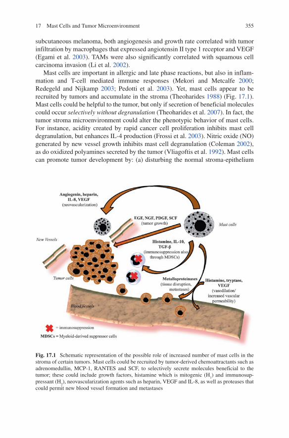
Mast cells are important in allergic and late phase reactions, but also in inflam-mation and T-cell mediated immune responses (Mekori and Metcalfe 2000; Redegeld and Nijkamp 2003; Pedotti et al. 2003). Yet, mast cells appear to be recruited by tumors and accumulate in the stroma (Theoharides 1988) (Fig. 17.1). Mast cells could be helpful to the tumor, but only if secretion of beneficial molecules could occur selectively without degranulation (Theoharides et al. 2007). In fact, the tumor stroma microenvironment could alter the phenotypic behavior of mast cells. For instance, acidity created by rapid cancer cell proliferation inhibits mast cell degranulation, but enhances IL-4 production (Frossi et al. 2003). Nitric oxide (NO) generated by new vessel growth inhibits mast cell degranulation (Coleman 2002), as do oxidized polyamines secreted by the tumor (Vliagoftis et al. 1992). Mast cells can promote tumor development by: (a) disturbing the normal stroma-epithelium
Fig. 17.1 Schematic representation of the possible role of increased number of mast cells in the stroma of certain tumors. Mast cells could be recruited by tumor-derived chemoattractants such as adrenomedullin, MCP-1, RANTES and SCF, to selectively secrete molecules beneficial to the tumor; these could include growth factors, histamine which is mitogenic (H
1) and immunosup-
pressant (H2), neovascularization agents such as heparin, VEGF and IL-8, as well as proteases that
could permit new blood vessel formation and metastases
356 T.C. Theoharides et al.
communication as was shown for matrix degradation at sites of tumor invasion in rat mammary adenocarcinoma, (b) facilitating tumor angiogenesis, (c) releasing growth factors (Conti et al. 2007), and (d) inducing a state of immunosuppression. The tumor enhancing effect of mast cells has been shown repeatedly with the use of W/Wv mast cell deficient mice, which developed fewer lung metastases to subcutaneous B16-BL6 tumors (Starkey et al. 1988), and in which mice premalignant angiogenesis of squamous epithelial carcinogenesis was blocked (Coussens et al. 1999). The development of 1, 2-dimethylhydrazine-induced intestinal tumors was slowed by 60% in W/Wv mice (Wedemeyer and Galli 2005). There was also reduced microvessel formation and tumor size in W/Wv mice injected with MB49 murine bladder carcinoma (Dethelfsen et al. 1994).
Mast Cell Biology
Mast cells derive from a specific bone marrow progenitor cell, they migrate into tissues where they mature depending upon microenvironmental conditions, and they participate in allergic reactions, as well as innate and acquired immunity (Mekori and Metcalfe 2000; Galli et al. 2005a, b). Mast cells are located perivas-cularly close to neurons and could have a critical role in neuroinflammatory diseases (Theoharides and Cochrane 2004), as well as in stress-induced brain metastases (Theoharides et al. 2008). Mast cells vary considerably in their cytokine and proteolytic enzyme content: connective tissue mast cells (CTMC) contain tryptase and chymase (CT mast cells), while mucosal mast cells (MMC) contain only tryptase (T mast cells). However, the phenotypic expression of mast cells is not fixed, since MMC can develop into CTMC given the appropriate microenviron-mental conditions (Galli et al. 2005a). Moreover, addition of IL-5 to human umbilical cord blood-derived cultured mast cells (hCBMCs) augmented IgE-induced production of distinct cytokines, such as tumor necrosis factor (TNF) and MIP-1a, but without histamine (Ochi et al. 2000).
In addition to IgE and antigen, the main trigger in allergic reactions, anaphyla-toxins (C3a, C5a), cytokines (IL-1, IL-33), hormones (CRH) and neuropeptides can stimulate mast cell activation; the latter include endorphins, substance P (SP), neurotensin (NT), and nerve growth factor (NGF) leading to secretion of numerous biologically active mediators, but through different pathways (Theoharides and Kalogeromitros 2006). In addition, thymic stromal lymphopoietin (TSLP) released from epithelial cells in response to infection, trauma, and inflammation activates mast cells in the absence of IgE, but in the presence of IL-1, to release IL-5 and IL-13 (Allakhverdi et al. 2007; Al-Shami et al. 2005).
Mast cells can secrete either the content of individual granules (Theoharides and Douglas 1978) or distinct mediators selectively (Theoharides et al. 1982), possibly through regulation by specific phosphoproteins (Sieghart et al. 1978; Theoharides et al. 1980). Vascular permeability/vascular endothelial cell growth factor (VPF/VEGF) can be secreted from bone marrow-derived mouse mast cells
35717 Mast Cells and Tumor Microenvironment
(Boesiger et al. 1998). In view of the fact that acute stress increased tumor size and decreased survival (Sklar and Anisman 1979; Antoni et al. 2006), we investigated if cortocotropin-releasing hormone (CRH), secreted under stress, could induce VEGF release from hCBMCs. We reported that CRH induced selective VEGF release without histamine (Cao et al. 2005). We further showed that IL-1 could induce selective secretion of IL-6 from hCBMCs without degranulation through a unique vesicular shuttle (Kandere-Grzybowska et al. 2003). IL-1 can further stimulate secretion of VEGF (Salven et al. 2002), thus promoting angiogenesis (Salven et al. 2002) and lung carcinoma growth (Saijo et al. 2002). Stem cell factor (SCF) can also induce selective release of IL-6 without histamine and without degranulation (Gagari et al. 1997). This process has been termed “differential release”, “intragranular activation” or “piecemeal degranulation” (Letourneau et al. 1996). Moreover, in certain diseases such as scleroderma and interstitial cystitis (Theoharides et al. 1995), mast cells could be almost totally depleted of their granule content, without classic degranulation, rendering them undetectable by light microscopy (“phantom mast cells”) (Claman et al. 1986).
Mast Cells Could Be Beneficial to the Tumor
Mast cells could accumulate at sites of tumor growth in response to numerous chemoattractants (Table 17.1) such as RANTES or MCP-1 (Conti et al. 1997), and are associated with poor prognosis (Molin et al. 2002). In addition, SCF at low doses mediates chemotaxis of mast cells, while a higher dose is necessary for release of mediators (Huang et al. 2008), such as metalloproteinase-9 (MMP-9) (Huang et al. 2008). Adrenomedullin can stimulate histamine release from rat peri-toneal mast cells (Yoshida et al. 2001), but can also be released from human cul-tured A549 lung carcinoma cells and stimulate human leukemic mast cells (HMC-1) (Zudaire et al. 2006). Moreover, adrenomedullin is also produced by HMC-1 cells, it augments growth of lung cancer cells (Zudaire et al. 2006), and adrenomedullin-producing mast cells were shown to infiltrate human lung cancers (Zudaire et al. 2006).
Mast cells have been consistently implicated in tumor angiogenesis (Crivellato et al. 2008), along with other myeloid cells (Murdoch et al. 2008). Mast cell-deficient W/Wv mice exhibited decreased rate of tumor angiogenesis (Starkey et al. 1988). Mast cells could facilitate tumor angiogenesis through heparin-like molecules that would also permit metastases through their anti-clotting effects (Fig. 17.1). Mast cells
• Adrenomedullin• MCP-1• RANTES• SCF
Table 17.1 Tumor-derived mast cell chemoattractants/triggers
358 T.C. Theoharides et al.
also generate and secrete IL-8 which has been shown to be an angiogenesis factor, as well as a tumor cell chemotactic factor and tumor mitogen (Waugh and Wilson 2008). Mast cells secrete a number of growth factors, such as EGF, PDGF, NGF, and SCF (Galli et al. 2005b). Moreover, VPF/VEGF is secreted from mouse bone marrow-derived and human cultured mast cells (Boesiger et al. 1998), as well as from HMC-1 cells (Grutzkau et al. 1998) (Table 17.2).
Mast cells are rich in metalloproteinases, such as MMP-9, that can facilitate tumor invasiveness (Almholt and Johnsen 2003). Such enzymes can disturb the normal stroma-epithelium communication, as was shown for matrix degradation at sites of tumor invasion in rat mammary adenocarcinoma (Dabbous et al. 1986). Mast cells and stress could also disrupt the blood-brain-barrier (BBB) and promote brain metastases (Theoharides et al. 2008). Acute stress can activate mast cells and increase BBB permeability that is mast cell dependent (Esposito et al. 2002a). These findings are important in view of the fact that acute stress increases metasta-ses in breast and other tumors (Sklar and Anisman 1979; Antoni et al. 2006), and over 30% of breast cancer patients develop brain metastases with poor associated prognosis (Schouten et al. 2002). In fact, a number of cancers express CRH recep-tors (Reubi et al. 2003), prompting the suggestion that CRH may affect tumor cell behavior. For instance, a human breast cancer line MCF7 expresses CRH mRNA and secrets immunoreactive CRH (Graziani et al. 2006b), prompting the possibility of autocrine or paracrine effects.
Another aspect of tumor microenvironment is immunosuppression. Histamine induces tumor cell proliferation through H1 receptors, while suppressing the immune system through H2 and possibly H4 receptors (Tiligada et al. 2009; Gutzmer et al. 2005). It was also shown that the histamine content of human breast
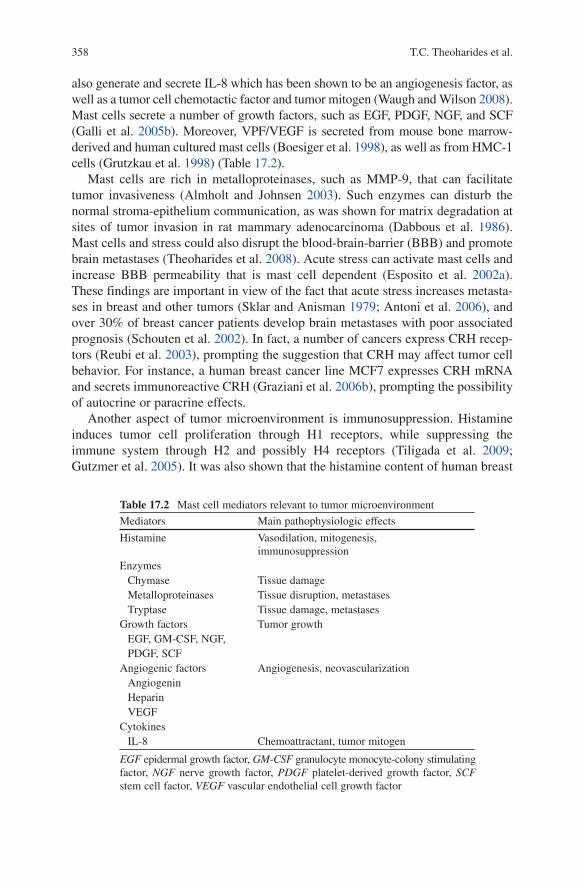
Table 17.2 Mast cell mediators relevant to tumor microenvironment
Mediators Main pathophysiologic effects
Histamine Vasodilation, mitogenesis, immunosuppression
Enzymes Chymase Tissue damage Metalloproteinases Tissue disruption, metastases Tryptase Tissue damage, metastasesGrowth factors Tumor growth EGF, GM-CSF, NGF, PDGF, SCFAngiogenic factors Angiogenesis, neovascularization Angiogenin Heparin VEGFCytokines IL-8 Chemoattractant, tumor mitogen
EGF epidermal growth factor, GM-CSF granulocyte monocyte-colony stimulating factor, NGF nerve growth factor, PDGF platelet-derived growth factor, SCF stem cell factor, VEGF vascular endothelial cell growth factor
35917 Mast Cells and Tumor Microenvironment
cancer tissue was much higher than adjacent normal tissue and sufficient to act as a local immunosuppressant (Reynolds et al. 1998). The H2 receptor antagonist, famotidine, given preoperatively enhanced tumor infiltrating lymphocytes and increased metastatic lymph node reactive changes in breast cancer in humans (Parshad et al. 2002). Mast cells can promote immunosuppression by also secreting the immunosuppressants transforming growth factor-b (TGF-b) and IL-10 (Conti et al. 2003). SCF-mediated mast cell infiltration of tumors further enhances immu-nosuppression (Huang et al. 2008). Mast cells may further contribute to tumor anergy by promoting the development/recruitment of “tolerogenic host antigen-presenting cells” (Wasiuk et al. 2009) and Treg cells (Wasiuk et al. 2009). Mast cells may also influence migration and function of dendritic cells through distinct prostaglandins (PGE
2 and PGD
2) (Wasiuk et al. 2009). In addition, mast cells could
indirectly down-regulate anti-tumor immunity by influencing the functions of immune suppressive myeloid-derived stem cells (MDSCs). MDSCs also secrete VEGF (Marx 2008), but this action requires MMP-9 (Yang et al. 2004), which is produced by mast cells. Mast cell-derived IL-1b induces MDSCs in mice with transplanted mammary carcinoma or fibrosarcoma (Bunt et al. 2006).
Breast Cancer
Disruption of the normal flow of information between stroma and parenchyma could permit neoplastic progression (Barcellos-Hoff 1998). Stromal matrix metal-loproteinases, rather than the target cell, were shown to promote mammary tumori-genesis (Sternlicht et al. 1999), while irradiated mammary gland stroma promoted carcinogenesis of unirradiated epithelial cells (Barcellos-Hoff and Ravani 2000). Mammary carcinogenesis in Wistar/Furth rats occurs when only the stroma of the mammary gland (fat pad) is exposed to the carcinogen nitrosomethylurea (NMU) (Maffini et al. 2004). The earliest effects of carcinogen administration in mammary gland carcinogenesis are manifested in the stroma with infiltration of inflammatory cells including mast cells (Maffini et al. 2004). The number of mast cells was significantly increased in malignant as compared to benign, lesions in human breast biopsies (Kashiwase et al. 2004). Moreover, the number of mast cells was greater in scirrhous than papillotubular carcinoma (Kashiwase et al. 2004). The histamine content of human breast cancer tissue, an index of mast cell presence, was much higher than adjacent normal tissue (Reynolds et al. 1998). Recent papers also con-firmed high number of mast cells in human mammary adenocarcinoma (Rajput et al. 2007; Ribatti et al. 2007). Some of these papers suggested that the presence of mast cells may indicate a favorable prognosis (Rajput et al. 2007; Dabiri et al. 2004); however, in these instances mast cells were “identified” by staining for c-kit (Rajput et al. 2007; Dabiri et al. 2004), which is not specific, as cancer cells also express c-kit (Charpin et al. 2009). Moreover, reduction of c-kit expression was associated with malignant transformation of breast epithelium in human breast cancer (Polat 2007), and in carcinogen-induced rat mammary carcinoma (Maffini
360 T.C. Theoharides et al.
et al. 2008). Consequently, tumor c-kit expression and not the presence of c-kit positive mast cells, appears to be associated with favorable outcomes.
An increased number of mast cells was reported in cis-hydroxyproline-induced mammary tumors in Buffalo rats (Strum et al. 1981). Similar findings were obtained in 7, 12-dimethylbenz(a)anthracene (DMBA)-induced mammary adenocarcinoma in which mast cells accumulated, but appeared to be intact and resistant to the mast cell degranulator compound 48/80 (Andersson et al. 1976). The mast cell inhibitor disodium cromoglycate increased blood clotting and hypoxia in invasive murine breast cancer (Samoszuk and Corwin 2003).
The location of mast cells in relation to tumor cells may also be important. Lymph nodes may behave differently (Munn and Mellor 2006) upon cancer cell entrapment. Tryptase-positive mast cells correlated with angiogenesis and presence of micrometastases in sentinel lymph nodes from 80 patients with breast cancer (Ribatti et al. 2007). In contrast, intermammary lymph node enlargement with mast cell infiltration was considered to be a positive prognostic sign (Quan et al. 2002).
Melanoma and Basal Cell Carcinoma
Mast cells accumulate especially around invasive melanoma (Reed et al. 1996; Dvorak et al. 1980), and their numbers correlate with increased neovascularization, mast cell overexpression of VEGF, tumor aggressiveness, and poor prognosis (Ch’ng et al. 2006). Tumor vascularity and tryptase-positive mast cells correlated with poor melanoma prognosis (Ribatti et al. 2003). Moreover, SCF splice variants were detected in melanoma (Welker et al. 2000), and could present new forms of mast cell growth factors related to melanoma growth. Mast cells have also been repeatedly noted to accumulate around basal cell carcinoma lesions and are thought to contribute to cancer growth by inducing immunosuppression (Grimbaldeston et al. 2000). Increased dermal mast cell numbers are associated with higher risk of developing basal cell carcinoma in humans possibly through UVB-induced immu-nosuppression (Grimbaldeston et al. 2002). Mast cells can also mediate TNF-a dependent dendritic cell migration and consequently increase skin tumor antigen presentation, but in a manner that does not elicit an immune response (Munn and Mellor 2006; Hart et al. 2002).
Pancreatic Cancer
Pancreatic ductal adenocarcinoma (PDAC) is the 4th cause of cancer-related deaths in the USA, with a prognosis of less than 6.0 months and 5-year survival of less than 5% (Welsch et al. 2007; Hezel et al. 2006). PDAC escapes early detection and resists treatment (Tuveson and Hingorani 2005). Even though PDAC had been
36117 Mast Cells and Tumor Microenvironment
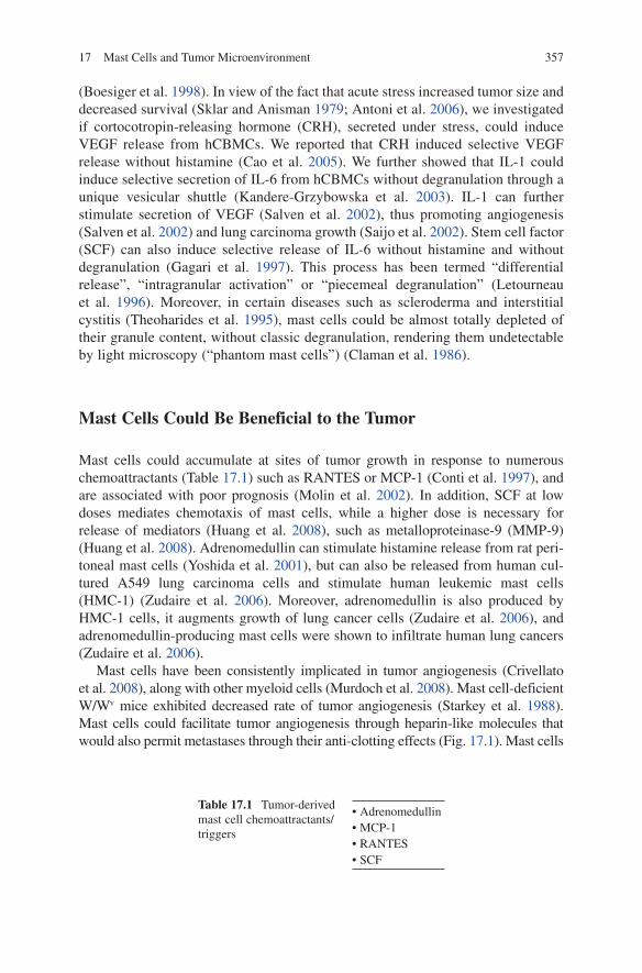
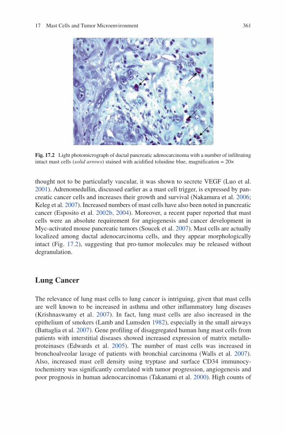
thought not to be particularly vascular, it was shown to secrete VEGF (Luo et al. 2001). Adrenomedullin, discussed earlier as a mast cell trigger, is expressed by pan-creatic cancer cells and increases their growth and survival (Nakamura et al. 2006; Keleg et al. 2007). Increased numbers of mast cells have also been noted in pancreatic cancer (Esposito et al. 2002b, 2004). Moreover, a recent paper reported that mast cells were an absolute requirement for angiogenesis and cancer development in Myc-activated mouse pancreatic tumors (Soucek et al. 2007). Mast cells are actually localized among ductal adenocarcinoma cells, and they appear morphologically intact (Fig. 17.2), suggesting that pro-tumor molecules may be released without degranulation.
Lung Cancer
The relevance of lung mast cells to lung cancer is intriguing, given that mast cells are well known to be increased in asthma and other inflammatory lung diseases (Krishnaswamy et al. 2007). In fact, lung mast cells are also increased in the epithelium of smokers (Lamb and Lumsden 1982), especially in the small airways (Battaglia et al. 2007). Gene profiling of disaggregated human lung mast cells from patients with interstitial diseases showed increased expression of matrix metallo-proteinases (Edwards et al. 2005). The number of mast cells was increased in bronchoalveolar lavage of patients with bronchial carcinoma (Walls et al. 2007). Also, increased mast cell density using tryptase and surface CD34 immunocy-tochemistry was significantly correlated with tumor progression, angiogenesis and poor prognosis in human adenocarcinomas (Takanami et al. 2000). High counts of
Fig. 17.2 Light photomicrograph of ductal pancreatic adenocarcinoma with a number of infiltrating intact mast cells (solid arrows) stained with acidified toluidine blue, magnification = 20×
362 T.C. Theoharides et al.
chymase-positive mast cells also correlated with worse prognosis in bronchoalveolar carcinoma (Nagata et al. 2003) and in lung adenocarcinoma (Ibaraki et al. 2005). This direct correlation between increased numbers of mast cells and lung cancer was apparently independent of tumor angiogenesis, as measured by the presence of endothelial cells stained with anti-human factor VIII antibody (Tomita et al. 2000). Nevertheless, increased mast cell density correlated with increased VEGF expres-sion and poor prognosis in 33/53 cases of non-small cell lung carcinomas (NSCLC) (Imada et al. 2000). Histidine decarboxylase (HDC) immunoreactivity, an index of mast cell presence/activation, could distinguish 18/23 cases of small cell lung carcinomas (SCLC), but only 6/12 cases of NSCLC (Matsuki et al. 2003), suggesting that higher number of mast cells were infiltrating the SCLC, known to be more aggressive.
Mast Cells Could be Detrimental to the Tumor
Even though mast cells could be detrimental to tumor growth, they apparently cannot secrete such mediators as they may be inhibited from degranulation by tumor-derived blockers, such as oxidized polyamines (Vliagoftis et al. 1992), or chondroitin sulfate, which inhibits mast cell activation (Theoharides et al. 2000). It would be fascinating if one could inhibit mast cells from secreting pro-tumor mediators, but promote secretion of anti-tumor molecules. For instance, tryptase stimulates pro-tease-activated receptors (PAR-1 and -2), also activated by thrombin and trypsin respectively, and induces widespread inflammation (D’Andrea et al. 2001). IL-4 binds to IL-4 receptors expressed by human breast carcinoma cells, and leads to apoptosis (Gooch et al. 1998). TNF-a could also induce tumor cell death (Gordon and Galli 1990). Histamine inhibited human primary melanoma cell proliferation presumably by acting through H1 receptors, an action enhanced by IL-6 (Lazar-Molnar et al. 2002). Heparan sulfate proteoglycans could block binding of heparin to the cell surface and prevent neovascularization (Fannon et al. 2003). For instance protamine, which binds to heparin and neutralizes its anticoagulant properties, induced selective thrombosis of blood vessels within mammary adenocarcinoma (Su et al. 2001). On the other hand, cancer cell-associated chondroitin sulfate accu-mulates in mammary gland tumors and in metastatic lesions (Hinrichs et al. 1999); in fact, tumor cells metastasize through binding of their surface chondroitin sulfate to the interstitial matrix (Kokenyesi 2001).
Interferon-a (IFN-a) or mast cell-derived interferon-g (IFN-g) may enhance TNF-related apoptosis-related ligand (TRAIL) gene expression and translation, leading to apoptosis of tumor cells in an autocrine and paracrine manner (Abadie et al. 2004; Wang et al. 2004). Moreover, treatment of melanoma patients with IFN-a increased TRAIL levels in serum (Tecchio et al. 2004). Receptor binding of TRAIL, in turn, activates an number of down steam events leading to regulation of the inflammatory response (Collison et al. 2009). In addition to its cytotoxic role,
36317 Mast Cells and Tumor Microenvironment
TRAIL can inhibit angiogenesis indirectly by facilitating apoptosis of endothelial cells (Li et al. 2003).
Certain studies indicate that CRH may inhibit growth of endometrial (Graziani et al. 2002) and breast cancer cells (Graziani et al. 2006b) in culture. Other studies suggest that endometrial cancer not expressing CRHR-1 may be associated with a more aggressive phenotype in humans (Graziani et al. 2006a). Mast cells (Kempuraj et al. 2004) and other inflammatory cells (Karalis et al. 1997) can synthesize and release CRH. It would, therefore, be interesting if mast cells could release CRH, but at the same time be prevented from CRH inducing VEGF release in an autocrine fashion.
Conclusion
Mounting evidence indicates that mast cells accumulate in tumor stroma and can promote tumor growth and metastases. Mast cells may, therefore, serve as a new target for the adjuvant treatment of tumors (Groot et al. 2009), such as mammary adenocarcinoma or pancreatic cancer (Theoharides 2008), through the selective inhibition of tumor-promoting molecules. A possible therapeutic approach could involve the use of select flavonoids. Flavonoids are naturally occurring polypheno-lic compounds present in green plants and seeds with anti-oxidant, anti-inflammatory and cancer-inhibiting properties (Middleton et al. 2000). A multiethnic epidemiologi-cal study identified an inverse relationship between flavonoid intake and pancreatic cancer (Nothlings et al. 2007). An inverse relationship has also been reported between intake of flavonoids, such as quercetin, and risk of lung cancer in general (Le Marchand et al. 2000), as well as in male smokers in particular (Hirvonen et al. 2001). The flavonoids quercetin, luteolin and epigallocatechin also decrease prolif-eration of pancreatic carcinoma cells in culture (Lee et al. 2002; Shankar et al. 2008; Takada et al. 2002), but the mechanism of this action is not known. We showed that these flavonoids inhibit proliferation and secretion from rat basophil leukemia (RBL-1) cells (Alexandrakis et al. 1999) and HMC-1 cells (Alexandrakis et al. 2003), as well as pro-inflammatory cytokine release from normal hCBMCs (Kempuraj et al. 2005). In fact, recent reviews have re-emphasized the potential use of select flavonoids, such as epigallocatechin, quercetin and curcumin in cancer treatment (Saif et al. 2009; Sogno et al. 2009; Jagtap et al. 2009).
Future studies should investigate any unique ability of select flavonoids with anti-cancer properties to also inhibit secretion of pro-cancer molecules, while permitting secretion of anti-cancer mediators from mast cells.
Acknowledgments Work discussed was supported in part by a “Concept Award” No. BC024430 from the United States Department of Defense to (TCT), and funds from Theta Biomedical Consulting and Development Co., Inc. (Brookline, MA). The possible therapeutic role of inhibit-ing mast cell-derived molecules beneficial to tumor growth is covered by patent application US 10/811,838 submitted by TCT and assigned to Theta, Inc.
364 T.C. Theoharides et al.
Many thanks are due to Dr. Eva Karamitropoulou (Attikon General Hospital, Athens Medical School, Athens, Greece) for the pancreatic cancer photo, and to Ms. Jessica Christian for her word processing skills.
References
Abadie A, Besancon F, Wietzerbin J (2004) Type I interferon and TNFalpha cooperate with type II interferon for TRAIL induction and triggering of apoptosis in SK-N-MC EWING tumor cells. Oncogene 23:4911–4920
Alexandrakis M, Singh L, Boucher W, Letourneau R, Theofilopoulos P et al (1999) Differential effect of flavonoids on inhibition of secretion and accumulation of secretory granules in rat basophilic leukemia cells. Int J Immunopharmacol 21:379–390
Alexandrakis MG, Letourneau R, Kempuraj D, Kandere K, Huang M et al (2003) Flavones inhibit proliferation and increase mediator content in human leukemic mast cells (HMC-1). Eur J Haematol 71:448–454
Allakhverdi Z, Comeau MR, Jessup HK, yoon BP, Breuer A et al (2007) Thymic stromal lym-phopoietin is released by human epithelial cell in response to microbes, trauma, or inflamma-tion and potently activates mast cells. J Exp Med 19:253–258
Almholt K, Johnsen M (2003) Stromal cell involvement in cancer. Cancer Res 162:31–42Al-Shami A, Spolski R, Kelly J, Keane-Myers A, Leonard WJ (2005) A role for TSLP in the
development of inflammation in an asthma model. J Exp Med 202:829–839Andersson A-C, Henningsson S, Lundell L, Rosengren E, Sundler F (1976) Diamines and
polyamines in DMBA-induced breast carcinoma containing mast cells resistant to compound 48/80. Agents Actions 6/5:577–583
Antoni MH, Lutgendorf SK, Cole SW, Dhabhar FS, Sephton SE et al (2006) The influence of bio-behavioural factors on tumour biology: pathways and mechanisms. Nat Rev Cancer 6:240–248
Barbera-Guillem E, Nyhus JK, Wolford CC, Friece CR, Sampsel JW (2002) Vascular endothelial growth factor secretion by tumor-infiltrating macrophages essentially supports tumor angio-genesis, and IgG immune complexes potentiate the process. Cancer Res 62:7042–7049
Barcellos-Hoff MH (1998) The potential influence of radiation-induced microenvironments in neoplastic progression. J Mammary Gland Biol Neoplasia 3:165–175
Barcellos-Hoff MH, Ravani SA (2000) Irradiated mammary gland stroma promotes the expression of tumorigenic potential by unirradiated epithelial cells. Cancer Res 60:1254–1260
Battaglia S, Mauad T, van Schadewijik AM, Vignola AM, Rabe KF et al (2007) Differential distribution of inflammatory cells in large and small airways in smokers. J Clin Pathol 60: 907–911
Boesiger J, Tsai M, Maurer M, Yamaguchi M, Brown LF et al (1998) Mast cells can secrete vas-cular permeability factor/vascular endothelial cell growth factor and exhibit enhanced release after immunoglobulin E-dependent upregulation of Fce receptor I expression. J Exp Med 188:1135–1145
Bunt SK, Sinha P, Clements VK, Leips J, Ostrand-Rosenberg S (2006) Inflammation induces myeloid-derived suppressor cells that facilitate tumor progression. J Immunol 176:284–290
Cao J, Papadopoulou N, Kempuraj D, Boucher WS, Sugimoto K et al (2005) Human mast cells express corticotropin-releasing hormone (CRH) receptors and CRH leads to selective secretion of vascular endothelial growth factor. J Immunol 174:7665–7675
Charpin C, Giusiano S, Charfi S, Secq V, Carpentier S et al (2009) Quantitative immunohis-tochemical expression of c Kit in breast carcinomas is predictive of patients’ outcome. Br J Cancer 101:48–54
36517 Mast Cells and Tumor Microenvironment
Ch’ng S, Waallis RA, Yuan L, Davis PF, Tan ST (2006) Mast cells and cutaneous malignancies. Mod Pathol 19:149–159
Claman HN, Choi KL, Sujansky W, Vatter AE (1986) Mast cell “disappearance” in chronic murine graft-vs-host disease (GVHD)-ultrastructural demonstration of “phantom mast cells”. J Immunol 137:2009–2013
Coleman JW (2002) Nitric oxide: a regulator of mast cell activation and mast cell-mediated inflammation. Clin Exp Immunol 129:4–10
Collison A, Foster PS, Mattes J (2009) The emerging role of TRAIL as key regulator of inflam-matory responses. Clin Exp Pharmacol Physiol 36:1049–1053
Conti P, Pang X, Boucher W, Letourneau R, Reale M et al (1997) Impact of Rantes and MCP-1 chemokines on in vivo basophilic mast cell recruitment in rat skin injection model and their role in modifying the protein and mRNA levels for histidine decarboxylase. Blood 89: 4120–4127
Conti P, Kempuraj D, Kandere K, Gioacchino MD, Barbacane RC et al (2003) IL-10, an inflam-matory/inhibitory cytokine, but not always. Immunol Lett 86:123–129
Conti P, Castellani ML, Kempuraj D, Salini V, Vecchiet J et al (2007) Role of mast cells in tumor growth. Ann Clin Lab Sci 37:315–322
Coussens LM, Raymond WW, Nergers G, Laig-Webster M, Behrendtsen O et al (1999) Inflammatory mast cells up-regulate angiogenesis during squamous epithelial carcinogenesis. Genes Dev 13:1382–1397
Crivellato E, Nico B, Ribatti D (2008) Mast cells and tumour angiogenesis: new insight from experimental carcinogenesis. Cancer Lett 269:1–6
D’Andrea MR, Derian CK, Santulli RJ, Andrade-Gordon P (2001) Differential expression of protease-activated receptors-1 and -2 in stromal fibroblasts of normal, benign, and malignant human tissues. Am J Pathol 158:2031–2041
Dabbous MK, Walker R, Haney L, Carter LM, Nicolson GL et al (1986) Mast cells and matrix degradation at sites of tumor invasion in rat mammary adenocarinoma. Br J Cancer 54:459–465
Dabiri S, Huntsman D, Makretsov N, Cheang M, Gilks B et al (2004) The presence of stromal mast cells identifies a subset of invasive breast cancers with a favorable prognosis. Mod Pathol 17:690–695
Dethelfsen SM, Matsumura N, Zetter BR (1994) Mast cell accumulation at sites of murine tumor implantation: implications for angiogenesis and tumor metastasis. Invasion Metastasis 14:395–408
Dvorak AM, Mihm MCJ, Osage JE, Dvorak HF (1980) Melanoma. An ultrastructural study of the host inflammatory and vascular responses. J Invest Dermatol 75:388–393
Edwards ST, Cruz AC, Donnelly S, Dazin PF, Schulman ES et al (2005) c-Kit immunophenotyp-ing and metalloproteinase expression profiles of mast cells in intestinal lung diseases. J Pathol 206:279–290
Egami K, Murohara T, Shimada T, Sasaki K, Shintani S et al (2003) Role of host angiotensin II type 1 receptor in tumor angiogenesis and growth. J Clin Invest 112:67–75
Esposito P, Chandler N, Kandere-Grzybowska K, Basu S, Jacobson S et al (2002a) Corticotropin-releasing hormone (CRH) and brain mast cells regulate blood-brain-barrier permeability induced by acute stress. J Pharmacol Exp Ther 303:1061–1066
Esposito I, Kleeff J, Bischoff SC, Fischer L, Collecchi P et al (2002b) The stem cell factor-c-kit system and mast cells in human pancreatic cancer. Lab Invest 82:1481–1492
Esposito I, Menicagli M, Funel N, Bergmann F, Boggi U et al (2004) Inflammatory cells contrib-ute to the generation of an angiogenic phenotype in pancreatic ductal adenocarcinoma. J Clin Pathol 57:630–636
Fannon M, Forsten-Williams K, Dowd CJ, Freedman DA, Folkman J et al (2003) Binding inhibi-tion of angiogenic factors by heparan sulfate proteoglycans in aqueous humor: potential mechanism for maintenance of an avascular environment. FASEB J 17:902–904
366 T.C. Theoharides et al.
Frossi B, De Carli M, Daniel KC, Rivera J, Pucillo C (2003) Oxidative stress stimulates IL-4 and IL-6 production in mast cells by an APE/Ref-1-dependent pathway. Eur J Immunol 33:2168–2177
Gagari E, Tsai M, Lantz CS, Fox LG, Galli SJ (1997) Differential release of mast cell interleu-kin-6 via c-kit. Blood 89:2654–2663
Galli SJ, Kalesnikoff J, Grimbaldeston MA, Piliponsky AM, Williams CM et al (2005a) Mast cells as “tunable” effector and immunoregulatory cells: recent advances. Annu Rev Immunol 23:749–786
Galli SJ, Nakae S, Tsai M (2005b) Mast cells in the development of adaptive immune responses. Nat Immunol 6:135–142
Gooch JL, Lee AV, Yee D (1998) Interleukin 4 inhibits growth and induces apoptosis in human breast cancer cells. Cancer Res 58:4199–4205
Gordon JR, Galli SJ (1990) Mast cells as a source of both preformed and immunologically induc-ible TNF-a/cachectin. Nature 346:274–276
Graziani G, Tentori L, Portarena I, Barbarino M, Tringali G et al (2002) CRH inhibits cell growth of human endometrial adenocarcinoma cells via CRH-receptor 1-mediated activation of cAMP-PKA pathway. Endocrinology 143:807–813
Graziani G, Ferrandina G, Pozzoli G, Vergati M, Muzi A et al (2006a) Corticotropin-releasing hormone receptor-1 in human endometrial cancer. Oncol Rep 15:375–379
Graziani G, Tentori L, Muzi A, Vergati M, Tringali G et al (2006b) Evidence that corticotropin-releasing hormone inhibits cell growth of human breast cancer cells via the activation of CRH-R1 receptor subtype. Mol Cell Endocrinol 264:44–49
Grimbaldeston MA, Skov L, Baadsgaard O, Skov BG, Marshman G et al (2000) Communications: high dermal mast cell prevalence is a predisposing factor for basal cell carcinoma in humans. J Invest Dermatol 115:317–320
Grimbaldeston MA, Skov L, Finlay-Jones JJ, Hart PH (2002) Increased dermal mast cell preva-lence and susceptibility to development of basal cell carcinoma in humans. Methods 28:90–96
Groot KT, Abudukelimu A, Redegeld FA (2009) Mast cells as target in cancer therapy. Curr Pharm Des 15:1868–1878
Grutzkau A, Kruger-Krasagakes S, Baumeister H, Schwarz C, Kogel H et al (1998) Synthesis, storage and release of vascular endothelial growth factor/vascular permeability factor (VEGF/VPF) by human mast cells: Implications for the biological significance of VEGF
206. Mol Biol
Cell 9:875–884Gutzmer R, Diestel C, Mommert S, Kother B, Stark H et al (2005) Histamine H4 receptor stimula-
tion suppresses IL-12p70 production and mediates chemotaxis in human monocyte-derived dendritic cells. J Immunol 174:5224–5232
Hart PH, Townley SL, Grimbaldeston MA, Khalil Z, Finlay-Jones JJ (2002) Mast cells, neuropep-tides, histamine, and prostaglandins in UV-induced systemic immunosuppression. Methods 28:79–89
Hezel AF, Kimmelman AC, Stanger BZ, Bardeesy N, Depinho RA (2006) Genetics and biology of pancreatic ductal adenocarcinoma. Genes Dev 20:1218–1249
Hinrichs U, Rutteman GR, Nederbragt H (1999) Stromal accumulation of chondroitin sulphate in mammary tumours of dogs. Br J Cancer 80:1359–1365
Hirvonen T, Tornwali ME, Pietinen P, Albanes D, Vitamo J (2001) Flavonol and flavone intake and the risk of cancer in male smokers (Finland). Cancer Causes Control 12:789–796
Huang B, Lei Z, Zhang GM, Li D, Song C et al (2008) SCF-mediated mast cell infiltration and activation exacerbate the inflammation and immunosuppression in tumor microenvironment. Blood 112:1269–1279
Ibaraki T, Muramatsu M, Takai S, Jin D, Maruyama H et al (2005) The relationship of tryptase and chymase positive mast cells to angiogenesis in stage 1 non-small cell lung cancer. Eur J Cardiothorac Surg 28:621
Imada A, Shijubo N, Kojima H, Abe S (2000) Mast cells correlate with angiogenesis and poor outcome in stage I lung adenocarcinoma. Eur Respir J 15:1087–1093
36717 Mast Cells and Tumor Microenvironment
Jagtap S, Meganathan K, Wagh V, Winkler J, Hescheler J et al (2009) Chemoprotective mecha-nism of the natural compounds, epigallocatechin-3-O-gallate, quercetin and curcumin against cancer and cardiovascular diseases. Curr Med Chem 16:1451–1462
Kandere-Grzybowska K, Letourneau R, Kempuraj D, Donelan J, Poplawski S et al (2003) IL-1 induces vesicular secretion of IL-6 without degranulation from human mast cells. J Immunol 171:4830–4836
Karalis K, Louis JM, Bae D, Hilderbrand H, Majzoub JA (1997) CRH and the immune system. J Neuroimmunol 72:131–136
Kashiwase Y, Morioka J, Inamura H, Yoshizawa Y, Usui R et al (2004) Quantitative analysis of mast cells in benign and malignant breast lesions. Immunohistochemical study on formalin-fixed, paraffin-embedded tissues. Int Arch Allergy Immunol 134:199–205
Kataki A, Scheid P, Piet M, Marie B, Martinet N et al (2002) Tumor infiltrating lymphocytes and macrophages have a potential dual role in lung cancer by supporting both host-defense and tumor progression. J Lab Clin Med 140:320–328
Keleg S, Kayed H, Jiang X, Penzel R, Giese T et al (2007) Adrenomedullin is induced by hypoxia and enhances pancreatic cancer cell invasion. Int J Cancer 121:21–32
Kempuraj D, Papadopoulou NG, Lytinas M, Huang M, Kandere-Grzybowska K et al (2004) Corticotropin-releasing hormone and its structurally related urocortin are synthesized and secreted by human mast cells. Endocrinology 145:43–48
Kempuraj D, Madhappan B, Christodoulou S, Boucher W, Cao J et al (2005) Flavonols inhibit proinflammatory mediator release, intracellular calcium ion levels and protein kinase C theta phosphorylation in human mast cells. Br J Pharmacol 145:934–944
Kokenyesi R (2001) Ovarian carcinoma cells synthesize both chondroitin sulfate and heparan sulfate cell surface proteoglycans that mediate cell adhesion to interstitial matrix. J Cell Biochem 83:259–270
Krishnaswamy G, Ajitawi O, Chi DS (2007) The human mast cells. Methods Mol Biol 315:13–34
Lamb D, Lumsden A (1982) Intra-epithelial mast cells in human airway epithelium: evidence for smoking-induced changes in their frequency. Thorax 37:334–342
Lazar-Molnar E, Hegyesi H, Pallinger E, Kovacs P, Toth S et al (2002) Inhibition of human primary melanoma cell proliferation by histamine is enhanced by interleukin-6. Eur J Clin Invest 32:743–749
Le Marchand L, Murphy SP, Hankin JH, Wilkens LR, Kolonel LN (2000) Intake of flavonoids and lung cancer. J Natl Cancer Inst 92:154–160
Lee LT, Huang YT, Hwang JJ, Lee PP, Ke FC et al (2002) Blockade of the epidermal growth factor receptor tyrosine kinase activity by quercetin and luteolin leads to growth inhibition and apoptosis of pancreatic tumor cells. Anticancer Res 22:1615–1627
Letourneau R, Pang X, Sant GR, Theoharides TC (1996) Intragranular activation of bladder mast cells and their association with nerve processes in interstitial cystitis. Br J Urol 77:41–54
Li C, Shintani S, Terakado N, Nakashiro K, Hamakawa H (2002) Infiltration of tumor-associated macrophages in human oral squamous cell carcinoma. Oncol Rep 9:1219–1223
Li JH, Kirkiles-Smith NC, McNiff JM, Pober JS (2003) TRAIL induces apoptosis and inflammatory gene expression in human endothelial cells. J Immunol 171:1526–1533
Luo J, Guo P, Matsuda K, Truong N, Lee A et al (2001) Pancreatic cancer cell-derived vascular endothelial growth factor is biologically active in vitro and enhances tumorigenicity in vivo. Int J Cancer 92:361–369
Maffini MV, Soto AM, Calabro JM, Ucol AA, Sonnenschein C (2004) The stroma as a crucial target in rat mammary gland carcinogenesis. J Cell Sci 117:1495–1502
Maffini MV, Soto AM, Sonnenschein C, Papadopoulos N, Theoharides TC (2008) Lack of c-kit receptor promotes mammary tumors in N-nitrosomethylurea-treated Ws/Ws rats. Cancer Cell Int 8:5
Mantovani A, Sozzani S, Locati M, Allavena P, Sica A (2002) Macrophage polarization: tumor-associated macrophages as a paradigm for polarized M2 mononuclear phagocytes. Trends Immunol 23:549–555
368 T.C. Theoharides et al.
Marx J (2008) Cancer immunology. Cancer’s bulwark against immune attack: MDS cells. Science 319:154–156
Matsuki Y, Tanimoto A, Hamada T, Sasaguri Y (2003) Histidine decarboxylase expression as a new sensitive and specific marker for small cell lung carcinoma. Mod Pathol 16:72–78
Mekori YA, Metcalfe DD (2000) Mast cells in innate immunity. Immunol Rev 173:131–140Middleton E Jr, Kandaswami C, Theoharides TC (2000) The effects of plant flavonoids on
mammalian cells: implications for inflammation, heart disease and cancer. Pharmacol Rev 52:673–751
Molin D, Edstrom A, Glimelius I, Glimelius B, Nilsson G et al (2002) Mast cell infiltration correlates with poor prognosis in Hodgkin’s lymphoma. Br J Haematol 119:122–124
Munn DH, Mellor AL (2006) The tumor-draining lymph node as an immune-privileged site. Immunol Rev 213:146–158
Murdoch C, Muthana M, Coffelt SB, Lewis CE (2008) The role of myeloid cells in the promotion of tumour angiogenesis. Nat Rev Cancer 8:618–631
Murphy PM (2001) Chemokines and the molecular basis of cancer metastasis. N Engl J Med 345:833–835
Nagata M, Shijubo N, Waalls AF, Ichimiya s, Abe S et al (2003) Chymase positive mast cells in small sized adenocarcinoma of the lung. Virchows Arch 443:565–573
Nakamura M, Han B, Nunobiki O, Kakudo K (2006) Adrenomedullin: a tumor progression factor via angiogenic control. Curr Cancer Drug Targets 6:635–643
Nothlings U, Murphy SP, Wilkens LR, Henderson BE, Kolonel LN (2007) Flavonols and pancre-atic cancer risk: the multiethnic cohort study. Am J Epidemiol 166:924–931
Ochi H, DeJesus NH, Hsieh FH, Austen KF, Boyce JA (2000) IL-4 and -5 prime human mast cells for different profiles of IgE-dependent cytokine production. Proc Natl Acad Sci U S A 97:10509–10513
Parshad R, Kapoor S, Gupta SD, Kumar A, Chattopadhyaya TK (2002) Does famotidine enhance tumor infiltrating lymphocytes in breast cancer? Results of a randomized prospective pilot study. Acta Oncol 41:362–365
Pedotti R, De Voss JJ, Steinman L, Galli SJ (2003) Involvement of both ‘allergic’ and ‘autoim-mune’ mechanisms in EAE, MS and other autoimmune diseases. Trends Immunol 24:479–484
Polat A (2007) c-KIT expression in columnar cell lesions of the breast accompanied by benign and malignant breast diseases. Pathol Res Pract 203:765–769
Quan C, Wang HL, Lu SJ (2002) Resistance to mammary carcinogenesis in Copenhagen rats: potential roles of vascular endothelial growth factor and mast cells. Cancer Lett 186:165–175
Rajput AB, Turbin DA, Cheang MCU, Voduc DK, Leung S et al (2007) Stromal mast cells in invasive breast cancer are a marker of favourable prognosis: a study of 4,444 cases. Breast Cancer Res 107:249–257
Redegeld FA, Nijkamp FP (2003) Immunoglobulin free light chains and mast cells: pivotal role in T-cell-mediated immune reactions? Trends Immunol 24:181–185
Reed JA, McNutt NS, Bogdany JK, Albino AP (1996) Expression of the mast cell growth factor interleukin-3 in melanocytic lesions correlates with an increased number of mast cells in the perilesional stroma: implications for melanoma progression. J Cutan Pathol 23:495–505
Reubi JC, Waser B, Vale W, Rivier J (2003) Expression of CRF1 and CRF2 receptors in human cancers. J Clin Endocrinol Metab 88:3312–3320
Reynolds JL, Akhter JA, Magarey CJ, Schwartz P, Adams WJ et al (1998) Histamine in human breast cancer. Br J Surg 85:538–541
Ribatti D, Ennas MG, Vacca A, Ferreli F, Nico B et al (2003) Tumor vascularity and tryptase-positive mast cells correlate with a poor prognosis in melanoma. Eur J Clin Invest 33: 420–425
Ribatti D, Finato N, Crivellato E, Guidolin D, Longo V et al (2007) Angiogenesis and mast cells in human breast cancer sentinel lymph nodes with and without micrometastases. Histopathology 51:837–842
36917 Mast Cells and Tumor Microenvironment
Saif MW, Tytler E, Lansigan F, Brown DM, Husband AJ (2009) Flavonoids, phenoxodiol, and a novel agent, triphendiol, for the treatment of pancreaticobiliary cancers. Expert Opin Investig Drugs 18:469–479
Saijo Y, Tanaka M, Miki M, Usui K, Suzuki T et al (2002) Proinflammatory cytokine IL-1 beta promotes tumor growth of Lewis lung carcinoma by induction of angiogenic factors: in vivo analysis of tumor-stromal interaction. J Immunol 169:469–475
Salven P, Hattori K, Heissig B, Rafii S (2002) Interleukin-1alpha promotes angiogenesis in vivo via VEGFR-2 pathway by inducing inflammatory cell VEGF synthesis and secretion. FASEB J 16:1471–1473
Samoszuk M, Corwin MA (2003) Mast cell inhibitor cromolyn increases blood clotting and hypoxia in murine breast cancer. Int J Cancer 107:159–163
Schouten LJ, Rutten J, Huveneers HA, Twijnstra A (2002) Incidence of brain metastases in a cohort of patients with carcinoma of the breast, colon, kidney, and lung and melanoma. Cancer 94:2698–2705
Shankar S, Ganapathy S, Hingorani SR, Srivastava RK (2008) EGCG inhibits growth, invasion, angiogenesis and metastasis of pancreatic cancer. Front Biosci 13:440–452
Sieghart W, Theoharides TC, Alper SL, Douglas WW, Greengard P (1978) Calcium-dependent protein phosphorylation during secretion by exocytosis in the mast cell. Nature 275:329–331
Silzle T, Kreutz M, Dobler MA, Brockhoff G, Knuechel R et al (2003) Tumor-associated fibro-blasts recruit blood monocytes into tumor tissue. Eur J Immunol 33:1311–1320
Sklar LS, Anisman H (1979) Stress and coping factors influence tumor growth. Science 205:513–515
Sogno I, Vannini N, Lorusso G, Cammarota R, Noonan DM et al (2009) Anti-angiogenic activity of a novel class of chemopreventive compounds: oleanic acid terpenoids. Recent Results Cancer Res 181:209–212
Soucek L, Lawlor ER, Soto D, Shchors K, Swigart LB et al (2007) Mast cells are required for angiogenesis and macroscopic expansion of Myc-induced pancreatic islet tumors. Nat Med 13:1211–1218
Starkey JR, Crowle PK, Taubenberger S (1988) Mast-cell-deficient W/Wv mice exhibit a decreased rate of tumor angiogenesis. Int J Cancer 42:48–52
Sternlicht MD, Lochter A, Sympson CJ, Huey B, Rougier JP et al (1999) The stromal proteinase MMP3/Stromelysin-1 promotes mammary carcinogenesis. Cell 98:134–146
Strum JM, Lewko WM, Kidwell WR (1981) Structural alterations within N-nitrosomethylurea-induced mammary tumors after in vivo treatment with cis-hydroxyproline. Lab Invest 45:347–354
Su MY, Najafi A, Nalcioglu O (2001) Selective thrombosis of tumor blood vessels in mammary adenocarcinoma implants in rats. Am J Pathol 159:245–251
Takada M, Nakamura Y, Koizumi T, Toyama H, Kamigaki T et al (2002) Suppression of human pancreatic carcinoma cell growth and invasion by epigallocatechin-3-gallate. Pancreas 25:45–48
Takanami I, Takeuchi K, Naruke M (2000) Mast cell density is associated with angiogenesis and poor prognosis in pulmonary adenocarcinoma. Cancer 88:2686–2692
Tecchio C, Huber V, Scapini P, Calzetti F, Margotto D et al (2004) IFNalpha-stimulated neutro-phils and monocytes release a soluble form of TNF-related apoptosis-inducing ligand (TRAIL/Apo-2 ligand) displaying apoptotic activity on leukemic cells. Blood 103:3837–3844
Theoharides TC (1988) Neuroimmunology of tumor growth: the role of mast cells. Int J Immunopath Pharmacol 1:89–98
Theoharides TC (2008) Mast cells and pancreatic cancer. N Engl J Med 358:1860–1861Theoharides TC, Cochrane DE (2004) Critical role of mast cells in inflammatory diseases and the
effect of acute stress. J Neuroimmunol 146:1–12Theoharides TC, Douglas WW (1978) Secretion in mast cells induced by calcium entrapped
within phospholipid vesicles. Science 201:1143–1145Theoharides TC, Kalogeromitros D (2006) The critical role of mast cell in allergy and inflamma-
tion. Ann N Y Acad Sci 1088:78–99
370 T.C. Theoharides et al.
Theoharides TC, Sieghart W, Greengard P, Douglas WW (1980) Antiallergic drug cromolyn may inhibit histamine secretion by regulating phosphorylation of a mast cell protein. Science 207:80–82
Theoharides TC, Bondy PK, Tsakalos ND, Askenase PW (1982) Differential release of serotonin and histamine from mast cells. Nature 297:229–231
Theoharides TC, Sant GR, El-Mansoury M, Letourneau RJ, Ucci AA Jr et al (1995) Activation of bladder mast cells in interstitial cystitis: a light and electron microscopic study. J Urol 153:629–636
Theoharides TC, Patra P, Boucher W, Letourneau R, Kempuraj D et al (2000) Chondroitin sulfate inhibits connective tissue mast cells. Br J Pharmacol 131:1039–1049
Theoharides TC, Kempuraj D, Tagen M, Conti P, Kalogeromitros D (2007) Differential release of mast cell mediators and the pathogenesis of inflammation. Immunol Rev 217:65–78
Theoharides TC, Rozniecki JJ, Sahagian G, Kempuraj D, Kalogeromitros D et al (2008) Impact of stress and mast cells on brain metastases. J Neuroimmunol 205:1–7
Tiligada E, Zampeli E, Sander K, Stark H (2009) Histamine H3 and H4 receptors as novel drug targets. Expert Opin Investig Drugs 18:1519–1531
Tomita M, Matsuzaki Y, Onitsuka T (2000) Effect of mast cells on tumor angiogenesis in lung cancer. Ann Thorac Surg 69:1686–1690
Tuveson DA, Hingorani SR (2005) Ductal pancreatic cancer in humans and mice. Cold Spring Harb Symp Quant Biol 70:65–72
Vliagoftis H, Boucher WS, Mak LL, Theoharides TC (1992) Inhibition of mast cell secretion by oxidation products of natural polyamines. Biochem Pharmacol 43:2237–2245
Walls AF, Roberts JA, Godfrey RC, Holgate ST (2007) Histochemical heterogeneity of human mast cells: disease-related differences in mast cell subsets recovered by bronchoalveolar lavaage. Int Arch Allergy Appl Immunol 92:233–241
Wang SH, Mezosi E, Wolf JM, Cao Z, Utsugi S et al (2004) IFNgamma sensitization to TRAIL-induced apoptosis in human thyroid carcinoma cells by upregulating Bak expres-sion. Oncogene 23:928–935
Wasiuk A, de V V, Hartmann K, Roers A, Noelle RJ (2009) Mast cells as regulators of adaptive immunity to tumours. Clin Exp Immunol 155:140–146
Waugh DJ, Wilson C (2008) The interleukin-8 pathway in cancer. Clin Cancer Res 14:6735–6741
Wedemeyer J, Galli SJ (2005) Decreased susceptibility of mast cell-deficient kit(W)Kit(W-v) mice to the development of 1,2-dimethyldrazine-induced intestinal tumors. Lab Invest 85:388–396
Welker P, Schadendorf D, Artuc M, Grabbe J, Henz BM (2000) Expression of SCF splice variants in human melanocytes and melanoma cell lines: potential prognostic implications. Br J Cancer 82:1453–1458
Welsch T, Kleeff J, Friess H (2007) Molecular pathogenesis of pancreatic cancer: advances and challenges. Curr Mol Med 7:504–521
Yang L, DeBusk LM, Fukuda K, Fingleton B, Green-Jarvis B et al (2004) Expansion of myeloid immune suppressor Gr+CD11b+ cells in tumor-bearing host directly promotes tumor angiogenesis. Cancer Cell 6:409–421
Yoshida M, Yoshida H, Kitaichi K, Hiramatsu K, Kimura T et al (2001) Adrenomedullin and proadrenomedullin N-terminal 20 peptide induce histamine release from rat peritoneal mast cells. Regul Pept 101:163–168
Yu JL, Rak JW (2003) Host microenvironment in breast cancer development: inflammatory and immune cells in tumour angiogenesis and arteriogenesis. Breast Cancer Res 5:83–88
Zudaire E, Martinez A, Garayoa M, Pio R, Kaur G et al (2006) Adrenomedullin is a cross-talk molecule that regulates tumor and mast cell function during human carcinogenesis. Am J Pathol 168:280–291


![Tumor Microenvironment Hijacking the Immune System [Read-Only]](https://img.pdfslide.net/doc/110x75/61bf372f43ec6023e9684384/tumor-microenvironment-hijacking-the-immune-system-read-only.jpg)