Embed Size (px)
Citation preview
Chapter 18PART IV: Molecular Pathology of Human Disease
Molecular Basis of Pulmonary Disease
Companion site for Molecular PathologyAuthor: William B. Coleman and Gregory J. Tsongalis
Companion site for Molecular Pathology Copyright © 2009 by Academic Press. All rights reserved.
2
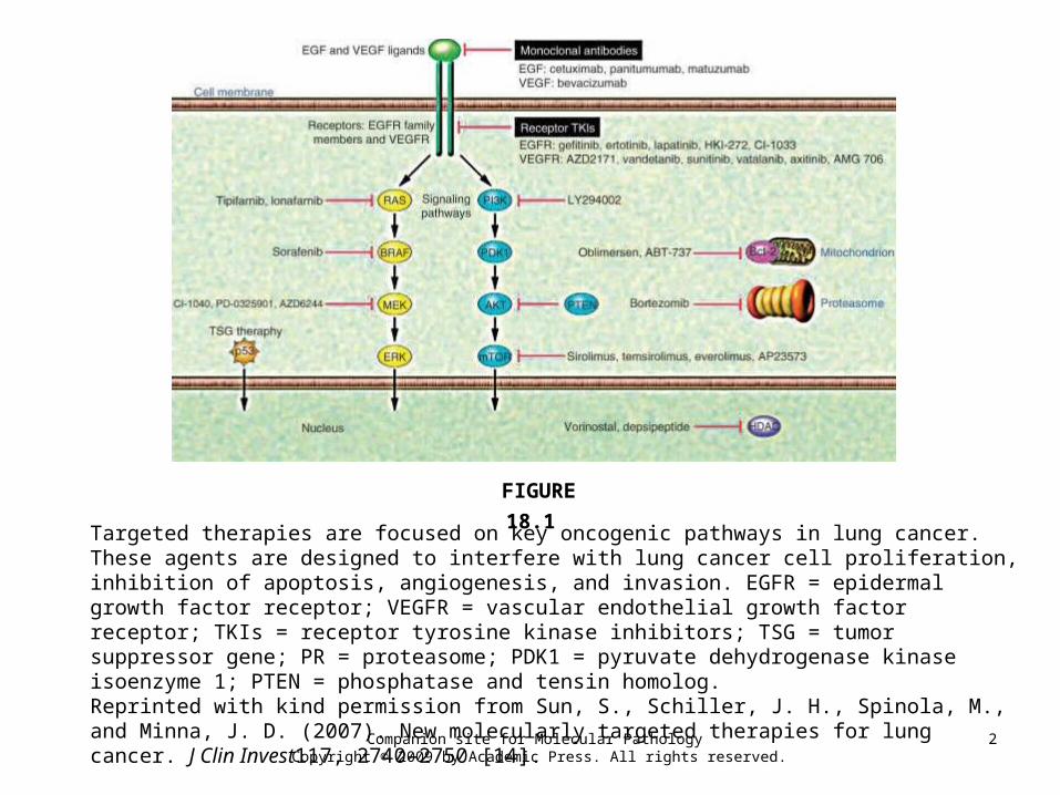
Targeted therapies are focused on key oncogenic pathways in lung cancer.These agents are designed to interfere with lung cancer cell proliferation, inhibition of apoptosis, angiogenesis, and invasion. EGFR = epidermal growth factor receptor; VEGFR = vascular endothelial growth factor receptor; TKIs = receptor tyrosine kinase inhibitors; TSG = tumor suppressor gene; PR = proteasome; PDK1 = pyruvate dehydrogenase kinase isoenzyme 1; PTEN = phosphatase and tensin homolog.Reprinted with kind permission from Sun, S., Schiller, J. H., Spinola, M., and Minna, J. D. (2007). New molecularly targeted therapies for lung cancer. J Clin Invest117, 2740–2750 [14].
FIGURE 18.1
Companion site for Molecular Pathology Copyright © 2009 by Academic Press. All rights reserved.
3
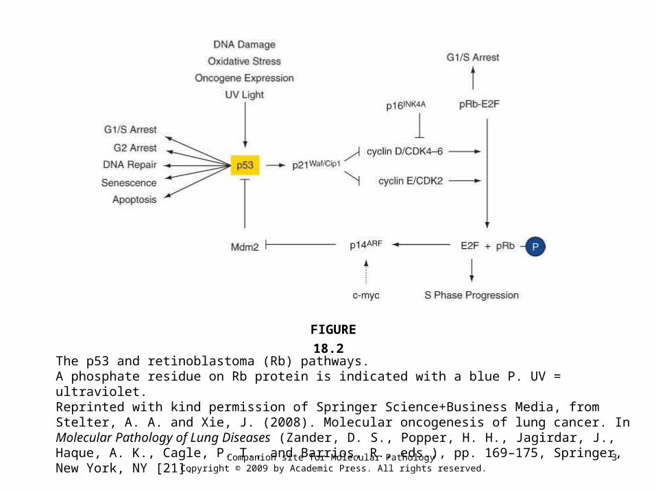
The p53 and retinoblastoma (Rb) pathways.A phosphate residue on Rb protein is indicated with a blue P. UV = ultraviolet.Reprinted with kind permission of Springer Science+Business Media, from Stelter, A. A. and Xie, J. (2008). Molecular oncogenesis of lung cancer. In Molecular Pathology of Lung Diseases (Zander, D. S., Popper, H. H., Jagirdar, J., Haque, A. K., Cagle, P. T., and Barrios, R., eds.), pp. 169–175, Springer, New York, NY [21].
FIGURE 18.2
Companion site for Molecular Pathology Copyright © 2009 by Academic Press. All rights reserved.
4
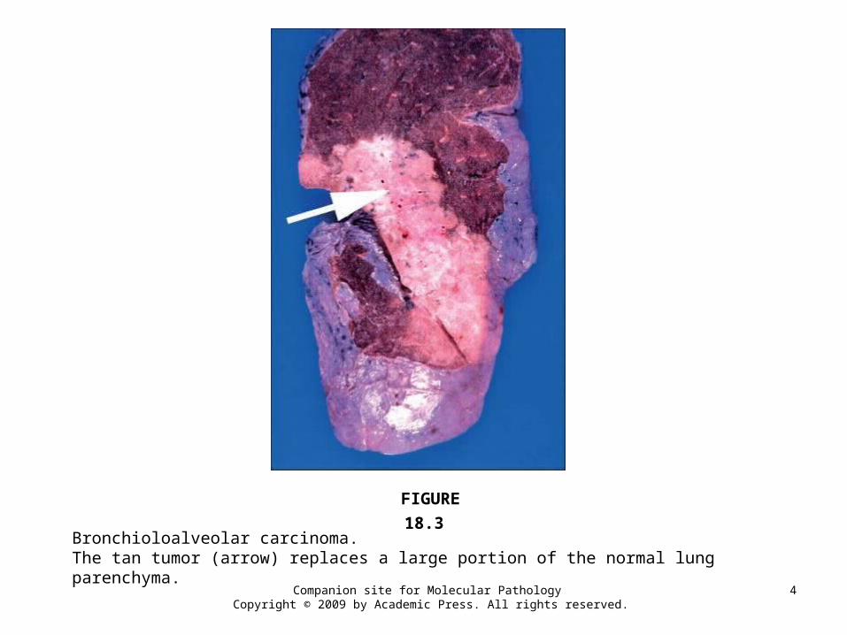
Bronchioloalveolar carcinoma.The tan tumor (arrow) replaces a large portion of the normal lung parenchyma.
FIGURE 18.3
Companion site for Molecular Pathology Copyright © 2009 by Academic Press. All rights reserved.
5
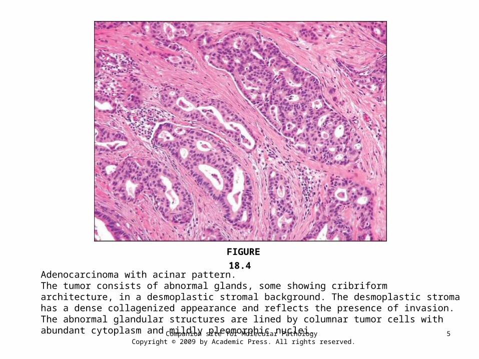
Adenocarcinoma with acinar pattern.The tumor consists of abnormal glands, some showing cribriform architecture, in a desmoplastic stromal background. The desmoplastic stroma has a dense collagenized appearance and reflects the presence of invasion. The abnormal glandular structures are lined by columnar tumor cells with abundant cytoplasm and mildly pleomorphic nuclei.
FIGURE 18.4
Companion site for Molecular Pathology Copyright © 2009 by Academic Press. All rights reserved.
6
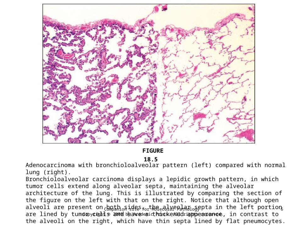
Adenocarcinoma with bronchioloalveolar pattern (left) compared with normal lung (right).Bronchioloalveolar carcinoma displays a lepidic growth pattern, in which tumor cells extend along alveolar septa, maintaining the alveolar architecture of the lung. This is illustrated by comparing the section of the figure on the left with that on the right. Notice that although open alveoli are present on both sides, the alveolar septa in the left portion are lined by tumor cells and have a thickened appearance, in contrast to the alveoli on the right, which have thin septa lined by flat pneumocytes.
FIGURE 18.5
Companion site for Molecular Pathology Copyright © 2009 by Academic Press. All rights reserved.
7
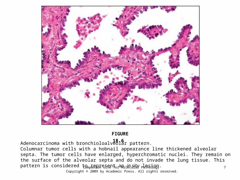
Adenocarcinoma with bronchioloalveolar pattern.Columnar tumor cells with a hobnail appearance line thickened alveolar septa. The tumor cells have enlarged, hyperchromatic nuclei. They remain on the surface of the alveolar septa and do not invade the lung tissue. This pattern is considered to represent an in situ lesion.
FIGURE 18.6
Companion site for Molecular Pathology Copyright © 2009 by Academic Press. All rights reserved.
8
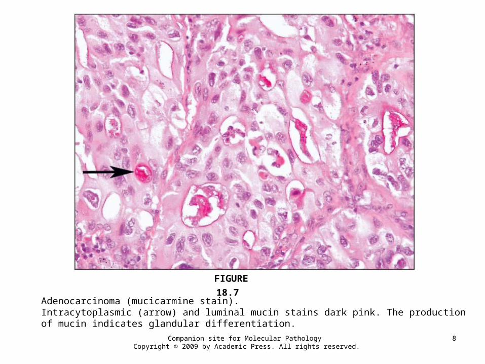
Adenocarcinoma (mucicarmine stain).Intracytoplasmic (arrow) and luminal mucin stains dark pink. The production of mucin indicates glandular differentiation.
FIGURE 18.7
Companion site for Molecular Pathology Copyright © 2009 by Academic Press. All rights reserved.
9
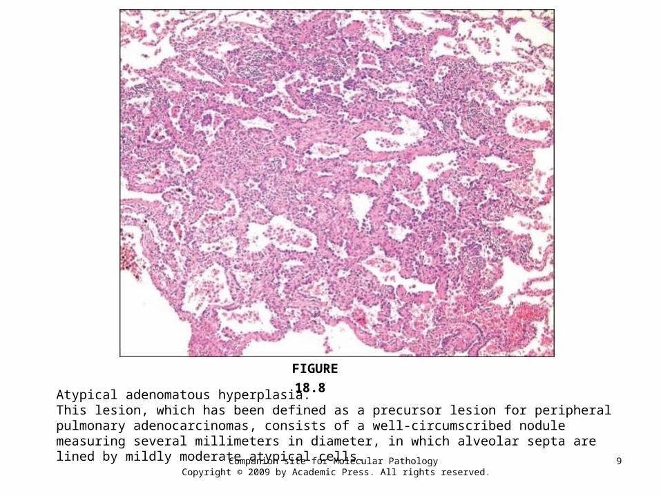
Atypical adenomatous hyperplasia.This lesion, which has been defined as a precursor lesion for peripheral pulmonary adenocarcinomas, consists of a well-circumscribed nodule measuring several millimeters in diameter, in which alveolar septa are lined by mildly moderate atypical cells.
FIGURE 18.8
Companion site for Molecular Pathology Copyright © 2009 by Academic Press. All rights reserved.
10
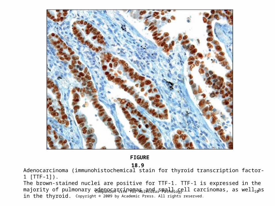
Adenocarcinoma (immunohistochemical stain for thyroid transcription factor-1 [TTF-1]).The brown-stained nuclei are positive for TTF-1. TTF-1 is expressed in the majority of pulmonary adenocarcinomas and small cell carcinomas, as well as in the thyroid.
FIGURE 18.9
Companion site for Molecular Pathology Copyright © 2009 by Academic Press. All rights reserved.
11
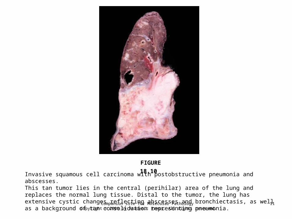
Invasive squamous cell carcinoma with postobstructive pneumonia and abscesses.This tan tumor lies in the central (perihilar) area of the lung and replaces the normal lung tissue. Distal to the tumor, the lung has extensive cystic changes reflecting abscesses and bronchiectasis, as well as a background of tan consolidation representing pneumonia.
FIGURE 18.10
Companion site for Molecular Pathology Copyright © 2009 by Academic Press. All rights reserved.
12
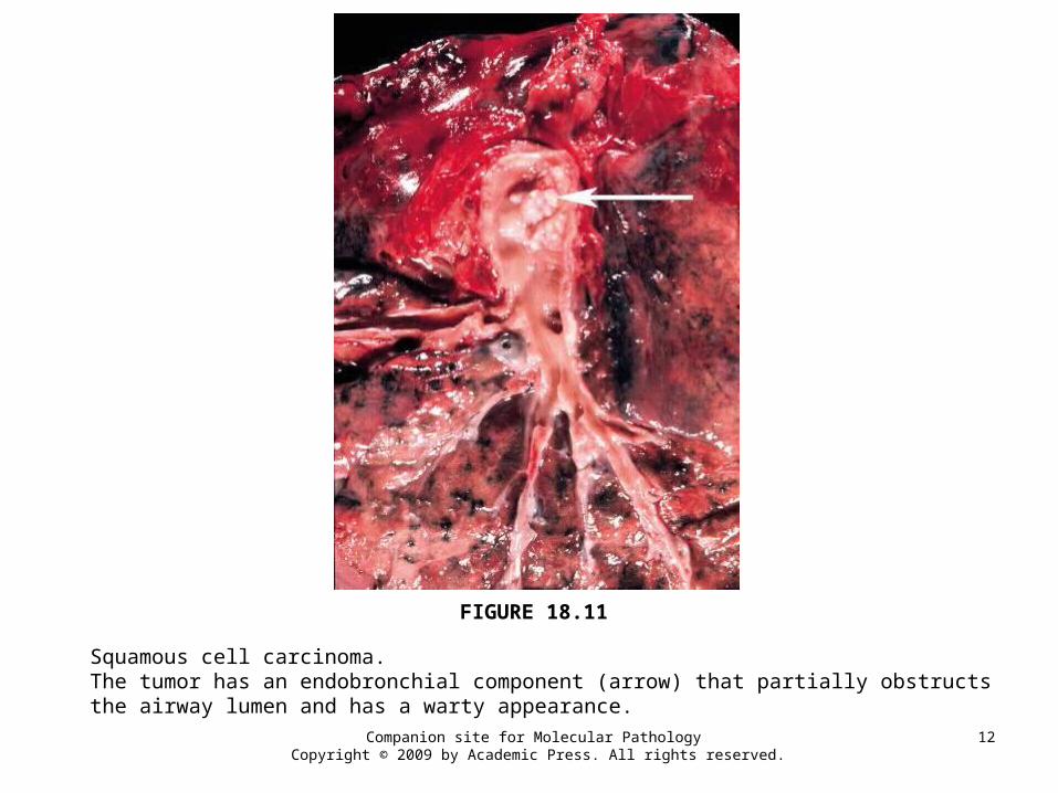
Squamous cell carcinoma.The tumor has an endobronchial component (arrow) that partially obstructs the airway lumen and has a warty appearance.
FIGURE 18.11
Companion site for Molecular Pathology Copyright © 2009 by Academic Press. All rights reserved.
13
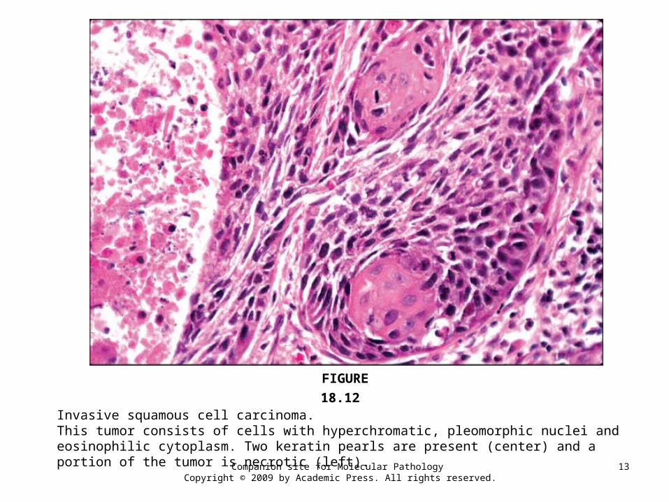
Invasive squamous cell carcinoma.This tumor consists of cells with hyperchromatic, pleomorphic nuclei and eosinophilic cytoplasm. Two keratin pearls are present (center) and a portion of the tumor is necrotic (left).
FIGURE 18.12
Companion site for Molecular Pathology Copyright © 2009 by Academic Press. All rights reserved.
14
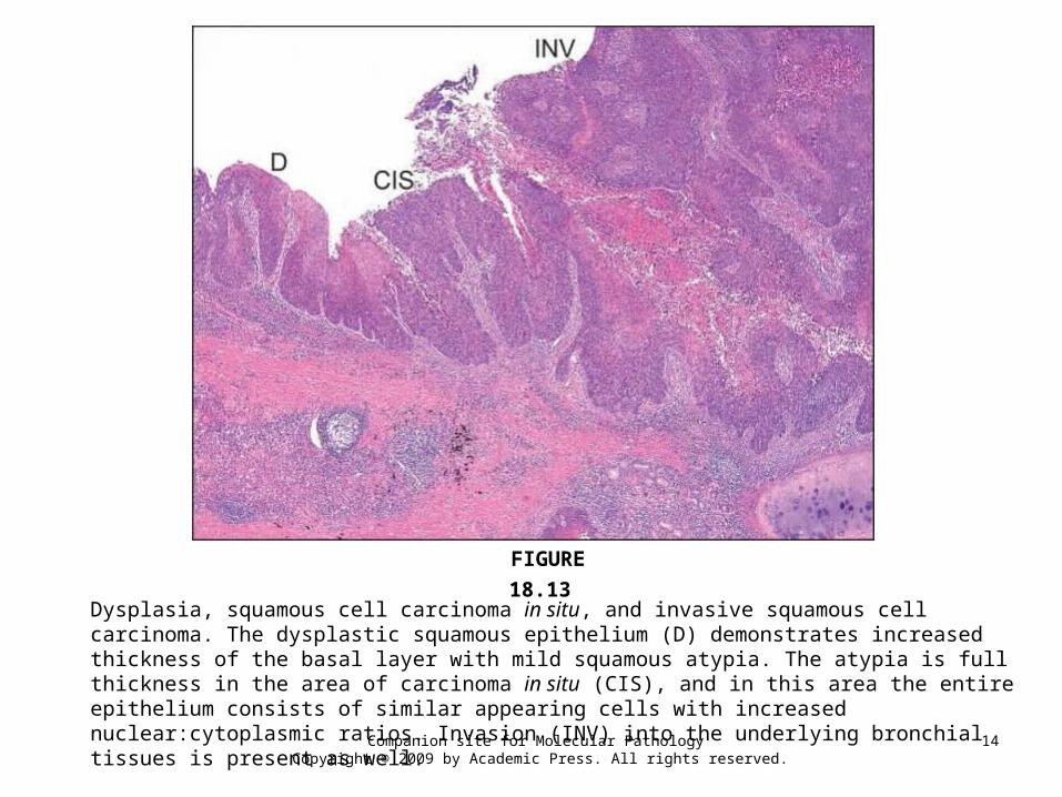
Dysplasia, squamous cell carcinoma in situ, and invasive squamous cell carcinoma. The dysplastic squamous epithelium (D) demonstrates increased thickness of the basal layer with mild squamous atypia. The atypia is full thickness in the area of carcinoma in situ (CIS), and in this area the entire epithelium consists of similar appearing cells with increased nuclear:cytoplasmic ratios. Invasion (INV) into the underlying bronchial tissues is present as well.
FIGURE 18.13
Companion site for Molecular Pathology Copyright © 2009 by Academic Press. All rights reserved.
15
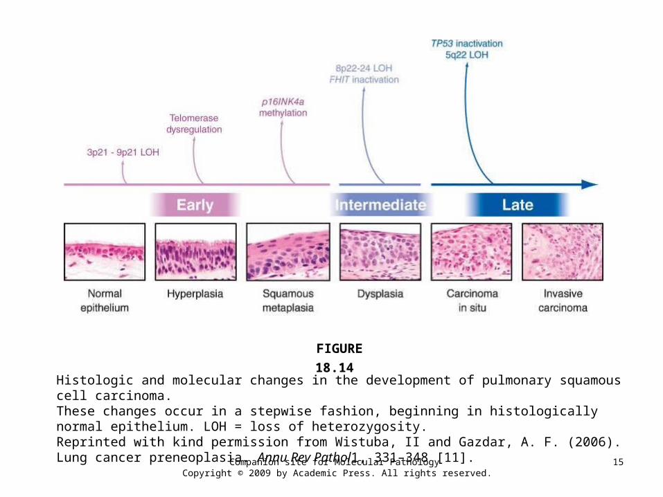
Histologic and molecular changes in the development of pulmonary squamous cell carcinoma.These changes occur in a stepwise fashion, beginning in histologically normal epithelium. LOH = loss of heterozygosity.Reprinted with kind permission from Wistuba, II and Gazdar, A. F. (2006). Lung cancer preneoplasia. Annu Rev Pathol1, 331–348 [11].
FIGURE 18.14
Companion site for Molecular Pathology Copyright © 2009 by Academic Press. All rights reserved.
16
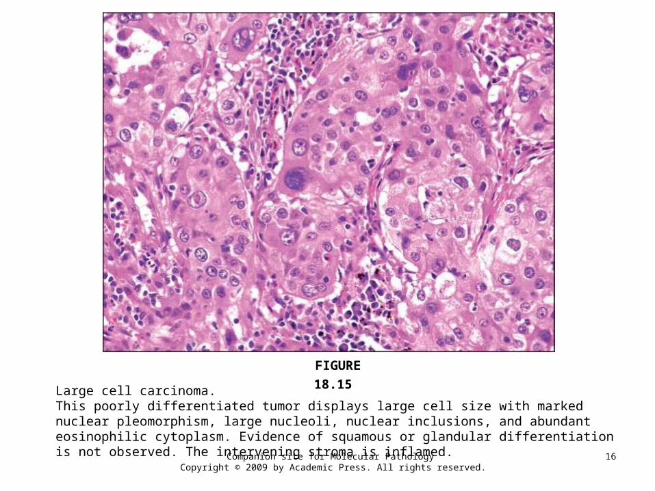
Large cell carcinoma.This poorly differentiated tumor displays large cell size with marked nuclear pleomorphism, large nucleoli, nuclear inclusions, and abundant eosinophilic cytoplasm. Evidence of squamous or glandular differentiation is not observed. The intervening stroma is inflamed.
FIGURE 18.15
Companion site for Molecular Pathology Copyright © 2009 by Academic Press. All rights reserved.
17
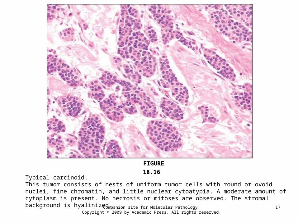
Typical carcinoid.This tumor consists of nests of uniform tumor cells with round or ovoid nuclei, fine chromatin, and little nuclear cytoatypia. A moderate amount of cytoplasm is present. No necrosis or mitoses are observed. The stromal background is hyalinized.
FIGURE 18.16
Companion site for Molecular Pathology Copyright © 2009 by Academic Press. All rights reserved.
18
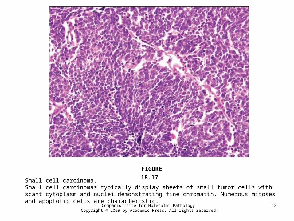
Small cell carcinoma.Small cell carcinomas typically display sheets of small tumor cells with scant cytoplasm and nuclei demonstrating fine chromatin. Numerous mitoses and apoptotic cells are characteristic.
FIGURE 18.17
Companion site for Molecular Pathology Copyright © 2009 by Academic Press. All rights reserved.
19
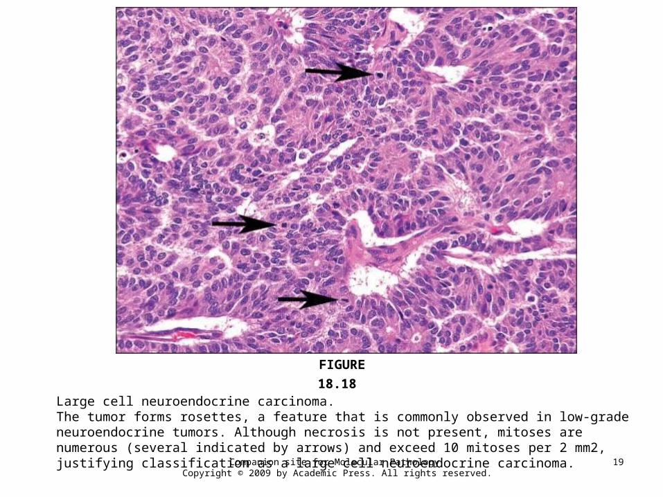
Large cell neuroendocrine carcinoma.The tumor forms rosettes, a feature that is commonly observed in low-grade neuroendocrine tumors. Although necrosis is not present, mitoses are numerous (several indicated by arrows) and exceed 10 mitoses per 2 mm2, justifying classification as a large cell neuroendocrine carcinoma.
FIGURE 18.18
Companion site for Molecular Pathology Copyright © 2009 by Academic Press. All rights reserved.
20
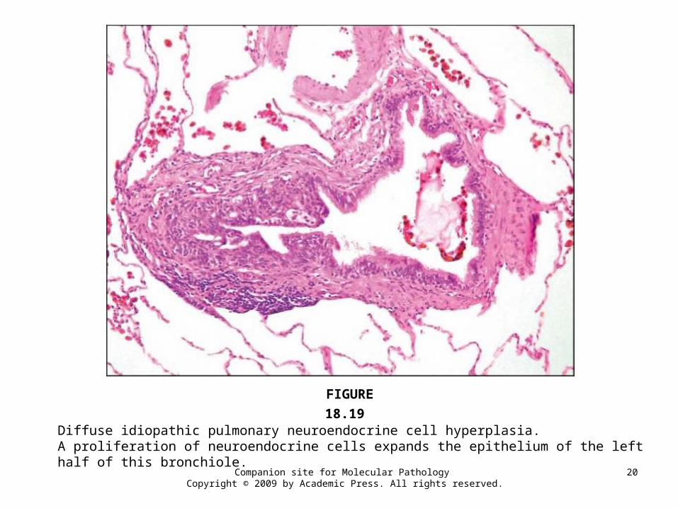
Diffuse idiopathic pulmonary neuroendocrine cell hyperplasia.A proliferation of neuroendocrine cells expands the epithelium of the left half of this bronchiole.
FIGURE 18.19
Companion site for Molecular Pathology Copyright © 2009 by Academic Press. All rights reserved.
21
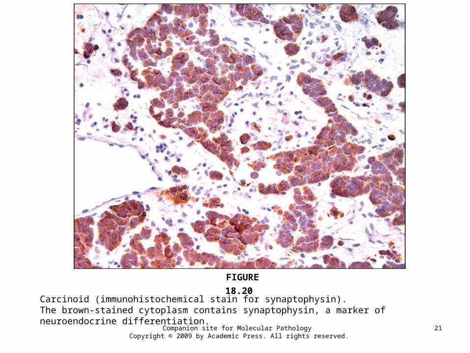
Carcinoid (immunohistochemical stain for synaptophysin).The brown-stained cytoplasm contains synaptophysin, a marker of neuroendocrine differentiation.
FIGURE 18.20
Companion site for Molecular Pathology Copyright © 2009 by Academic Press. All rights reserved.
22
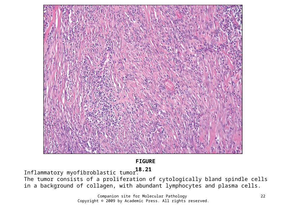
Inflammatory myofibroblastic tumor.The tumor consists of a proliferation of cytologically bland spindle cells in a background of collagen, with abundant lymphocytes and plasma cells.
FIGURE 18.21
Companion site for Molecular Pathology Copyright © 2009 by Academic Press. All rights reserved.
23
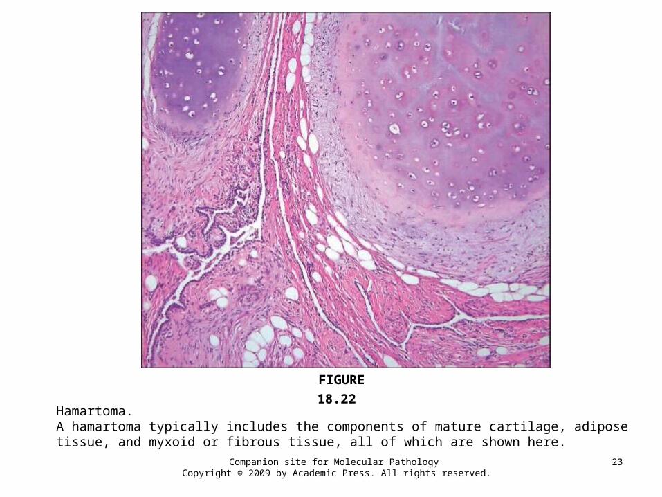
Hamartoma.A hamartoma typically includes the components of mature cartilage, adipose tissue, and myxoid or fibrous tissue, all of which are shown here.
FIGURE 18.22
Companion site for Molecular Pathology Copyright © 2009 by Academic Press. All rights reserved.
24
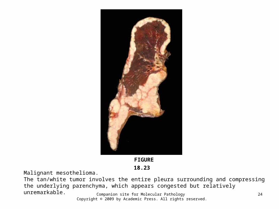
Malignant mesothelioma.The tan/white tumor involves the entire pleura surrounding and compressing the underlying parenchyma, which appears congested but relatively unremarkable.
FIGURE 18.23
Companion site for Molecular Pathology Copyright © 2009 by Academic Press. All rights reserved.
25
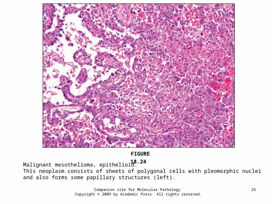
Malignant mesothelioma, epithelioid.This neoplasm consists of sheets of polygonal cells with pleomorphic nuclei and also forms some papillary structures (left).
FIGURE 18.24
Companion site for Molecular Pathology Copyright © 2009 by Academic Press. All rights reserved.
26
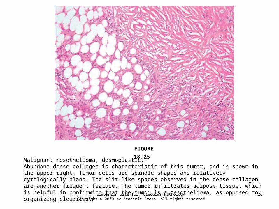
Malignant mesothelioma, desmoplastic.Abundant dense collagen is characteristic of this tumor, and is shown in the upper right. Tumor cells are spindle shaped and relatively cytologically bland. The slit-like spaces observed in the dense collagen are another frequent feature. The tumor infiltrates adipose tissue, which is helpful in confirming that the tumor is a mesothelioma, as opposed to organizing pleuritis.
FIGURE 18.25
Companion site for Molecular Pathology Copyright © 2009 by Academic Press. All rights reserved.
27
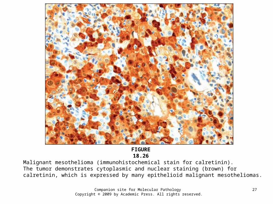
Malignant mesothelioma (immunohistochemical stain for calretinin).The tumor demonstrates cytoplasmic and nuclear staining (brown) for calretinin, which is expressed by many epithelioid malignant mesotheliomas.
FIGURE 18.26
Companion site for Molecular Pathology Copyright © 2009 by Academic Press. All rights reserved.
28
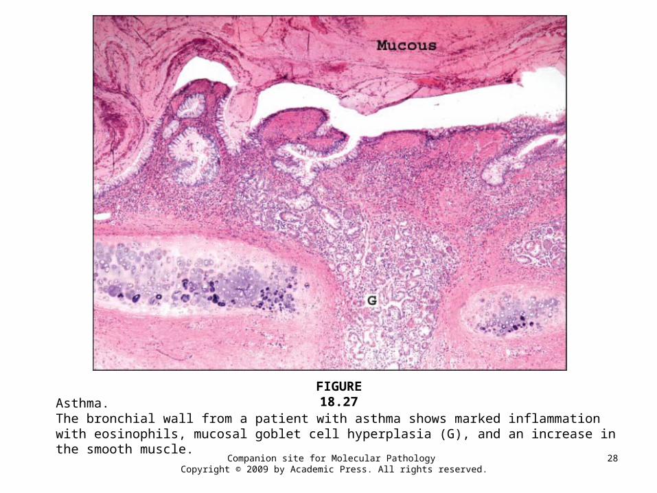
Asthma.The bronchial wall from a patient with asthma shows marked inflammation with eosinophils, mucosal goblet cell hyperplasia (G), and an increase in the smooth muscle.
FIGURE 18.27
Companion site for Molecular Pathology Copyright © 2009 by Academic Press. All rights reserved.
29
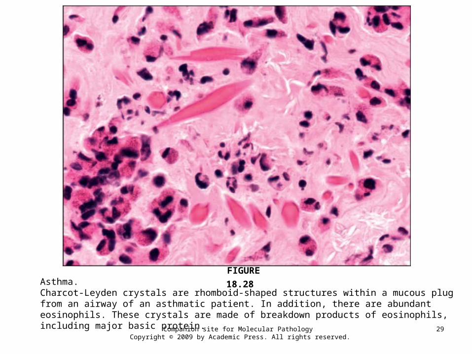
Asthma.Charcot-Leyden crystals are rhomboid-shaped structures within a mucous plug from an airway of an asthmatic patient. In addition, there are abundant eosinophils. These crystals are made of breakdown products of eosinophils, including major basic protein.
FIGURE 18.28
Companion site for Molecular Pathology Copyright © 2009 by Academic Press. All rights reserved.
30
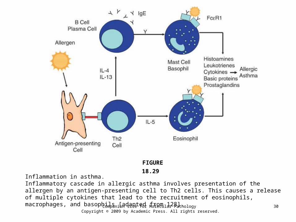
Inflammation in asthma.Inflammatory cascade in allergic asthma involves presentation of the allergen by an antigen-presenting cell to Th2 cells. This causes a release of multiple cytokines that lead to the recruitment of eosinophils, macrophages, and basophils [adapted from 128].
FIGURE 18.29
Companion site for Molecular Pathology Copyright © 2009 by Academic Press. All rights reserved.
31
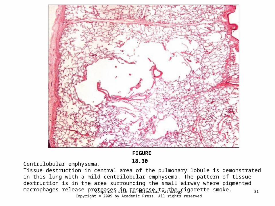
Centrilobular emphysema.Tissue destruction in central area of the pulmonary lobule is demonstrated in this lung with a mild centrilobular emphysema. The pattern of tissue destruction is in the area surrounding the small airway where pigmented macrophages release proteases in response to the cigarette smoke.
FIGURE 18.30
Companion site for Molecular Pathology Copyright © 2009 by Academic Press. All rights reserved.
32
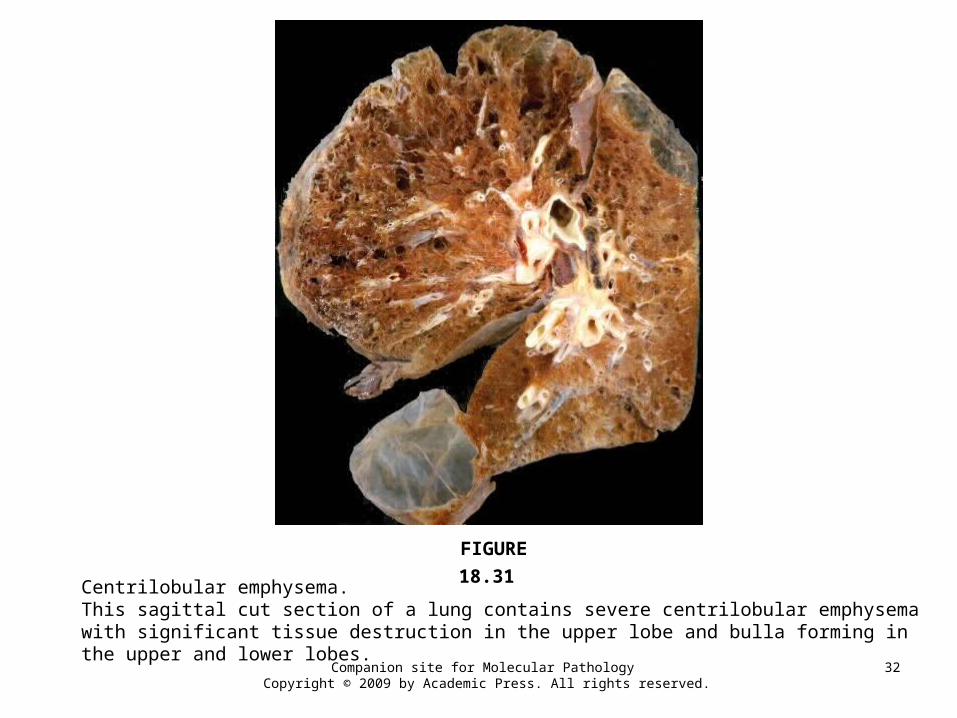
Centrilobular emphysema.This sagittal cut section of a lung contains severe centrilobular emphysema with significant tissue destruction in the upper lobe and bulla forming in the upper and lower lobes.
FIGURE 18.31
Companion site for Molecular Pathology Copyright © 2009 by Academic Press. All rights reserved.
33
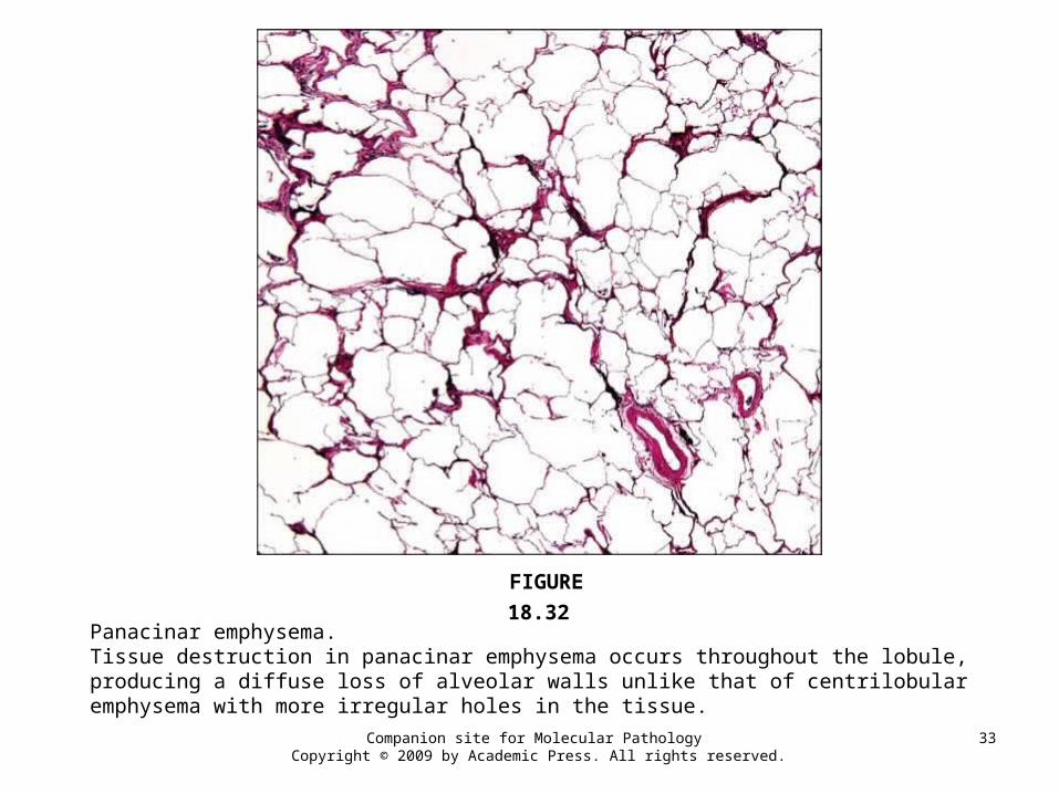
Panacinar emphysema.Tissue destruction in panacinar emphysema occurs throughout the lobule, producing a diffuse loss of alveolar walls unlike that of centrilobular emphysema with more irregular holes in the tissue.
FIGURE 18.32
Companion site for Molecular Pathology Copyright © 2009 by Academic Press. All rights reserved.
34
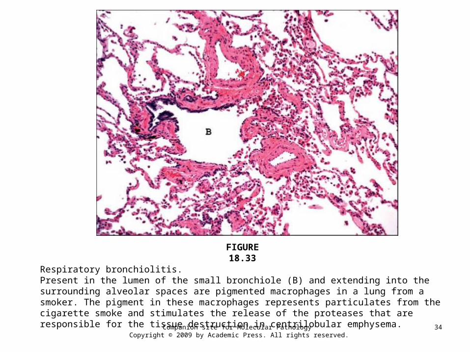
Respiratory bronchiolitis.Present in the lumen of the small bronchiole (B) and extending into the surrounding alveolar spaces are pigmented macrophages in a lung from a smoker. The pigment in these macrophages represents particulates from the cigarette smoke and stimulates the release of the proteases that are responsible for the tissue destruction in centrilobular emphysema.
FIGURE 18.33
Companion site for Molecular Pathology Copyright © 2009 by Academic Press. All rights reserved.
35
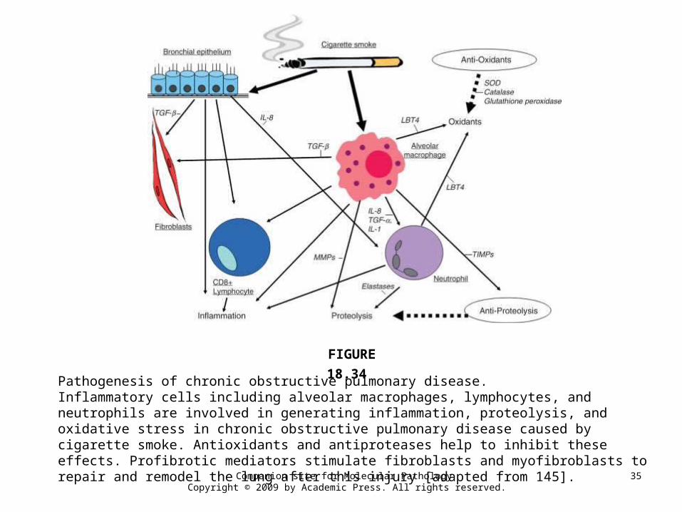
Pathogenesis of chronic obstructive pulmonary disease. Inflammatory cells including alveolar macrophages, lymphocytes, and neutrophils are involved in generating inflammation, proteolysis, and oxidative stress in chronic obstructive pulmonary disease caused by cigarette smoke. Antioxidants and antiproteases help to inhibit these effects. Profibrotic mediators stimulate fibroblasts and myofibroblasts to repair and remodel the lung after this injury [adapted from 145].
FIGURE 18.34
Companion site for Molecular Pathology Copyright © 2009 by Academic Press. All rights reserved.
36
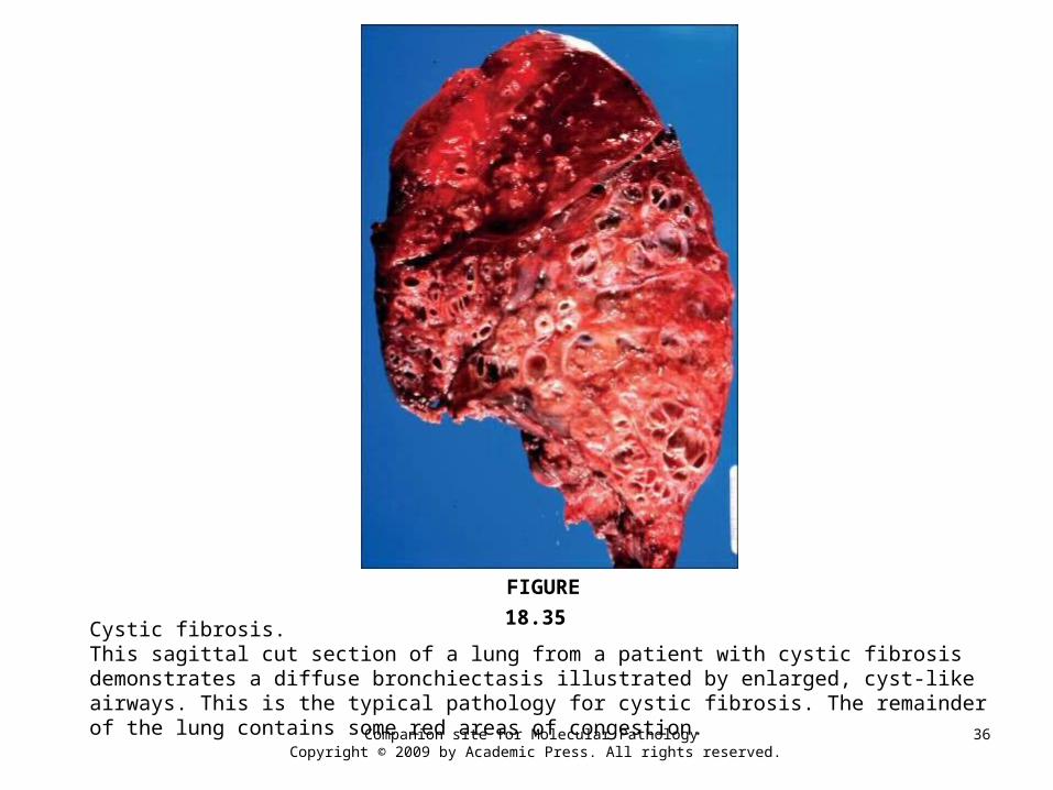
Cystic fibrosis.This sagittal cut section of a lung from a patient with cystic fibrosis demonstrates a diffuse bronchiectasis illustrated by enlarged, cyst-like airways. This is the typical pathology for cystic fibrosis. The remainder of the lung contains some red areas of congestion.
FIGURE 18.35
Companion site for Molecular Pathology Copyright © 2009 by Academic Press. All rights reserved.
37
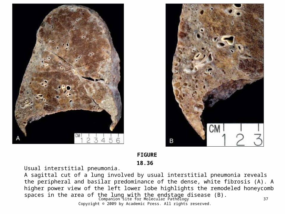
Usual interstitial pneumonia.A sagittal cut of a lung involved by usual interstitial pneumonia reveals the peripheral and basilar predominance of the dense, white fibrosis (A). A higher power view of the left lower lobe highlights the remodeled honeycomb spaces in the area of the lung with the endstage disease (B).
FIGURE 18.36
Companion site for Molecular Pathology Copyright © 2009 by Academic Press. All rights reserved.
38
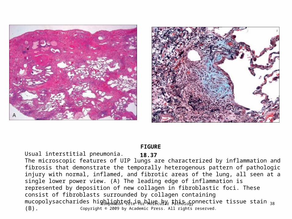
Usual interstitial pneumonia.The microscopic features of UIP lungs are characterized by inflammation and fibrosis that demonstrate the temporally heterogenous pattern of pathologic injury with normal, inflamed, and fibrotic areas of the lung, all seen at a single lower power view. (A) The leading edge of inflammation is represented by deposition of new collagen in fibroblastic foci. These consist of fibroblasts surrounded by collagen containing mucopolysaccharides highlighted in blue by this connective tissue stain (B).
FIGURE 18.37
Companion site for Molecular Pathology Copyright © 2009 by Academic Press. All rights reserved.
39
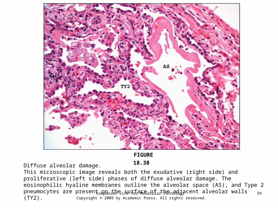
Diffuse alveolar damage.This microscopic image reveals both the exudative (right side) and proliferative (left side) phases of diffuse alveolar damage. The eosinophilic hyaline membranes outline the alveolar space (AS), and Type 2 pneumocytes are present on the surface of the adjacent alveolar walls (TY2).
FIGURE 18.38
Companion site for Molecular Pathology Copyright © 2009 by Academic Press. All rights reserved.
40
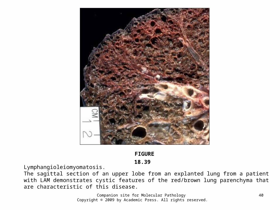
Lymphangioleiomyomatosis.The sagittal section of an upper lobe from an explanted lung from a patient with LAM demonstrates cystic features of the red/brown lung parenchyma that are characteristic of this disease.
FIGURE 18.39
Companion site for Molecular Pathology Copyright © 2009 by Academic Press. All rights reserved.
41
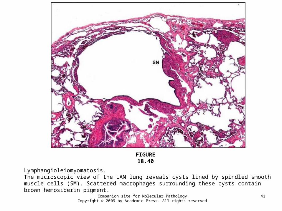
Lymphangioleiomyomatosis.The microscopic view of the LAM lung reveals cysts lined by spindled smooth muscle cells (SM). Scattered macrophages surrounding these cysts contain brown hemosiderin pigment.
FIGURE 18.40
Companion site for Molecular Pathology Copyright © 2009 by Academic Press. All rights reserved.
42
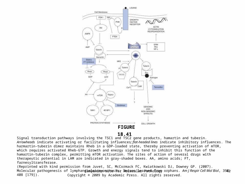
Signal transduction pathways involving the TSC1 and TSC2 gene products, hamartin and tuberin.Arrowheads indicate activating or facilitating influences; flat-headed lines indicate inhibitory influences. The harmartin-tuberin dimer maintains Rheb in a GDP-loaded state, thereby preventing activation of mTOR, which requires activated Rheb-GTP. Growth and energy signals tend to inhibit this function of the hamartin-tuberin complex, permitting mTOR activation. The sites of action of several drugs with therapeutic potential in LAM are indicated in gray-shaded boxes. AA, amino acids; FT, farnesyltransferase. (Reprinted with kind permission from Juvet, SC, McCormack FC, Kwiatkowski DJ, Downey GP. (2007). Molecular pathogenesis of lymphangioleiomyomatosis: lesson learned from orphans. Am J Respir Cell Mol Biol, 398–408 [179]).
FIGURE 18.41
Companion site for Molecular Pathology Copyright © 2009 by Academic Press. All rights reserved.
43
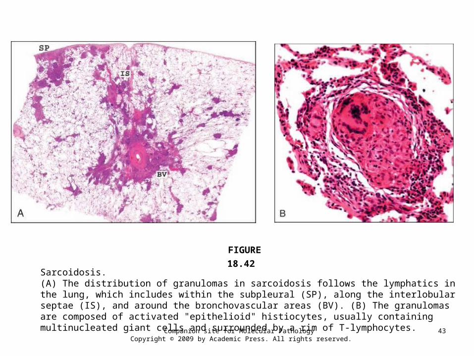
Sarcoidosis.(A) The distribution of granulomas in sarcoidosis follows the lymphatics in the lung, which includes within the subpleural (SP), along the interlobular septae (IS), and around the bronchovascular areas (BV). (B) The granulomas are composed of activated "epithelioid" histiocytes, usually containing multinucleated giant cells and surrounded by a rim of T-lymphocytes.
FIGURE 18.42
Companion site for Molecular Pathology Copyright © 2009 by Academic Press. All rights reserved.
44
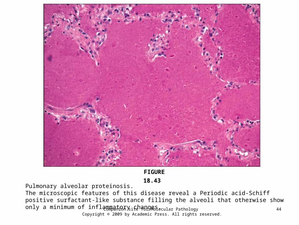
Pulmonary alveolar proteinosis.The microscopic features of this disease reveal a Periodic acid-Schiff positive surfactant-like substance filling the alveoli that otherwise show only a minimum of inflammatory changes.
FIGURE 18.43
Companion site for Molecular Pathology Copyright © 2009 by Academic Press. All rights reserved.
45
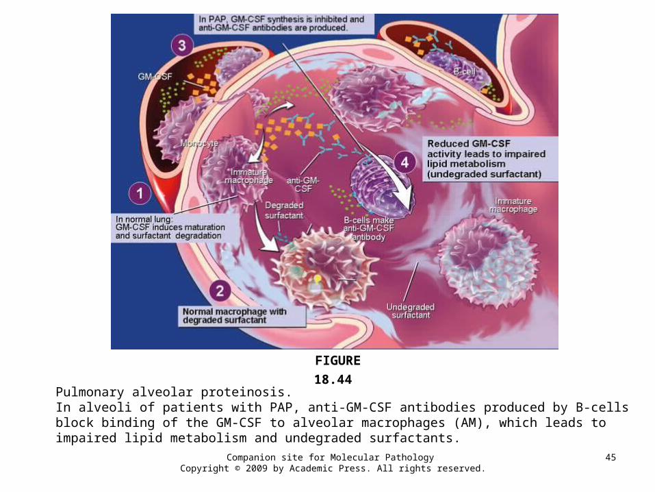
Pulmonary alveolar proteinosis.In alveoli of patients with PAP, anti-GM-CSF antibodies produced by B-cells block binding of the GM-CSF to alveolar macrophages (AM), which leads to impaired lipid metabolism and undegraded surfactants.
FIGURE 18.44
Companion site for Molecular Pathology Copyright © 2009 by Academic Press. All rights reserved.
46
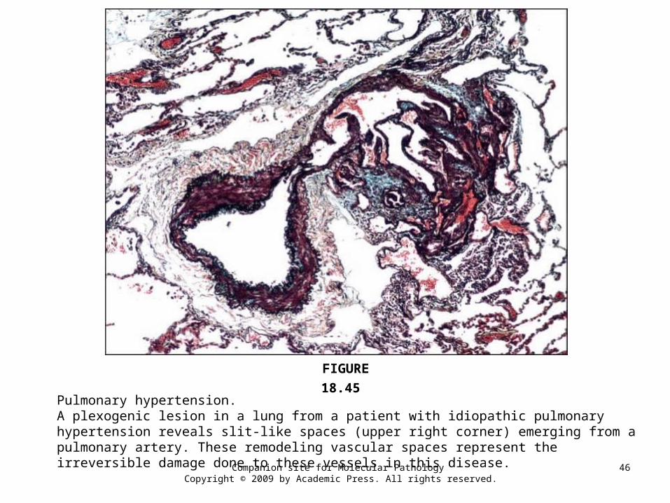
Pulmonary hypertension.A plexogenic lesion in a lung from a patient with idiopathic pulmonary hypertension reveals slit-like spaces (upper right corner) emerging from a pulmonary artery. These remodeling vascular spaces represent the irreversible damage done to these vessels in this disease.
FIGURE 18.45
Companion site for Molecular Pathology Copyright © 2009 by Academic Press. All rights reserved.
47
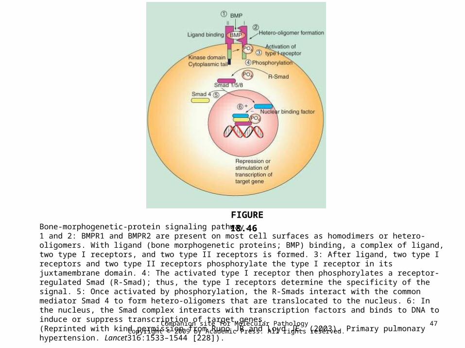
Bone-morphogenetic-protein signaling pathway.1 and 2: BMPR1 and BMPR2 are present on most cell surfaces as homodimers or hetero-oligomers. With ligand (bone morphogenetic proteins; BMP) binding, a complex of ligand, two type I receptors, and two type II receptors is formed. 3: After ligand, two type I receptors and two type II receptors phosphorylate the type I receptor in its juxtamembrane domain. 4: The activated type I receptor then phosphorylates a receptor-regulated Smad (R-Smad); thus, the type I receptors determine the specificity of the signal. 5: Once activated by phosphorylation, the R-Smads interact with the common mediator Smad 4 to form hetero-oligomers that are translocated to the nucleus. 6: In the nucleus, the Smad complex interacts with transcription factors and binds to DNA to induce or suppress transcription of target genes.(Reprinted with kind permission from Runo JR and Loyd JE. (2003). Primary pulmonary hypertension. Lancet316:1533–1544 [228]).
FIGURE 18.46
Companion site for Molecular Pathology Copyright © 2009 by Academic Press. All rights reserved.
48
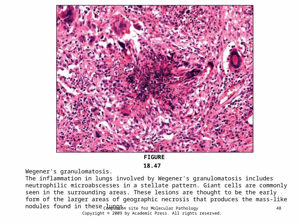
Wegener's granulomatosis.The inflammation in lungs involved by Wegener's granulomatosis includes neutrophilic microabscesses in a stellate pattern. Giant cells are commonly seen in the surrounding areas. These lesions are thought to be the early form of the larger areas of geographic necrosis that produces the mass-like nodules found in these lungs.
FIGURE 18.47
Companion site for Molecular Pathology Copyright © 2009 by Academic Press. All rights reserved.
49
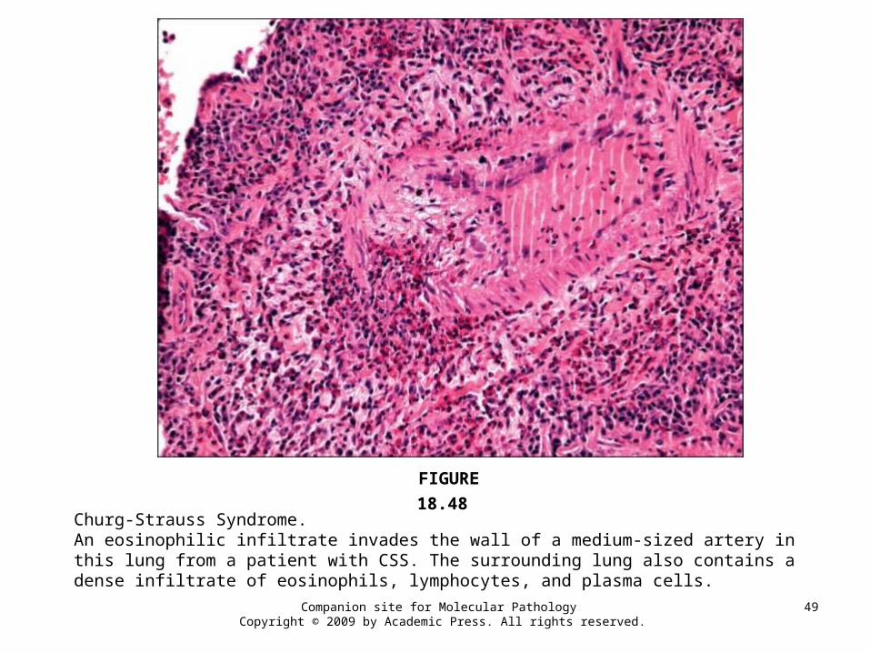
Churg-Strauss Syndrome.An eosinophilic infiltrate invades the wall of a medium-sized artery in this lung from a patient with CSS. The surrounding lung also contains a dense infiltrate of eosinophils, lymphocytes, and plasma cells.
FIGURE 18.48
Companion site for Molecular Pathology Copyright © 2009 by Academic Press. All rights reserved.
50
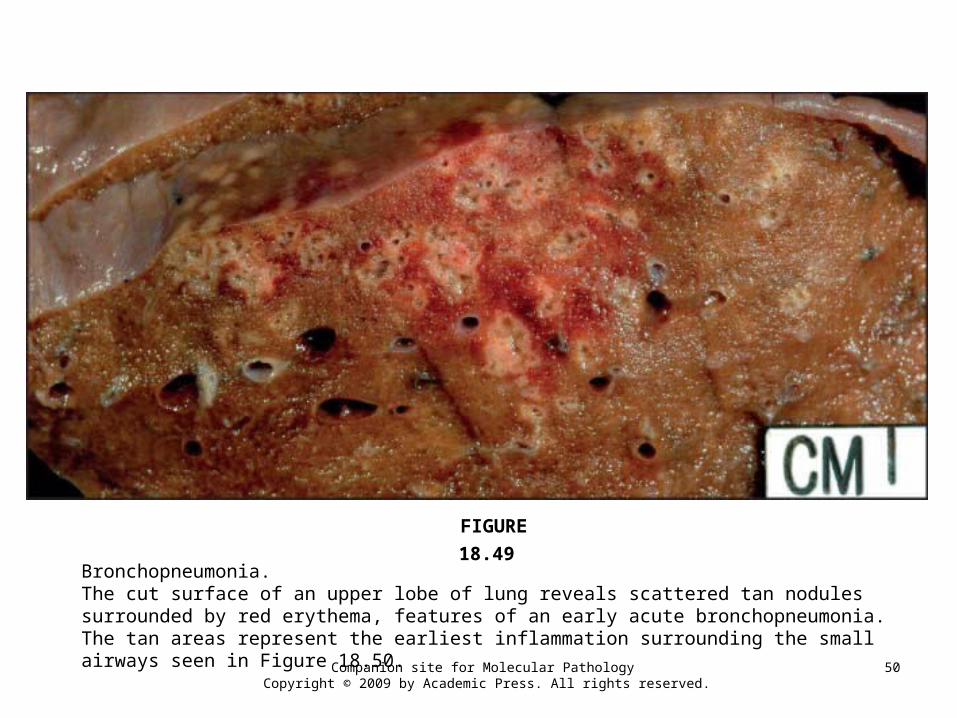
Bronchopneumonia.The cut surface of an upper lobe of lung reveals scattered tan nodules surrounded by red erythema, features of an early acute bronchopneumonia. The tan areas represent the earliest inflammation surrounding the small airways seen in Figure 18.50.
FIGURE 18.49
Companion site for Molecular Pathology Copyright © 2009 by Academic Press. All rights reserved.
51
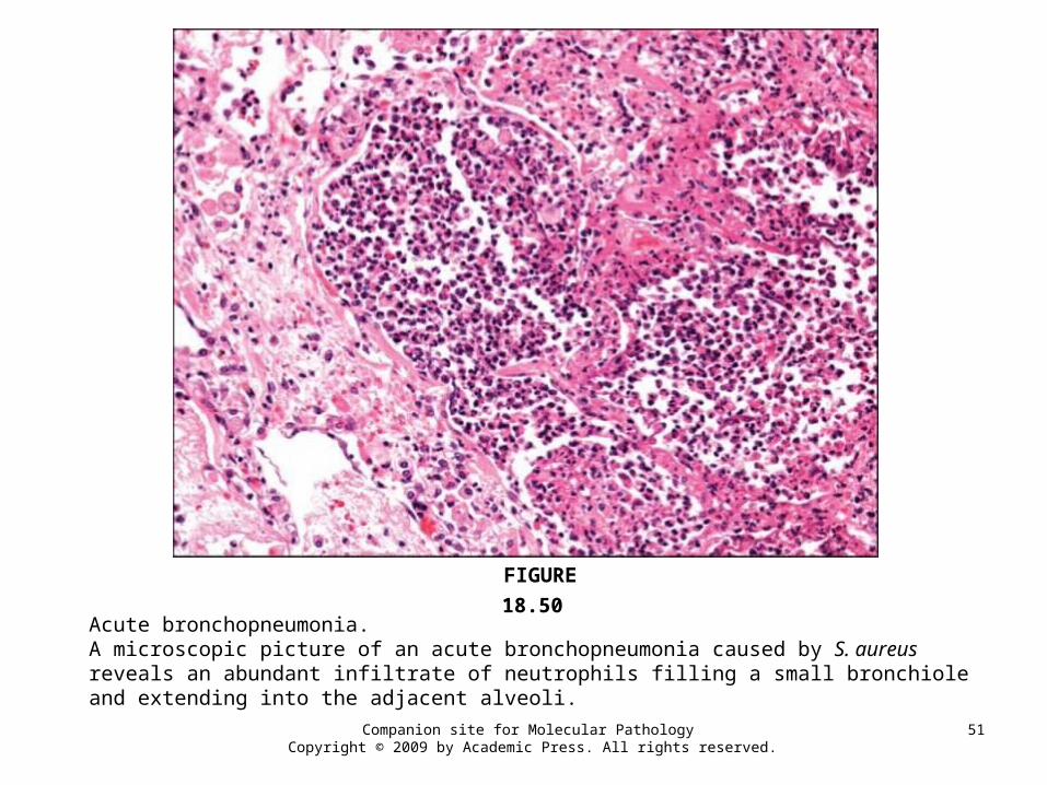
Acute bronchopneumonia.A microscopic picture of an acute bronchopneumonia caused by S. aureus reveals an abundant infiltrate of neutrophils filling a small bronchiole and extending into the adjacent alveoli.
FIGURE 18.50
Companion site for Molecular Pathology Copyright © 2009 by Academic Press. All rights reserved.
52
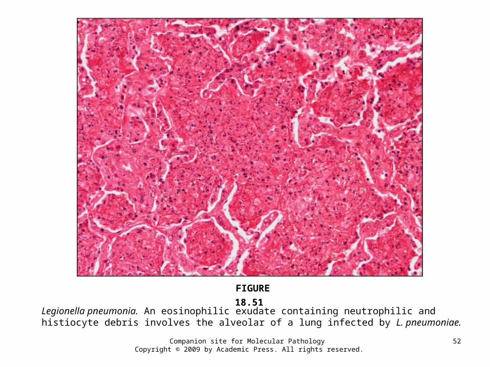
Legionella pneumonia. An eosinophilic exudate containing neutrophilic and histiocyte debris involves the alveolar of a lung infected by L. pneumoniae.
FIGURE 18.51
Companion site for Molecular Pathology Copyright © 2009 by Academic Press. All rights reserved.
53
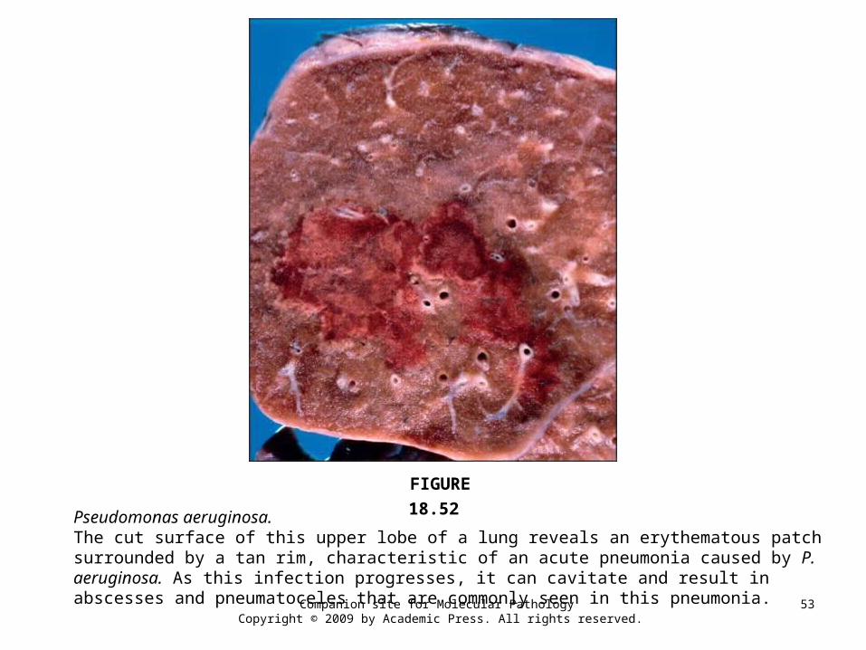
Pseudomonas aeruginosa.The cut surface of this upper lobe of a lung reveals an erythematous patch surrounded by a tan rim, characteristic of an acute pneumonia caused by P. aeruginosa. As this infection progresses, it can cavitate and result in abscesses and pneumatoceles that are commonly seen in this pneumonia.
FIGURE 18.52
Companion site for Molecular Pathology Copyright © 2009 by Academic Press. All rights reserved.
54
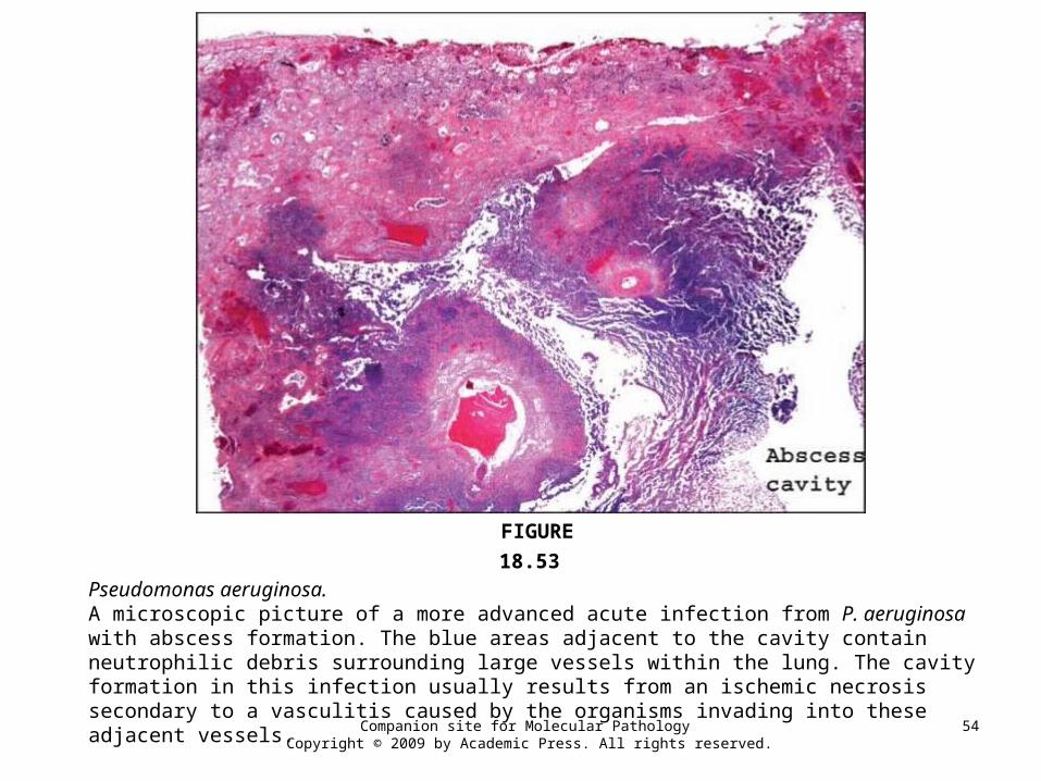
Pseudomonas aeruginosa.A microscopic picture of a more advanced acute infection from P. aeruginosa with abscess formation. The blue areas adjacent to the cavity contain neutrophilic debris surrounding large vessels within the lung. The cavity formation in this infection usually results from an ischemic necrosis secondary to a vasculitis caused by the organisms invading into these adjacent vessels.
FIGURE 18.53
Companion site for Molecular Pathology Copyright © 2009 by Academic Press. All rights reserved.
55
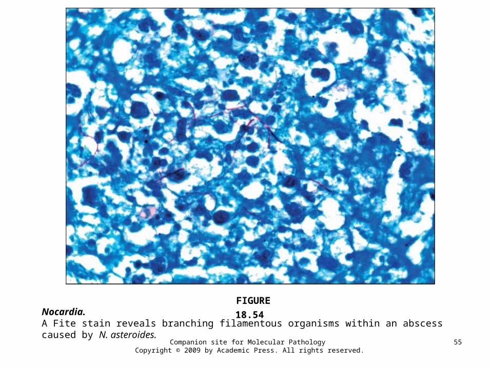
Nocardia.A Fite stain reveals branching filamentous organisms within an abscess caused by N. asteroides.
FIGURE 18.54
Companion site for Molecular Pathology Copyright © 2009 by Academic Press. All rights reserved.
56
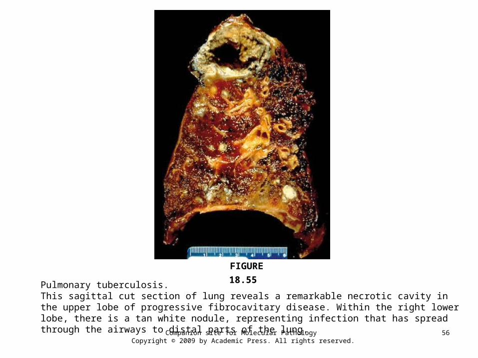
Pulmonary tuberculosis.This sagittal cut section of lung reveals a remarkable necrotic cavity in the upper lobe of progressive fibrocavitary disease. Within the right lower lobe, there is a tan white nodule, representing infection that has spread through the airways to distal parts of the lung.
FIGURE 18.55
Companion site for Molecular Pathology Copyright © 2009 by Academic Press. All rights reserved.
57
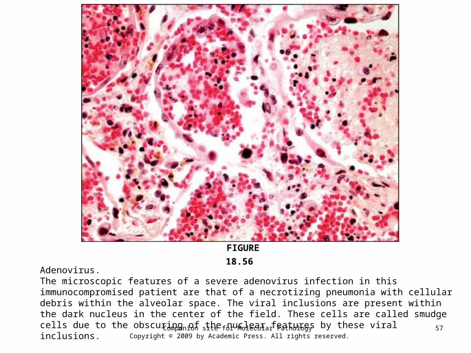
Adenovirus.The microscopic features of a severe adenovirus infection in this immunocompromised patient are that of a necrotizing pneumonia with cellular debris within the alveolar space. The viral inclusions are present within the dark nucleus in the center of the field. These cells are called smudge cells due to the obscuring of the nuclear features by these viral inclusions.
FIGURE 18.56
Companion site for Molecular Pathology Copyright © 2009 by Academic Press. All rights reserved.
58
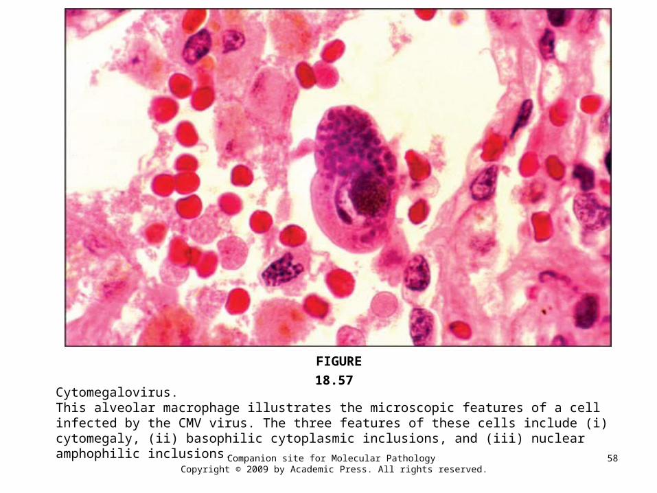
Cytomegalovirus.This alveolar macrophage illustrates the microscopic features of a cell infected by the CMV virus. The three features of these cells include (i) cytomegaly, (ii) basophilic cytoplasmic inclusions, and (iii) nuclear amphophilic inclusions.
FIGURE 18.57
Companion site for Molecular Pathology Copyright © 2009 by Academic Press. All rights reserved.
59
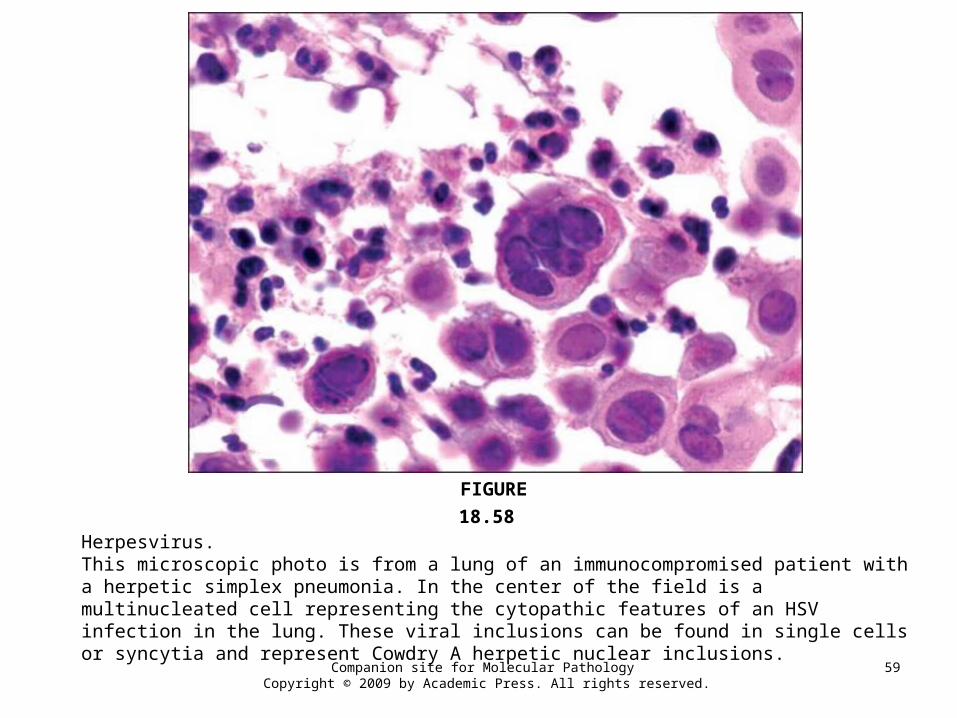
Herpesvirus.This microscopic photo is from a lung of an immunocompromised patient with a herpetic simplex pneumonia. In the center of the field is a multinucleated cell representing the cytopathic features of an HSV infection in the lung. These viral inclusions can be found in single cells or syncytia and represent Cowdry A herpetic nuclear inclusions.
FIGURE 18.58
Companion site for Molecular Pathology Copyright © 2009 by Academic Press. All rights reserved.
60
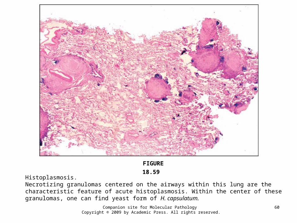
Histoplasmosis.Necrotizing granulomas centered on the airways within this lung are the characteristic feature of acute histoplasmosis. Within the center of these granulomas, one can find yeast form of H. capsulatum.
FIGURE 18.59
Companion site for Molecular Pathology Copyright © 2009 by Academic Press. All rights reserved.
61
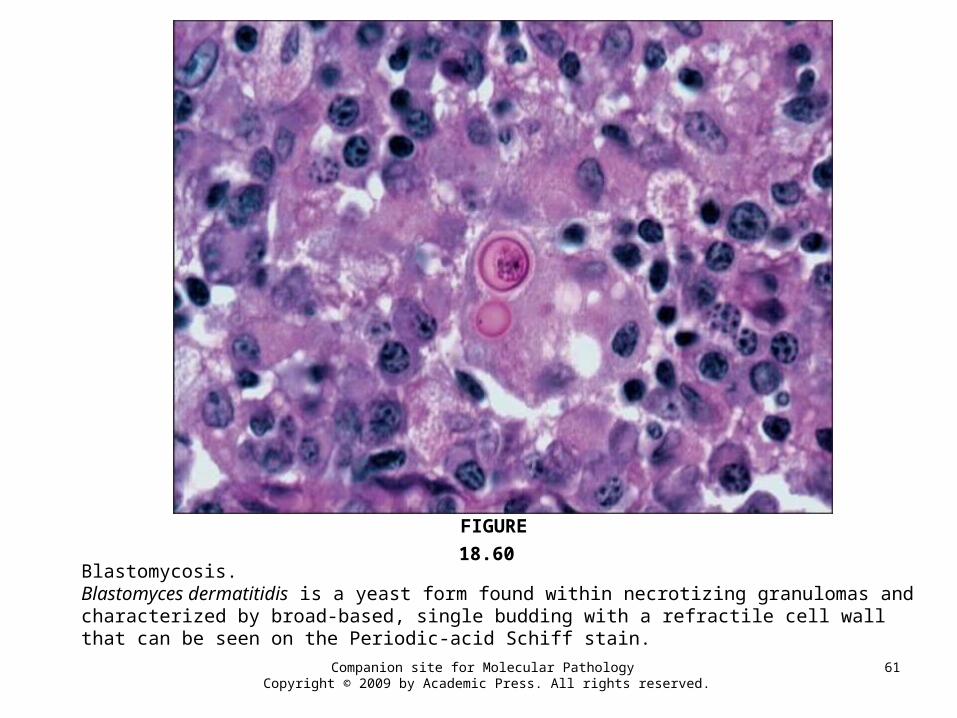
Blastomycosis.Blastomyces dermatitidis is a yeast form found within necrotizing granulomas and characterized by broad-based, single budding with a refractile cell wall that can be seen on the Periodic-acid Schiff stain.
FIGURE 18.60
Companion site for Molecular Pathology Copyright © 2009 by Academic Press. All rights reserved.
62
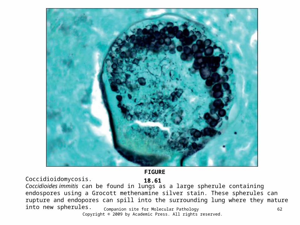
Coccidioidomycosis.Coccidioides immitis can be found in lungs as a large spherule containing endospores using a Grocott methenamine silver stain. These spherules can rupture and endopores can spill into the surrounding lung where they mature into new spherules.
FIGURE 18.61
Companion site for Molecular Pathology Copyright © 2009 by Academic Press. All rights reserved.
63
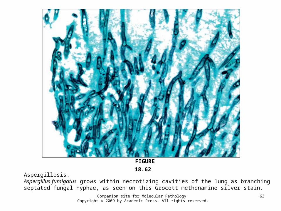
Aspergillosis.Aspergillus fumigatus grows within necrotizing cavities of the lung as branching septated fungal hyphae, as seen on this Grocott methenamine silver stain.
FIGURE 18.62
Companion site for Molecular Pathology Copyright © 2009 by Academic Press. All rights reserved.
64
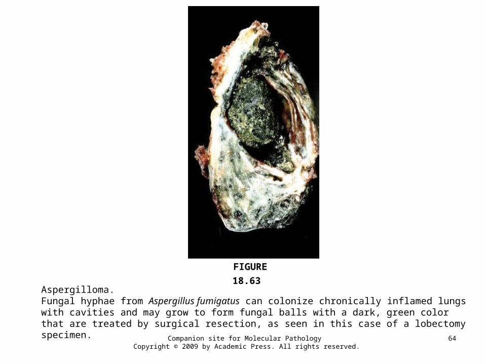
Aspergilloma.Fungal hyphae from Aspergillus fumigatus can colonize chronically inflamed lungs with cavities and may grow to form fungal balls with a dark, green color that are treated by surgical resection, as seen in this case of a lobectomy specimen.
FIGURE 18.63
Companion site for Molecular Pathology Copyright © 2009 by Academic Press. All rights reserved.
65
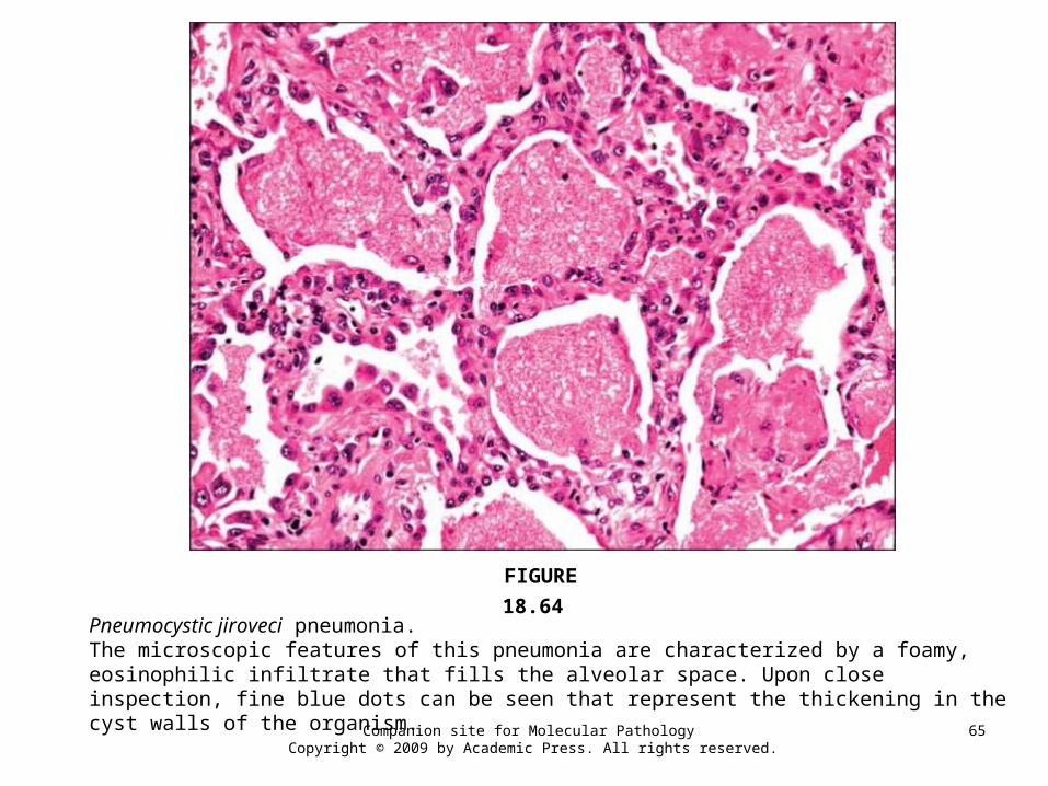
Pneumocystic jiroveci pneumonia.The microscopic features of this pneumonia are characterized by a foamy, eosinophilic infiltrate that fills the alveolar space. Upon close inspection, fine blue dots can be seen that represent the thickening in the cyst walls of the organism.
FIGURE 18.64
Companion site for Molecular Pathology Copyright © 2009 by Academic Press. All rights reserved.
66
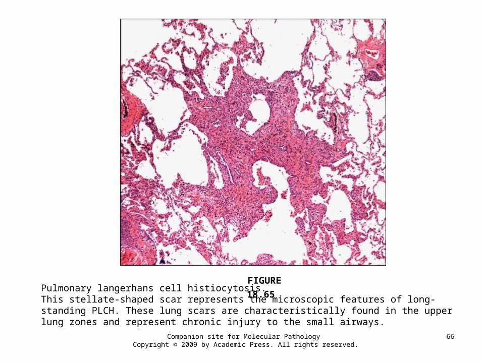
Pulmonary langerhans cell histiocytosis.This stellate-shaped scar represents the microscopic features of long-standing PLCH. These lung scars are characteristically found in the upper lung zones and represent chronic injury to the small airways.
FIGURE 18.65
Companion site for Molecular Pathology Copyright © 2009 by Academic Press. All rights reserved.
67
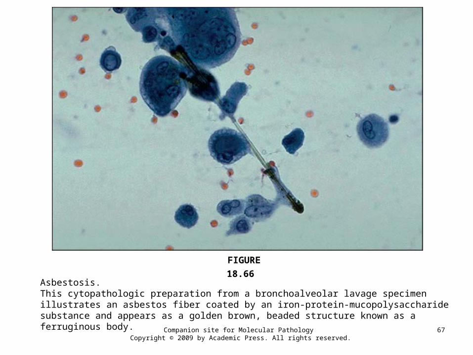
Asbestosis.This cytopathologic preparation from a bronchoalveolar lavage specimen illustrates an asbestos fiber coated by an iron-protein-mucopolysaccharide substance and appears as a golden brown, beaded structure known as a ferruginous body.
FIGURE 18.66
Companion site for Molecular Pathology Copyright © 2009 by Academic Press. All rights reserved.
68
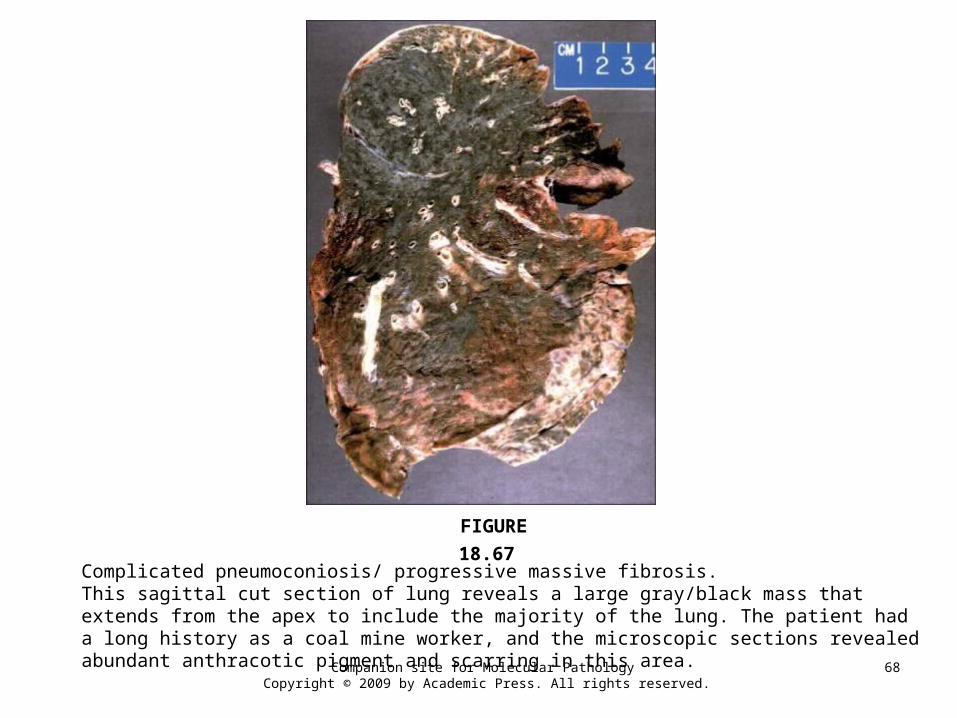
Complicated pneumoconiosis/ progressive massive fibrosis.This sagittal cut section of lung reveals a large gray/black mass that extends from the apex to include the majority of the lung. The patient had a long history as a coal mine worker, and the microscopic sections revealed abundant anthracotic pigment and scarring in this area.
FIGURE 18.67
Companion site for Molecular Pathology Copyright © 2009 by Academic Press. All rights reserved.
69
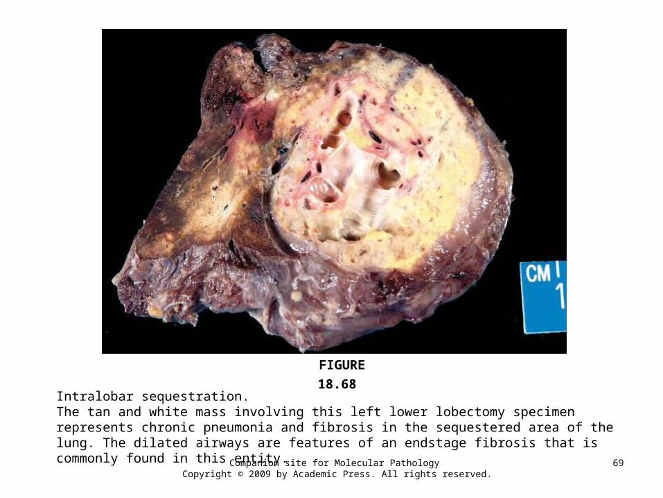
Intralobar sequestration.The tan and white mass involving this left lower lobectomy specimen represents chronic pneumonia and fibrosis in the sequestered area of the lung. The dilated airways are features of an endstage fibrosis that is commonly found in this entity.
FIGURE 18.68


![Valvular Heart Disease 10.24.ppt [Read-Only] · 1 Valvular Heart Disease General Principles • Etiology • Cellular and molecular mechanism of valve damage • Structural pathology](https://img.pdfslide.net/doc/110x75/5af1fadb7f8b9a8b4c8f76d1/valvular-heart-disease-1024ppt-read-only-valvular-heart-disease-general-principles.jpg)