Embed Size (px)
Citation preview
Chapter 19 Aminoacids and Proteins
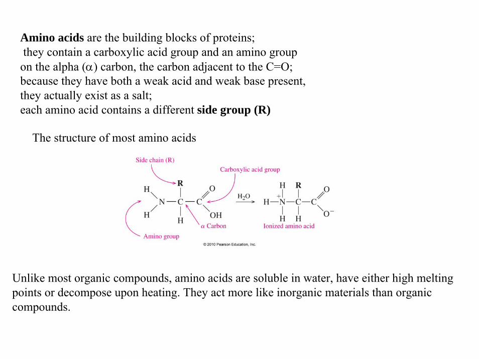
The structure of most amino acids
Amino acids are the building blocks of proteins;they contain a carboxylic acid group and an amino group on the alpha (α) carbon, the carbon adjacent to the C=O;because they have both a weak acid and weak base present, they actually exist as a salt;each amino acid contains a different side group (R)
Unlike most organic compounds, amino acids are soluble in water, have either high melting points or decompose upon heating. They act more like inorganic materials than organic compounds.
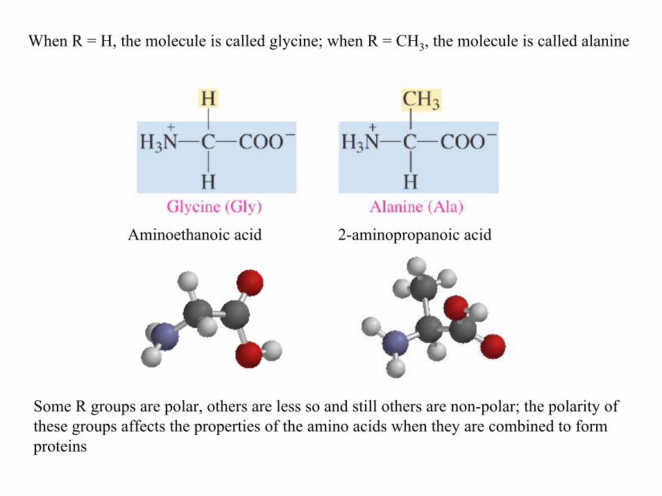
When R = H, the molecule is called glycine; when R = CH3, the molecule is called alanine
Some R groups are polar, others are less so and still others are non-polar; the polarity of these groups affects the properties of the amino acids when they are combined to form proteins
Aminoethanoic acid 2-aminopropanoic acid

The polar aminoacids
less polar
non-polar
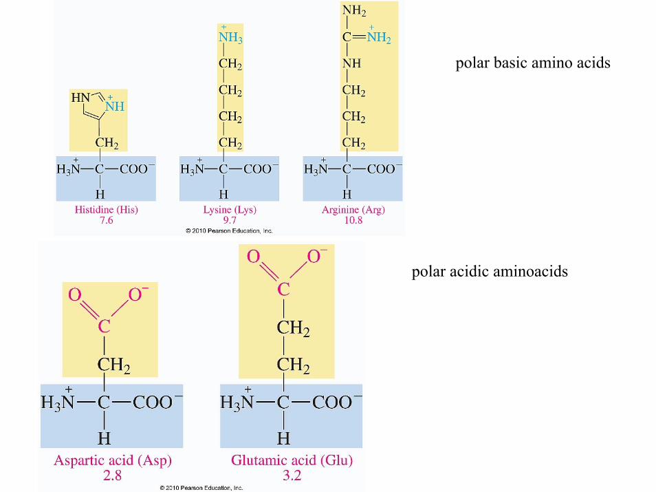
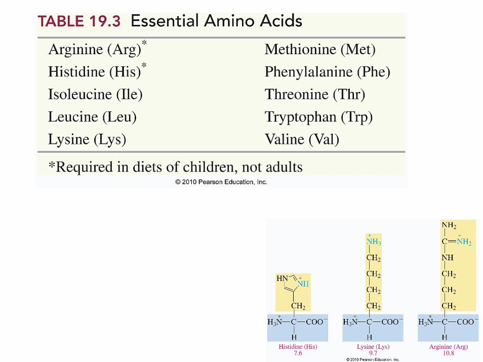
polar basic amino acids
polar acidic aminoacids
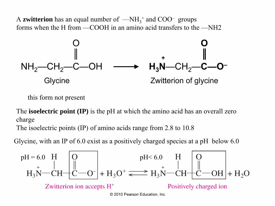
A zwitterion has an equal number of —NH3+ and COO– groups
forms when the H from —COOH in an amino acid transfers to the —NH2
this form not present
The isoelectric point (IP) is the pH at which the amino acid has an overall zero charge The isoelectric points (IP) of amino acids range from 2.8 to 10.8
Glycine, with an IP of 6.0 exist as a positively charged species at a pH below 6.0
pH = 6.0 pH< 6.0
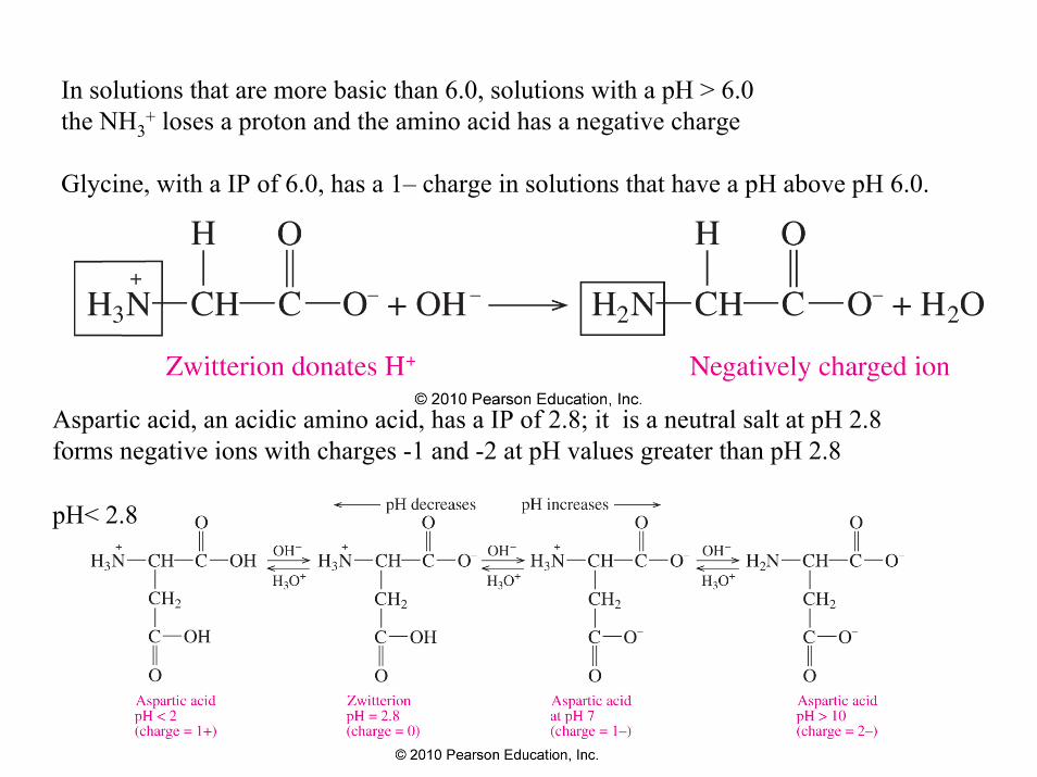
In solutions that are more basic than 6.0, solutions with a pH > 6.0 the NH3
+ loses a proton and the amino acid has a negative charge
Glycine, with a IP of 6.0, has a 1– charge in solutions that have a pH above pH 6.0.
Aspartic acid, an acidic amino acid, has a IP of 2.8; it is a neutral salt at pH 2.8forms negative ions with charges -1 and -2 at pH values greater than pH 2.8
pH< 2.8
In electrophoresis, an electric current is used to separate a mixture of amino acids;the positively charged amino acids move toward the negative electrode,the negatively charged amino acids move toward the positive electrode an amino acid at its pI does not migrate;the amino acids are identified as separate bands on the filter paper or thin layer plate
Why is the isolectric points of amino acids significant?
They provide a means of separating and identifying them
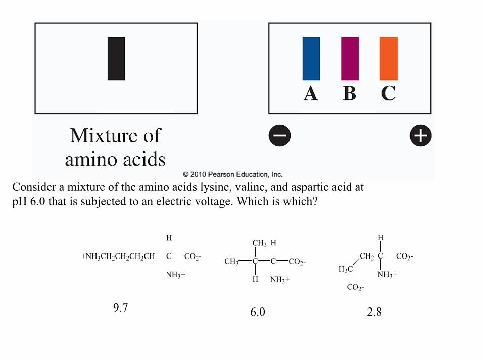
Consider a mixture of the amino acids lysine, valine, and aspartic acid at pH 6.0 that is subjected to an electric voltage. Which is which?
CO2-C
NH3+
+NH3CH2CH2CH2CH
H
CO2-C
NH3+
C
HCH3
H
CH3CO2-C
NH3+
CH2
H
H2C
CO2-
9.7 6.0 2.8
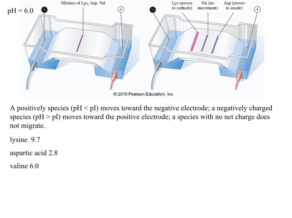
A positively species (pH < pI) moves toward the negative electrode; a negatively charged species (pH > pI) moves toward the positive electrode; a species with no net charge does not migrate.
lysine 9.7
aspartic acid 2.8
valine 6.0
pH = 6.0
lysine 9.7
aspartic acid 2.8
valine 6.0
The numbers associated with each amino acid is called the isoelectric point. It represents the pH at which the amino acid is neutral. For valine at a pH of 6 we have
CH3
CH3
H NH3+
O
O-
pH > 6 pH = 6 pH < 6
CH3
CH3
H NH3+
O
OH
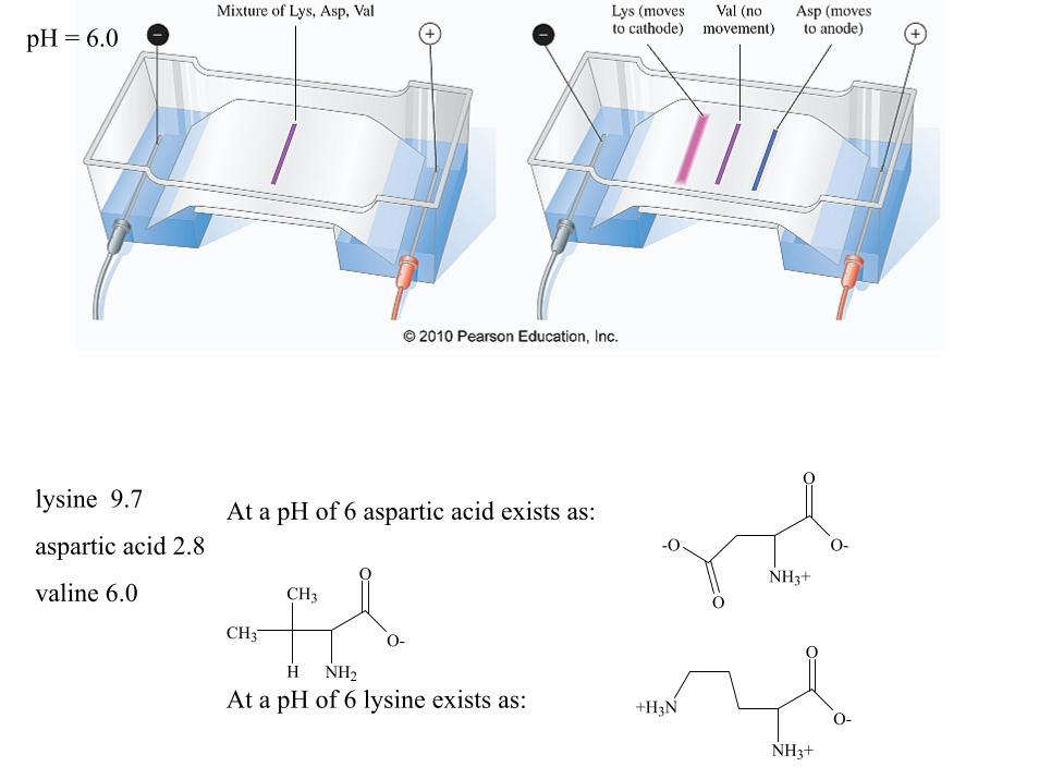
At a pH of 6 aspartic acid exists as:
At a pH of 6 lysine exists as:
NH3+
O
O--O
O
NH3+
O
O-+H3N
CH3
CH3
H NH2
O
O-
pH = 6.0
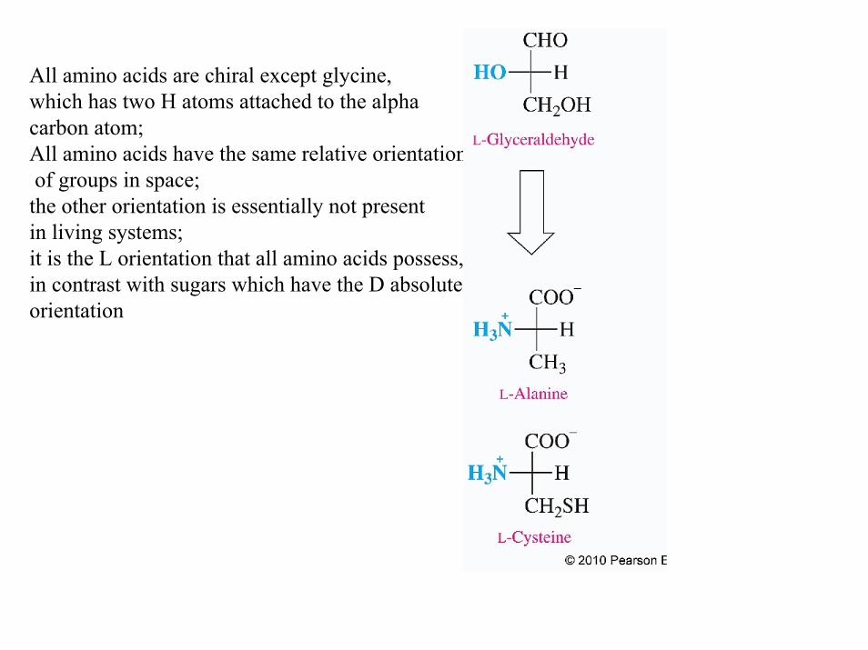
All amino acids are chiral except glycine, which has two H atoms attached to the alpha carbon atom; All amino acids have the same relative orientationof groups in space; the other orientation is essentially not present in living systems;it is the L orientation that all amino acids possess,in contrast with sugars which have the D absolute orientation
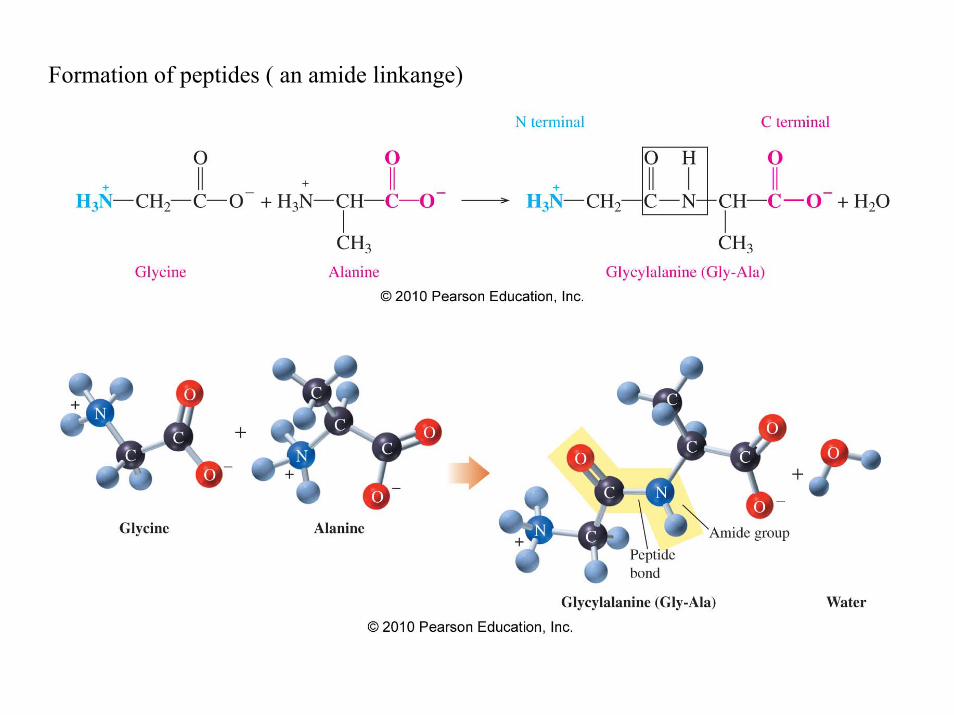
Formation of peptides ( an amide linkange)

A dipeptide is named with a yl ending for the N-terminal (free H3N+) amino acid in sequencethe full amino acid name of the free carboxyl group (COO–) at the C-terminal end
N -terminal C-terminal
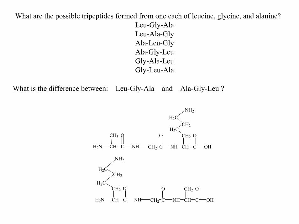
What are the possible tripeptides formed from one each of leucine, glycine, and alanine?Leu-Gly-AlaLeu-Ala-GlyAla-Leu-GlyAla-Gly-LeuGly-Ala-LeuGly-Leu-Ala
What is the difference between: Leu-Gly-Ala and Ala-Gly-Leu ?
CHH2N
CH3
C NH
O
CH2 C
O
NH CH C OH
OCH2
H2CCH2
H2CNH2
CHH2N
CH2
C NH
OH2C
CH2H2C
NH2
CH2 C
O
NH CH C OH
OCH2
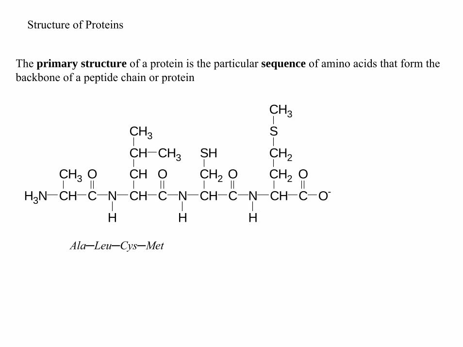
The primary structure of a protein is the particular sequence of amino acids that form the backbone of a peptide chain or protein
Structure of Proteins
CH3
SHCH2
CH3
S
CH2
CH2CH O
O-CCH
H
N
O
CCH
H
N
O
CCH
H
N
O
CCHH3N
CH3
CH3CH
Ala─Leu─Cys─Met
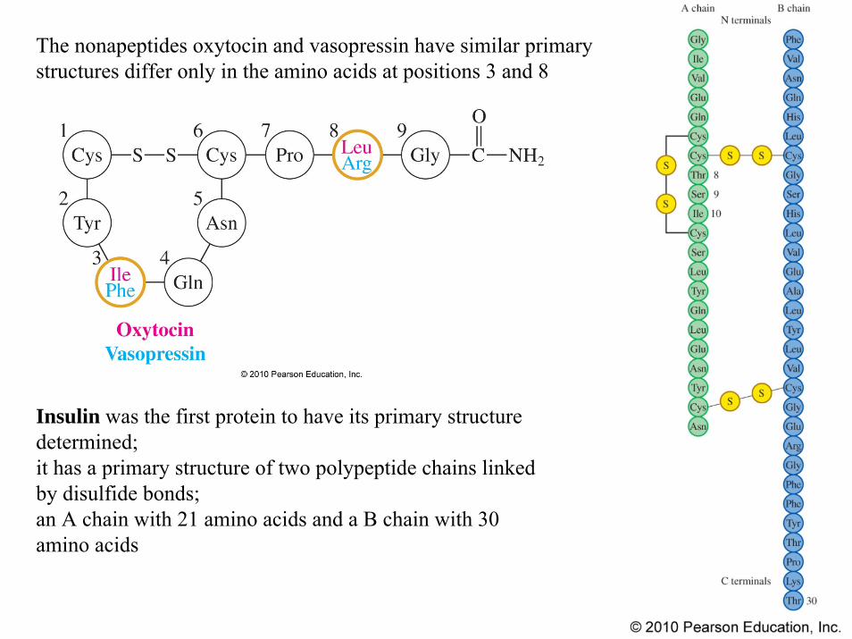
The nonapeptides oxytocin and vasopressin have similar primary structures differ only in the amino acids at positions 3 and 8
Insulin was the first protein to have its primary structure determined;it has a primary structure of two polypeptide chains linked by disulfide bonds; an A chain with 21 amino acids and a B chain with 30 amino acids
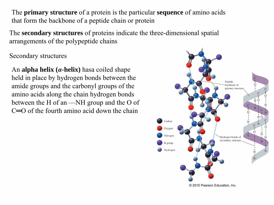
The secondary structures of proteins indicate the three-dimensional spatial arrangements of the polypeptide chains
The primary structure of a protein is the particular sequence of amino acids that form the backbone of a peptide chain or protein
An alpha helix (α-helix) hasa coiled shape held in place by hydrogen bonds between the amide groups and the carbonyl groups of the amino acids along the chain hydrogen bonds between the H of an —NH group and the O of C═O of the fourth amino acid down the chain
Secondary structures
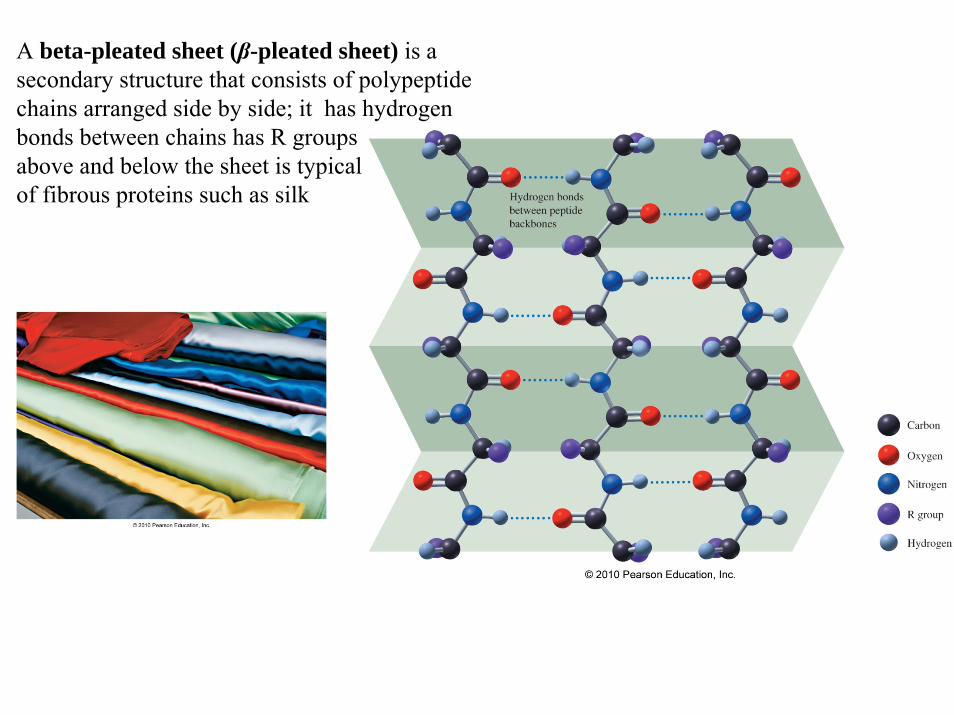
A beta-pleated sheet (β-pleated sheet) is a secondary structure that consists of polypeptide chains arranged side by side; it has hydrogen bonds between chains has R groups above and below the sheet is typical of fibrous proteins such as silk
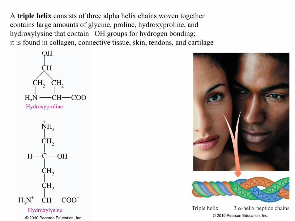
A triple helix consists of three alpha helix chains woven together contains large amounts of glycine, proline, hydroxyproline, and hydroxylysine that contain –OH groups for hydrogen bonding;it is found in collagen, connective tissue, skin, tendons, and cartilage

The tertiary structure of a protein gives a specific three-dimensional shape to the polypeptide chain including interactions and cross-links between different parts of the peptide chainThe tertiary structure is stabilized by: hydrophobic and hydrophilic interactions, salt bridgeshydrogen bonds and disulfide bonds
The primary structure of a protein is the particular sequence of amino acids that form the backbone of a peptide chain or protein
The secondary structures of proteins indicate the three-dimensional spatial arrangements of the polypeptide chains
secondarystructure

Globular proteins have compact, spherical shapescarry out synthesis, transport, and metabolism in the cellsA protein such as myoglobin stores and transports oxygen in muscle

The tertiary structure of a protein gives a specific three-dimensional shape to the polypeptide chain including interactions and cross-links between different parts of the peptide chain
The secondary structures of proteins indicate the three-dimensional spatial arrangements of the polypeptide chains
The primary structure of a protein is the particular sequence of amino acids that form the backbone of a peptide chain or protein
The quaternary structure is the combination of two or more tertiary units;it is stabilized by the same interactions found in tertiary structures;hemoglobin consists of two alpha chains and two beta chains with heme groups in each subunit that pick up oxygen for transport in the blood to the tissues
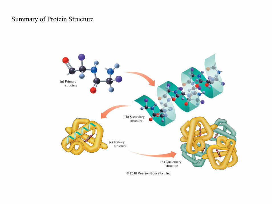
Summary of Protein Structure
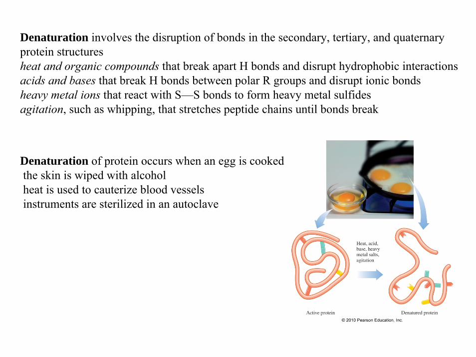
Denaturation involves the disruption of bonds in the secondary, tertiary, and quaternary protein structuresheat and organic compounds that break apart H bonds and disrupt hydrophobic interactionsacids and bases that break H bonds between polar R groups and disrupt ionic bondsheavy metal ions that react with S—S bonds to form heavy metal sulfidesagitation, such as whipping, that stretches peptide chains until bonds break
Denaturation of protein occurs when an egg is cooked the skin is wiped with alcoholheat is used to cauterize blood vesselsinstruments are sterilized in an autoclave
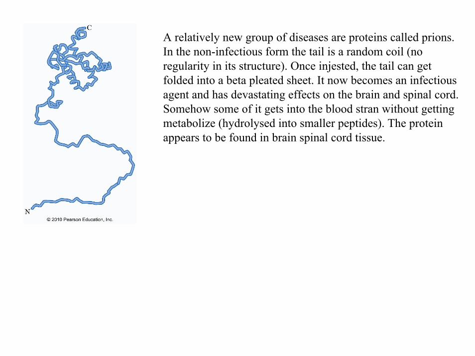
A relatively new group of diseases are proteins called prions. In the non-infectious form the tail is a random coil (no regularity in its structure). Once injested, the tail can get folded into a beta pleated sheet. It now becomes an infectious agent and has devastating effects on the brain and spinal cord. Somehow some of it gets into the blood stran without getting metabolize (hydrolysed into smaller peptides). The protein appears to be found in brain spinal cord tissue.
![Differential diagnosis of (inherited) amino acid metabolism or ...26 W. Blom and J. G. M. Huijmans TRANSPORT METABOLISM t SYNTHESIS AMINO ~ AMINOACIDS ] ACIDS l] DE~GRADATIO:EGRADATION](https://img.pdfslide.net/doc/110x75/5fe66b7cd8754e792944ccfb/differential-diagnosis-of-inherited-amino-acid-metabolism-or-26-w-blom-and.jpg)