Embed Size (px)
Citation preview
CHAPTER 21 – INFECTIONS OF THE RESPIRATORY SYSTEM
© Dennis Kunkel
WHY IS THIS IMPORTANT?
The respiratory system is the most commonly infected system.
Health care providers will see more respiratory infections than any other type.
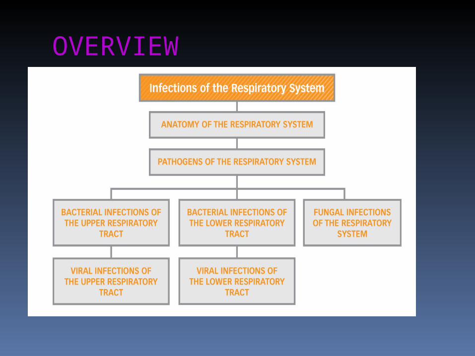
OVERVIEW
THE RESPIRATORY SYSTEM
A major portal of entry for infectious organisms
It is divided into two tracts – upper and lower. The division is based on structures and functions in
each part. The two parts have different types of infection.
…THE RESPIRATORY SYSTEM
The upper respiratory tract: Nasal cavity, sinuses, pharynx, and larynx Infections are fairly common. Usually nothing more than an irritation
The lower respiratory tract: Lungs and bronchi Infections are more dangerous. Can be very difficult to treat
ANATOMY OF THE RESPIRATORY SYSTEM The most accessible system in the body
Breathing brings in clouds of potentially infectious pathogens.
The body has a variety of host defense mechanisms. Innate immune response -the cells and mechanisms
that defend the host from infection by other organisms, in a non-specific manner
Adaptive immune It is adaptive immunity because the body's immune system prepares itself for future challenges.
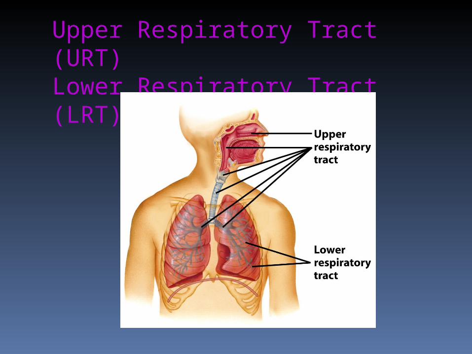
Upper Respiratory Tract (URT)Lower Respiratory Tract (LRT)
..ANATOMY OF THE RESPIRATORY SYSTEM Upper respiratory tract is continuously
exposed to potential pathogens. Lower respiratory tract is essentially a sterile
environment.
PATHOGENS OF THE RESPIRATORY SYSTEM Many bacterial organisms infect the
respiratory system. Upper respiratory tract also portal of entry for
viral pathogens. Vaccination has eliminated many respiratory
infections. Some still seen in underdeveloped parts of the
world.
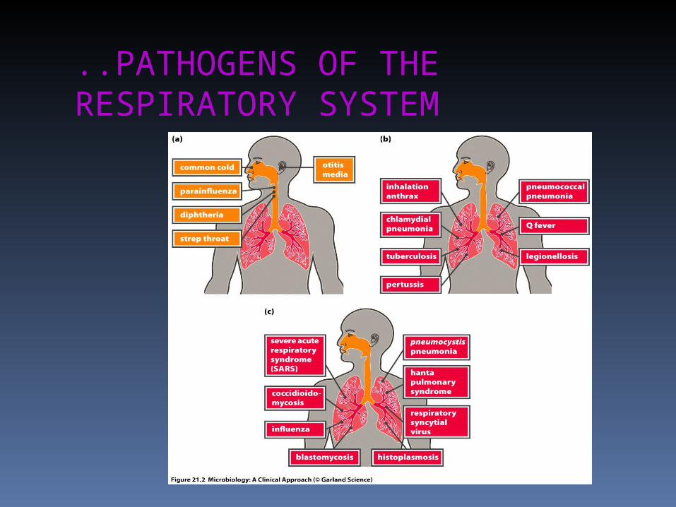
..PATHOGENS OF THE RESPIRATORY SYSTEM
…PATHOGENS OF THE RESPIRATORY SYSTEM Respiratory pathogens are easily transmitted from
human to human. They circulate within a community. Infections spread easily.
Some respiratory pathogens exist as part of the normal flora.
Others are acquired from animal source, water, air etc Fungi are also a source of respiratory infection.
Usually in immunocompromised patients Most dangerous are Aspergillus and Pneumocystis.
…PATHOGENS OF THE RESPIRATORY SYSTEM Some pathogens are restricted to certain sites.
Legionella only infects the lung. Other pathogens cause infection in multiple sites.
Streptococcus can cause: Middle ear infections. Sinusitis. Pneumonia.
SITES OF INFECTION
Frequent sites of infection are: Middle ear. Mastoid cavity. Nasal sinuses. Nasopharynx.
DEFENSES OF THE RESPIRATORY SYSTEM The respiratory system has significant defenses. The upper respiratory tract has:
Mucociliary escalator. Coughing.
The lower respiratory tract has: Alveolar macrophages.
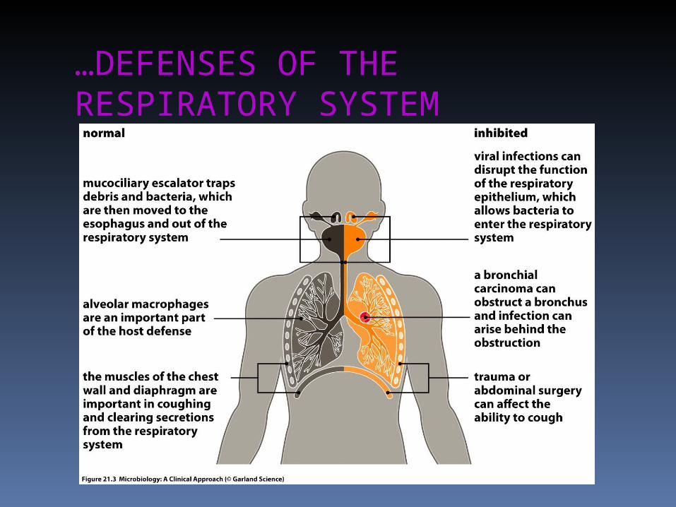
…DEFENSES OF THE RESPIRATORY SYSTEM
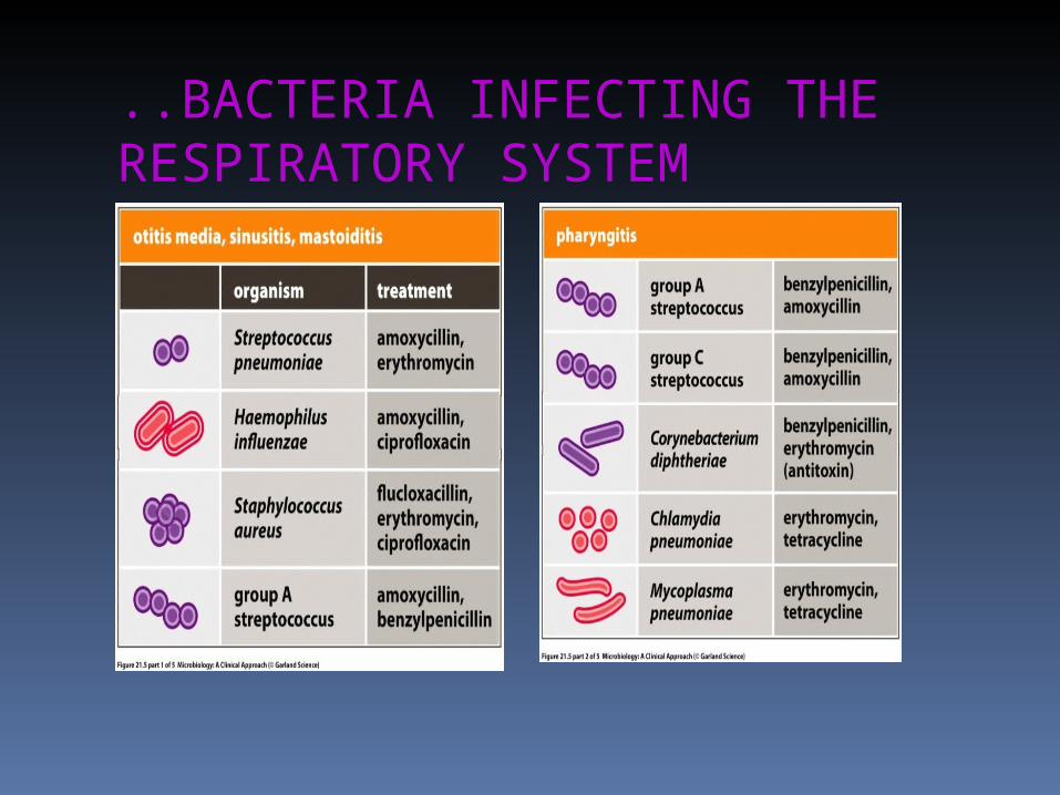
BACTERIA INFECTING THE RESPIRATORY SYSTEM Can be divided into groups depending on the
infections they cause Otitis media, sinusitis, and mastoiditis (the
portion of the temporal bone of the skull that is behind the ear which contains open, air-containing spaces).
Pharyngitis Typical and atypical community-acquired
pneumonia Hospital-acquired (nosocomial) pneumonia
..BACTERIA INFECTING THE RESPIRATORY SYSTEM
Detection of bacteria typeDetected by Blood Agar Cultures
Hemolytic Reactions: Blood agar is a solid growth medium that contains red blood cells. The medium is used to detect bacteria that produce enzymes to break apart the blood cells. This process is also termed hemolysis. The degree to which the blood cells are hemolyzed is used to distinguish bacteria from one another. Beta Hemolysis
Complete Hemolysis Clear Zone Around Colonies on Blood Agar
Alpha Hemolysis Incomplete Hemolysis Greenish Zone Around Colonies on Blood Agar
Gamma Reaction: Absence of a Hemolytic Reaction No Change Around Colonies on Blood Agar
Lancefield GroupsRebecca Craighill Lancefield (January 5, 1895 – March 3,
1981) was a prominent American microbiologist- serological classification of beta-hemolytic streptococcal bacteria. Based on Serological Groupings:
Group A
Streptococcus pyogenes
The most virulent human pathogen of the genus
Beta hemolytic
Often identified by rapid serological tests or by antibiotic resistance
…Lancefield Groups Streptococcus agalactiae Mildly to moderately virulent;
esp. in children & elderly Usually beta or alpha hemolytic;
some strains are gamma Detected biochemically.
Group C
-Includes S. equi, which causes strangles in horses, and S. zooepidemicus- a subspecies of S. equi
- Causes infections in several species of mammals including cattle and horses. This can also cause death in chickens and moose.
….Lancefield Groups
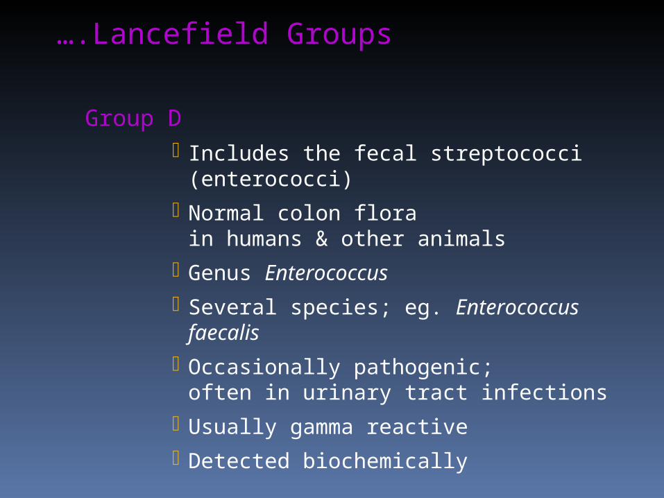
Group D Includes the fecal streptococci (enterococci)
Normal colon flora in humans & other animals
Genus Enterococcus
Several species; eg. Enterococcus faecalis
Occasionally pathogenic; often in urinary tract infections
Usually gamma reactive
Detected biochemically
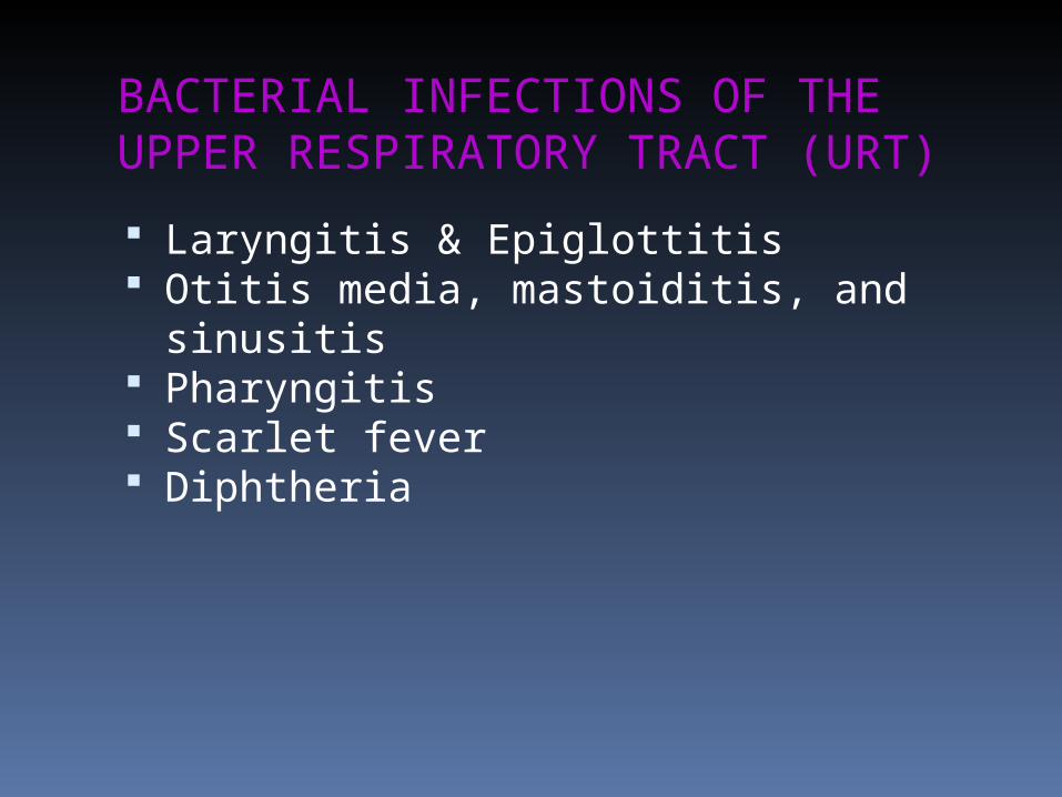
BACTERIAL INFECTIONS OF THE UPPER RESPIRATORY TRACT (URT)
Laryngitis & Epiglottitis Otitis media, mastoiditis, and sinusitis Pharyngitis Scarlet fever Diphtheria
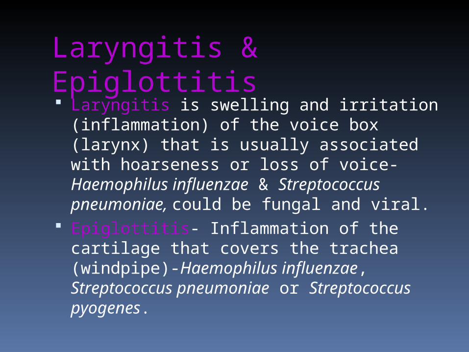
Laryngitis & Epiglottitis
Laryngitis is swelling and irritation (inflammation) of the voice box (larynx) that is usually associated with hoarseness or loss of voice-Haemophilus influenzae & Streptococcus pneumoniae, could be fungal and viral.
Epiglottitis- Inflammation of the cartilage that covers the trachea (windpipe)-Haemophilus influenzae, Streptococcus pneumoniae or Streptococcus pyogenes.
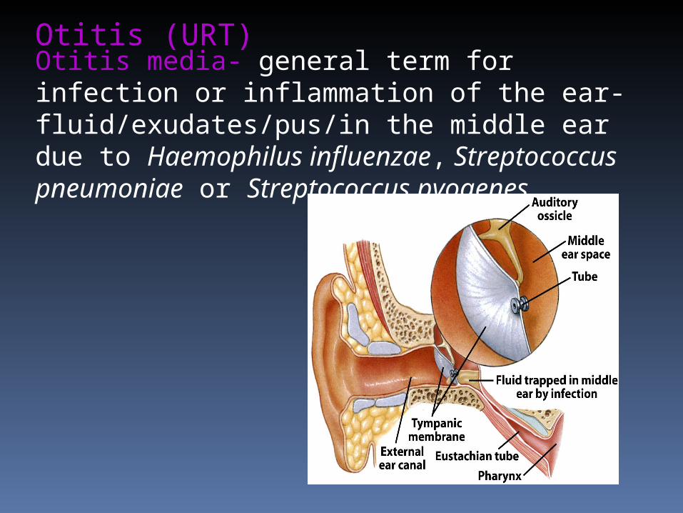
Otitis (URT)Otitis media- general term for infection or inflammation of the ear-fluid/exudates/pus/in the middle ear due to Haemophilus influenzae, Streptococcus pneumoniae or Streptococcus pyogenes.
OTITIS MEDIA, MASTOIDITIS, AND SINUSITIS
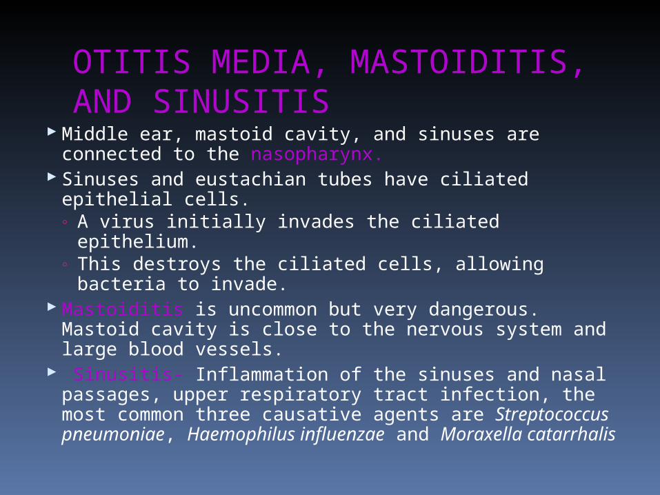
Middle ear, mastoid cavity, and sinuses are connected to the nasopharynx.
Sinuses and eustachian tubes have ciliated epithelial cells.◦ A virus initially invades the ciliated epithelium.◦ This destroys the ciliated cells, allowing bacteria to invade.
Mastoiditis is uncommon but very dangerous. Mastoid cavity is close to the nervous system and large blood vessels.
Sinusitis- Inflammation of the sinuses and nasal passages, upper respiratory tract infection, the most common three causative agents are Streptococcus pneumoniae, Haemophilus influenzae and Moraxella catarrhalis
PHARYNGITIS A variety of bacteria can cause infection in the
pharynx. A classic infection is strep throat.
Caused by Streptococcus pyogenes Contains M proteins which inhibits phagocytosis Produces pyrogenic toxins which cause the
symptoms seen with pharyngitis Group A streptococci can cause abscesses on the
tonsils. S. pyogenes can cause scarlet fever and toxic shock
syndrome.
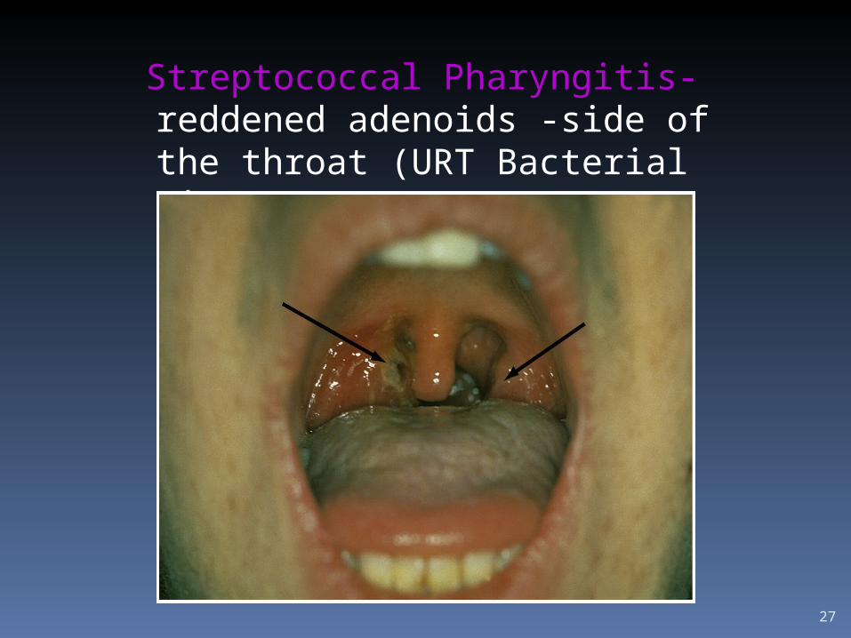
Streptococcal Pharyngitis-reddened adenoids -side of the throat (URT Bacterial Diseases)
27
SCARLET FEVER
Caused by Group A streptococci Usually seen in children under age of 18 years Symptoms usually begin with appearance of a
rash. Tiny bumps on the chest and abdomen Can spread over the entire body
Appears redder in armpits and groin Rash lasts 2-5 days
..SCARLET FEVER
Symptoms can also include: Very sore throat with yellow or white papules Fever of 101˚F or higher Lymphadenopathy in neck Headache, body aches, and nausea A variety of antibiotic therapies is available
DIPHTHERIA
Caused by the toxin produced by Corynebacterium diphtheriae A potent inhibitor of protein synthesis
It is a localized infection. Presents as severe pharyngitis Can be accompanied by plaque-like
pseudomembrane in the throat
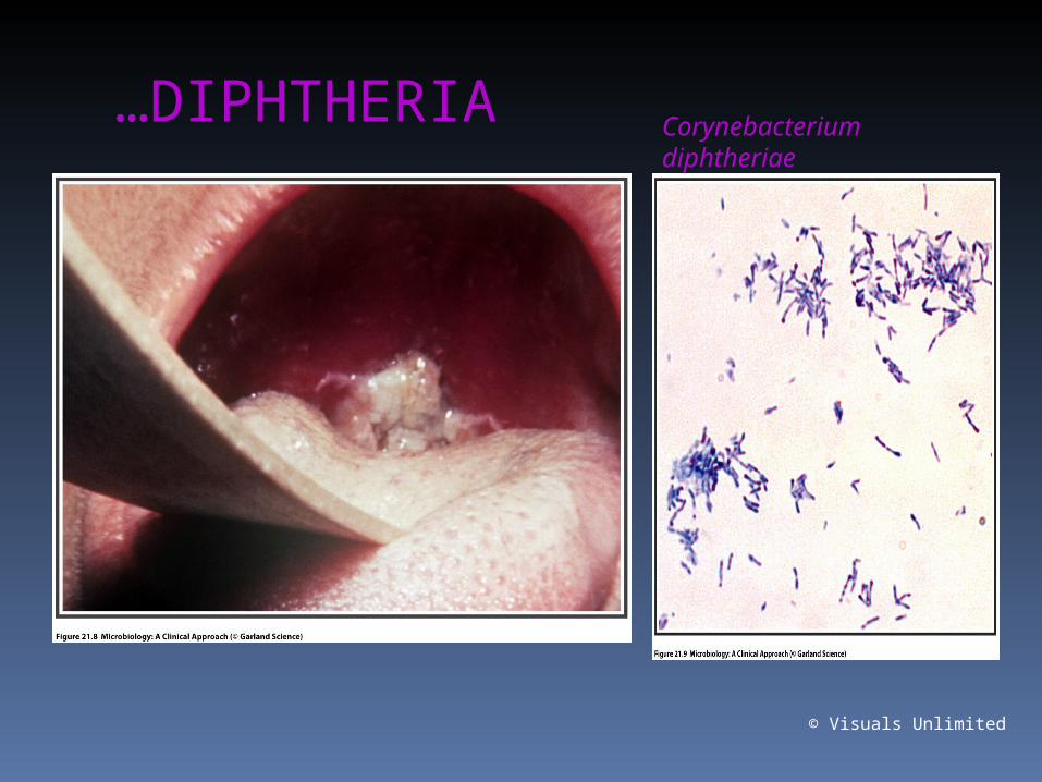
…DIPHTHERIA
© Visuals Unlimited
Corynebacterium diphtheriae
….DIPHTHERIA
Toxemia can make diphtheria life threatening. Can involve multiple organ systems Can cause acute myocarditis
Diphtheria is transmitted by: Droplet aerosol. Direct contact with skin. Fomites (to a lesser degree).
…DIPHTHERIA:Vaccination
Vaccination against diphtheria- Infection is rare when vaccination is in place.
Diphtheria still occurs frequently in some parts of the world, particularly where conditions do not permit vaccination.
Toxin neutralization (exotoxin) is the most important. Must be done as quickly as possible Antitoxin can only neutralize free toxin.
Pathogen elimination is also important. Corynebacterium diphtheriae is sensitive to many
antibiotics
…DIPHTHERIA:Pathogenesis Corynebacterium diphtheriae is a small Gram-
positive bacillus. Has V and L forms Forms are caused by a unique cell division
process – snapping. Corynebacterium is poorly invasive.
Effects of infection are due to the exotoxin.
…DIPHTHERIA:PathogenesisLocal effects include epithelial cell necrosis and
inflammation.Pseudomembrane is composed of a mixture of fibrin,
leukocytes, cell debris.◦ Size varies from small and localized to extensive◦ An extensive membrane can cover the trachea.
Incubation takes two to four days.Disease usually presents as pharyngitis or tonsillitis with
fever, sore throat, and malaise.Pseudomembrane can develop on tonsils, uvula, soft
palate, or pharyngeal walls.◦ May extend downward toward larynx and trachea.
VIRAL INFECTIONS OF THE UPPER RESPIRATORY TRACT (URT)
RHINOVIRUS INFECTION -There are several hundred serotypes of rhinovirus. Fewer than half have been characterized. 50% that have are all picornaviruses. Extremely small, non-enveloped, single-
stranded RNA viruses Optimum temperature for picornavirus growth is
33˚C. The temperature in the nasopharynx
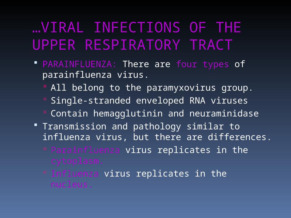
…VIRAL INFECTIONS OF THE UPPER RESPIRATORY TRACT PARAINFLUENZA: There are four types of
parainfluenza virus. All belong to the paramyxovirus group. Single-stranded enveloped RNA viruses Contain hemagglutinin and neuraminidase
Transmission and pathology similar to influenza virus, but there are differences. Parainfluenza virus replicates in the cytoplasm. Influenza virus replicates in the nucleus.
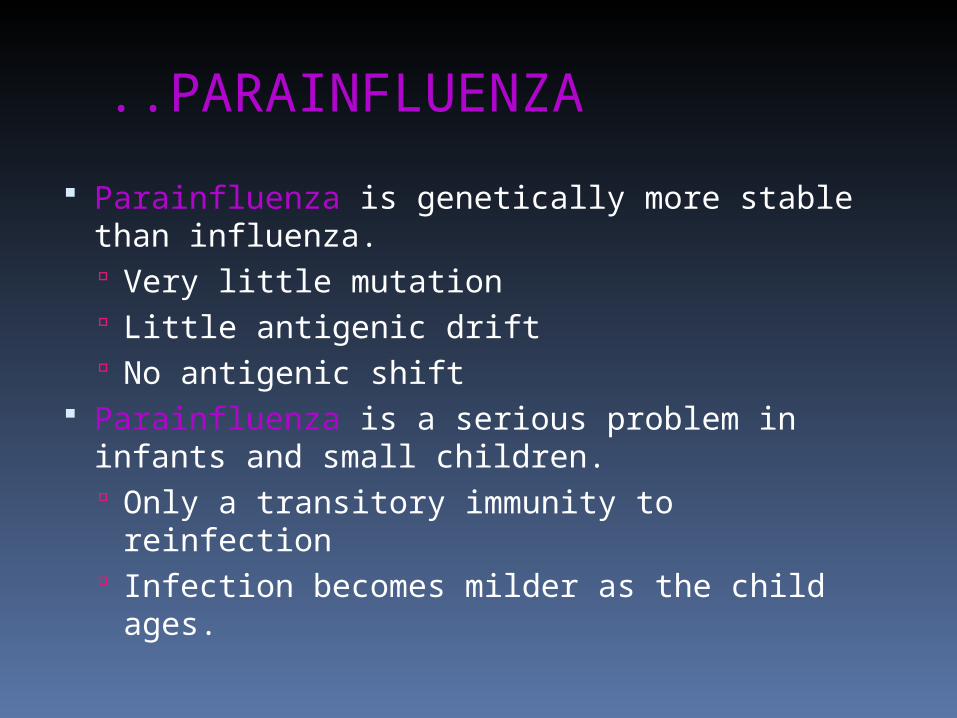
..PARAINFLUENZA
Parainfluenza is genetically more stable than influenza. Very little mutation Little antigenic drift No antigenic shift
Parainfluenza is a serious problem in infants and small children. Only a transitory immunity to reinfection Infection becomes milder as the child ages.
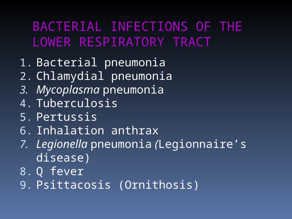
BACTERIAL INFECTIONS OF THE LOWER RESPIRATORY TRACT
1. Bacterial pneumonia2. Chlamydial pneumonia3. Mycoplasma pneumonia4. Tuberculosis5. Pertussis6. Inhalation anthrax7. Legionella pneumonia (Legionnaire’s disease)8. Q fever9. Psittacosis (Ornithosis)
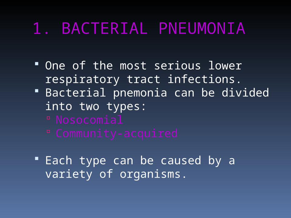
1. BACTERIAL PNEUMONIA
One of the most serious lower respiratory tract infections.
Bacterial pnemonia can be divided into two types: Nosocomial Community-acquired
Each type can be caused by a variety of organisms.
….BACTERIAL PNEUMONIA Nosocomial pneumonia
Occurs approximately 48 hours after admission to hospital
Usually associated with Staphylococcus aureus Also caused by Gram-negative bacteria Particularly difficult to deal with if pathogen is
resistant to antibiotics Community-acquired pneumonia
Usually presents as a lobar pneumonia Accompanied by fever, chest pain, and production
of purulent sputum
…BACTERIAL PNEUMONIA Pathogenesis
COMMUNITY-ACQUIRED PNEUMONIA : Usually occurs after the aspiration of pathogens
Requires enough pathogens to overwhelm resident defenses
Establishment of an infection in the lungs depends on: The number of pathogens entering and the
competence of the mucociliary escalator.
….COMMUNITY-ACQUIRED BACTERIAL PNEUMONIA
Classical lobar pneumonia has four stages: Acute congestion
Local capillaries become engorged with neutrophils. Red hepatization
Red blood cells from the capillaries flow into the alveolar spaces.
Grey hepatization Large numbers of dead neutrophils (are the first immune
cells that reach the site of infection through a process known as chemotaxis) and degenerating red cells
Resolution Adaptive immune response begins to produce antibodies. Which control the infection.
…BACTERIAL PNEUMONIA
TWO types:A . Atypical pneumonia
Coughing without sputum Caused by a variety of bacteria
Bacterial pneumonia can progress to the production of lung abscesses.
…BACTERIAL PNEUMONIA
B. Typical or Classic Pneumonia Typical bacterial pneumonia is a respiratory
condition with inflammation of the lung. Often characterized as inflammation of the
parenchyma of the lung (the alveoli) and abnormal alveolar filling with fluid.
Typical symptoms associated with pneumonia include cough, chest pain, fever, and difficulty in breathing.
Bacterial pneumonia is treated with antibiotics.
..Further Classification of PneumoniaLobar Pneumonia: Streptococcus pneumonia that affects a part of a lobe in the
lung or it may affect more than one lobes.Bronchial Pneumonia: pneumonia spreads to several patches in one or both lungs is most prevalant in infants, young children and aged
adults cough (with or without mucus), chest pain, rapid breathing,
and shortness of breath Transmitted by respiratory droplets
Types of bacteria causing pneumonia
Gram-positive bacteria:
Streptococcus pneumoniae, often called "pneumococcus" , Staphylococcus aureus, with Streptococcus agalactiae.
Gram-negative bacteria: Haemophilus influenzae, Klebsiella pneumoniae,
Escherichia coli, Pseudomonas aeruginosa and Moraxella catarrhalis.
…..BACTERIAL PNEUMONIATreatment
Course of treatment depends on: Severity of the infection. Type of organism causing the infection.
Most common pathogen is Streptococcus pneumoniae. Treated with penicillin, amoxicillin-clavulanate,
and erythromycin.
2. CHLAMYDIAL PNEUMONIA Caused by Chlamydia pneumoniae:
Found throughout the world Responsible for 10% of pneumonia cases-
pharyngitis Lower-respiratory-tract infection Infection occurs throughout the year. Spread by person-to-person contact More infections in the elderly Can cause both community-acquired and
nosocomial infections Similar to Mycoplasma pneumonia. Tetracycline or erythromycin
3. MYCOPLASMA PNEUMONIA
Mild form of pneumonia Accounts for about 10% of all pneumonias Referred to as walking pneumonia
No need for hospitalization. Most common age for infections between 5 and 15
years. Causes approximately 30% of all teenage
pneumonias.
..MYCOPLASMA PNEUMONIA
Caused by Mycoplasma pneumoniae◦ Lacks a cell wall◦ Acquired by droplet transmission◦ Infectious dose fewer than 100 pathogens◦ Found throughout the world, especially in temperate
climates Infection affects the trachea, bronchi, and bronchioles.◦ May extend down to the alveoli
Fever, headache, and malaise for 2 to 4 days◦ Mild tracheobronchitis.◦ Fever, cough, headache, and malaise.◦ Sore throat.◦ Otitis media◦ Treatment : erythromycin or tetracycline
4. TUBERCULOSIS
An estimated 1.7 billion people are infected. 3 million die each year
AIDS and HIV infection have had a significant role in the increase of tuberculosis. They increase the efficiency of the tuberculosis
transmission cycle. Poverty and poor socioeconomic conditions
are breeding grounds for tuberculosis.
…TUBERCULOSIS
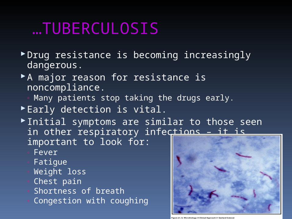
Drug resistance is becoming increasingly dangerous.A major reason for resistance is noncompliance.◦ Many patients stop taking the drugs early.
Early detection is vital.Initial symptoms are similar to those seen in other
respiratory infections – it is important to look for: ◦ Fever◦ Fatigue◦ Weight loss◦ Chest pain◦ Shortness of breath◦ Congestion with coughing
…TUBERCULOSIS
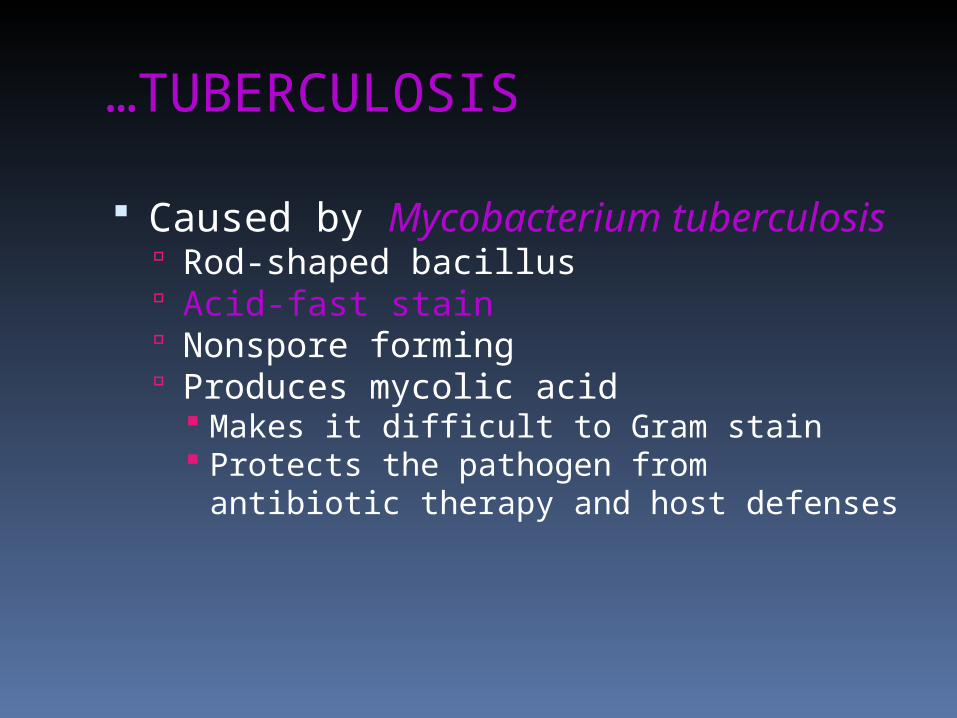
Caused by Mycobacterium tuberculosis Rod-shaped bacillus Acid-fast stain Nonspore forming Produces mycolic acid
Makes it difficult to Gram stain Protects the pathogen from antibiotic therapy and
host defenses
…TUBERCULOSIS: Pathogenesis M. tuberculosis cell wall interferes with macrophage
function (white blood cells which acts as phagocytes) and immune cells activation. Inhibits the formation of the phagolysosome (a phagosome
is a vacuole formed around a particle absorbed by phagocytosis; Lysosomes are cellular organelles which contain acid hydrolase enzymes to break up waste materials and cellular debris
This allows Mt to escape into the cytoplasm where it: Increases in number & eventually spreads to the lymph
nodes. From here it enters the blood and is distributed
throughout the body.
…TUBERCULOSIS: Pathogenesis
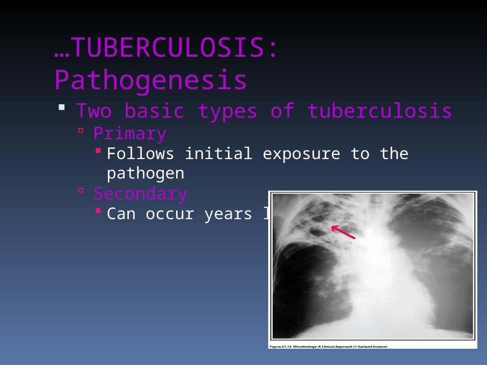
Two basic types of tuberculosis Primary
Follows initial exposure to the pathogen Secondary
Can occur years later
..PRIMARY TUBERCULOSIS:
Occurs when a host encounters pathogen for the first time.
Organisms find their way to the alveoli. A localized inflammatory response develops. Phagocytosis of the bacilli by macrophages.
…PRIMARY TUBERCULOSIS:
Pathogens are not killed, they: Are transported by white cells to the regional
lymph nodes. Continue to divide intracellularly.
Cell mediated immune response begins. If the primary lesion is not contained,
tubercles form. Tubercles are aggregates of enlarged
macrophages filled with bacteria. Readily seen on X-rays
..SECONDARY TUBERCULOSIS:Pathogenesis
Secondary tuberculosis can be due to: Reactivation of old lesions. Gradual progression of primary tuberculosis into chronic
disease.
Recurrence of disease occurs in a small percentage of patients. Usually manifests itself in the apices of the lungs Usually occurs within two years of the primary infection It can evolve decades later when innate resistance is
diminished.
…TUBERCULOSIS: Treatment
Usually a triple therapy containing: Isoniazid (INH) Pyrazinamide (PZA) Rifampicin (RFP)
All three are taken once a day for two months. INH and RFP are taken for nine more months. If the strain is drug-resistant, initial treatment
includes ethambutol.
…TUBERCULOSIS: Treatment
Compliance with the drug therapy is very important.
Compliance can be difficult because of side effects. The drugs are very toxic. Most serious is liver toxicity.
5. PERTUSSIS (WHOOPING COUGH)
Spread by airborne droplets from patients in the early stages.
Highly contagious Infects 80-100% of exposed susceptible
individuals. Spreads rapidly in schools, hospitals, offices, and
homes – just about anywhere.
….PERTUSSIS
Caused by Bordetella pertussis Gram-negative coccobacillus Does not survive in the environment Reservoir is humans.
Symptoms can be similar to those of a cold. Infected adults often spread the infection to schools
and nurseries.
…PERTUSSIS
Mortality is highest in infants and children under 1 year old.
Immunization against pertussis started in the 1940s. Continues today as part of DTaP vaccination
Pertussis appears to be making a comeback. Epidemics are occurring every 3-5 years. Greatest numbers of infections are among 10-20 year-olds.
People who were not immunized Shows a relationship between lack of vaccination and
infection
..PERTUSSIS: Pathogenesis
Bordetella pertussis has an affinity for ciliated bronchial epithelium.
After attaching, it produces a tracheal toxin. Immobilizes and progressively destroys the ciliated
cells. Causes persistent coughing
Caused by the inability to move the mucus that builds up
Pertussis does not invade cells of the respiratory tract or deeper tissues.
Incubation period is 7 to 10 days.
..PERTUSSIS: Pathogenesis
Infection has three stages: Persistent perfuse and mucoid rhinorrhea (runny nose) May have sneezing, malaise, and anorexia Most communicable during this stage
Complication of pertussis can lead to superinfection with Streptococcus pneumonia.
PERTUSSIS: Pathogenesis
Most common complications of pertussis are: Superinfection with Streptococcus pneumonia. Convulsions. Subconjunctival and cerebral bleeding and anoxia.
.. PERTUSSIS: Treatment
Antibiotics can be used in the early stages. Limits the spread of infection.
Once the paroxysmal stage is reached, therapy is only supportive.
Vaccination is the best option.
6. INHALATION ANTHRAX
Produces a fulminate pneumonia Comes on suddenly with great severity Leads to respiratory failure and death
Anthrax primarily a disease of herbivores Acquired from spores found in pastures If spores are inhaled, anthrax can occur in the
respiratory tract.
..INHALATION ANTHRAX
Infection is infrequently seen in healthy individuals. Usually presents as localized lesions where it
occurs. Has been recent interest in inhalation anthrax
as a biological weapon In October 2001, letters contaminated with
powdered anthrax spores were mailed to various locations in the US. Several deaths resulted.
..INHALATION ANTHRAX: Pathogenesis The causative agent is Bacillus anthracis.
Gram-positive rod Spore-forming
Spores germinate in human tissues. Antiphagocytic properties of the capsule aid
its survival and growth in large numbers.
...INHALATION ANTHRAX Pathogenesis
Pathogenesis results from the powerful exotoxin produced.
Symptoms of pulmonary anthrax are: 1-5 days of nonspecific malaise, mild fever, nonproductive
cough. Progressive respiratory distress and cyanosis.
Rapid and massive spread to the central nervous system and bloodstream is followed by death.
Antibiotic therapy can be successful. B. anthracis is susceptible to penicillin. Doxicycline and ciprofloxacin are alternative prophylactics.
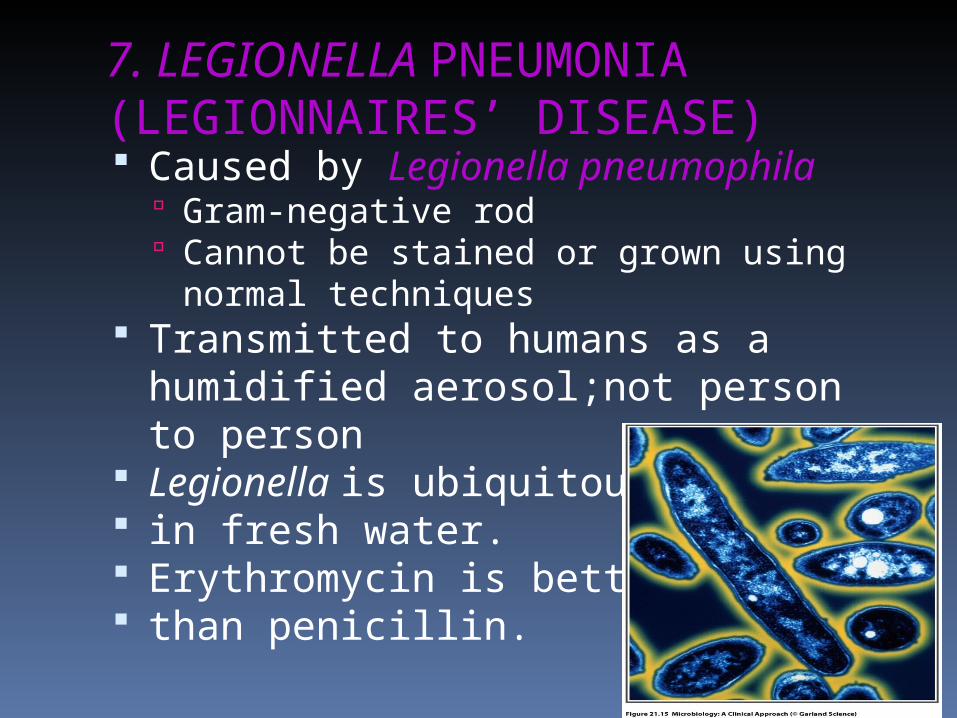
7. LEGIONELLA PNEUMONIA (LEGIONNAIRES’ DISEASE) Caused by Legionella pneumophila
Gram-negative rod Cannot be stained or grown using normal
techniques Transmitted to humans as a humidified
aerosol;not person to person Legionella is ubiquitous in fresh water. Erythromycin is better than penicillin.
8. Q FEVER A zoonotic infection seen wordwide
Cattle, sheep, and goats are the primary reservoirs for humans.
Caused by Coxiella burnetii Gram-negative Spore-forming Grows well in placenta of animals
Large numbers of Coxiella can be transmitted by inhalation during animal births.
Transmission can also be by ingestion of unpasteurized milk. Most cases resolve spontaneously.
Tetracycline can be given to shorten fever
….Q FEVER: Pathogenesis
Not clearly understood Begins 9 to 20 days after inhalation Abrupt onset of chills, fever, and headache Can also be a mild hacking cough and patchy
interstitial pneumonia Some cases show abnormal liver function
9. PSITTACOSIS (ORNITHOSIS)
Zoonotic pneumonia Contracted by inhalation of bird droppings
infected with Chlamydia psittaci. Found in many birds, including turkeys
Some strains of C. psittaci are extremely contagious. Acute onset of fever, headache, malaise, muscle
aches, dry hacking cough, and bilateral pneumonia Tetracycline and erythromycin are effective if
given early.
VIRAL INFECTIONS OF THE LOWER RESPIRATORY TRACT 75-80% of all acute respiratory tract infections
in the US are of viral origin. Everyone has 3 or 4 per year
Incidence varies inversely with age. Greatest in young children
..VIRAL INFECTIONS OF THE LOWER RESPIRATORY TRACT
Majority of acute viral infections are in the lower respiratory tract and caused by: Influenza virus. Respiratory syncytial virus.
Common characteristics of infection are: Short incubation period of 1 to 4 days. Transmission from person to person.
Transmission can be direct or indirect. Direct – through droplets Indirect – through hand transfer of contaminated secretions
1. INFLUENZA
Influenza virus is an orthomyxovirus. Virions are surrounded by an envelope.
Genome is single-stranded RNA Allows a high rate of mutation
Three major serotypes of virus: A, B, and C. Differences are based on antigens associated with the
nucleoprotein.
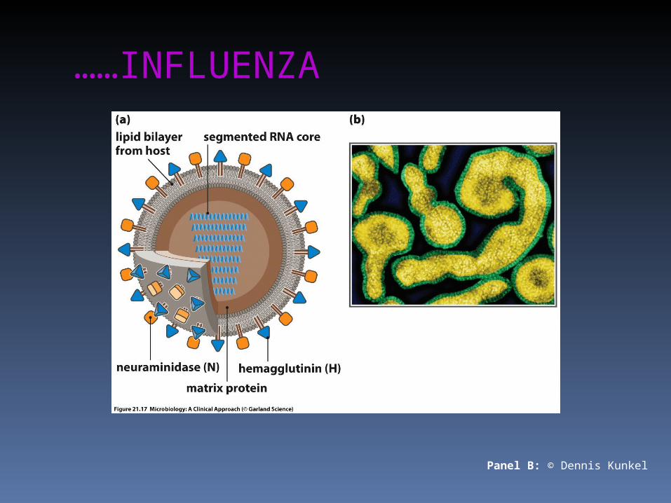
……INFLUENZA
Panel B: © Dennis Kunkel
..INFLUENZA
Influenza is a significant health concern. Human virus can combine with an avian virus to
produce a highly pathogenic virus. Humans are the hosts for influenza.
Aquatic birds are the reservoir.
..INFLUENZA
Primary manifestation of infection is severe respiratory problems.
Outbreaks have been described since the sixteenth century. Differ in severity nearly every year Occur more frequently in the winter
Direct droplet transmission most common method of spreading.
..INFLUENZA
A major outbreak occurs every 2 to 3 years. Typical epidemic lasts 3 to 6 weeks. Up to 10% of the general population is affected. Illness rates exceed 30% in certain groups.
In school-aged children Residents of closed institutions
…INFLUENZA: Pathogenesis
Influenza virus prefers the respiratory epithelium. Viremia is rare.
Virus multiplies in the ciliated cells of lower respiratory tract. Results in functional and structural abnormalities
Cellular synthesis of nucleic acids and proteins is shut down.
Ciliated and mucus-producing epithelial cells are shed. Substantial interference with clearance mechanisms Localized inflammation
…INFLUENZA: Pathogenesis
Three bacteria are common causes of superinfection. Streptococcus pneumoniae Haemophilus influenzae Staphylococcus aureus
..INFLUENZA: Treatment
Two basic approaches Symptomatic care Anticipation of potential complications
The best treatments are: Rest and fluid intake Conservative use of analgesics for myalgia and
headache Cough suppressants.
Amantidine and rimantadine are useful only if the infection is diagnosed within 12-24 hours.
FUNGAL INFECTIONS OF THE RESPIRATORY SYSTEM
Two major factors govern the incidence and spread of fungal infection. Ubiquity of the infectious organisms
Found in soil Resident flora
The adaptive immune response Usually keeps these infections under control Immunocompromised patients at much greater risk
1. PNEUMOCYSTIS PNEUMONIA (PCP) A lethal pneumonia
Common in AIDS patients Caused by the fungus Pneumocystis (carinii)
jiroveci Never been grown in culture Most information comes from clinical information
from patients.
2. BLASTOMYCOSIS
Caused by Blastomyces dermatitidis. Spores of the fungi enter through the respiratory
system. Primarily affect the lungs
Can spread through bloodstream and affect other parts.
Men between ages of 20 and 40 years are the most commonly infected.
Blastomycosis is not increased in AIDS.
..BLASTOMYCOSIS: Pathogenesis
Infection of the lungs is gradual. Fever, chills, and drenching sweats develop. Chest pain, difficulty breathing, and cough may
also develop. Can sometimes heal without treatment. Skin – warty patches develop surrounded by tiny
painless abscesses Bones – painful swellings Genitounrinary tract – prostatitis or painful
swelling of epididymis
3. HISTOPLASMOSIS
Caused by Histoplasma capsulatum Occurs in soil contaminated with bat or bird droppings Commonly found in temperate, subtropical, and
tropical zones 50% - 90% of residents in these areas test positive for
exposure. People who live and work in the vicinity of bat or bird
droppings are at increased risk of infection. Amphotericin B is the treatment of choice if necessary.
..HISTOPLASMOSIS: Pathogenesis
Transmission is through inhalation of conidia. Small enough to reach bronchioles and alveoli Intravenous amphotericin B or oral itraconazole
After inhalation: Microconidia convert to yeast form. These are phagocytosed. Tubercles form. Severe cases may develop chills, malaise, chest pain,
and extensive pulmonary infiltration
5. COCCIDIOIDOMYCOSIS
Caused by Coccidioides immitis Infection can be symptomatic or
asymptomatic. Symptomatic form known as Valley Fever.
Restricted to certain geographical areas.
..COCCIDIOIDOMYCOSIS: Pathogenesis Arthroconidia of the fungus are inhaled.
Small enough to bypass defenses of the upper tract. Lodge directly in bronchioles.
Fungal outer wall has antiphagocytic properties. Prevents elimination
Arthroconidia convert to spherules which grow slowly. Completely inhibit phagocytosis
…COCCIDIOIDOMYCOSIS: Pathogenesis Disseminated coccidioidomycosis is seen in
patients with AIDS and on immunosuppressive therapy.
Can also cause a form of coccidioidal meningitis Can be fatal if not treated aggressively
..COCCIDIOIDOMYCOSIS:Treatment Usually self-limiting and no treatment is
required. Progressive pulmonary infection or infection
of central nervous system is treated with amphotericin B.
6. ASPERGILLOSIS Invasive aspergillosis shows a rapid
progression to death. Typically seen in the immunocompromised.
Particularly patients with leukemia or AIDS. Patients undergoing bone marrow transplantation.
Also seen in individuals with preexisting pulmonary disease Chronic bronchitis, asthma, and tuberculosis Fungus produces extracellular proteases,
phospholipases, and toxic metabolites.
…ASPERGILLOSIS
Caused by the fungus Aspergillus Widely distributed and found throughout world Dispersal is through inhalation of resistant conidia. Seen more and more in nosocomial infections
associated with air-conditioning systems.
..ASPERGILLOSIS: Pathogenesis
Colonization with Aspergillus leads to invasion of tissues. Invasion of lung tissue causes penetration of blood
vessels. This causes hemoptysis and/or acute pneumonia.
Pneumonia is accompanied by multifocal pulmonary infiltrates and high fever. Prognosis is grave. Mortality for invasive aspergillosis is 100%. Amphotericin B and itraconazole can be used but
are usually ineffective.

References
Microbiology, A clinical Approach -
Danielle Moszyk-Strelkauskas-Garland
Science 2010 http://en.wikipedia.org/wiki/Scientific_method