Embed Size (px)
Citation preview
824
The Chemistry of Life
Connective tissue—proteins
Skin cells— nucleic acids
Fat cells—lipids
ChemFacts
• Fats provide more than twice the energy per gram as carbohydrates and proteins.
• Special lipids, called phospholipids, make up the cellular membranes of living cells.
• A single human chromosome consists of a DNA molecule that, if stretched out, would measure nearly 5 cm.
BIG Idea Biological molecules—proteins, carbohydrates, lipids, and nucleic acids—interact to carry out activities necessary to living cells.
23.1 ProteinsMAIN Idea Proteins perform essential functions, including regulation of chemical reactions, structural support, transport of materials, and muscle contractions.
23.2 CarbohydratesMAIN Idea Carbohydrates provide energy and structural material for living things.
23.3 LipidsMAIN Idea Lipids make cell membranes, store energy, and regulate cellular processes.
23.4 Nucleic AcidsMAIN Idea Nucleic acids store and transmit genetic information.
23.5 MetabolismMAIN Idea Metabolism involves many thousands of reactions in living cells.
(t)©EYE OF SCIENCE/SCIENCE PHOTO LIBRARY/Photo Researchers, Inc., (c)©Dr. Kessel & Dr. Kardon/Tissues & Organs/Visuals Unlimited, (b)©Steve Gschmeissner/Photo Researchers, Inc., (bkgd)©AK PhotoLibrary/Alamy

Nucleic Acids
Lipids
Carbohydrates
Proteins
Biolo
gical
Molec
ules
Chapter 23 • The Chemistry of Life 825
Start-Up ActivitiesStart-Up Activities
LLAAUUNCH NCH LabLabHow do you test for simple sugars?Many different food sources supply the energy that your body uses constantly. This energy is stored in the bonds of molecules called simple sugars.
Procedure 1. Read and complete the lab safety form.2. Fill a 400-mL beaker until it is one-third full of
water. Place it on a hot plate and heat it to boiling.3. Use a graduated cylinder to measure 5.0 mL
10% glucose solution. Pour it into a test tube.4. Add 3.0 mL Benedict’s solution to the test tube.
Mix the two solutions using a stirring rod. Add a boiling chip to the test tube.
WARNING: Benedict’s solution is an eye and skin irritant.
5. Using tongs, place the test tube in the boiling water bath for 5 min.
6. A color change to yellow or orange indicates the presence of a simple sugar. Record your observations.
7. Repeat the procedure using a 10% starch solu-tion, a 10% gelatin suspension, and a few drops of honey suspended in water.
Analysis1. Describe the color changes you observed. 2. Classify Which foods contained a simple sugar?
Inquiry Think about what you had for dinner last night. Which foods likely contained simple sugars? How could you test the food to find out?
Biological Molecules Make the following Foldable to help you organize information about biological molecules.
Visit glencoe.com to: ▶ ▶ study the entire chapter online
▶ ▶ explore
▶ ▶ take Self-Check Quizzes
▶ ▶ use the Personal Tutor to work Example Problems step-by-step
▶ ▶ access Web Links for more information, projects, and activities
▶ ▶ find the Try at Home Lab, Modeling Sugars
STEP 1 Fold a sheet of notebook paper lengthwise, keeping the margin visible on the left side.
STEP 2 Cut the top flap into four tabs.
STEP 3 Label as follows: Biological Molecules, Proteins, Carbohydrates, Lipids, and Nucleic Acids.
Use this Foldable with Sections 23.1, 23.2, 23.3, and 23.4. As you read, summarize the general structure and function of the biological molecules, and give examples of each.
Matt Meadows
826 Chapter 23 • The Chemistry of Life
Section 23.123.1
Objectives
◗ Describe the structures of amino acids and proteins.
◗ Explain the roles of proteins in cells.
Review Vocabularypolymer: large molecules composed of many repeating units called monomers
New Vocabularyproteinamino acidpeptide bondpeptidedenaturationenzymesubstrateactive site
ProteinsMAIN Idea Proteins perform essential functions, including regulation of chemical reactions, structural support, transport of materials, and muscle contractions.
Real-World Reading Link Some cleaning products, such as contact lens cleaning solution, contain enzymes. Did you ever wonder what an enzyme was?
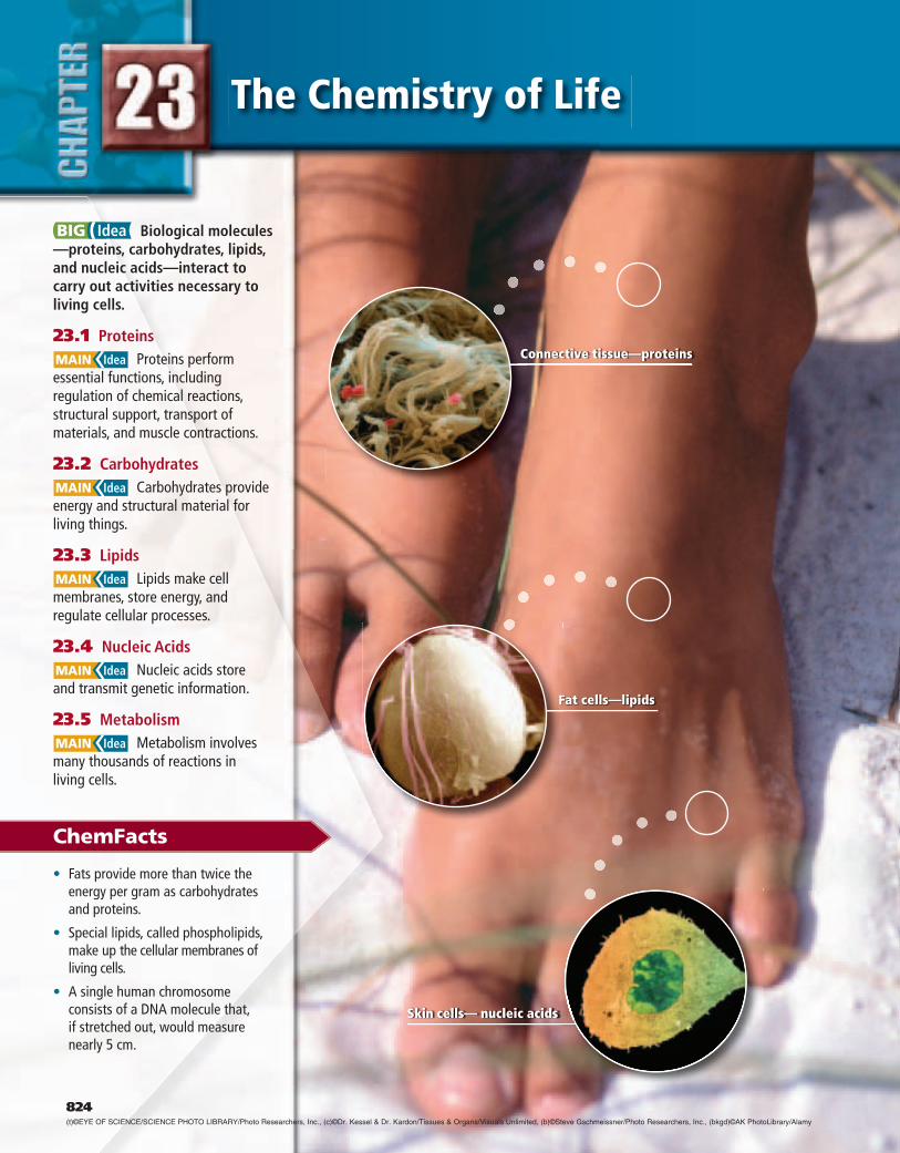
Protein StructureEnzymes form a class of proteins. Proteins are organic polymers made of amino acids linked together in a specific order. Proteins are not just large, randomly arranged chains of amino acids. To function properly, each protein must be folded into a specific three-dimensional structure. All living organisms, including the mountain goat and the plants shown in Figure 23.1, are composed of proteins. In this section, you will read about how proteins are made from their amino-acid building blocks and how different types of proteins function.
Amino acids As you read in Chapter 22, many different functional groups are found in organic compounds. Amino acids, as their name implies, are organic molecules that have both an amino group and an acidic carboxyl group. The general structure of an amino acid is shown below.
— —
R
Amino group Carboxyl group
Variable side chain
Hydrogen atom H O
H2N — C — C — OH
——
Each amino acid has a central carbon atom around which four groups are arranged: an amino group (—N H 2 ), a carboxyl group (—COOH), a hydrogen atom, and a variable side chain, R. The side chains range from a single hydrogen atom to a complex double-ring structure.
■ Figure 23.1 All living organisms con-tain proteins. A goat’s hair, hooves, and muscles are made up of structural proteins, as are the roots and leaves of plants.
(l)© John Conrad/CORBIS, (r)©Ron Niebrugge/Alamy
Section 23.1 • Proteins 827
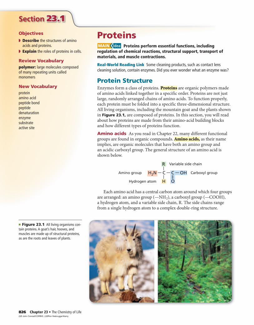
Examine the different side chains of the amino acids shown in Table 23.1. Identify the nonpolar alkanes, polar hydroxyl groups, acidic and basic groups such as carboxyl and amino groups, aromatic rings, and sulfur-containing groups. This wide range of side chains gives the dif-ferent amino acids a large variety of chemical and physical properties and is an important reason why proteins can perform so many different functions.
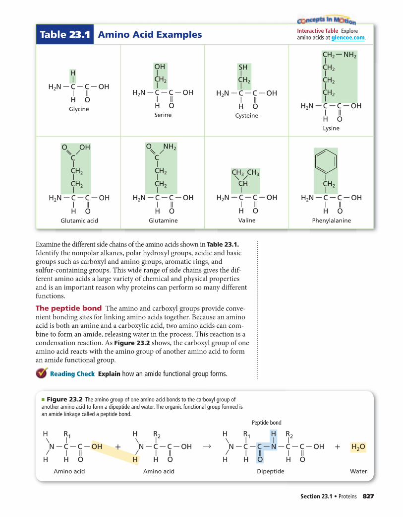
The peptide bond The amino and carboxyl groups provide conve-nient bonding sites for linking amino acids together. Because an amino acid is both an amine and a carboxylic acid, two amino acids can com-bine to form an amide, releasing water in the process. This reaction is a condensation reaction. As Figure 23.2 shows, the carboxyl group of one amino acid reacts with the amino group of another amino acid to form an amide functional group.
Reading Check Explain how an amide functional group forms.
Table 23.1 Amino Acid Examples
Glycine
H2N — C — C — OH
H—
H
—
O
——
Serine
H2N — C — C — OH
CH2
—
OH
—
H
—
O
——
Cysteine
H2N — C — C — OH
CH2
—
SH
—
H
—
O
——
Lysine
H2N — C — C — OH
CH2
CH2
CH2 — NH2
CH2
——
——
H
—
O
——
Glutamic acid
H2N — C — C — OH
H
—
O
——
—O OH——
CH2
CH2
C
——
—
Glutamine
H2N — C — C — OH
H
—
O
——
CH2
CH2
O —NH2——C
——
—
Valine
H2N — C — C — OH
CH3
—
H
—
O
——
—— CH3
CH
Phenylalanine
H2N — C — C — OH
CH2
——
H
—
O
——
Interactive Table Explore amino acids at glencoe.com.
Amino acid
Peptide bond
H R1
H OH
— —
N — C — C — OH
——
Amino acid
H R2
H OH
— —
N — C — C — OH
——
Dipeptide Water
H HR1
H OH
— —
N — C — C — N — C — C — OH
—
OH
— ——
—
R2
——
+→ H2O+
■ Figure 23.2 The amino group of one amino acid bonds to the carboxyl group of another amino acid to form a dipeptide and water. The organic functional group formed is an amide linkage called a peptide bond.

828 Chapter 23 • The Chemistry of Life
The amide bond that joins two amino acids, shown in Figure 23.3, is referred to by biochemists as a peptide bond. A chain of two or more amino acids linked together by peptide bonds is called a peptide. A molecule that consists of two amino acids bound together by a peptide bond is called a dipeptide. Figure 23.4a shows the structure of a dipeptide that is formed from the amino acids glycine (Gly) and phenylalanine (Phe). Figure 23.4b shows a different dipeptide, also formed by linking together glycine and phenylalanine. Is Gly-Phe the same compound as Phe-Gly? No, they’re different. Examine these two dipeptides to see that the order in which amino acids are linked in a dipeptide is important.
Each end of the two-amino-acid unit in a dipeptide still has a free group—one end has a free amino group and the other end has a free carboxyl group. Each of those groups can be linked to the opposite end of yet another amino acid, forming more peptide bonds. Living cells always build peptides by adding amino acids to the carboxyl end of a growing chain.
Reading Check Explain the difference between a peptide and a dipeptide.
Polypeptides As peptide chains increase in length, other ways of referring to them become necessary. A chain of ten or more amino acids joined by peptide bonds is referred to as a polypeptide. An example of a polypeptide is shown in Figure 23.5. When a chain reaches a length of about 50 amino acids, it is called a protein.
Because there are only 20 different amino acids that form proteins, it might seem reasonable to think that only a limited number of different protein structures are possible. However, a protein can have as few as 50 or more than a 1000 amino acids, arranged in any possible sequence. To calculate the number of possible sequences these amino acids can have, consider that each position on the chain can have any of 20 possible amino acids. For a peptide that contains n amino acids, there are 2 0 n possible sequences of the amino acids. So a dipeptide, with only two amino acids, can have 2 0 2 , or 400, different possible amino acid sequences. Even the smallest protein, containing only 50 amino acids, has 2 0 50 , or more than 1 × 1 0 65 , possible arrangements of amino acids! It is estimated that human cells make between 80,000 and 100,000 dif-ferent proteins. You can see that this is only a small fraction of the total number of proteins possible.
Reading Check Calculate the possible number of sequences for a peptide chain comprised of four amino acids.
Glycylphenylalanine (Gly-Phe)
Gly Phe
Phenylalanylglycine (Phe-Gly)
Phe Gly
H HH
H OH
— —
N — C — C — N — C — C — OH
—
OH
— ——
—
CH2
——
H H
H OH
— —
N — C — C — N — C — C — OH
—
OH
— ——
H
——
—
CH2
——
■ Figure 23.4 Glycine (Gly) and phenylalanine (Phe) can combine in two configurations.Explain Why are these two structures different substances?
a b
■ Figure 23.3 A peptide bond joins two amino acids to form a dipeptide.
— —
H
Peptide bond
O
— C — N—
—
Section 23.1 • Proteins 829
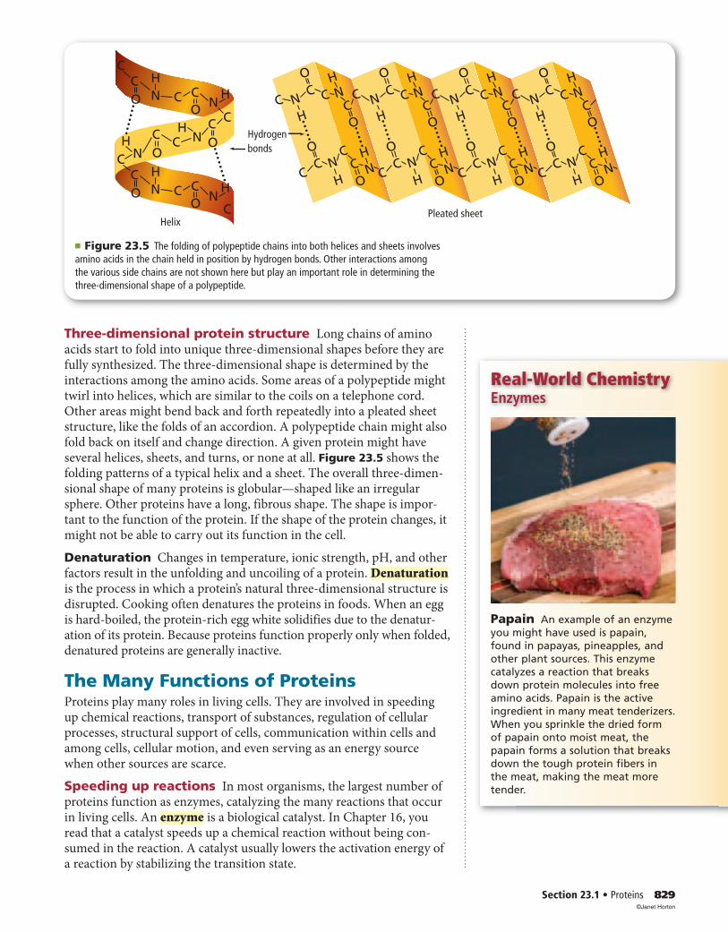
Three-dimensional protein structure Long chains of amino acids start to fold into unique three-dimensional shapes before they are fully synthesized. The three-dimensional shape is determined by the interactions among the amino acids. Some areas of a polypeptide might twirl into helices, which are similar to the coils on a telephone cord. Other areas might bend back and forth repeatedly into a pleated sheet structure, like the folds of an accordion. A polypeptide chain might also fold back on itself and change direction. A given protein might have several helices, sheets, and turns, or none at all. Figure 23.5 shows the folding patterns of a typical helix and a sheet. The overall three-dimen-sional shape of many proteins is globular—shaped like an irregular sphere. Other proteins have a long, fibrous shape. The shape is impor-tant to the function of the protein. If the shape of the protein changes, it might not be able to carry out its function in the cell.
Denaturation Changes in temperature, ionic strength, pH, and other factors result in the unfolding and uncoiling of a protein. Denaturationis the process in which a protein’s natural three-dimensional structure is disrupted. Cooking often denatures the proteins in foods. When an egg is hard-boiled, the protein-rich egg white solidifies due to the denatur-ation of its protein. Because proteins function properly only when folded, denatured proteins are generally inactive.
The Many Functions of ProteinsProteins play many roles in living cells. They are involved in speeding up chemical reactions, transport of substances, regulation of cellular processes, structural support of cells, communication within cells and among cells, cellular motion, and even serving as an energy source when other sources are scarce.
Speeding up reactions In most organisms, the largest number of proteins function as enzymes, catalyzing the many reactions that occur in living cells. An enzyme is a biological catalyst. In Chapter 16, you read that a catalyst speeds up a chemical reaction without being con-sumed in the reaction. A catalyst usually lowers the activation energy of a reaction by stabilizing the transition state.
Real-World Chemistry Enzymes
Papain An example of an enzyme you might have used is papain, found in papayas, pineapples, and other plant sources. This enzyme catalyzes a reaction that breaks down protein molecules into free amino acids. Papain is the active ingredient in many meat tenderizers. When you sprinkle the dried form of papain onto moist meat, the papain forms a solution that breaks down the tough protein fibers in the meat, making the meat more tender.
Hydrogenbonds
HelixPleated sheet
N
NN
N
CC
CC
CC
C
H
H
H
O
O
O
H
O
C N
NN
N
CC
CC
CC
C
H
H
H
O
O
O
H
O
C C CN
NN
N
CC
CC
CC
C
H
H
H
O
O
O
H
O
N
NN
N
CC
CC
CC
C
H
H
H
O
O
O
H
O
C
CC NH
HN
O
CO
HCC
CC
C
CC
N
N
O
HHN
HN O
O
CO
C
■ Figure 23.5 The folding of polypeptide chains into both helices and sheets involves amino acids in the chain held in position by hydrogen bonds. Other interactions among the various side chains are not shown here but play an important role in determining the three-dimensional shape of a polypeptide.
©Janet Horton

830 Chapter 23 • The Chemistry of Life
How do enzymes function? The term substrate refers to a reactant in an enzyme-catalyzed reaction, as shown in Figure 23.6. Substrates bind to specific sites on enzyme molecules, usually pockets or crevices. The spot to which the substrates bind is called the active site of the enzyme. After the substrates bind to the active site, the active site chang-es shape slightly to fit more tightly around the substrates. This recogni-tion process is called induced fit. The shapes of the substrates must fit the shape of the active site, in the same way that puzzle pieces or a lock and key fit together. A molecule that is only slightly different in shape from an enzyme’s normal substrate will not bind as well to the active site and might not undergo the catalyzed reaction.
The structure that forms when substrates are bound to an enzyme is called an enzyme-substrate complex. The large size of enzyme mole-cules allows them to form multiple bonds with their substrates, and the large variety of amino acid side chains in the enzyme allows a number of different intermolecular forces to form. These intermolecular forces lower the activation energy needed for the reaction in which bonds are broken and the substrates are converted to product.
Reading Check Describe in your own words how an enzyme works.
Transport proteins Some proteins are involved in transporting smaller particles throughout the body. Figure 23.7 shows the protein hemoglobin, which carries oxygen in the blood from the lungs to the rest of the body. Other proteins combine with biological molecules called lipids to transport them from one part of the body to another through the bloodstream. You will learn about lipids later in this chapter.
Heme
■ Figure 23.7 Hemoglobin is a glob-ular protein with four polypeptide chains, each containing an iron group (called a heme) to which oxygen binds.
■ Figure 23.6 Enzymes lower the activation energy needed for a reaction to occur. Enzymes change the speed at which chemical reactions occur without being altered themselves in the reaction.
Complex sugar
Enzyme(protein)
Products
Induced fit
Activesites
Simplesugar
Simplesugar
+H2O
Activesites
Enzyme
Each substrate fits into the active site. The enzyme changes shape slightly to fit with the substrate.
The products are released; in this case the complex sugar is divided into less complex sugars.
After the reaction, the enzyme released is in its original shape and can carry out the same reaction repeatedly.
Enzymes act on specific substrates, such as a complex sugar.
Section 23.123.1 Assessment
Section 23.1 • Proteins 831Self-Check Quiz glencoe.com
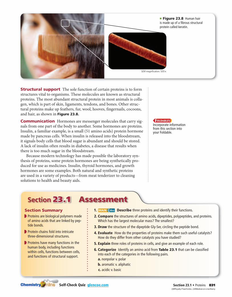
Structural support The sole function of certain proteins is to form structures vital to organisms. These molecules are known as structural proteins. The most abundant structural protein in most animals is colla-gen, which is part of skin, ligaments, tendons, and bones. Other struc-tural proteins make up feathers, fur, wool, hooves, fingernails, cocoons, and hair, as shown in Figure 23.8.
Communication Hormones are messenger molecules that carry sig-nals from one part of the body to another. Some hormones are proteins. Insulin, a familiar example, is a small (51 amino acids) protein hormone made by pancreas cells. When insulin is released into the bloodstream, it signals body cells that blood sugar is abundant and should be stored. A lack of insulin often results in diabetes, a disease that results when there is too much sugar in the bloodstream.
Because modern technology has made possible the laboratory syn-thesis of proteins, some protein hormones are being synthetically pro-duced for use as medicines. Insulin, thyroid hormones, and growth hormones are some examples. Both natural and synthetic proteins are used in a variety of products—from meat tenderizer to cleaning solutions to health and beauty aids.
Section Summary◗ ◗ Proteins are biological polymers made
of amino acids that are linked by pep-tide bonds.
◗ ◗ Protein chains fold into intricate three-dimensional structures.
◗ ◗ Proteins have many functions in the human body, including functions within cells, functions between cells, and functions of structural support.
1. MAIN Idea Describe three proteins and identify their functions.
2. Compare the structures of amino acids, dipeptides, polypeptides, and proteins. Which has the largest molecular mass? The smallest?
3. Draw the structure of the dipeptide Gly-Ser, circling the peptide bond.
4. Evaluate How do the properties of proteins make them such useful catalysts? How do they differ from other catalysts you have studied?
5. Explain three roles of proteins in cells, and give an example of each role.
6. Categorize Identify an amino acid from Table 23.1 that can be classified into each of the categories in the following pairs.
a. nonpolar v. polar b. aromatic v. aliphatic c. acidic v. basic
Incorporate information from this section into your Foldable.
■ Figure 23.8 Human hair is made up of a fibrous structural protein called keratin.
SEM magnification: 500×
(l)©Royalty-Free/Corbis, (r)©Medical-on-Line/Alamy
832 Chapter 23 • The Chemistry of Life
Section 23.223.2
CarbohydratesMAIN Idea Carbohydrates provide energy and structural material for living things.
Real-World Reading Link A lot of media attention has been focused on carbohydrates. Low-carb diets have become a popular way of controlling weight. However, carbohydrates are an important energy source for the body.
Kinds of CarbohydratesAnalyzing the term carbohydrate offers a hint about the structure of this group of molecules. Early observations that these compounds have the general chemical formula C n ( H 2 O ) n and appear to be hydrates of car-bon led to their being called carbohydrates. Although scientists now know that there are no full water molecules attached to carbohydrates, the name has stayed.
The main function of carbohydrates in living organisms is as a source of energy, both immediate and stored. Foods rich in carbohy-drates include pasta, milk, fruit, bread, and potatoes. Carbohydrates are compounds that contain multiple hydroxyl groups (—OH) as well as a carbonyl functional group (C=O). These molecules range in size from single monomers to polymers made of hundreds or even thou-sands of monomer units.
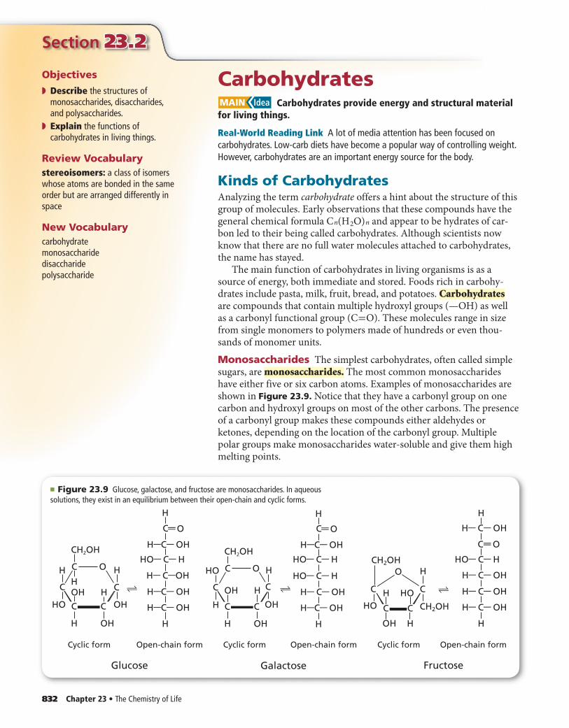
Monosaccharides The simplest carbohydrates, often called simple sugars, are monosaccharides. The most common monosaccharides have either five or six carbon atoms. Examples of monosaccharides are shown in Figure 23.9. Notice that they have a carbonyl group on one carbon and hydroxyl groups on most of the other carbons. The presence of a carbonyl group makes these compounds either aldehydes or ketones, depending on the location of the carbonyl group. Multiple polar groups make monosaccharides water-soluble and give them high melting points.
Objectives
◗ Describe the structures of monosaccharides, disaccharides, and polysaccharides.
◗ Explain the functions of carbohydrates in living things.
Review Vocabularystereoisomers: a class of isomers whose atoms are bonded in the same order but are arranged differently in space
New Vocabularycarbohydratemonosaccharidedisaccharidepolysaccharide
■ Figure 23.9 Glucose, galactose, and fructose are monosaccharides. In aqueous solutions, they exist in an equilibrium between their open-chain and cyclic forms.
Cyclic form Open-chain form
H OH
H
HO H
OH
OH
CH2OH
H CC
C O
C C
Galactose
Cyclic form Open-chain form
OH H
HOH HO
O
CC
C C
CH2OH
CH2OH
H
Fructose
H
H— C— OH
C— O——
H— C— OH
H
⇌
H
C
H—C— OH
O——
H— C— OH
H—C— OH
H
⇌
Cyclic form Open-chain form
H
H
OH
HO
H H
OHOH
CH2OH
H CC
C O
C C
Glucose
H
C
H—C— OH
O——
H— C—OH
HO— C— H HO— C— H HO— C— H
H— C— OH
H— C— OH
HO— C— H
H—C— OH
H—C— OH
H
⇌
Section 23.2 • Carbohydrates 833
Glucosesubunit
Glycogen
Glucose is a six-carbon sugar that has an aldehyde structure. Glucose is present in high concentration in blood because it serves as the major source of immediate energy for the body. For this reason, glucose is often called blood sugar. Closely related to glucose is galactose, which differs only in how a hydrogen and a hydroxyl group are oriented in space around one of the six carbon atoms. Recall from Chapter 21 that this relationship makes glucose and galactose stereoisomers. Fructose, also known as fruit sugar because it is the major carbohydrate in most fruits, is a six-carbon monosaccharide that has a ketone structure. Fructose is a structural isomer of glucose.
When monosaccharides are in aqueous solution, they exist in both open-chain and cyclic structures, but they rapidly interconvert forms. The cyclic structures are more stable and are the predominant form of monosaccharides at equilibrium. Note in Figure 23.9 that the carbonyl groups are present only in the open-chain structures. In the cyclic struc-tures, they are converted to hydroxyl groups.
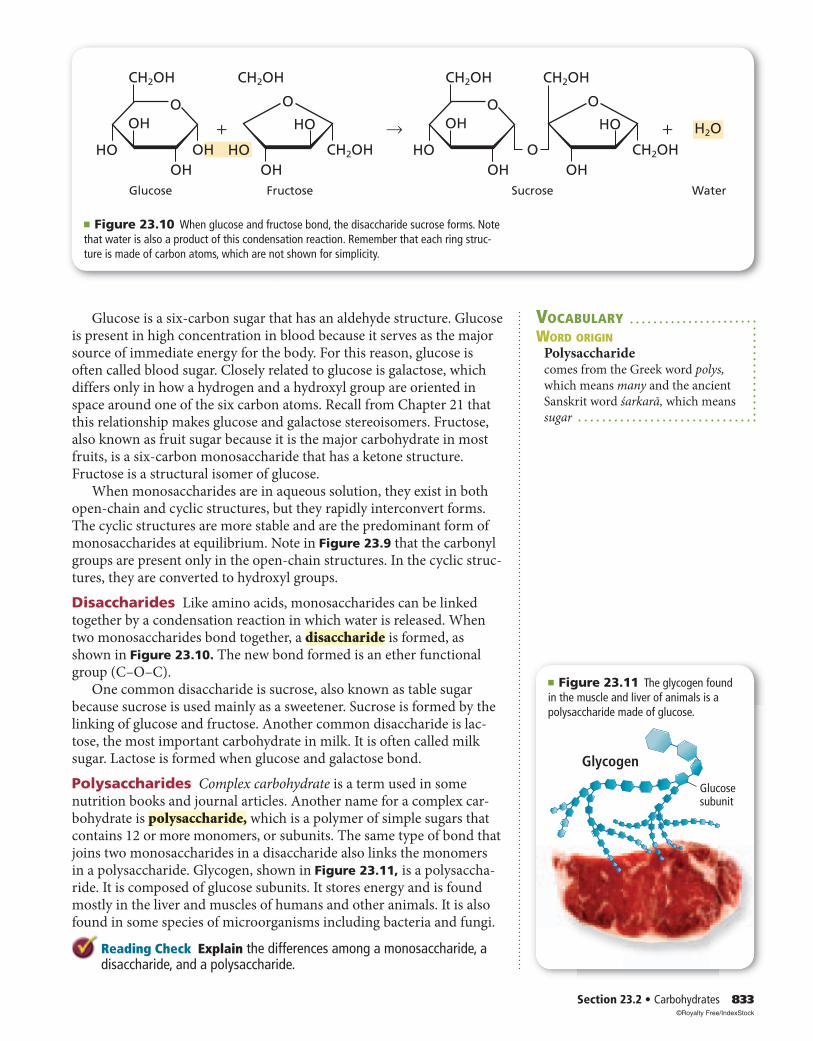
Disaccharides Like amino acids, monosaccharides can be linked together by a condensation reaction in which water is released. When two monosaccharides bond together, a disaccharide is formed, as shown in Figure 23.10. The new bond formed is an ether functional group (C–O–C).
One common disaccharide is sucrose, also known as table sugar because sucrose is used mainly as a sweetener. Sucrose is formed by the linking of glucose and fructose. Another common disaccharide is lac-tose, the most important carbohydrate in milk. It is often called milk sugar. Lactose is formed when glucose and galactose bond.
Polysaccharides Complex carbohydrate is a term used in some nutrition books and journal articles. Another name for a complex car-bohydrate is polysaccharide, which is a polymer of simple sugars that contains 12 or more monomers, or subunits. The same type of bond that joins two monosaccharides in a disaccharide also links the monomers in a polysaccharide. Glycogen, shown in Figure 23.11, is a polysaccha-ride. It is composed of glucose subunits. It stores energy and is found mostly in the liver and muscles of humans and other animals. It is also found in some species of microorganisms including bacteria and fungi.
Reading Check Explain the differences among a monosaccharide, a disaccharide, and a polysaccharide.
VOCABULARYWORD ORIGIN
Polysaccharidecomes from the Greek word polys,
which means many and the ancient
Sanskrit word śarkarā, which means
sugar
■ Figure 23.10 When glucose and fructose bond, the disaccharide sucrose forms. Note that water is also a product of this condensation reaction. Remember that each ring struc-ture is made of carbon atoms, which are not shown for simplicity.
OH
CH2OH CH2OH
HO
Glucose
OHO
HO+ →
OH
HO
CH2OH
Fructose
O
OHO
CH2OH CH2OH
HO
OHO
+
OHCH2OH
H2O
Sucrose Water
O
HO
OH
■ Figure 23.11 The glycogen found in the muscle and liver of animals is a polysaccharide made of glucose.
©Royalty Free/IndexStock
Section 23.223.2 Assessment
834 Chapter 23 • The Chemistry of Life Self-Check Quiz glencoe.com
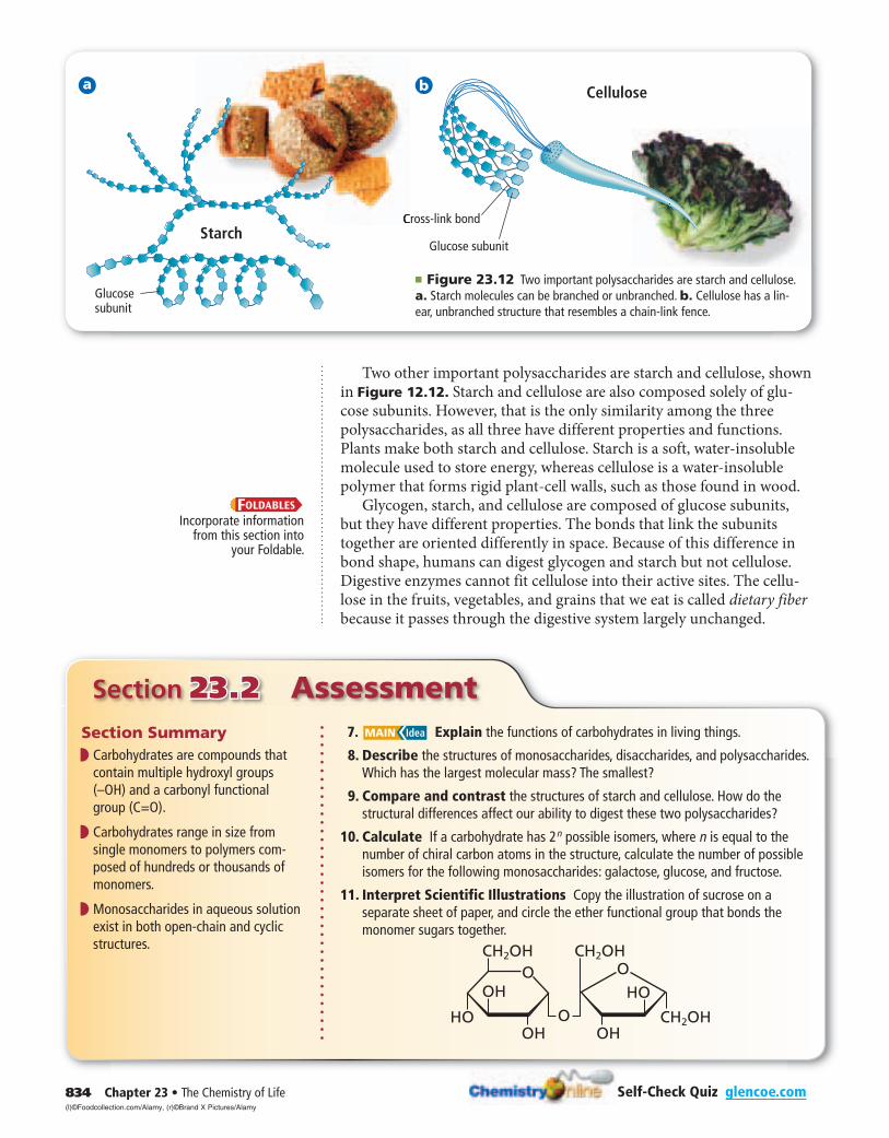
Two other important polysaccharides are starch and cellulose, shown in Figure 12.12. Starch and cellulose are also composed solely of glu-cose subunits. However, that is the only similarity among the three polysaccharides, as all three have different properties and functions. Plants make both starch and cellulose. Starch is a soft, water-insoluble molecule used to store energy, whereas cellulose is a water-insoluble polymer that forms rigid plant-cell walls, such as those found in wood.
Glycogen, starch, and cellulose are composed of glucose subunits, but they have different properties. The bonds that link the subunits together are oriented differently in space. Because of this difference in bond shape, humans can digest glycogen and starch but not cellulose. Digestive enzymes cannot fit cellulose into their active sites. The cellu-lose in the fruits, vegetables, and grains that we eat is called dietary fiber because it passes through the digestive system largely unchanged.
Incorporate information from this section into
your Foldable.
Section Summary◗ ◗ Carbohydrates are compounds that
contain multiple hydroxyl groups (–OH) and a carbonyl functional group (C=O).
◗ ◗ Carbohydrates range in size from single monomers to polymers com-posed of hundreds or thousands of monomers.
◗ ◗ Monosaccharides in aqueous solution exist in both open-chain and cyclic structures.
7. MAIN Idea Explain the functions of carbohydrates in living things.
8. Describe the structures of monosaccharides, disaccharides, and polysaccharides. Which has the largest molecular mass? The smallest?
9. Compare and contrast the structures of starch and cellulose. How do the structural differences affect our ability to digest these two polysaccharides?
10. Calculate If a carbohydrate has 2 n possible isomers, where n is equal to the number of chiral carbon atoms in the structure, calculate the number of possible isomers for the following monosaccharides: galactose, glucose, and fructose.
11. Interpret Scientific Illustrations Copy the illustration of sucrose on a separate sheet of paper, and circle the ether functional group that bonds the monomer sugars together.
a
OHO
CH2OH CH2OH
HO
OHO
OHCH2OH
OHO
Cellulose
Cross-link bond
Glucose subunit
b
Glucosesubunit
Starch
■ Figure 23.12 Two important polysaccharides are starch and cellulose. a. Starch molecules can be branched or unbranched. b. Cellulose has a lin-ear, unbranched structure that resembles a chain-link fence.
(l)©Foodcollection.com/Alamy, (r)©Brand X Pictures/Alamy
Section 23.3 • Lipids 835
Section 23.323.3
Objectives
◗ ◗ Describe the structures of fatty acids, triglycerides, phospholipids, and steroids.
◗ ◗ Explain the functions of lipids in living organisms.
◗ ◗ Identify some reactions that fatty acids undergo.
◗ ◗ Relate the structure and function of cell membranes.
Review Vocabularynonpolar: without separate positive and negative areas or dipoles
New Vocabularylipidfatty acidtriglyceridesaponificationphospholipidwaxsteroid
LipidsMAIN Idea Lipids make cell membranes, store energy, and regulate cellular processes.
Real-World Reading Link The wax used to polish cars, the fat that drips out of hamburgers, and the vitamin D that fortifies the milk people drink—what do these things have in common? They are all lipids.
What is a lipid?A lipid is a large, nonpolar biological molecule. Because lipids are non-polar, they are insoluble in water. Lipids have two major functions in living organisms. They store energy efficiently, and they make up most of the structure of cell membranes. Unlike proteins and carbohydrates, lipids are not polymers with repeated monomer subunits.
Fatty acids Although lipids are not polymers, many lipids have a major building block in common. This building block is the fatty acid, a long-chain carboxylic acid. Most naturally occurring fatty acids contain between 12 and 24 carbon atoms. Their structure can be represented by the following formula.
C H 3 (C H 2 ) n COOH
Most fatty acids have an even number of carbon atoms, which is a result of being constructed two carbons at a time in enzymatic reactions.
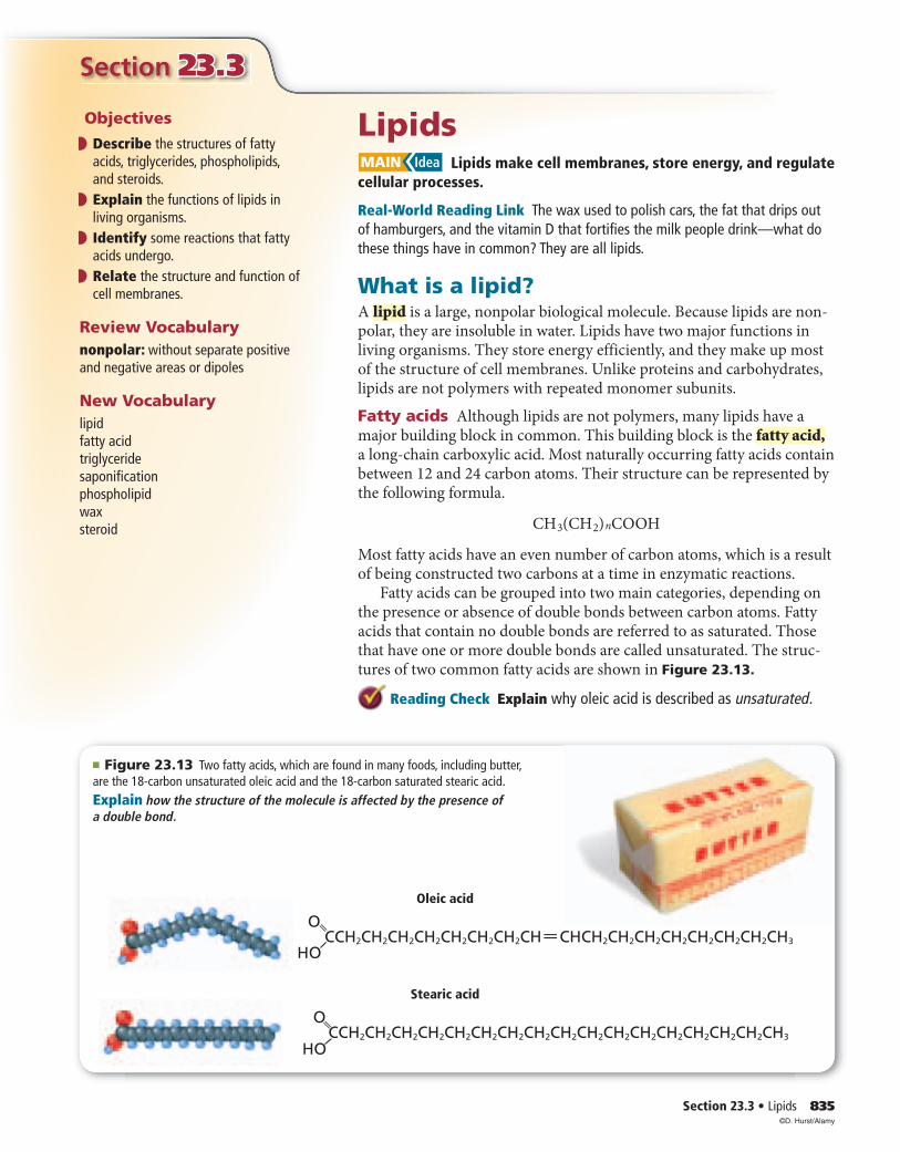
Fatty acids can be grouped into two main categories, depending on the presence or absence of double bonds between carbon atoms. Fatty acids that contain no double bonds are referred to as saturated. Those that have one or more double bonds are called unsaturated. The struc-tures of two common fatty acids are shown in Figure 23.13.
Reading Check Explain why oleic acid is described as unsaturated.
■ Figure 23.13 Two fatty acids, which are found in many foods, including butter, are the 18-carbon unsaturated oleic acid and the 18-carbon saturated stearic acid. Explain how the structure of the molecule is affected by the presence of a double bond.
CCH2CH2CH2CH2CH2CH2CH2CH CHCH2CH2CH2CH2CH2CH2CH2CH3
O
HO
CCH2CH2CH2CH2CH2CH2CH2CH2CH2CH2CH2CH2CH2CH2CH2CH2CH3
O
HO
Oleic acid
Stearic acid
——
©D. Hurst/Alamy
836 Chapter 23 • The Chemistry of Life
CH2OH
CHOH
CH2OH
HOC(CH2)14CH3
HOC(CH2)16CH3
HOC(CH2)18CH3
+
Glycerol 3 Fatty acids Triglyceride Water
— —
O
→
— —
O
— —
O
— —
O
CH — O — C — (CH2)16 — CH3 + 3H2O
— —
O
CH2 — O — C — (CH2)14 — CH3
— —
O
CH2 — O — C — (CH2)18 — CH3
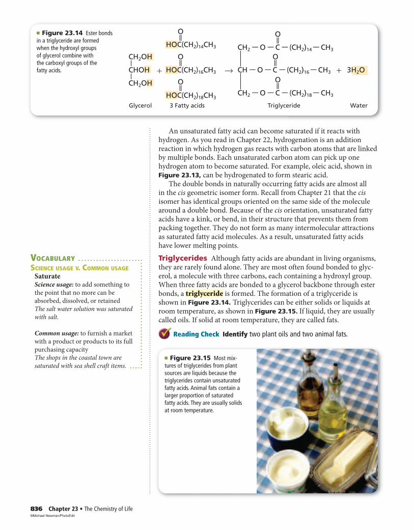
■ Figure 23.14 Ester bonds in a triglyceride are formedwhen the hydroxyl groups of glycerol combine with the carboxyl groups of the fatty acids.
An unsaturated fatty acid can become saturated if it reacts with hydrogen. As you read in Chapter 22, hydrogenation is an addition reaction in which hydrogen gas reacts with carbon atoms that are linked by multiple bonds. Each unsaturated carbon atom can pick up one hydrogen atom to become saturated. For example, oleic acid, shown in Figure 23.13, can be hydrogenated to form stearic acid.
The double bonds in naturally occurring fatty acids are almost all in the cis geometric isomer form. Recall from Chapter 21 that the cis isomer has identical groups oriented on the same side of the molecule around a double bond. Because of the cis orientation, unsaturated fatty acids have a kink, or bend, in their structure that prevents them from packing together. They do not form as many intermolecular attractions as saturated fatty acid molecules. As a result, unsaturated fatty acids have lower melting points.
Triglycerides Although fatty acids are abundant in living organisms, they are rarely found alone. They are most often found bonded to glyc-erol, a molecule with three carbons, each containing a hydroxyl group. When three fatty acids are bonded to a glycerol backbone through ester bonds, a triglyceride is formed. The formation of a triglyceride is shown in Figure 23.14. Triglycerides can be either solids or liquids at room temperature, as shown in Figure 23.15. If liquid, they are usually called oils. If solid at room temperature, they are called fats.
Reading Check Identify two plant oils and two animal fats.
■ Figure 23.15 Most mix-tures of triglycerides from plant sources are liquids because the triglycerides contain unsaturated fatty acids. Animal fats contain a larger proportion of saturated fatty acids. They are usually solids at room temperature.
VOCABULARYSCIENCE USAGE V. COMMON USAGE
SaturateScience usage: to add something to
the point that no more can be
absorbed, dissolved, or retained
The salt water solution was saturated
with salt.
Common usage: to furnish a market
with a product or products to its full
purchasing capacity
The shops in the coastal town are
saturated with sea shell craft items.
©Michael Newman/PhotoEdit
Section 23.3 • Lipids 837
Fatty acids are stored in the fat cells of your body as triglycerides. When energy is abundant, fat cells store the excess energy in the fatty acids of triglycerides. When energy is scarce, the cells break down the triglycerides, releasing the energy used to form them.
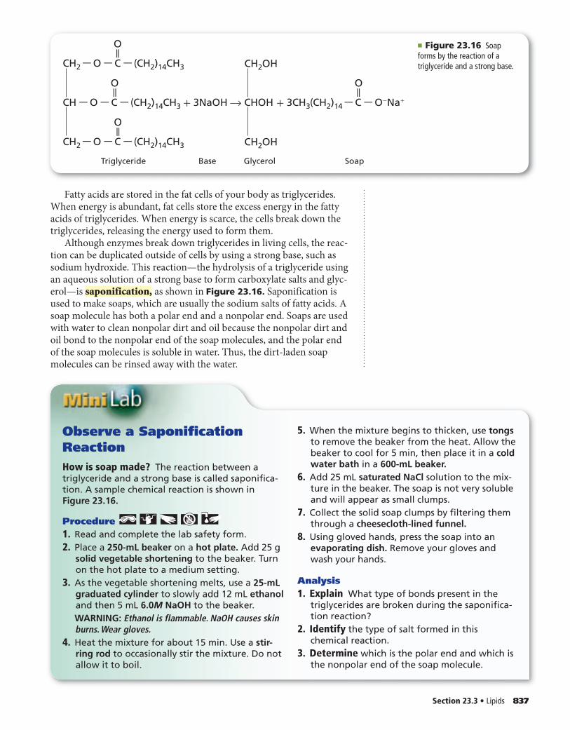
Although enzymes break down triglycerides in living cells, the reac-tion can be duplicated outside of cells by using a strong base, such as sodium hydroxide. This reaction—the hydrolysis of a triglyceride using an aqueous solution of a strong base to form carboxylate salts and glyc-erol—is saponification, as shown in Figure 23.16. Saponification is used to make soaps, which are usually the sodium salts of fatty acids. A soap molecule has both a polar end and a nonpolar end. Soaps are used with water to clean nonpolar dirt and oil because the nonpolar dirt and oil bond to the nonpolar end of the soap molecules, and the polar end of the soap molecules is soluble in water. Thus, the dirt-laden soap molecules can be rinsed away with the water.
Triglyceride Glycerol SoapBase
— —
O
— —
O
CH — O — C — (CH2)14CH3 + 3NaOH→ CHOH + 3CH3(CH2)14 — C — O-Na+
— —O
— —
O
CH2 — O — C — (CH2)14CH3
CH2 — O — C — (CH2)14CH3 CH2OH
CH2OH
■ Figure 23.16 Soap forms by the reaction of atriglyceride and a strong base.
Observe a Saponification ReactionHow is soap made? The reaction between a triglyceride and a strong base is called saponifica-tion. A sample chemical reaction is shown in Figure 23.16.
Procedure 1. Read and complete the lab safety form.2. Place a 250-mL beaker on a hot plate. Add 25 g
solid vegetable shortening to the beaker. Turn on the hot plate to a medium setting.
3. As the vegetable shortening melts, use a 25-mL graduated cylinder to slowly add 12 mL ethanol and then 5 mL 6.0M NaOH to the beaker.
WARNING: Ethanol is flammable. NaOH causes skin burns. Wear gloves.
4. Heat the mixture for about 15 min. Use a stir-ring rod to occasionally stir the mixture. Do not allow it to boil.
5. When the mixture begins to thicken, use tongs to remove the beaker from the heat. Allow the beaker to cool for 5 min, then place it in a cold water bath in a 600-mL beaker.
6. Add 25 mL saturated NaCl solution to the mix-ture in the beaker. The soap is not very soluble and will appear as small clumps.
7. Collect the solid soap clumps by filtering them through a cheesecloth-lined funnel.
8. Using gloved hands, press the soap into an evaporating dish. Remove your gloves and wash your hands.
Analysis1. Explain What type of bonds present in the
triglycerides are broken during the saponifica-tion reaction?
2. Identify the type of salt formed in this chemical reaction.
3. Determine which is the polar end and which is the nonpolar end of the soap molecule.
838 Chapter 23 • The Chemistry of Life
■ Figure 23.18 Plants produce a wax that coats their leaves. The wax protects the leaves from drying out.
Outside the cell
Inside the cell
Phospholipidbilayer
Polar heads Nonpolar tails
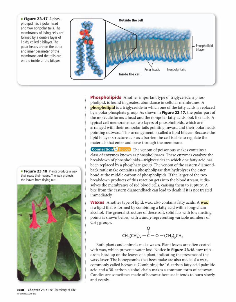
■ Figure 23.17 A phos-pholipid has a polar head and two nonpolar tails. The membranes of living cells are formed by a double layer of lipids, called a bilayer. The polar heads are on the outer and inner perimeter of the membrane and the tails are on the inside of the bilayer.
Phospholipids Another important type of triglyceride, a phos-pholipid, is found in greatest abundance in cellular membranes. A phospholipid is a triglyceride in which one of the fatty acids is replaced by a polar phosphate group. As shown in Figure 23.17, the polar part of the molecule forms a head and the nonpolar fatty acids look like tails. A typical cell membrane has two layers of phospholipids, which are arranged with their nonpolar tails pointing inward and their polar heads pointing outward. This arrangement is called a lipid bilayer. Because the lipid bilayer structure acts as a barrier, the cell is able to regulate the materials that enter and leave through the membrane.
Connection Biology The venom of poisonous snakes contains a class of enzymes known as phospholipases. These enzymes catalyze the breakdown of phospholipids—triglycerides in which one fatty acid has been replaced by a phosphate group. The venom of the eastern diamond-back rattlesnake contains a phospholipase that hydrolyzes the ester bond at the middle carbon of phospholipids. If the larger of the two breakdown products of this reaction gets into the bloodstream, it dis-solves the membranes of red blood cells, causing them to rupture. A bite from the eastern diamondback can lead to death if it is not treated immediately.
Waxes Another type of lipid, wax, also contains fatty acids. A wax is a lipid that is formed by combining a fatty acid with a long-chain alcohol. The general structure of these soft, solid fats with low melting points is shown below, with x and y representing variable numbers of C H 2 groups.
CH3(CH2)x — C — O — (CH2)yCH3
O
——
Both plants and animals make waxes. Plant leaves are often coated with wax, which prevents water loss. Notice in Figure 23.18 how rain-drops bead up on the leaves of a plant, indicating the presence of the waxy layer. The honeycombs that bees make are also made of a wax, commonly called beeswax. Combining the 16-carbon fatty acid palmitic acid and a 30-carbon alcohol chain makes a common form of beeswax. Candles are sometimes made of beeswax because it tends to burn slowly and evenly.
©Pat O’Hara/CORBIS

Section 23.323.3 Assessment
Section 23.3 • Lipids 839Self-Check Quiz glencoe.com
■ Figure 23.19 This Giant Marine toad uses a steroid toxin called bufotoxin as a defense mechanism. The toxin is fatal to some animals, including dogs and cats.
◗ ◗ Fatty acids are long-chain carboxylic acids that usually have between 12 and 24 carbon atoms.
◗ ◗ Saturated fatty acids have no double bonds; unsaturated fatty acids have one or more double bonds.
◗ ◗ Fatty acids can be linked to glycerol backbones to form triglycerides.
◗ ◗ Steroids are lipids that have multiple-ring structures.
12. MAIN Idea Describe the function of lipids.
13. Describe the structures of fatty acids, triglycerides, phospholipids, and steroids.
14. List an important function of each of these types of lipids. a. triglycerides c. waxes b. phospholipids d. steroids
15. Identify two reactions that fatty acids undergo.
16. Describe the structure and function of cell membranes.
17. Compare and contrast the structures of a steroid, a phospholipid, and a wax.
18. Write the equation for the complete hydrogenation of the polyunsaturated fatty acid linoleic acid, C H 3 (C H 2 ) 4 CH=CHC H 2 CH=CH(C H 2 ) 7 COOH.
19. Interpret Scientific Illustrations Draw the general structure of a phospho-lipid. Label the polar and nonpolar portions of the structure.
Steroids Not all lipids contain fatty acid chains. Steroids are lipids that have multiple cyclic rings in their structures. All steroids are built from the basic four-ring steroid structure shown below.
Some hormones, such as many sex hormones, are steroids that function to regulate metabolic processes. Cholesterol, another steroid, is an important structural component of cell membranes. Vitamin D also contains the four-ring steroid structure and plays a role in the formation of bones. The Giant Marine toad, Bufo marinus, shown in Figure 23.19 uses a steroid called bufotoxin as a defense mechanism. The toad secretes the toxin from warts on its back and from glands just behind the eye. The toxin is only an irritant for humans, but in small animals the toxin causes drooling, loss of coordination, convulsions, and death.
Incorporate information from this section into your Foldable.
©MC DONALD, JOE/Animals Animals/Earth Scene

840 Chapter 23 • The Chemistry of Life
Section 23.423.4
■ Figure 23.20 Nucleotides are the monomers from which nucleic acid polymers are formed.
Each nucleotide contains a nitrogen-containing base, a five-carbon sugar, and a phosphate group.
Nucleic acids are linear chains of alternating sugars and phos-phates. Attached to every sugar is a nitrogen base. Because the nucleotides are offset, the chains resemble steps in a staircase.
Sugar
Nucleotide Nucleic acid
— —
OH OH
HHH H
O
HO
HO — P — O —— CH2
— O
CC
C C
C
H
N
C N NH2
C
C
H
——
——
Phosphate groupNitrogen-containingbase
Phosphate
Sugar Base
Base
Base
Phosphate
Sugar
Phosphate
Sugar
——Oa b
Objectives
◗ Identify the structural components of nucleic acids.
◗ Relate the function of DNA to its structure.
◗ Describe the structure and function of RNA.
Review Vocabularygenetic information: an inherited sequence of RNA or DNA that causes traits or characteristics to pass from one generation to the next
New Vocabularynucleic acidnucleotide
Nucleic AcidsMAIN Idea Nucleic acids store and transmit genetic information.
Real-World Reading Link DNA testing is becoming more routine in medicine, forensic science, genealogy, and identification of victims in disasters. Modern techniques have made it possible to get a useful DNA sample from surprising sources, such as a strand of hair or dried saliva on a postage stamp.
Structure of Nucleic AcidsNucleic acids comprise a fourth class of biological molecules. They are the information-storage molecules of the cell. This group of molecules got its name from the cellular location in which the molecules are pri-marily found—the nucleus. It is from this control center of cells that nucleic acids carry out their major functions. A nucleic acid is a nitrogen-containing biological polymer that is involved in the storage and trans-mission of genetic information. The monomer that makes up a nucleic acid is called a nucleotide. Each nucleotide has three parts: an inorganic phosphate group, a five-carbon monosaccharide sugar, and a nitrogen-containing structure called a nitrogenous base. Examine each part of Figure 23.20a. Although the phosphate group is the same in all nucleo-tides, the sugar and the nitrogen base vary.
In a nucleic acid, the sugar of one nucleotide is bonded to the phos-phate of another nucleotide, as shown in Figure 23.20b. Thus, the nucleotides are strung together in a chain, or strand, containing alter-nating sugar and phosphate groups. Each sugar is also bonded to a nitrogen base that sticks out from the chain. The nitrogen bases on adjoining nucleotide units are stacked one above the other in a slightly askew position, much like the steps in a staircase. This orientation is shown in Figure 23.20b. Intermolecular forces hold each nitrogen base close to the nitrogen bases above and below it.

Section 23.4 • Nucleic Acids 841
DNA: The Double HelixYou might have heard of DNA (deoxyribonucleic acid), one of the two kinds of nucleic acids found in living cells. DNA contains the master plans for building all the proteins in an organism’s body.
The structure of DNA DNA consists of two long chains of nucleotides wound together to form a spiral structure, as shown in Figure 23.21. Each nucleotide in DNA contains a phosphate group, the five-carbon sugar deoxyribose, and a nitrogenous base. The alternating sugar and phosphate groups in each chain make up the outside, or backbone, of the spiral structure, The nitrogen bases are on the inside of the struc-ture. Because the spiral structure is composed of two chains, it is known as a double helix.
Reading Check Describe what forms the teeth of the DNA zipper.
DNA contains four different nitrogenous bases: adenine (A), thy-mine (T), cytosine (C), and guanine (G). As Figure 23.21 shows, both adenine and guanine contain a double ring. Thymine and cytosine are single-ring structures. Looking again at Figure 23.21, notice that each nitrogen base on one strand of the helix is oriented next to a nitrogen base on the opposite strand, in the same way that the teeth of a zipper are oriented. The side-by-side base pairs are close enough so that hydrogen bonds form between them. Because each nitrogen base has a unique arrangement of organic functional groups that can form hydrogen bonds, the nitrogen bases always pair in a specific way so that the optimum number of hydrogen bonds form. As Figure 23.22 shows, guanine always binds to cytosine, and adenine always binds to thymine. The G–C and A–T pairs are called complementary base pairs.
Because of complementary base pairing, the amount of adenine in a molecule of DNA always equals the amount of thymine, and the amount of cytosine always equals the amount of guanine. In 1953, James Watson and Francis Crick used this observation to make one of the greatest sci-entific discoveries of the twentieth century when they determined the double-helix structure of DNA. They accomplished this feat without performing many laboratory experiments themselves. Instead, they ana-lyzed and synthesized the work of numerous scientists who had careful-ly carried out studies on DNA.
H—
H—......
.....
Deoxyribose
Deoxyribose
DeoxyriboseO
O O
——
—— ——CH3
H
H
N—
H
N
N
H
NNH— H—......
H—.......
Hydrogenbond
O——
H
H
N
......H —
NN
N
N
H
N
Thymine Adenine Cytosine Guanine
N
N
Deoxyribose
HN
N
N
■ Figure 23.22 In DNA, base pairing exists between a double-ringed base and a single-ringed base. Adenine and thymine always pair, forming two hydrogen bonds between them. Guanine and cytosine always form three hydrogen bonds when they pair.
■ Figure 23.21 The structure of DNA is a double helix that resembles a twisted zipper. The two sugar-phosphate backbones form the outsides of the zipper.
C
GA
C
C
G
C
G
G
G
TA
T A
T
A T
A T
Interactive Figure To see an animation of the structure of DNA, visit glencoe.com.

Problem-solving labProblem-solving lab
842 Chapter 23 • The Chemistry of Life
The function of DNA Watson and Crick used their model to predict how DNA’s chemical structure enables it to function. DNA stores the genetic information of a cell in the cell’s nucleus. Before the cell divides, the DNA is copied so that the new generation of cells gets the same genetic information. Having determined that the two chains of the DNA helix are complementary, Watson and Crick realized that complementary base pairing provides a mechanism by which the genetic material of a cell is copied.
The four nitrogenous bases of DNA serve as the letters of the alpha-bet in the information-storage language of living cells. The specific sequence of these letters represents an organism’s master instructions, just as the sequence of letters in the words of this sentence convey special meaning. The sequence of bases is different in every species of organism, allowing for an enormous diversity of life-forms—all from a language that uses only four letters. It is estimated that the DNA in a human cell has about three billion complementary base pairs, arranged in a sequence unique to humans.
Formulate a ModelHow does DNA replicate? DNA rep-licates, before a cell divides so that each of the two newly formed cells has a complete set of genetic instructions. When DNA begins to repli-cate, the two nucleotide strands start to unzip. An enzyme breaks the hydrogen bonds between the nitrogenous bases, and the strands separate. Other enzymes deliver free nucleotides from the surrounding medium to the exposed nitrogenous bases, adenine hydrogen-bonding with thymine, and cytosine bonding with guanine. Thus, each strand builds a complementary strand by base-pairing with free nucleotides. This process is shown in the top diagram at the right. When the free nucleotides have been hydrogen-bonded into place, their sugars and phosphates bond cova-lently to those on adjacent nucleotides to form the new backbone. Each strand of the original DNA molecule is now bonded to a new strand.
AnalysisThe bottom diagram shows a small segment of a DNA molecule. Copy the base sequence onto a clean sheet of paper, being careful not to make copying errors. Show the steps of replication to produce two segments of the DNA.
Think Critically1. Describe how the base sequence of a newly
synthesized strand compares with the original strand to which it is bonded.
C
C
C
CC
C
TT
T
T
TT
T
T
T
AA
A
A
A
ATA
TA TA
TAT A
A
A
A
G
GG C G
CG
G
GG
A T C G G T T A A A C GT A G C C A A T T T G C
2. Explain If the original DNA segment is col-ored red and the free nucleotides are colored blue, what pattern of colors will the newly replicated DNA segments have? Will all new segments have the same color pattern?
3. Explain how an organism might be affected if an error occurs during replication of its DNA. Are the affects permanent? Explain.
Section 23.423.4 Assessment
Section 23.4 • Nucleic Acids 843Self-Check Quiz glencoe.com
— —
H O
Thymine
O
——
UracilDeoxyriboseHOH
H H
O
H
OH
H
HOCH2
OH
H H
O
H
OH
H
HOCH2
Ribose
N
— — H HN
— —
O
O
——
H
N
— —
N
H
H3C H
OHH
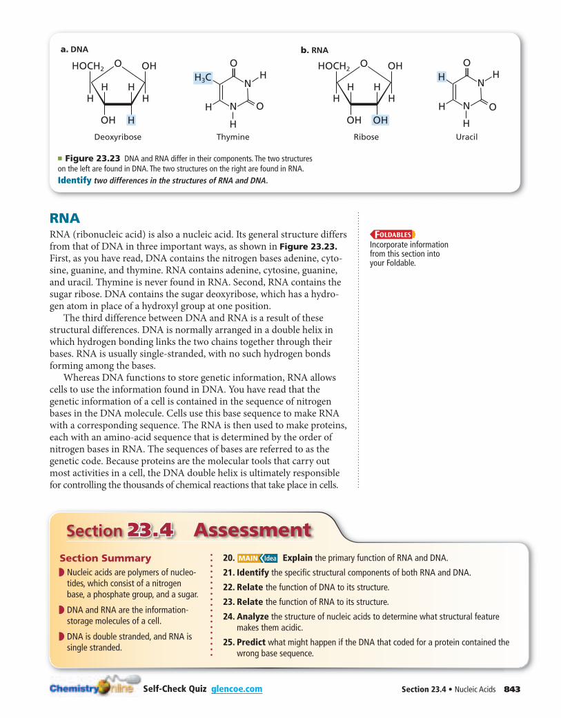
■ Figure 23.23 DNA and RNA differ in their components. The two structures on the left are found in DNA. The two structures on the right are found in RNA.Identify two differences in the structures of RNA and DNA.
b. RNAa. DNA
RNARNA (ribonucleic acid) is also a nucleic acid. Its general structure differs from that of DNA in three important ways, as shown in Figure 23.23. First, as you have read, DNA contains the nitrogen bases adenine, cyto-sine, guanine, and thymine. RNA contains adenine, cytosine, guanine, and uracil. Thymine is never found in RNA. Second, RNA contains the sugar ribose. DNA contains the sugar deoxyribose, which has a hydro-gen atom in place of a hydroxyl group at one position.
The third difference between DNA and RNA is a result of these structural differences. DNA is normally arranged in a double helix in which hydrogen bonding links the two chains together through their bases. RNA is usually single-stranded, with no such hydrogen bonds forming among the bases.
Whereas DNA functions to store genetic information, RNA allows cells to use the information found in DNA. You have read that the genetic information of a cell is contained in the sequence of nitrogen bases in the DNA molecule. Cells use this base sequence to make RNA with a corresponding sequence. The RNA is then used to make proteins, each with an amino-acid sequence that is determined by the order of nitrogen bases in RNA. The sequences of bases are referred to as the genetic code. Because proteins are the molecular tools that carry out most activities in a cell, the DNA double helix is ultimately responsible for controlling the thousands of chemical reactions that take place in cells.
Section Summary◗ ◗ Nucleic acids are polymers of nucleo-
tides, which consist of a nitrogen base, a phosphate group, and a sugar.
◗ ◗ DNA and RNA are the information-storage molecules of a cell.
◗ ◗ DNA is double stranded, and RNA is single stranded.
20. MAIN Idea Explain the primary function of RNA and DNA.
21. Identify the specific structural components of both RNA and DNA.
22. Relate the function of DNA to its structure.
23. Relate the function of RNA to its structure.
24. Analyze the structure of nucleic acids to determine what structural feature makes them acidic.
25. Predict what might happen if the DNA that coded for a protein contained the wrong base sequence.
Incorporate information from this section into your Foldable.
844 Chapter 23 • The Chemistry of Life
Section 23.523.5
Objectives
◗ Distinguish between anabolism and catabolism.
◗ Describe the role of ATP in metabolism.
◗ Compare and contrast the processes of photosynthesis, cellular respiration, and fermentation.
Review Vocabularyredox process: a chemical reaction in which electrons are transferred from one atom to another
New VocabularymetabolismcatabolismanabolismATPphotosynthesiscellular respirationfermentation
MetabolismMAIN Idea Metabolism involves many thousands of reactions in living cells.
Real-World Reading Link You have studied the four major kinds of biological molecules and learned that they are all present in the food you eat. What happens to these molecules after they enter your body?
Anabolism and CatabolismMany thousands of chemical reactions take place in the cells of a living organism. The set of chemical reactions that occur within an organism is its metabolism. Why are so many reactions involved in metabolism? Living organisms must accomplish two major functions in order to survive. They have to extract energy from nutrients in forms that they can use immediately as well as store for future use. In addition, they have to use nutrients to make building blocks for synthesizing all of the molecules needed to perform their life functions. These processes are summarized in Figure 23.24.
The term catabolism refers to the metabolic reactions that break down complex biological molecules such as proteins, polysaccharides, triglycerides, and nucleic acids for the purposes of forming smaller building blocks and extracting energy. After you eat a meal of spaghetti and meatballs, your body immediately begins to break down the starch polymer in the pasta into glucose. The glucose is then broken down into smaller molecules in a series of energy-releasing catabolic reactions. Meanwhile, the protein polymers in the meatballs are catabolized into amino acids.
The term anabolism refers to the metabolic reactions that use energy and small building blocks to synthesize the complex molecules needed by an organism. After your body has extracted the energy from the starch in the pasta, it uses that energy and the amino-acid building blocks produced from the meat proteins to synthesize the specific pro-teins that allow your muscles to contract, catalyze metabolic reactions, and perform many other functions in your body.
Nutrients ingestedCarbohydrates
FatsProteins
Intermediate productsAmino acids
Simple sugarsFatty acidsNucleotides
ATP
Complex cellularmolecules
ProteinsPolysaccharides
TriglyceridesNucleic acids
ADP + P
Catabolism(nutrients broken
down)
Anabolism(new molecules
synthesized)
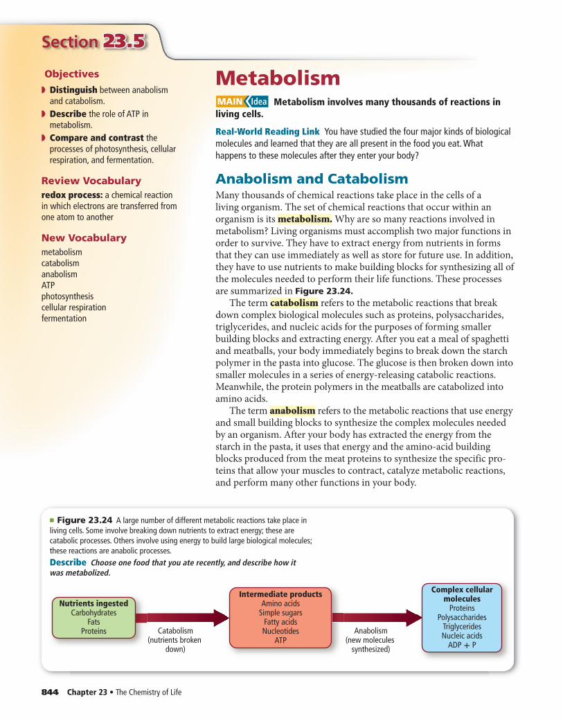
■ Figure 23.24 A large number of different metabolic reactions take place in living cells. Some involve breaking down nutrients to extract energy; these are catabolic processes. Others involve using energy to build large biological molecules; these reactions are anabolic processes.Describe Choose one food that you ate recently, and describe how it was metabolized.
Section 23.5 • Metabolism 845
Figure 23.24 shows the relationship between catabolism and anabo-lism. The nutrients listed on the left side of the diagram are broken down into intermediate products. These intermediate products are used as building blocks for the products listed on the right side of the dia-gram. Another way of conceptualizing this process is to view the nutri-ents ingested as the raw materials for the complex cellular molecules formed in a living organism.
Reading Check Explain how the terms metabolism, catabolism, and anabolism are related.
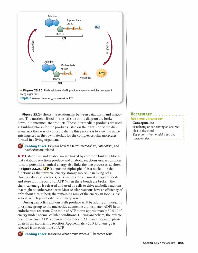
ATP Catabolism and anabolism are linked by common building blocks that catabolic reactions produce and anabolic reactions use. A common form of potential chemical energy also links the two processes, as shown in Figure 23.25. ATP (adenosine triphosphate) is a nucleotide that functions as the universal energy-storage molecule in living cells. During catabolic reactions, cells harness the chemical energy of foods and store it in the bonds of ATP. When these bonds are broken, the chemical energy is released and used by cells to drive anabolic reactions that might not otherwise occur. Most cellular reactions have an efficiency of only about 40% at best; the remaining 60% of the energy in food is lost as heat, which your body uses to keep warm.
During catabolic reactions, cells produce ATP by adding an inorganic phosphate group to the nucleotide adenosine diphosphate (ADP) in an endothermic reaction. One mole of ATP stores approximately 30.5 kJ of energy under normal cellular conditions. During anabolism, the reverse reaction occurs. ATP is broken down to form ADP and inorganic phos-phate in an exothermic reaction. Approximately 30.5 kJ of energy is released from each mole of ATP.
Reading Check Describe what occurs when ATP becomes ADP.
+ +
+
ATP
P P P
P P
Triphosphategroup
ADP
P
Diphosphategroup
Anabolism
Phosphate
Energy
H2O
Adenine
Adenine
Cata
bolis
mRibose
Ribose
■ Figure 23.25 The breakdown of ATP provides energy for cellular processes in living organisms.Explain where the energy is stored in ATP.
VOCABULARYACADEMIC VOCABULARY
Conceptualizevisualizing or conceiving an abstract
idea in the mind
The atomic cloud model is hard to
conceptualize.
846 Chapter 23 • The Chemistry of Life
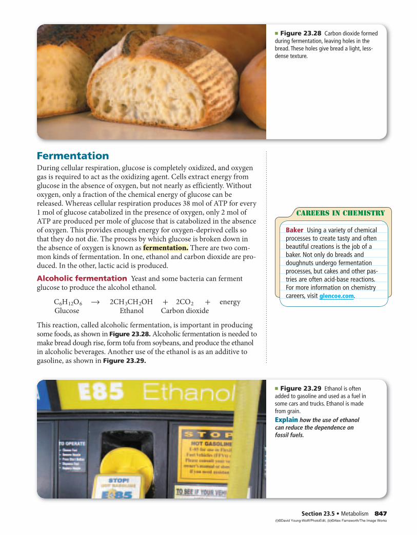
■ Figure 23.27 Swimmers need large amounts of energy when they compete in a race. This energy is stored in the bonds of ATP in their cells.
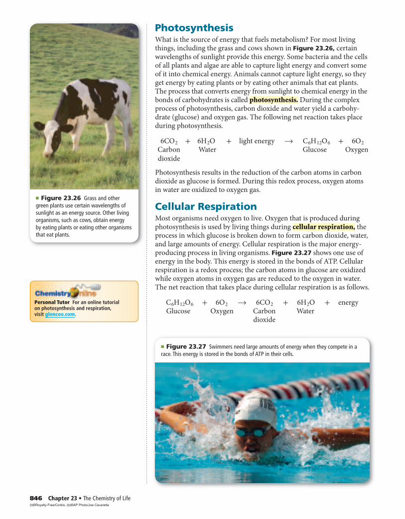
PhotosynthesisWhat is the source of energy that fuels metabolism? For most living things, including the grass and cows shown in Figure 23.26, certain wavelengths of sunlight provide this energy. Some bacteria and the cells of all plants and algae are able to capture light energy and convert some of it into chemical energy. Animals cannot capture light energy, so they get energy by eating plants or by eating other animals that eat plants. The process that converts energy from sunlight to chemical energy in the bonds of carbohydrates is called photosynthesis. During the complex process of photosynthesis, carbon dioxide and water yield a carbohy-drate (glucose) and oxygen gas. The following net reaction takes place during photosynthesis.
6C O 2 + 6 H 2 O + light energy → C 6 H 12 O 6 + 6 O 2 Carbon Water Glucose Oxygen
dioxide
Photosynthesis results in the reduction of the carbon atoms in carbon dioxide as glucose is formed. During this redox process, oxygen atoms in water are oxidized to oxygen gas.
Cellular RespirationMost organisms need oxygen to live. Oxygen that is produced during photosynthesis is used by living things during cellular respiration, the process in which glucose is broken down to form carbon dioxide, water, and large amounts of energy. Cellular respiration is the major energy-producing process in living organisms. Figure 23.27 shows one use of energy in the body. This energy is stored in the bonds of ATP. Cellular respiration is a redox process; the carbon atoms in glucose are oxidized while oxygen atoms in oxygen gas are reduced to the oxygen in water. The net reaction that takes place during cellular respiration is as follows.
C 6 H 12 O 6 + 6 O 2 → 6C O 2 + 6 H 2 O + energy Glucose Oxygen Carbon Water dioxide
■ Figure 23.26 Grass and other green plants use certain wavelengths of sunlight as an energy source. Other living organisms, such as cows, obtain energy by eating plants or eating other organisms that eat plants.
Personal Tutor For an online tutorial on photosynthesis and respiration, visit glencoe.com.
(t)©Royalty-Free/Corbis, (b)©AP Photo/Joe Cavaretta
Section 23.5 • Metabolism 847
FermentationDuring cellular respiration, glucose is completely oxidized, and oxygen gas is required to act as the oxidizing agent. Cells extract energy from glucose in the absence of oxygen, but not nearly as efficiently. Without oxygen, only a fraction of the chemical energy of glucose can be released. Whereas cellular respiration produces 38 mol of ATP for every 1 mol of glucose catabolized in the presence of oxygen, only 2 mol of ATP are produced per mole of glucose that is catabolized in the absence of oxygen. This provides enough energy for oxygen-deprived cells so that they do not die. The process by which glucose is broken down in the absence of oxygen is known as fermentation. There are two com-mon kinds of fermentation. In one, ethanol and carbon dioxide are pro-duced. In the other, lactic acid is produced.
Alcoholic fermentation Yeast and some bacteria can ferment glucose to produce the alcohol ethanol.
C 6 H 12 O 6 → 2C H 3 C H 2 OH + 2C O 2 + energy Glucose Ethanol Carbon dioxide
This reaction, called alcoholic fermentation, is important in producing some foods, as shown in Figure 23.28. Alcoholic fermentation is needed to make bread dough rise, form tofu from soybeans, and produce the ethanol in alcoholic beverages. Another use of the ethanol is as an additive to gasoline, as shown in Figure 23.29.
■ Figure 23.28 Carbon dioxide formed during fermentation, leaving holes in the bread. These holes give bread a light, less-dense texture.
Careers In chemistry
Baker Using a variety of chemical processes to create tasty and often beautiful creations is the job of a baker. Not only do breads and doughnuts undergo fermentation processes, but cakes and other pas-tries are often acid-base reactions. For more information on chemistry careers, visit glencoe.com.
■ Figure 23.29 Ethanol is often added to gasoline and used as a fuel in some cars and trucks. Ethanol is made from grain.Explain how the use of ethanol can reduce the dependence on fossil fuels.
(t)©David Young-Wolff/PhotoEdit, (b)©Alex Farnsworth/The Image Works
Section 23.523.5 Assessment
848 Chapter 23 • The Chemistry of Life Self-Check Quiz glencoe.com
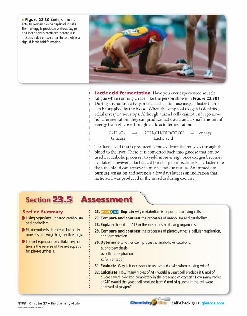
■ Figure 23.30 During strenuous activity, oxygen can be depleted in cells. Then, energy is produced without oxygen and lactic acid is produced. Soreness in muscles a day or two after the activity is a sign of lactic acid formation.
Section Summary◗ ◗ Living organisms undergo catabolism
and anabolism.
◗ ◗ Photosynthesis directly or indirectly provides all living things with energy.
◗ ◗ The net equation for cellular respira-tion is the reverse of the net equation for photosynthesis.
26. MAIN Idea Explain why metabolism is important to living cells.
27. Compare and contrast the processes of anabolism and catabolism.
28. Explain the role of ATP in the metabolism of living organisms.
29. Compare and contrast the processes of photosynthesis, cellular respiration, and fermentation.
30. Determine whether each process is anabolic or catabolic. a. photosynthesis b. cellular respiration c. fermentation
31. Evaluate Why is it necessary to use sealed casks when making wine?
32. Calculate How many moles of ATP would a yeast cell produce if 6 mol of glucose were oxidized completely in the presence of oxygen? How many moles of ATP would the yeast cell produce from 6 mol of glucose if the cell were deprived of oxygen?
Lactic acid fermentation Have you ever experienced muscle fatigue while running a race, like the person shown in Figure 23.30? During strenuous activity, muscle cells often use oxygen faster than it can be supplied by the blood. When the supply of oxygen is depleted, cellular respiration stops. Although animal cells cannot undergo alco-holic fermentation, they can produce lactic acid and a small amount of energy from glucose through lactic acid fermentation.
C 6 H 12 O 6 → 2C H 3 CH(OH)COOH + energy Glucose Lactic acid
The lactic acid that is produced is moved from the muscles through the blood to the liver. There, it is converted back into glucose that can be used in catabolic processes to yield more energy once oxygen becomes available. However, if lactic acid builds up in muscle cells at a faster rate than the blood can remove it, muscle fatigue results. An immediate burning sensation and soreness a few days later is an indication that lactic acid was produced in the muscles during exercise.
©Wally McNamee/CORBIS
In the Field 849
“No right-thinking paleontologist would do what Mary did. We don’t go to all this effort to dig this stuff out of the ground and then destroy it in acid.” So says a colleague about Mary Schweitzer, the scientist who used the techniques of molecular biology to discover soft tissue where none should be—in the thighbone of a 68-million-year-old Tyrannosaurus rex.
Mother Bob When the fossilized T. rex, nicknamed Bob, was recovered in 2003 from a remote section of Montana by paleontologists, the bones were encased in plaster for protection during transport. However, the bones and plaster weighed more than the helicopter could lift. So the paleontologists were forced to break the intact thighbone to move the dinosaur out of the remote area. Schweitzer took small fragments from the broken thighbone for further study.
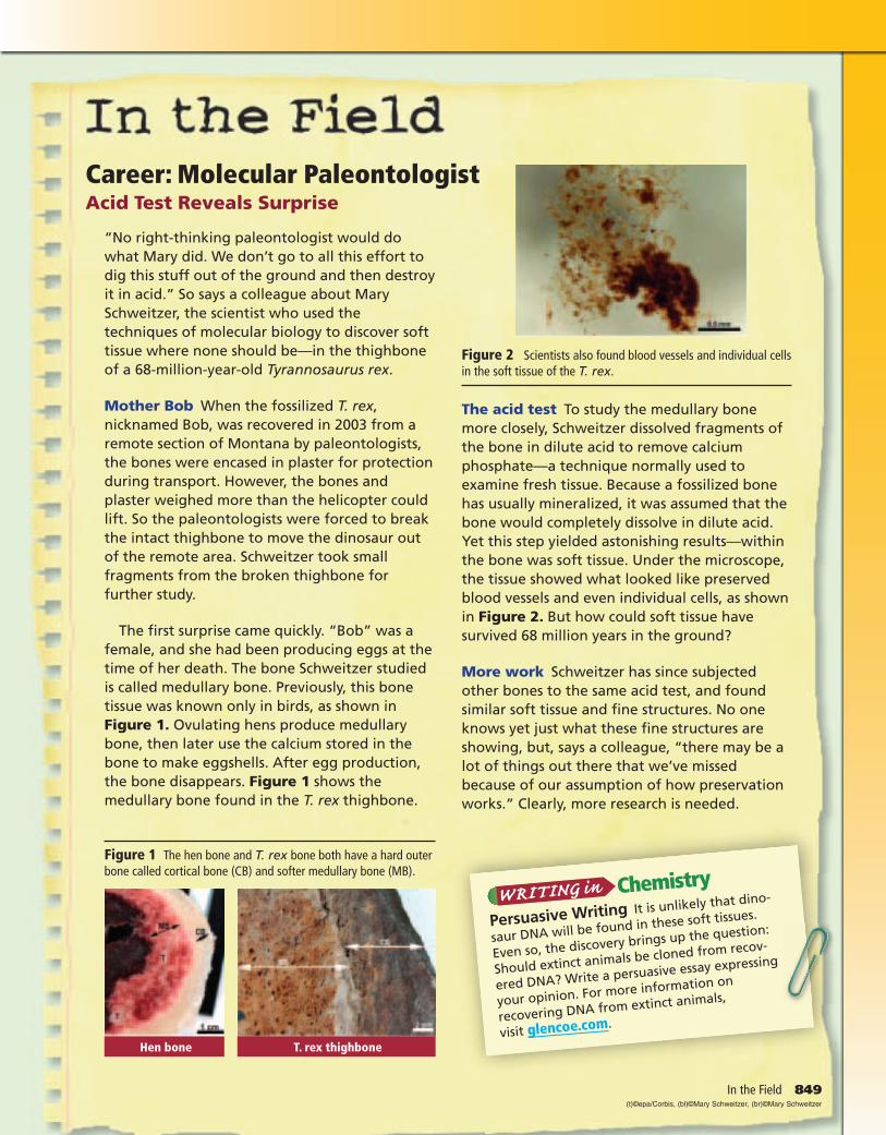
The first surprise came quickly. “Bob” was a female, and she had been producing eggs at the time of her death. The bone Schweitzer studied is called medullary bone. Previously, this bone tissue was known only in birds, as shown in Figure 1. Ovulating hens produce medullary bone, then later use the calcium stored in the bone to make eggshells. After egg production, the bone disappears. Figure 1 shows the medullary bone found in the T. rex thighbone.
Career: Molecular PaleontologistAcid Test Reveals Surprise
Figure 1 The hen bone and T. rex bone both have a hard outer bone called cortical bone (CB) and softer medullary bone (MB).
Figure 2 Scientists also found blood vessels and individual cells in the soft tissue of the T. rex.
Hen bone T. rex thighbone
The acid test To study the medullary bone more closely, Schweitzer dissolved fragments of the bone in dilute acid to remove calcium phosphate—a technique normally used to examine fresh tissue. Because a fossilized bone has usually mineralized, it was assumed that the bone would completely dissolve in dilute acid. Yet this step yielded astonishing results—within the bone was soft tissue. Under the microscope, the tissue showed what looked like preserved blood vessels and even individual cells, as shown in Figure 2. But how could soft tissue have survived 68 million years in the ground?
More work Schweitzer has since subjected other bones to the same acid test, and found similar soft tissue and fine structures. No one knows yet just what these fine structures are showing, but, says a colleague, “there may be a lot of things out there that we’ve missed because of our assumption of how preservation works.” Clearly, more research is needed.
Chemistry
Persuasive Writing It is unlikely that dino-
saur DNA will be found in these soft tissues.
Even so, the discovery brings up the question:
Should extinct animals be cloned from recov-
ered DNA? Write a persuasive essay expressing
your opinion. For more information on
recovering DNA from extinct animals,
visit glencoe.com.
(t)©epa/Corbis, (bl)©Mary Schweitzer, (br)©Mary Schweitzer
850 Chapter 23 • The Chemistry of Life
Probeware Alternate CBL instructions can be found at glencoe.com.
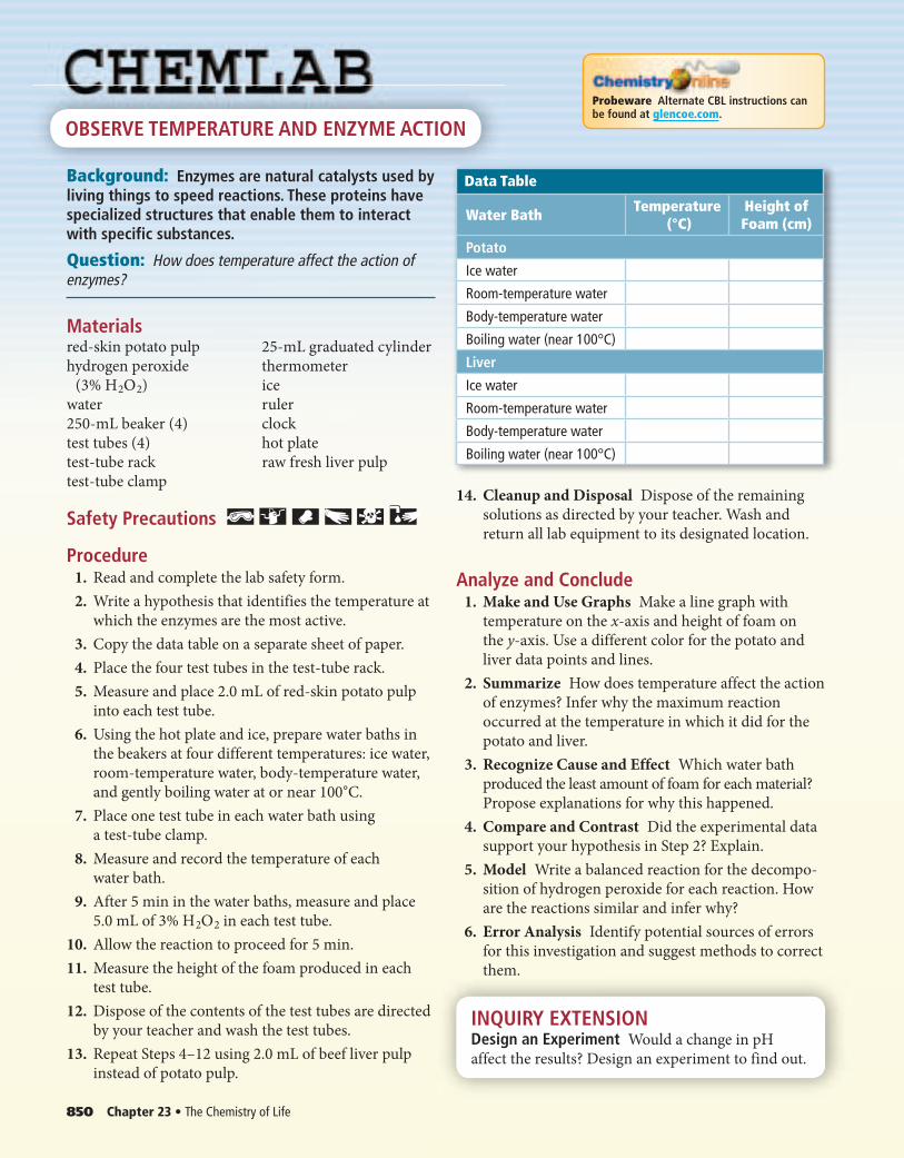
OBSERVE TEMPERATURE AND ENZYME ACTION
Background: Enzymes are natural catalysts used by living things to speed reactions. These proteins have specialized structures that enable them to interact with specific substances.
Question: How does temperature affect the action of enzymes?
Materialsred-skin potato pulp 25-mL graduated cylinder
hydrogen peroxide thermometer
(3% H 2 O 2 ) ice
water ruler
250-mL beaker (4) clock
test tubes (4) hot plate
test-tube rack raw fresh liver pulp
test-tube clamp
Safety Precautions
Procedure 1. Read and complete the lab safety form.
2. Write a hypothesis that identifies the temperature at
which the enzymes are the most active.
3. Copy the data table on a separate sheet of paper.
4. Place the four test tubes in the test-tube rack.
5. Measure and place 2.0 mL of red-skin potato pulp
into each test tube.
6. Using the hot plate and ice, prepare water baths in
the beakers at four different temperatures: ice water,
room-temperature water, body-temperature water,
and gently boiling water at or near 100°C.
7. Place one test tube in each water bath using
a test-tube clamp.
8. Measure and record the temperature of each
water bath.
9. After 5 min in the water baths, measure and place
5.0 mL of 3% H 2 O 2 in each test tube.
10. Allow the reaction to proceed for 5 min.
11. Measure the height of the foam produced in each
test tube.
12. Dispose of the contents of the test tubes are directed
by your teacher and wash the test tubes.
13. Repeat Steps 4–12 using 2.0 mL of beef liver pulp
instead of potato pulp.
Data Table
Water Bath Temperature (°C)
Height of Foam (cm)
Potato
Ice water
Room-temperature water
Body-temperature water
Boiling water (near 100°C)
Liver
Ice water
Room-temperature water
Body-temperature water
Boiling water (near 100°C)
14. Cleanup and Disposal Dispose of the remaining
solutions as directed by your teacher. Wash and
return all lab equipment to its designated location.
Analyze and Conclude 1. Make and Use Graphs Make a line graph with
temperature on the x-axis and height of foam on
the y-axis. Use a different color for the potato and
liver data points and lines.
2. Summarize How does temperature affect the action
of enzymes? Infer why the maximum reaction
occurred at the temperature in which it did for the
potato and liver.
3. Recognize Cause and Effect Which water bath
produced the least amount of foam for each material?
Propose explanations for why this happened.
4. Compare and Contrast Did the experimental data
support your hypothesis in Step 2? Explain.
5. Model Write a balanced reaction for the decompo-
sition of hydrogen peroxide for each reaction. How
are the reactions similar and infer why?
6. Error Analysis Identify potential sources of errors
for this investigation and suggest methods to correct
them.
INQUIRY EXTENSIONDesign an Experiment Would a change in pH
affect the results? Design an experiment to find out.
Chapter 23 • Study Guide 851Vocabulary PuzzleMaker glencoe.com
Download quizzes, key terms, and flash cards from glencoe.com.
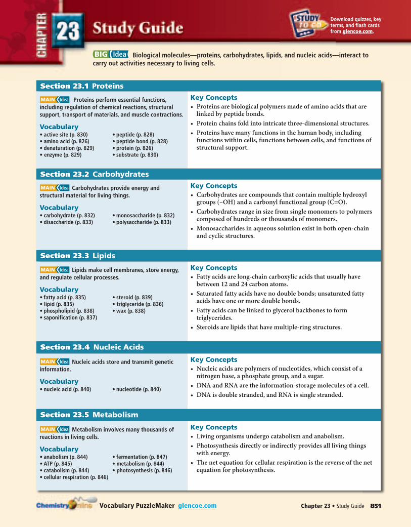
BIG Idea Biological molecules—proteins, carbohydrates, lipids, and nucleic acids—interact to carry out activities necessary to living cells.
Section 23.1Section 23.1 Proteins
MAIN Idea Proteins perform essential functions, including regulation of chemical reactions, structural support, transport of materials, and muscle contractions.
Vocabulary• active site (p. 830) • peptide (p. 828)• amino acid (p. 826) • peptide bond (p. 828)• denaturation (p. 829) • protein (p. 826)• enzyme (p. 829) • substrate (p. 830)
Key Concepts• Proteins are biological polymers made of amino acids that are
linked by peptide bonds.
• Protein chains fold into intricate three-dimensional structures.
• Proteins have many functions in the human body, including functions within cells, functions between cells, and functions of structural support.
Section 23.2Section 23.2 Carbohydrates
MAIN Idea Carbohydrates provide energy and structural material for living things.
Vocabulary• carbohydrate (p. 832) • monosaccharide (p. 832)• disaccharide (p. 833) • polysaccharide (p. 833)
Key Concepts• Carbohydrates are compounds that contain multiple hydroxyl
groups (–OH) and a carbonyl functional group (C=O).
• Carbohydrates range in size from single monomers to polymers composed of hundreds or thousands of monomers.
• Monosaccharides in aqueous solution exist in both open-chain and cyclic structures.
Section 23.3Section 23.3 Lipids
MAIN Idea Lipids make cell membranes, store energy, and regulate cellular processes.
Vocabulary• fatty acid (p. 835) • steroid (p. 839)• lipid (p. 835) • triglyceride (p. 836)• phospholipid (p. 838) • wax (p. 838)• saponification (p. 837)
Key Concepts• Fatty acids are long-chain carboxylic acids that usually have
between 12 and 24 carbon atoms.
• Saturated fatty acids have no double bonds; unsaturated fatty acids have one or more double bonds.
• Fatty acids can be linked to glycerol backbones to form triglycerides.
• Steroids are lipids that have multiple-ring structures.
Section 23.4Section 23.4 Nucleic Acids
MAIN Idea Nucleic acids store and transmit genetic information.
Vocabulary• nucleic acid (p. 840) • nucleotide (p. 840)
Key Concepts• Nucleic acids are polymers of nucleotides, which consist of a
nitrogen base, a phosphate group, and a sugar.
• DNA and RNA are the information-storage molecules of a cell.
• DNA is double stranded, and RNA is single stranded.
Section 23.5Section 23.5 Metabolism
MAIN Idea Metabolism involves many thousands of reactions in living cells.
Vocabulary• anabolism (p. 844) • fermentation (p. 847)• ATP (p. 845) • metabolism (p. 844)• catabolism (p. 844) • photosynthesis (p. 846)• cellular respiration (p. 846)
Key Concepts• Living organisms undergo catabolism and anabolism.
• Photosynthesis directly or indirectly provides all living things with energy.
• The net equation for cellular respiration is the reverse of the net equation for photosynthesis.

852 Chapter 23 • The Chemistry of Life Chapter Test glencoe.com
Section 23.1Mastering Concepts
33. What should you call a chain of eight amino acids? A
chain of 200 amino acids?
34. Name the two types of functional groups that react
together to form a peptide bond, and name the
functional group in the peptide bond itself.
35. Using the four symbols shown to represent four differ-
ent amino acids, draw structures four possible peptides
for a four-member chain that can be made by linking
them together in different orders.
Amino acid 1: � Amino acid 3: �
Amino acid 2: � Amino acid 4: �
36. Human Anatomy Name five parts of the body that con-
tain structural proteins.
37. List four major functions of proteins, and give one
example of a protein that carries out each function.
38. Describe two common shapes found in the three-
dimensional folding of proteins.
39. Name the organic functional groups in the side chains of
the following amino acids.
a. glutamine c. glutamic acid
b. serine d. lysine
40. Explain how the active site of an enzyme functions.
41. Name an example of an amino acid that has an aromatic
ring in its side chain.
42. Name two nonpolar and two polar amino acids.
O
CH2
H2N OH
— —
CC
NHC
H■ Figure 23.31
43. The structure shown in Figure 23.31 is tryptophan.
Describe some of the properties you would expect tryp-
tophan to have, based on its structure. In what class of
large molecules is tryptophan a member? Explain.
44. Is the dipeptide lysine-valine the same compound as the
dipeptide valine-lysine? Explain.
45. Enzymes How do enzymes lower the activation energy
for a reaction?
46. Cellular Chemistry Most proteins with a globular shape
are oriented so that they have mostly nonpolar amino
acids on the inside and polar amino acids located on the
outer surface. Does this make sense in terms of the
nature of the cellular environment? Explain.
Mastering Problems 47. How many different ways can you arrange three differ-
ent amino acids in a peptide? Four amino acids? Five
amino acids?
48. How many peptide bonds are present in a peptide that
has five amino acids?
49. Proteins The average molecular weight of an amino
acid residue in a polypeptide is 110. What is the approx-
imate molecular weight of the following proteins?
a. Insulin (51 amino acids)
b. Myosin (1750 amino acids)
H
—
H — N — C — C
H
—
O
——
OH SH
OH
CH2
H—
N — C — C
H—
O
——
CHH3C CH3
H
—
N — C — C
H
—
O
——
CH2
—
H O
—
N — C — C —OH
H
—
——
CH2
—
■ Figure 23.32
50. Determine how many amino acids and peptide bonds
are in the peptide shown in Figure 23.32.
51. The average molecular mass of an amino acid is 110 g/mol.
Calculate the approximate number of amino acids in a
protein that has a molecular mass of 36,500 g/mol.
Section 23.2Mastering Concepts
52. Carbohydrates Classify the following carbohydrates as
monosaccharides, disaccharides, or polysaccharides.
a. starch d. ribose g. fructose
b. glucose e. cellulose h. lactose
c. sucrose f. glycogen
53. Name two isomers of glucose.
54. What kind of bond is formed when two monosaccha-
rides combine to form a disaccharide?
55. Sugars Give a scientific term for each of the following.
a. blood sugar
b. fruit sugar
c. table sugar
d. milk sugar

Chapter 23 • Assessment 853Chapter Test glencoe.com
OH
OH
CH2OH
O
O
H
HH
H OH
CH2OH
H
OH
OH
CH2OHO
H
HH
H
H
OH
OH
CH2OHO
H
HH
H
HO
OO
O
O O
O
H H H H H H
H
OH
OH
H
OH
CH2OH
O
H
OH
OH
H
OH
CH2OH
O
H
OH
OH
H
O
CH2OH
O
O
CH2OH
O
O
CH2OH
O
O
CH2OH
O
O
CH2
O
O
CH2OH
O
OOH
■ Figure 23.33
56. Cellulose and Starch The molecular structures of
cellulose and starch are shown in Figure 23.33.Compare and contrast their molecular structures.
57. Chemistry in Plants Compare and contrast the func-
tions of starch and cellulose in plants. Explain why their
molecular structures are important to their functions.
58. Infer how the different bonding arrangements in cellu-
lose and starch give them such different properties.
59. The disaccharide maltose is formed from two glucose
monomers. Draw its structure.
60. The hydrolysis of cellulose, glycogen, and starch produc-
es only one monosaccharide. Why is this so? What
monosaccharide is produced?
61. Digestion Disaccharides and polysaccharides cannot
be broken down in the absence of water. Why do you
think this is so? Include an equation in your answer.
62. Draw the structure of the open-chain form of fructose.
Circle all chiral carbons, and then calculate the number
of stereoisomers with the same formula as fructose.
63. Sugars Compare and contrast the molecular formula,
molecular weight, and functional groups found in glu-
cose and fructose.
64. Historical Perspective Carbohydrates are not hydrates
of carbon as the name suggests. Explain how this mis-
conception occurred.
Mastering Problems 65. Complex Carbohydrates Stachyose is a tetrasaccharide
that contains two D-galactose units, one D-glucose unit,
and one D-fructose unit. Each sugar unit has a molecular
weight of 180 g/mol before it is linked together in this
tetrasaccharide, and one water molecule is released for
each two sugar units that come together. What is the
molecular weight of stachyose?
Section 23.3Mastering Concepts
66. Compare and contrast the structures of a triglyceride
and a phospholipid.
67 Predict whether a triglyceride from beef fat or a triglyc-
eride from olive oil will have a higher melting point.
Explain your reasoning.
68. Soaps and Detergents Explain how the structure of
soaps makes them effective cleaning agents.
69. Draw a portion of a lipid bilayer membrane, labeling the
polar and nonpolar parts of the membrane.
70. Where and in what form are fatty acids stored in the
human body?
71. What type of lipid does not contain fatty acid chains?
Why are these molecules classified as lipids?
72. Soap Draw the structure of the soap sodium palmitate
(palmitate is the conjugate base of the 16-carbon satu-
rated fatty acid, palmitic acid). Label its polar and non-
polar ends.
73. Determine whether each structure is a fatty acid,
triglyceride, phospholipid, steroid, or wax. Explain
your reasoning.
a.
H3C
H3CO
O
CH2OHC
O
OH——
——
——
b. — O —
-O
— P
—
O
-O
——
O
——
O
——
CH2
CH — O — C — CH2
H2C — O — C — CH2
Mastering Problems 74. The fatty acid palmitic acid has a density of 0.853 g/mL
at 62°C. What will be the mass of a 0.886-L sample of
palmitic acid at that temperature?
75. Polyunsaturated Fats How many moles of hydrogen
gas are required for complete hydrogenation of 1 mol of
linolenic acid, whose structure is shown below? Write a
balanced equation for the hydrogenation reaction.
C H 3 C H 2 CH=CHC H 2 CH=CHC H 2 CH=CH
(C H 2 ) 7 COOH
Cellulose
Starch
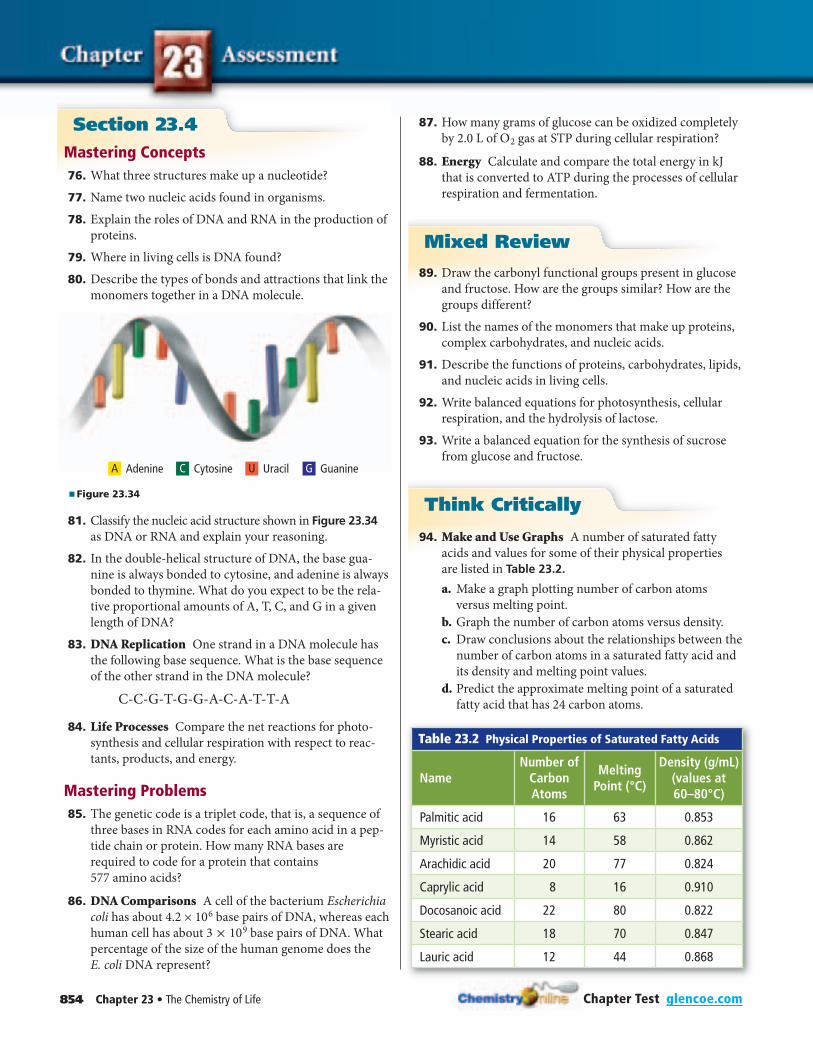
854 Chapter 23 • The Chemistry of Life Chapter Test glencoe.com
Section 23.4Mastering Concepts
76. What three structures make up a nucleotide?
77. Name two nucleic acids found in organisms.
78. Explain the roles of DNA and RNA in the production of
proteins.
79. Where in living cells is DNA found?
80. Describe the types of bonds and attractions that link the
monomers together in a DNA molecule.
AdenineA Cytosine Uracil GuanineC U G
■ Figure 23.34
81. Classify the nucleic acid structure shown in Figure 23.34 as DNA or RNA and explain your reasoning.
82. In the double-helical structure of DNA, the base gua-
nine is always bonded to cytosine, and adenine is always
bonded to thymine. What do you expect to be the rela-
tive proportional amounts of A, T, C, and G in a given
length of DNA?
83. DNA Replication One strand in a DNA molecule has
the following base sequence. What is the base sequence
of the other strand in the DNA molecule?
C-C-G-T-G-G-A-C-A-T-T-A
84. Life Processes Compare the net reactions for photo-
synthesis and cellular respiration with respect to reac-
tants, products, and energy.
Mastering Problems 85. The genetic code is a triplet code, that is, a sequence of
three bases in RNA codes for each amino acid in a pep-
tide chain or protein. How many RNA bases are
required to code for a protein that contains
577 amino acids?
86. DNA Comparisons A cell of the bacterium Escherichia
coli has about 4.2 × 1 0 6 base pairs of DNA, whereas each
human cell has about 3 × 1 0 9 base pairs of DNA. What
percentage of the size of the human genome does the
E. coli DNA represent?
87. How many grams of glucose can be oxidized completely
by 2.0 L of O 2 gas at STP during cellular respiration?
88. Energy Calculate and compare the total energy in kJ
that is converted to ATP during the processes of cellular
respiration and fermentation.
Mixed Review
89. Draw the carbonyl functional groups present in glucose
and fructose. How are the groups similar? How are the
groups different?
90. List the names of the monomers that make up proteins,
complex carbohydrates, and nucleic acids.
91. Describe the functions of proteins, carbohydrates, lipids,
and nucleic acids in living cells.
92. Write balanced equations for photosynthesis, cellular
respiration, and the hydrolysis of lactose.
93. Write a balanced equation for the synthesis of sucrose
from glucose and fructose.
Think Critically
94. Make and Use Graphs A number of saturated fatty
acids and values for some of their physical properties
are listed in Table 23.2.
a. Make a graph plotting number of carbon atoms
versus melting point.
b. Graph the number of carbon atoms versus density.
c. Draw conclusions about the relationships between the
number of carbon atoms in a saturated fatty acid and
its density and melting point values.
d. Predict the approximate melting point of a saturated
fatty acid that has 24 carbon atoms.
Table 23.2 Physical Properties of Saturated Fatty Acids
NameNumber of
Carbon Atoms
Melting Point (°C)
Density (g/mL) (values at 60–80°C)
Palmitic acid 16 63 0.853
Myristic acid 14 58 0.862
Arachidic acid 20 77 0.824
Caprylic acid 8 16 0.910
Docosanoic acid 22 80 0.822
Stearic acid 18 70 0.847
Lauric acid 12 44 0.868
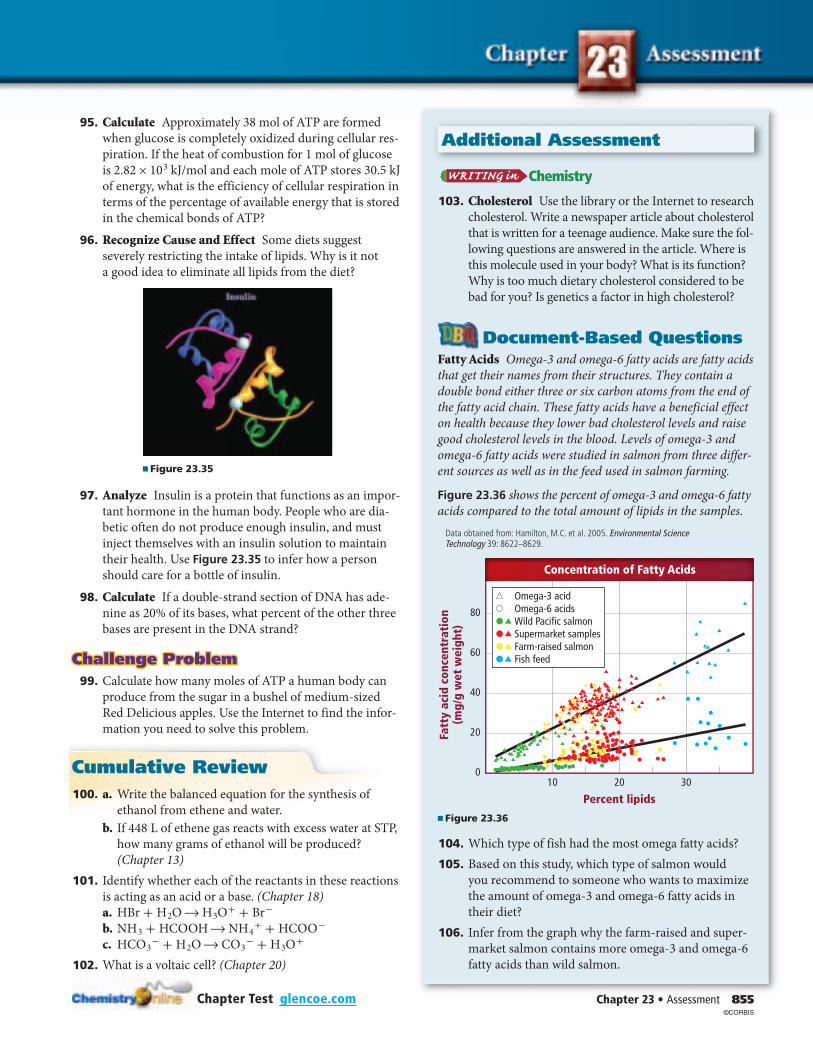
Chapter 23 • Assessment 855
Additional Assessment
Chapter Test glencoe.com
95. Calculate Approximately 38 mol of ATP are formed
when glucose is completely oxidized during cellular res-
piration. If the heat of combustion for 1 mol of glucose
is 2.82 × 1 0 3 kJ/mol and each mole of ATP stores 30.5 kJ
of energy, what is the efficiency of cellular respiration in
terms of the percentage of available energy that is stored
in the chemical bonds of ATP?
96. Recognize Cause and Effect Some diets suggest
severely restricting the intake of lipids. Why is it not
a good idea to eliminate all lipids from the diet?
■ Figure 23.35
97. Analyze Insulin is a protein that functions as an impor-
tant hormone in the human body. People who are dia-
betic often do not produce enough insulin, and must
inject themselves with an insulin solution to maintain
their health. Use Figure 23.35 to infer how a person
should care for a bottle of insulin.
98. Calculate If a double-strand section of DNA has ade-
nine as 20% of its bases, what percent of the other three
bases are present in the DNA strand?
Challenge ProblemChallenge Problem 99. Calculate how many moles of ATP a human body can
produce from the sugar in a bushel of medium-sized
Red Delicious apples. Use the Internet to find the infor-
mation you need to solve this problem.
Cumulative Review 100. a. Write the balanced equation for the synthesis of
ethanol from ethene and water.
b. If 448 L of ethene gas reacts with excess water at STP,
how many grams of ethanol will be produced?
(Chapter 13)
101. Identify whether each of the reactants in these reactions
is acting as an acid or a base. (Chapter 18)
a. HBr + H 2 O → H 3 O + + B r -
b. N H 3 + HCOOH → N H 4 + + HCO O -
c. HC O 3 - + H 2 O → C O 3 - + H 3 O +
102. What is a voltaic cell? (Chapter 20)
Chemistry
103. Cholesterol Use the library or the Internet to research
cholesterol. Write a newspaper article about cholesterol
that is written for a teenage audience. Make sure the fol-
lowing questions are answered in the article. Where is
this molecule used in your body? What is its function?
Why is too much dietary cholesterol considered to be
bad for you? Is genetics a factor in high cholesterol?
Document-Based QuestionsFatty Acids Omega-3 and omega-6 fatty acids are fatty acids
that get their names from their structures. They contain a
double bond either three or six carbon atoms from the end of
the fatty acid chain. These fatty acids have a beneficial effect
on health because they lower bad cholesterol levels and raise
good cholesterol levels in the blood. Levels of omega-3 and
omega-6 fatty acids were studied in salmon from three differ-
ent sources as well as in the feed used in salmon farming.
Figure 23.36 shows the percent of omega-3 and omega-6 fatty
acids compared to the total amount of lipids in the samples.
Data obtained from: Hamilton, M.C. et al. 2005. Environmental Science Technology 39: 8622–8629.
Fatt
y ac
id c
once
ntra
tion
(mg/
g w
et w
eigh
t)
80
60
40
20
0
Percent lipids10 20 30
Concentration of Fatty Acids
Omega-3 acidOmega-6 acidsWild Pacific salmonSupermarket samplesFarm-raised salmonFish feed
■ Figure 23.36
104. Which type of fish had the most omega fatty acids?
105. Based on this study, which type of salmon would
you recommend to someone who wants to maximize
the amount of omega-3 and omega-6 fatty acids in
their diet?
106. Infer from the graph why the farm-raised and super-
market salmon contains more omega-3 and omega-6
fatty acids than wild salmon.
©CORBIS
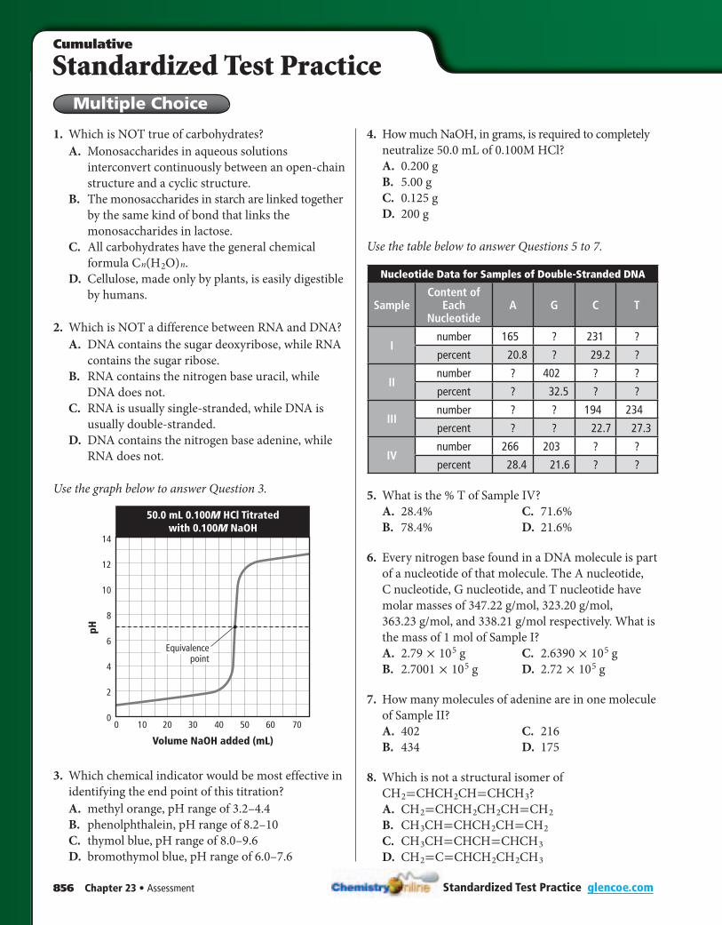
856 Chapter 23 • Assessment
Standardized Test PracticeCumulative
Multiple Choice
Standardized Test Practice glencoe.com
1. Which is NOT true of carbohydrates?
A. Monosaccharides in aqueous solutions
interconvert continuously between an open-chain
structure and a cyclic structure.
B. The monosaccharides in starch are linked together
by the same kind of bond that links the
monosaccharides in lactose.
C. All carbohydrates have the general chemical
formula C n ( H 2 O ) n .
D. Cellulose, made only by plants, is easily digestible
by humans.
2. Which is NOT a difference between RNA and DNA?
A. DNA contains the sugar deoxyribose, while RNA
contains the sugar ribose.
B. RNA contains the nitrogen base uracil, while
DNA does not.
C. RNA is usually single-stranded, while DNA is
usually double-stranded.
D. DNA contains the nitrogen base adenine, while
RNA does not.
Use the graph below to answer Question 3.
100 20 30 50 60 7040
pH
Volume NaOH added (mL)
14
12
10
8
6
4
2
0
Equivalencepoint
50.0 mL 0.100M HCl Titratedwith 0.100M NaOH
3. Which chemical indicator would be most effective in
identifying the end point of this titration?
A. methyl orange, pH range of 3.2–4.4
B. phenolphthalein, pH range of 8.2–10
C. thymol blue, pH range of 8.0–9.6
D. bromothymol blue, pH range of 6.0–7.6
4. How much NaOH, in grams, is required to completely
neutralize 50.0 mL of 0.100M HCl?
A. 0.200 g
B. 5.00 g
C. 0.125 g
D. 200 g
Use the table below to answer Questions 5 to 7.
Nucleotide Data for Samples of Double-Stranded DNA
SampleContent of
Each Nucleotide
A G C T
Inumber 165 ? 231 ?
percent 20.8 ? 29.2 ?
IInumber ? 402 ? ?
percent ? 32.5 ? ?
IIInumber ? ? 194 234
percent ? ? 22.7 27.3
IVnumber 266 203 ? ?
percent 28.4 21.6 ? ?
5. What is the % T of Sample IV?
A. 28.4% C. 71.6%
B. 78.4% D. 21.6%
6. Every nitrogen base found in a DNA molecule is part
of a nucleotide of that molecule. The A nucleotide,
C nucleotide, G nucleotide, and T nucleotide have
molar masses of 347.22 g/mol, 323.20 g/mol,
363.23 g/mol, and 338.21 g/mol respectively. What is
the mass of 1 mol of Sample I?
A. 2.79 × 1 0 5 g C. 2.6390 × 1 0 5 g
B. 2.7001 × 1 0 5 g D. 2.72 × 1 0 5 g
7. How many molecules of adenine are in one molecule
of Sample II?
A. 402 C. 216
B. 434 D. 175
8. Which is not a structural isomer of
C H 2 =CHC H 2 CH=CHC H 3 ?
A. C H 2 =CHC H 2 C H 2 CH=C H 2
B. C H 3 CH=CHC H 2 CH=C H 2
C. C H 3 CH=CHCH=CHC H 3
D. C H 2 =C=CHC H 2 C H 2 C H 3
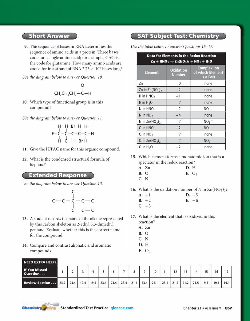
Chapter 23 • Assessment 857
Short Answer SAT Subject Test: Chemistry
Standardized Test Practice glencoe.com
9. The sequence of bases in RNA determines the
sequence of amino acids in a protein. Three bases
code for a single amino acid; for example, CAG is
the code for glutamine. How many amino acids are
coded for in a strand of RNA 2.73 × 1 0 4 bases long?
Use the diagram below to answer Question 10.
O
CH3CH2CH2 HC
10. Which type of functional group is in this
compound?
Use the diagram below to answer Question 11.
H
CF C C
Br HH
H Cl
H
C H
HH Br
C— — — —— —
— — —
— — —
— —
— —
11. Give the IUPAC name for this organic compound.
12. What is the condensed structural formula of
heptane?
Extended ResponseUse the diagram below to answer Question 13.
C C — C
C
C — C — C — C — C
—— —
13. A student records the name of the alkane represented
by this carbon skeleton as 2-ethyl 3,3-dimethyl
pentane. Evaluate whether this is the correct name
for the compound.
14. Compare and contrast aliphatic and aromatic
compounds.
Use the table below to answer Questions 15–17.
Data for Elements in the Redox Reaction Zn + HN O 3 → Zn(N O 3 ) 2 + N O 2 + H 2 O
Element Oxidation Number
Complex Ion of which Element
is a Part
Zn 0 none
Zn in Zn(N O 3 ) 2 +2 none
H in HN O 3 +1 none
H in H 2 O ? none
N in HN O 3 ? N O 3 -
N in N O 2 +4 none
N in Zn(N O 3 ) 2 ? N O 3 -
O in HN O 3 -2 N O 3 -
O in N O 2 ? none
O in Zn(N O 3 ) 2 ? N O 3 -
O in H 2 O -2 none
15. Which element forms a monatomic ion that is a
spectator in the redox reaction?
A. Zn D. H
B. O E. O 2
C. N
16. What is the oxidation number of N in Zn(N O 3 ) 2 ?
A. +1 D. +5
B. +2 E. +6
C. +3
17. What is the element that is oxidized in this
reaction?
A. Zn
B. O
C. N
D. H
E. O 3
NEED EXTRA HELP?
If You Missed Question . . . 1 2 3 4 5 6 7 8 9 10 11 12 13 14 15 16 17
Review Section . . . 23.2 23.4 19.4 19.4 23.4 23.4 23.4 21.4 23.4 22.1 23.1 21.2 21.2 21.5 9.3 19.1 19.1