Embed Size (px)
Citation preview

Chapter 26: Transcription and Translation
The central dogma of molecular biology• Information contained in DNA molecules is expressed in the
structure of proteins.• Gene expression is the turning on or activation of a gene.

26-2
TranscriptionTranscription:Transcription: The process by which information encoded in
a DNA molecule is copied into an mRNA molecule.• Transcription takes place in the nucleus.• Transcription starts when the DNA double helix begins
to unwind near the gene to be transcribed.• Only one strand of the DNA is transcribed.• Ribonucleotides assemble along the unwound DNA
strand in a complementary sequence.• Enzymes called polymerasespolymerases (poly)(poly) catalyze
transcription: poly I for rRNA formation, poly II for mRNA formation, and poly III for tRNA formation.

26-3
TranscriptionFigure 26.2 The information in one DNA strand is transcribed to a strand of RNA. The termination site is the locus of termination of transcription.

26-4
Transcription• A eukaryotic gene has two parts:• A structural genestructural gene that is transcribed into RNA; the
structural gene is made of exons and introns.• A regulatory generegulatory gene that controls transcription; the
regulatory gene is not transcribed but has control elements, one of which is the promoter.promoter.
A promoter is unique to each gene.• There is always a sequence of bases on the DNA
strand called an initiation signal.initiation signal.• Promoters also contain consensus sequences,consensus sequences, such as
the TATA box,TATA box, in which the two nucleotides T and A are repeated many times.

26-5
Transcription• The RNA products of transcription are not necessarily
functional RNAs.• They are made functional by post-transcription post-transcription
modification.modification.• Transcribed mRNA is capped at both ends.• The 5’ end acquires a methylated guanine.• The 3’ end acquires a polyA tail that may contain from
100 to 200 adenine residues.• Once the two ends are capped, the introns are spliced
out.• tRNA is similarly trimmed, capped, and methylated.• Functional rRNA also undergoes post-transcription
methylation.

26-6
Transcription• Figure 26.4 Organization and transcription of a split
eukaryote gene.

26-7
Role of RNA in Translation• mRNA, rRNA, and tRNA all participate in translation.• Protein synthesis takes place on ribosomes.• A ribosome dissociates into larger and a smaller body.• In higher organisms, including humans, the larger body is
called a 60S ribosome; the smaller body is called a 40S ribosome.
• The 5’ end of the mature mRNA is bonded to the 40S ribosome and this unit then joined to the 60S ribosome.
• Together the 40S and 60S ribosomes form a unit on which mRNA is stretched out.
• Triplets of bases on mRNA are called codons.codons.• The 20 amino acids are then brought to the mRNA-ribosome
complex, each amino acid by its own particular tRNA.

26-8
tRNA• Each tRNA is specific for only one amino acid.• Each cell carries at least 20 specific enzymes, each
specific for one amino acid.• Each enzyme recognizes only one tRNA.• The enzyme bonds the activated amino acid to the 3’
terminal -OH group of the appropriate tRNA by an ester bond.
• At the opposite end of the tRNA molecule is a codon codon recognition site.recognition site.
• The codon recognition site is a sequence of three bases called an anticodon.anticodon.
• This triplet of bases aligns itself in a complementary fashion to the codon triplet on mRNA.

26-9
tRNA• Figure 26.5
The three-dimensional structure of a tRNA.

26-10
The Genetic Code
A
U
U C
C
A
LeuLeuLeuLeu
ValValValVal
SerSerSerSer
ProProProPro
ThrThrThrThr
AlaAlaAlaAla
ArgArgArgArg
GlyGlyGlyGly
UUUUUC
PhePheLeuLeu
IleIleIle
AUUAUCAUA
UAUUAC
TyrTyr
CAUCAC
HisHisGlnGln
AAUAAC
AsnAsnLysLys
AspAspGluGlu
Trp
CysCys
SerSerArgArg
UCUUCCUCAUCG
G
UCAG
CUUCUCCUACUG
CCUCCCCCACCG
CGUCGCCGACGG
UCAG
G
ACUACCACAACG
UCAG
UCAG
GUUGUCGUAGUG
GCUGCCGCAGCG
GGUGGCGGAGGG
StopUUAUUG
Met*AUG
UAA UAG
StopStop
CAACAG
AAAAAG
GAUGACGAAGAG
UGAUGG
UGUUGC
AGUAGCAGAAGG
*AUG signals translation initiation as well as coding for Met
FirstPosition(5' end)
Third Position(3' end)

26-11
Features of the Code• All 64 codons have been assigned.• 61 code for amino acids.• 3 (UAA, UAG, and UGA) serve as termination signals.• AUG also serves as an initiation signal.• Only Trp and Met have one codon each.• More than one triplet can code for the same amino
acid; Leu, Ser, and Arg, for example, are each coded for by six triplets.
• The third base is irrelevant for Leu, Val, Ser, Pro, Thr, Ala, Gly, and Arg.
• It is said to be continuous and unpunctuated. There are no overlapping codons and no nucleotides interspersed.

26-12
Features of the Code• For the 15 amino acids coded for by 2, 3, or 4 triplets, it
is only the third letter of the codon that varies. Gly, for example, is coded for by GGA, GGG, GGC, and GGU.
• The code is almost universal: it the same in viruses, prokaryotes, and eukaryotes; the only exceptions are some codons in mitochondria

26-13
Translation• Translation:Translation: The process whereby a base sequence of
mRNA is used to create a protein.• There are four major stages in protein synthesis:• Activation• Initiation• Elongation• Termination

26-14
Protein Synthesis• Molecular components of reactions at four stages of
protein synthesis:
Stage Molecular Components
Activation
Initiation
Elongation
Termination
Amino acids, ATP, tRNAs,aminoacyl-tRNA synthases
fMet-tRNAfMet, 30S ribosome, initiation
factor proteins, mRNA with
Shine-Dalgarno sequence, 50S ribosome, GTP30S and 50S ribosomes, aminoacyl-tRNAs,elongation factors, mRNA, GTPReleasing factors, GTP

26-15
Amino Acid Activation• Requires• amino acids• tRNAs• aminoacyl-tRNA synthetases• ATP, Mg2+
• Activation of an amino acid (formation of an amino acid-tRNA)
+ ATP
+
+ +ATP
+ AMP
+
+
AMP
+
PPi
PPi
amino acid-tRNA
amino acid
tRNA amino acid-tRNA
amino acid tRNA
an amino acid-AMP
amino acid-AMP

26-16
Amino Acid ActivationThe activated amino acid is bound to its own particular tRNA by an ester bond between the carboxyl group of the amino acid and the 3’-OH of the tRNA.

26-17
Amino Acid ActivationThis two-stage reaction allows selectivity at two levels• The amino acid:The amino acid: The amino acid-AMP remains bound to
the enzyme and binding of the correct amino acid is verified by an editing site on the tRNA synthetase
• tRNA:tRNA: There are specific binding sites on tRNAs that are recognized by aminoacyl-tRNA synthetases.
• This stage is very important and accuracy is vital. Once the amino acid is on its tRNA, there is no other opportunity to check for correct pairing. The anticodon of the tRNA will match up with its correct codon on the mRNA regardless of whether it is carrying the correct amino acid.

26-18
Chain Initiation• Figure 26.6 Formation of an initiation complex.• Step 1: Formation of the pre-initiation complex.

26-19
Chain Initiation• Figure 26.6
Formation of the Initiation complex.
• Step 2: Migration to mRNA

26-20
Chain Initiation• Figure 26.6 Formation of an initiation complex.• Step 3: Translocation from the A site to the P site.

26-21
Chain Elongation• Figure 26.7 The steps of chain elongation.

26-22
Peptide Bond Formation• Figure 26.9 Peptide bond formation in protein synthesis.

26-23
Chain Termination• Chain termination requires:• Termination codons (UAA, UAG, or UGA) of mRNA.• Releasing factors that cleave the polypeptide chain
from the last tRNA and release the tRNA from the ribosome.

26-24
Alternate SplicingFigure 26.15 Alternate splicing. A gene’s primary transcript can be edited in several different ways where splicing activity is indicated by dashed lines.

26-25
Alternate Splicing• Figure 26.15 cont’d

26-26
Gene Regulation• Control at the translational level to ensure quality control.• 1. The specificity of a tNRA for its unique amino acid.• 2. Recognition of the stop codon.• 3. Post-translational control.• (a) Removal of methionine.• (b) Chaperoning• (c) Degradation of misfolded proteins.

26-27
Mutations and Mutagens• Mutation:Mutation: An error in the copying of a sequence of bases.• It is estimated that, on average, there is one copying
error for every 1010 bases.• Mutations can occur during replication.• Base errors can also occur during transcription in
protein synthesis (a nonheritable error).• Consider the mRNA codons for Val, which are CAT,
CAC, CAG, and CAA.• If the original codon is CAT, it may be transcribed onto
mRNA as GUC which codes for Val.• Other errors in replication may lead to a change in
protein structure and be very harmful.

26-28
Mutations and Mutagens• Mutagen:Mutagen: a chemical that causes a base change or
mutation in DNA.• Many changes in base sequence caused by radiation and
mutagens do not become mutations because cells have repair mechanisms called nucleotide excision repair nucleotide excision repair (NER).(NER).• NER can prevent mutations by cutting out damaged
areas and resynthesizing the proper sequence.• Not all mutations are harmful.• Certain ones may be beneficial because they enhance
the survival rate of the species.

26-29
Recombinant DNA• Recombinant DNA:Recombinant DNA: DNA from two sources that have been
combined into one molecule.• One example of the technique begins with plasmids found
in the cells of Escherichia coli.• Plasmid:Plasmid: a small, circular, double-stranded DNA
molecule of bacterial origin.• A class of enzymes called restriction endonucleasesrestriction endonucleases
cleave DNA at specific locations.• One, for example, may be specific for cleavage of the
bond between A-G in the sequence -CTTAAAG-.

26-30
Recombinant DNA• In this example “B ” stands for bacterial gene, and “H
for human gene.
• The DNA is now double-stranded with two “sticky ends”, each with free bases that can pair with a complementary section of DNA.
• Next, we cut a human gene with the same restriction endonuclease; for example, the gene for human insulin.
B GAATTC BB CTTAAG B
B GB CTTAA
AATTC BG B
restrictionendonuclease +
H GAATTC HH CTTAAG H
H GH CTTAA
AATTC HG H
restrictionendonuclease +
26-31
Recombinant DNA• The human gene is now spliced into the plasmid by the
enzyme DNA ligase.DNA ligase.
• Splicing takes place at both ends of the human gene and the plasmid is once again circular.
• The modified plasmid is then put back into the bacterial cell where it replicates naturally every time the cell divides.
• These cells now manufacture the human protein, in our example human insulin, by transcription and translation.
H GH CTTAA
AATTC BG B
H GAATTC BH CTTAAG B
+ DNA ligase
26-32
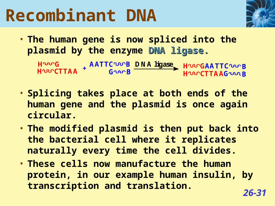
Recombinant DNA• Figure 26.17 The recombinant DNA
technique used to turn a bacterium into an insulin “factory”.
26-33
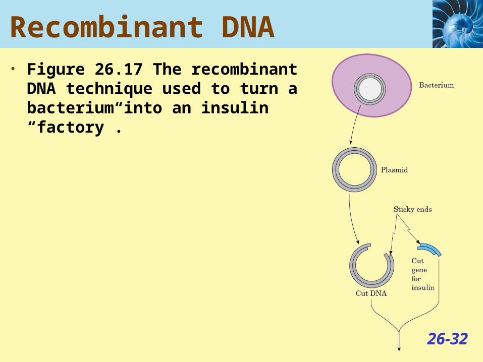
Recombinant DNA• Figure 26.17 Continued

26-34
Cloning DNA• Figure 26.18
The cloning of human DNA fragments with a viral vector.

26-35
Cloning DNA• Figure 26.18 The cloning of human DNA fragments with
a viral vector.

26-36
Gene Therapy• Figure 26.19 Gene therapy via retroviruses

26-37
Gene Therapy• Figure 16.19
Gene therapy via retroviruses.

26-38
Gene Therapy• Gene therapy is a technique whereby a missing gene is
replaced by a viral vector.• In ex vivo gene therapy, cells are removed from a
patient, given the missing gene, and then the cells are given back to the patient.
• In in vivo gene therapy, the patient is given the virus directly.