Embed Size (px)
DESCRIPTION
Chapter 28. From Egg to Embryo. Fertilization. ~ 300 million sperm enter female reproductive tract, most are lost ~2000-5000 reach egg Fertilization occurs when a sperm fuses with an egg to form a zygote. - PowerPoint PPT Presentation
Citation preview

Chapter 28
From Egg to Embryo
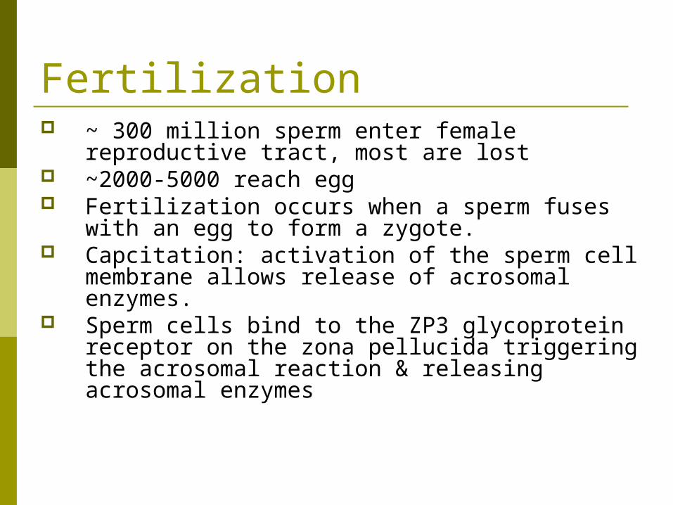
Fertilization ~ 300 million sperm enter female reproductive
tract, most are lost ~2000-5000 reach egg Fertilization occurs when a sperm fuses with an
egg to form a zygote. Capcitation: activation of the sperm cell membrane
allows release of acrosomal enzymes. Sperm cells bind to the ZP3 glycoprotein receptor
on the zona pellucida triggering the acrosomal reaction & releasing acrosomal enzymes
From Egg to Embryo
Hundreds of sperm cells must release their acrosomal enzymes before fertilization can occur
Acrosomal enzymes cut through the zona pelucida
Figure 28.2

From Egg to Embryo
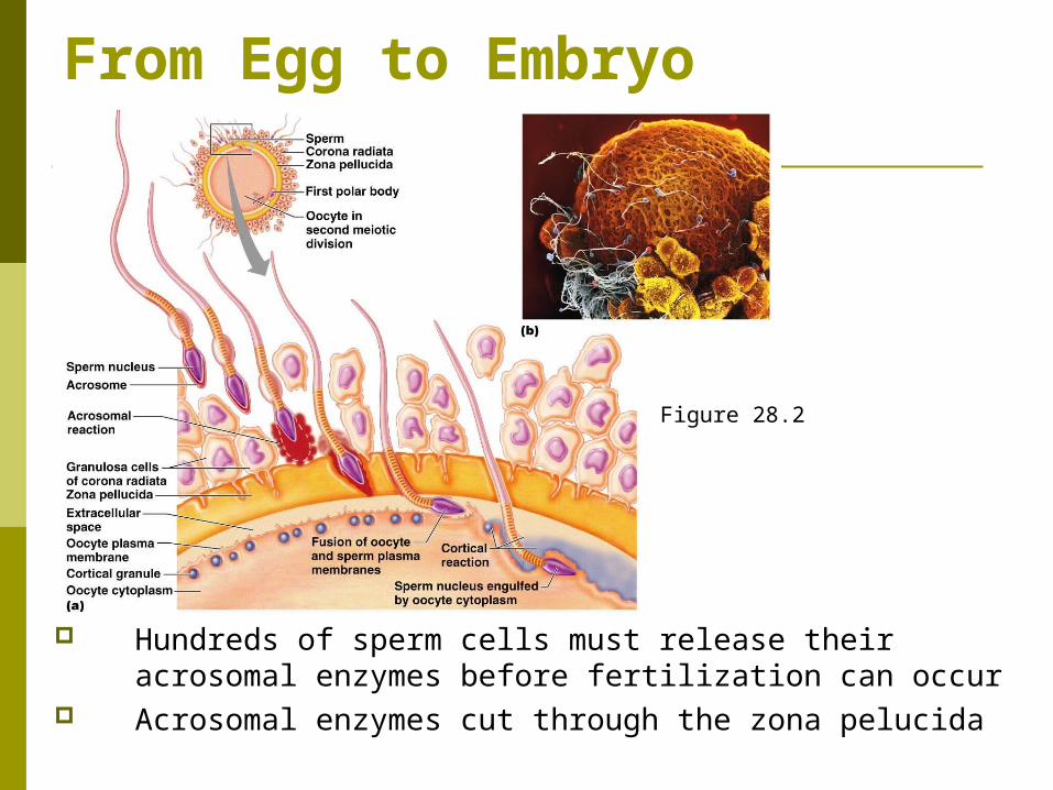
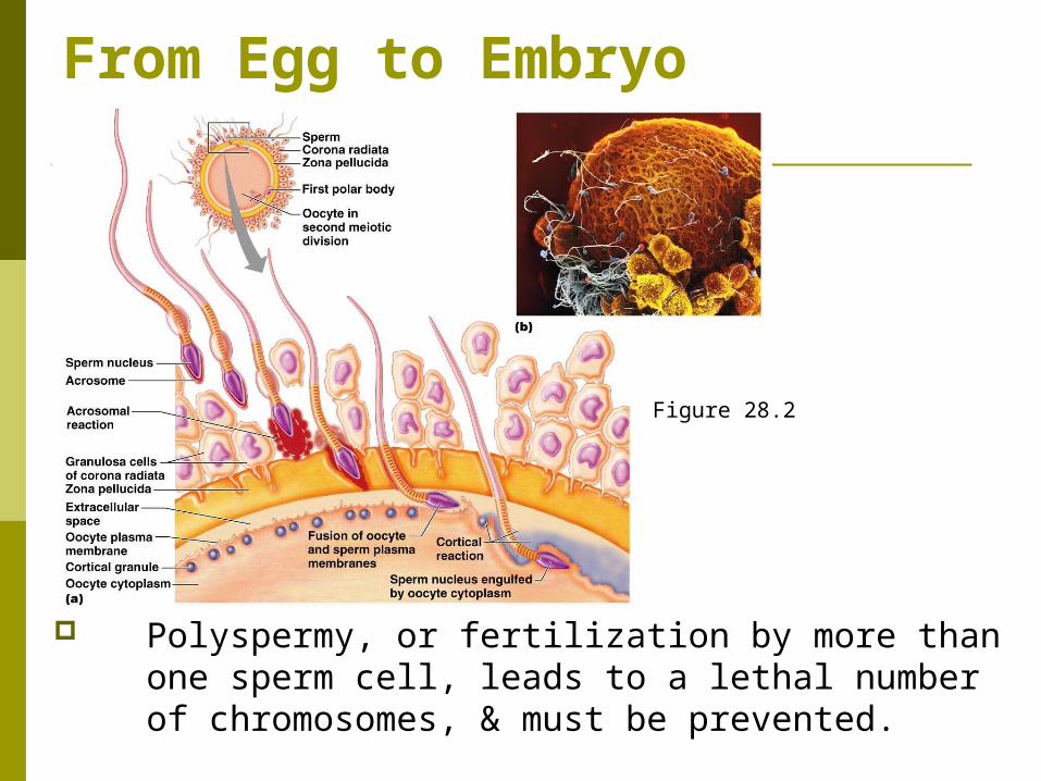
A protein on sperm binds to membrane receptors of oocyte activating the egg receptor to cause fusion of the egg & sperm membranes. sperm nucleus is pulled into the oocyte cytoplasm.
Figure 28.2
From Egg to Embryo
Polyspermy, or fertilization by more than one sperm cell, leads to a lethal number of chromosomes, & must be prevented.
Figure 28.2
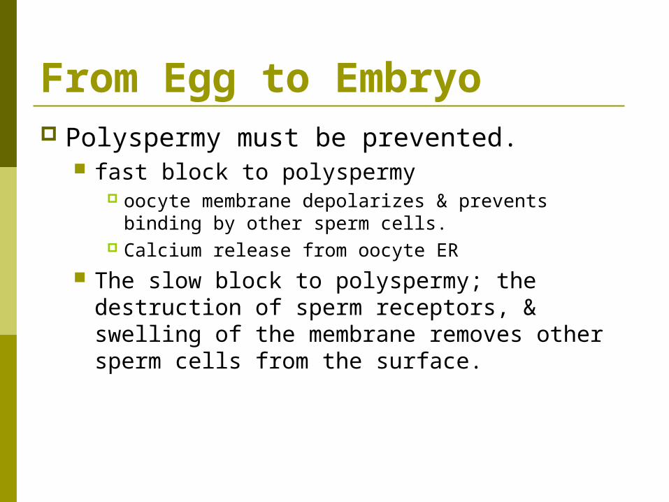
From Egg to Embryo Polyspermy must be prevented.
fast block to polyspermy oocyte membrane depolarizes & prevents binding by
other sperm cells. Calcium release from oocyte ER
The slow block to polyspermy; the destruction of sperm receptors, & swelling of the membrane removes other sperm cells from the surface.

From Egg to Embryo
After a sperm enters an oocyte, it loses its tail & midpiece, & migrates to the center of the oocyte while the oocyte completes meiosis II.
Figure 28.3

From Egg to Embryo
After meiosis II is completed, male & female pronuclei fuse & produce a zygote, which almost immediately enters into mitosis.
Figure 28.3
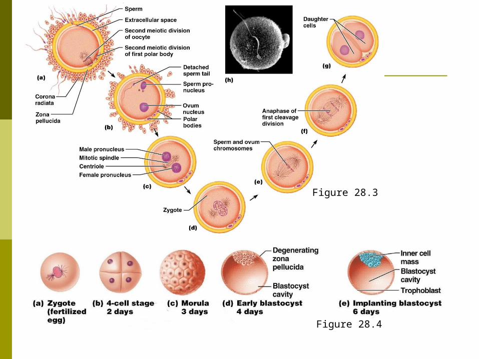
Figure 28.3
Figure 28.4
From Egg to Embryo
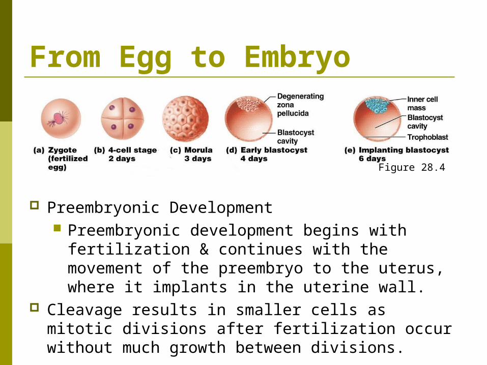
Preembryonic Development Preembryonic development begins with
fertilization & continues with the movement of the preembryo to the uterus, where it implants in the uterine wall.
Cleavage results in smaller cells as mitotic divisions after fertilization occur without much growth between divisions.
Figure 28.4
From Egg to Embryo
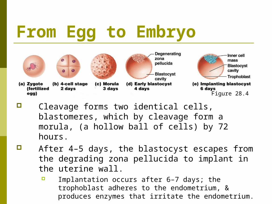
Cleavage forms two identical cells, blastomeres, which by cleavage form a morula, (a hollow ball of cells) by 72 hours.
After 4–5 days, the blastocyst escapes from the degrading zona pellucida to implant in the uterine wall. Implantation occurs after 6–7 days; the
trophoblast adheres to the endometrium, & produces enzymes that irritate the endometrium.
Figure 28.4
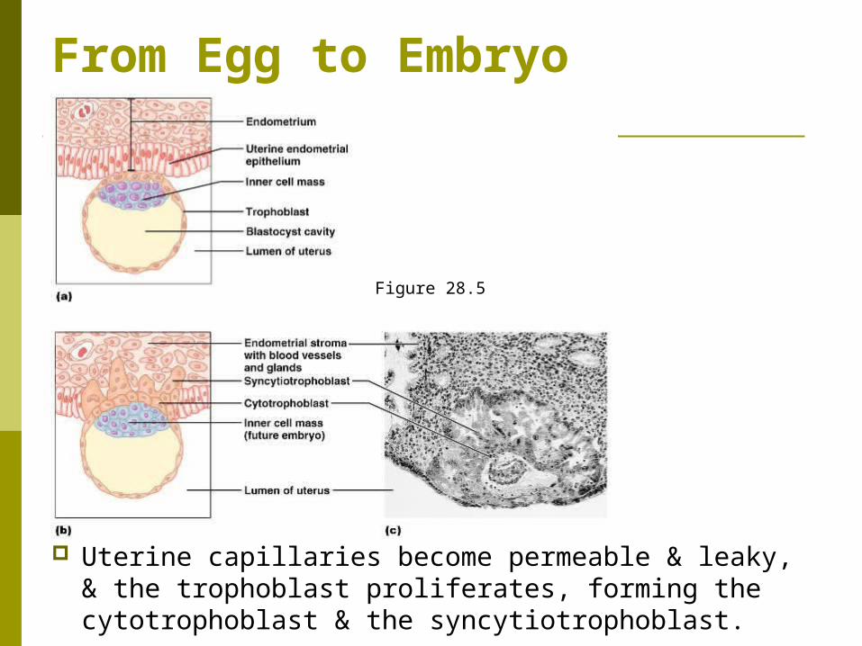
From Egg to Embryo
Uterine capillaries become permeable & leaky, & the trophoblast proliferates, forming the cytotrophoblast & the syncytiotrophoblast.
Figure 28.5
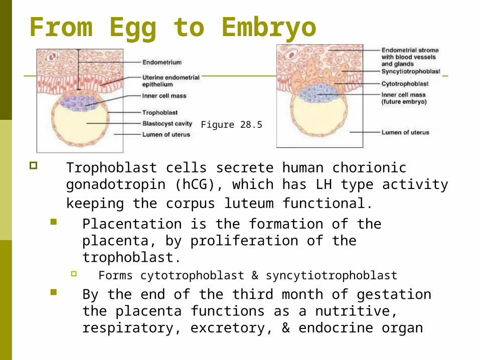
From Egg to Embryo
Trophoblast cells secrete human chorionic gonadotropin (hCG), which has LH type activity keeping the corpus luteum functional.
Placentation is the formation of the placenta, by proliferation of the trophoblast.
Forms cytotrophoblast & syncytiotrophoblast By the end of the third month of gestation
the placenta functions as a nutritive, respiratory, excretory, & endocrine organ
Figure 28.5
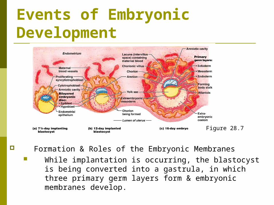
Events of Embryonic Development
Formation & Roles of the Embryonic Membranes While implantation is occurring, the blastocyst is being
converted into a gastrula, in which three primary germ layers form & embryonic membranes develop.
Figure 28.7
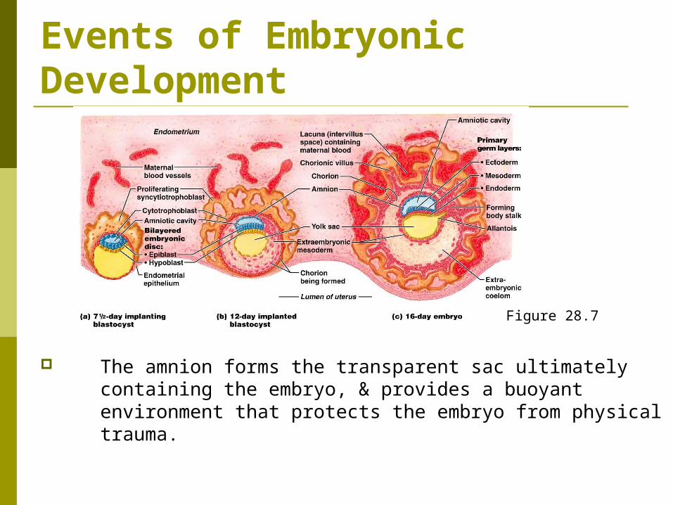
Events of Embryonic Development
The amnion forms the transparent sac ultimately containing the embryo, & provides a buoyant environment that protects the embryo from physical trauma.
Figure 28.7
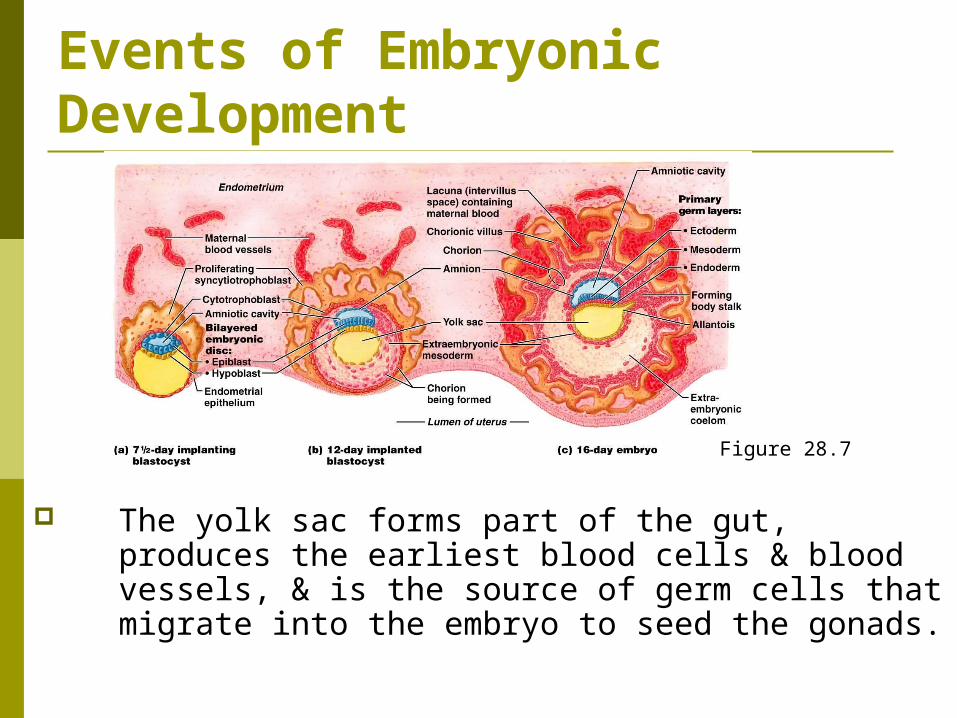
Events of Embryonic Development
The yolk sac forms part of the gut, produces the earliest blood cells & blood vessels, & is the source of germ cells that migrate into the embryo to seed the gonads.
Figure 28.7

Events of Embryonic Development
The allantois is the structural base for the umbilical cord that links the embryo to the placenta, & becomes part of the urinary bladder.
Figure 28.7
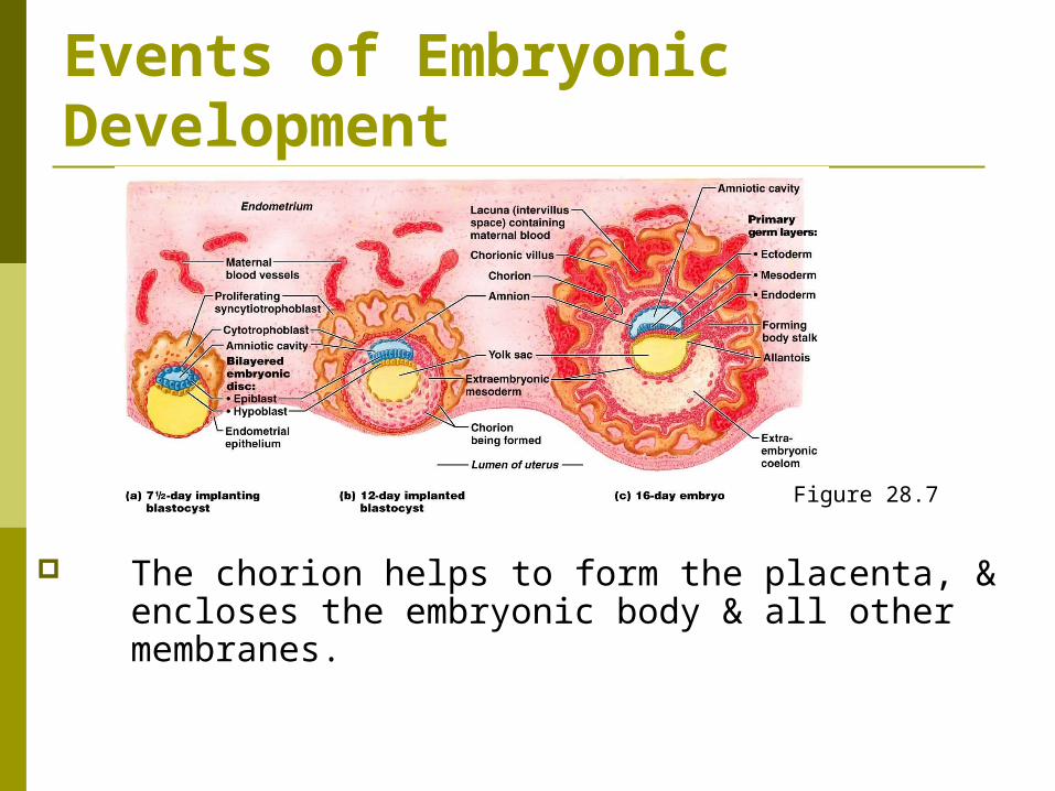
Events of Embryonic Development
The chorion helps to form the placenta, & encloses the embryonic body & all other membranes.
Figure 28.7
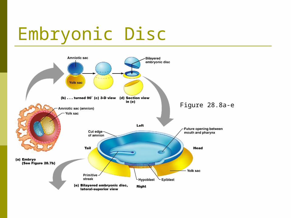
Embryonic Disc
Figure 28.8a-e
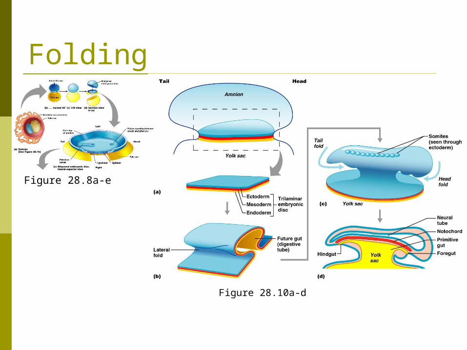
Folding
Figure 28.8a-e
Figure 28.10a-d
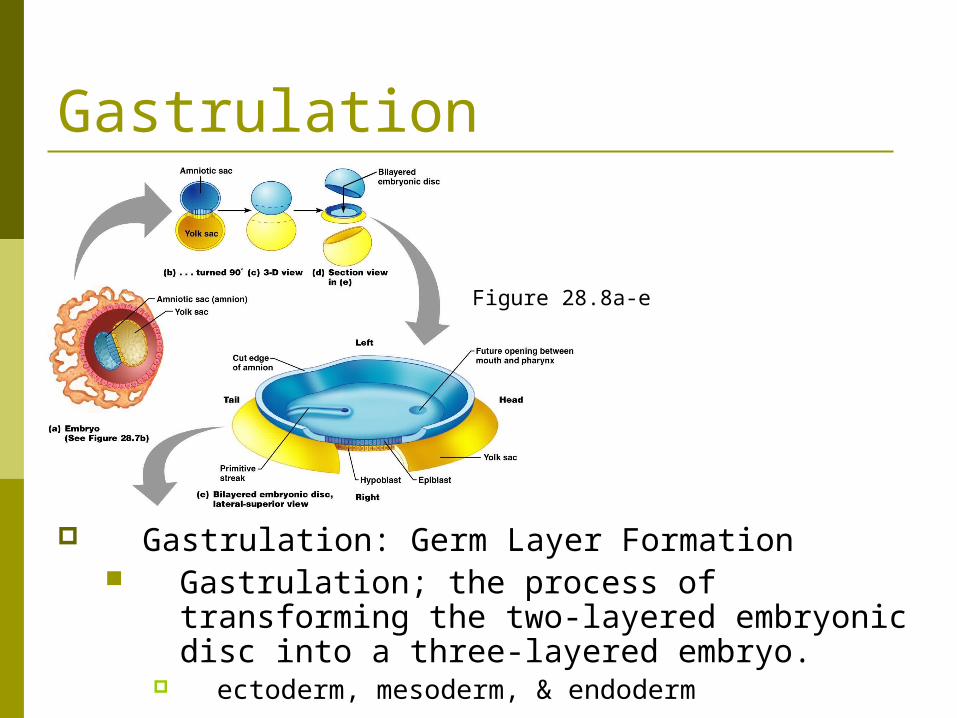
Gastrulation
Gastrulation: Germ Layer Formation Gastrulation; the process of transforming the
two-layered embryonic disc into a three-layered embryo.
ectoderm, mesoderm, & endoderm
Figure 28.8a-e
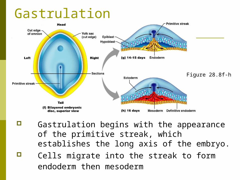
Gastrulation
Gastrulation begins with the appearance of the primitive streak, which establishes the long axis of the embryo.
Cells migrate into the streak to form endoderm then mesoderm
Fig. 28.8 Figure 28.8f-h

Organogenesis Organogenesis: Differentiation of the Germ Layers
Organogenesis is the formation of organs & organ systems; by the end of the embryonic period, all organ systems are recognizable.

Organogenesis
The ectoderm gives rise to structures of the nervous system & the epidermis.
Neurulation, the formation of the brain & spinal cord, is the first event of organogenesis.
Figure 28.9a-d
Figure 28.8f-h
Organogenesis
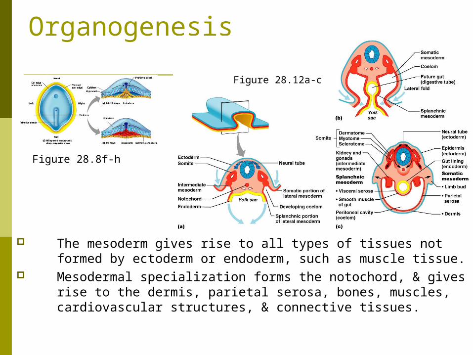
The mesoderm gives rise to all types of tissues not formed by ectoderm or endoderm, such as muscle tissue.
Mesodermal specialization forms the notochord, & gives rise to the dermis, parietal serosa, bones, muscles, cardiovascular structures, & connective tissues.
Fig. 28.8Figure 28.8f-h
Figure 28.12a-c
Organogenesis
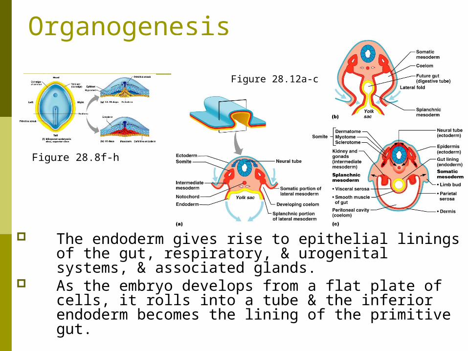
The endoderm gives rise to epithelial linings of the gut, respiratory, & urogenital systems, & associated glands.
As the embryo develops from a flat plate of cells, it rolls into a tube & the inferior endoderm becomes the lining of the primitive gut.
Fig. 28.8Figure 28.8f-h
Figure 28.12a-c

Folding
Figure 28.8f-h
Figure 28.10a-d
Organogenesis
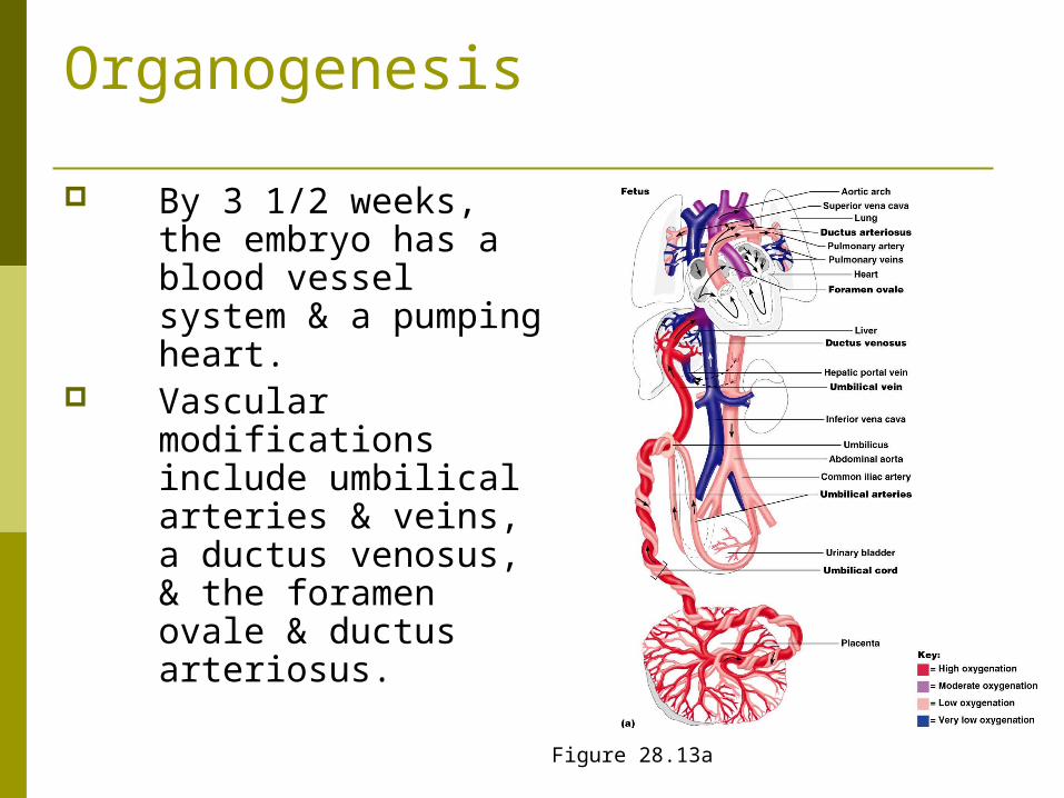
By 3 1/2 weeks, the embryo has a blood vessel system & a pumping heart.
Vascular modifications include umbilical arteries & veins, a ductus venosus, & the foramen ovale & ductus arteriosus.
Figure 28.13a
Events of Fetal Development
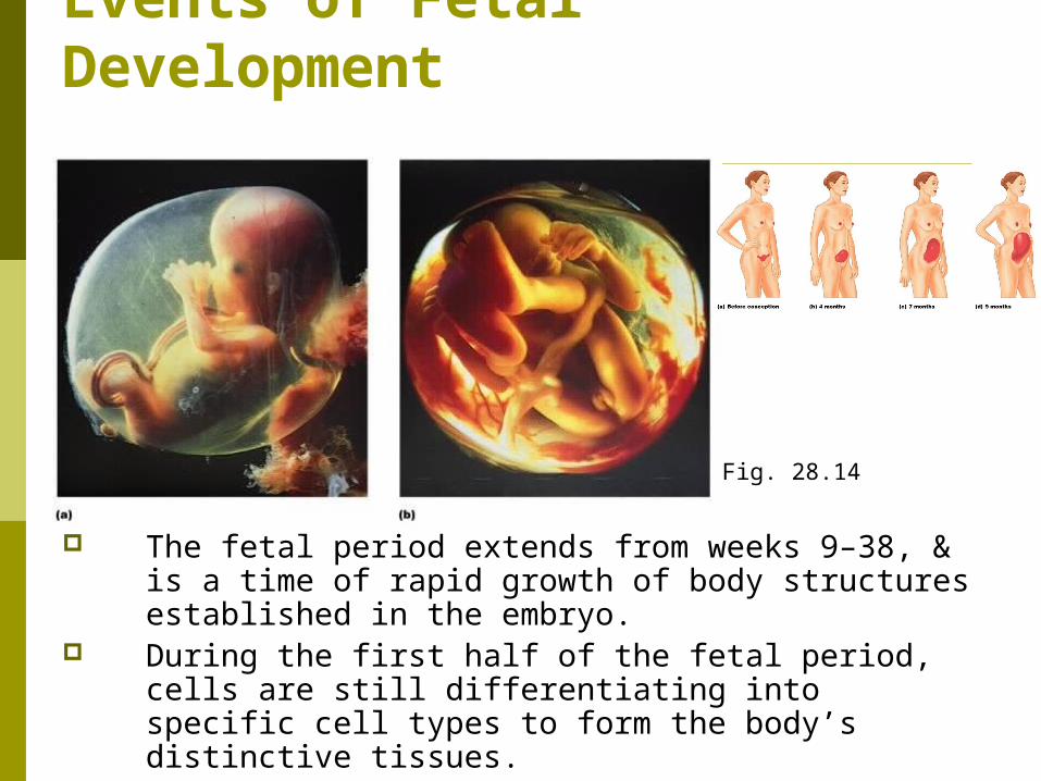
The fetal period extends from weeks 9–38, & is a time of rapid growth of body structures established in the embryo.
During the first half of the fetal period, cells are still differentiating into specific cell types to form the body’s distinctive tissues.
Fig. 28.14

Effects of Pregnancy on the Mother
Anatomical Changes Metabolic Changes Physiological Changes
Effects of Pregnancy on the Mother
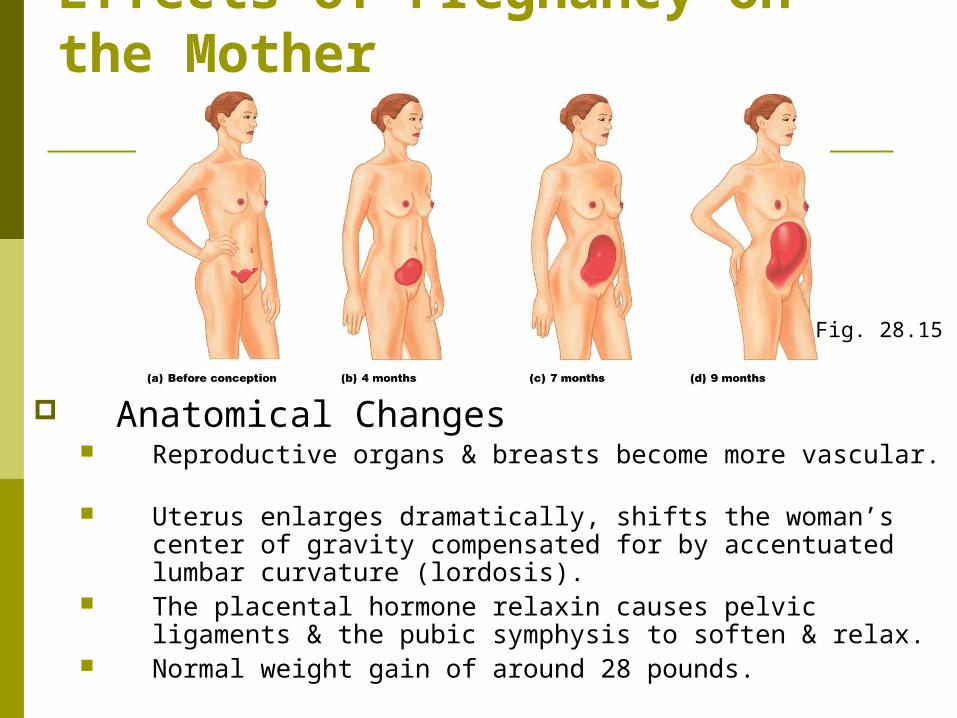
Anatomical Changes Reproductive organs & breasts become more vascular. Uterus enlarges dramatically, shifts the woman’s center of
gravity compensated for by accentuated lumbar curvature (lordosis).
The placental hormone relaxin causes pelvic ligaments & the pubic symphysis to soften & relax.
Normal weight gain of around 28 pounds.
Fig. 28.15
Effects of Pregnancy on the Mother Metabolic Changes
The placenta produces; Human placental lactogen
Promotes breast maturation (with estrogen & progesterone).
Promotes the growth of the fetus, & exerts a glucose-sparing effect on maternal metabolism.
Human chorionic thyrotropin which increases maternal metabolic rate.
Effects of Pregnancy on the Mother Physiological Changes (p. 1135)
Morning sickness may be present during the first few months of pregnancy, until adaptation to elevated levels of estrogen & progesterone occurs.
Heartburn due to esophageal displacement Constipation may result due to the decreased
motility of the digestive tract.
Effects of Pregnancy on the Mother Physiological Changes (p. 1135)
Increased urine production to dispose of additional fetal metabolic waste.
Vital capacity & respiratory rate increases Decrease in residual volume Many women experience dyspnea.
Blood pressure & heart rate rise. Blood volume increases to accommodate the needs of the fetus.
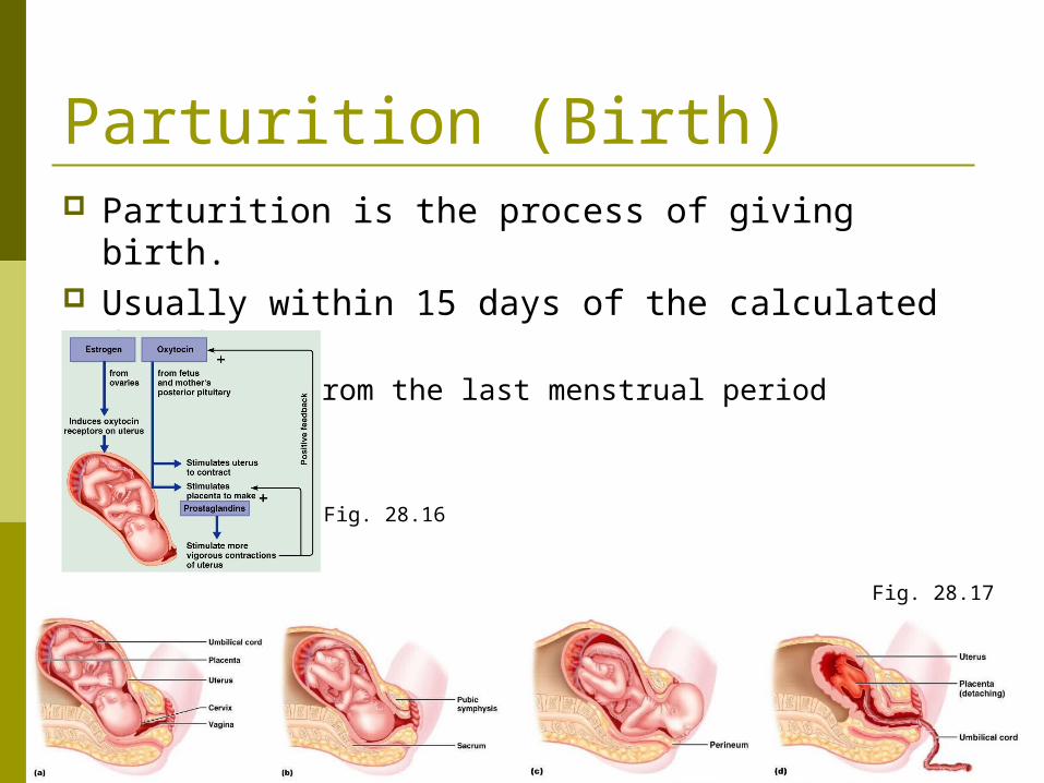
Parturition (Birth) Parturition is the process of giving birth. Usually within 15 days of the calculated due date.
280 days from the last menstrual period
Fig. 28.17
Fig. 28.16
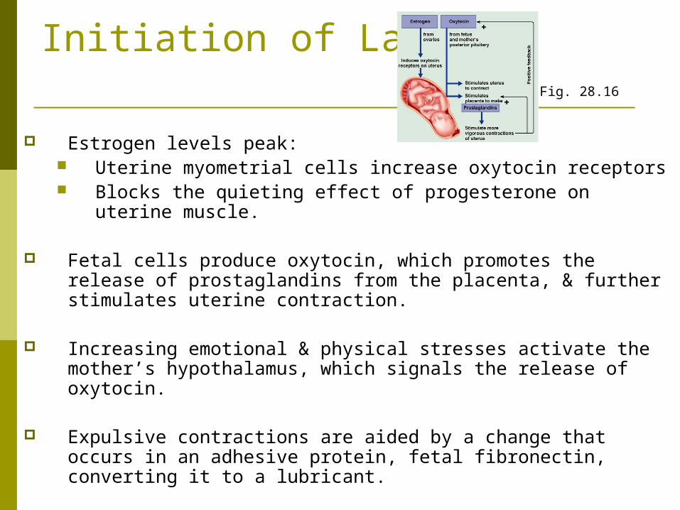
Initiation of Labor
Estrogen levels peak: Uterine myometrial cells increase oxytocin receptors Blocks the quieting effect of progesterone on uterine
muscle.
Fetal cells produce oxytocin, which promotes the release of prostaglandins from the placenta, & further stimulates uterine contraction.
Increasing emotional & physical stresses activate the mother’s hypothalamus, which signals the release of oxytocin.
Expulsive contractions are aided by a change that occurs in an adhesive protein, fetal fibronectin, converting it to a lubricant.
Fig. 28.16
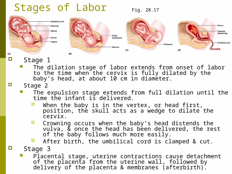
Stages of Labor
Stage 1 The dilation stage of labor extends from onset of labor to the time
when the cervix is fully dilated by the baby’s head, at about 10 cm in diameter.
Stage 2 The expulsion stage extends from full dilation until the time the
infant is delivered. When the baby is in the vertex, or head first, position, the skull
acts as a wedge to dilate the cervix. Crowning occurs when the baby’s head distends the vulva, &
once the head has been delivered, the rest of the baby follows much more easily.
After birth, the umbilical cord is clamped & cut. Stage 3
Placental stage, uterine contractions cause detachment of the placenta from the uterine wall, followed by delivery of the placenta & membranes (afterbirth).
Fig. 28.17
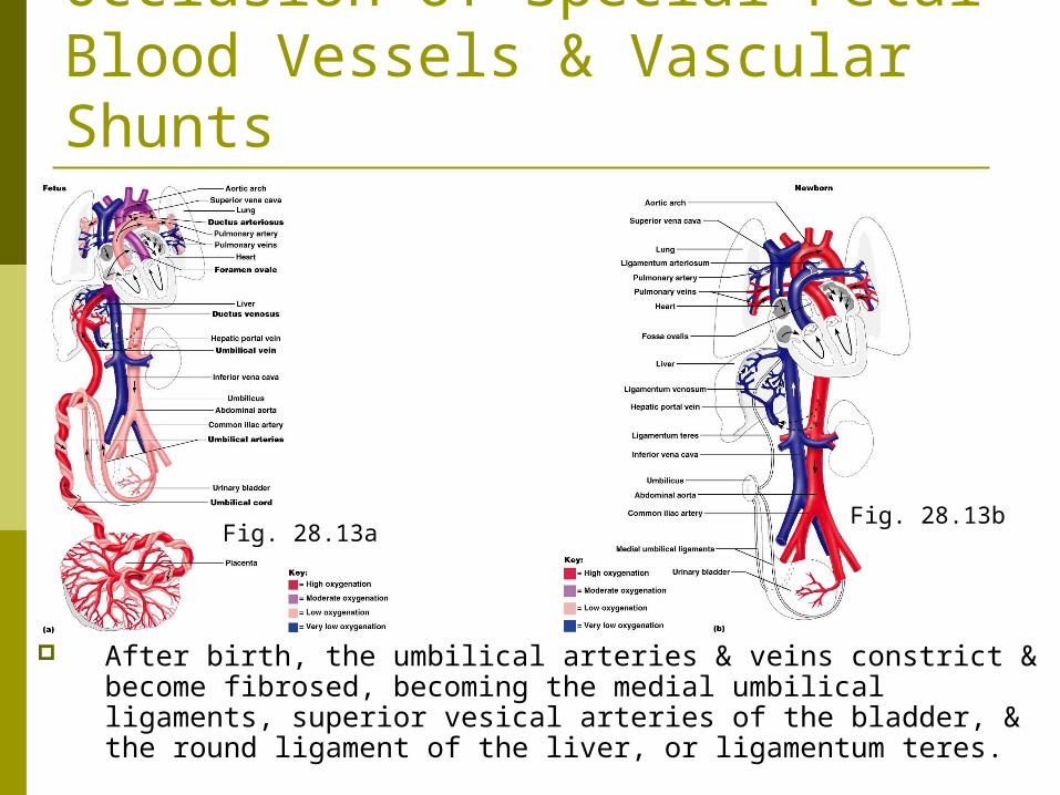
Occlusion of Special Fetal Blood Vessels & Vascular Shunts
After birth, the umbilical arteries & veins constrict & become fibrosed, becoming the medial umbilical ligaments, superior vesical arteries of the bladder, & the round ligament of the liver, or ligamentum teres.
Fig. 28.13aFig. 28.13b
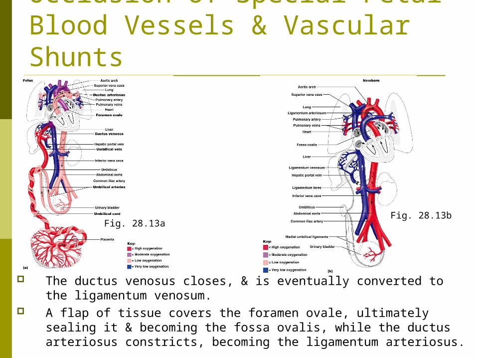
Occlusion of Special Fetal Blood Vessels & Vascular Shunts
The ductus venosus closes, & is eventually converted to the ligamentum venosum.
A flap of tissue covers the foramen ovale, ultimately sealing it & becoming the fossa ovalis, while the ductus arteriosus constricts, becoming the ligamentum arteriosus.
Fig. 28.13aFig. 28.13b
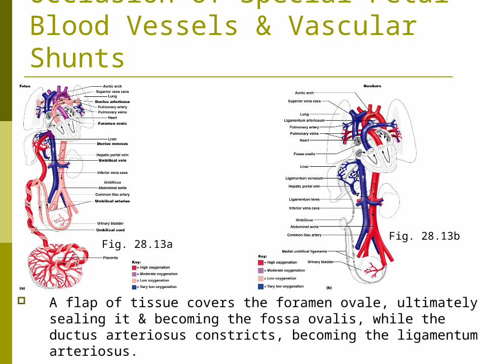
Occlusion of Special Fetal Blood Vessels & Vascular Shunts
A flap of tissue covers the foramen ovale, ultimately sealing it & becoming the fossa ovalis, while the ductus arteriosus constricts, becoming the ligamentum arteriosus.
Fig. 28.13aFig. 28.13b
Lactation Lactation is the production of milk by the
hormone-prepared mammary glands.
Lactation
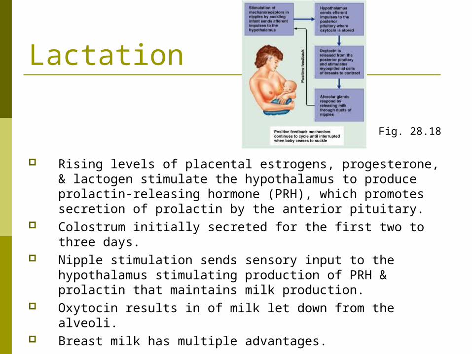
Rising levels of placental estrogens, progesterone, & lactogen stimulate the hypothalamus to produce prolactin-releasing hormone (PRH), which promotes secretion of prolactin by the anterior pituitary.
Colostrum initially secreted for the first two to three days. Nipple stimulation sends sensory input to the
hypothalamus stimulating production of PRH & prolactin that maintains milk production.
Oxytocin results in of milk let down from the alveoli. Breast milk has multiple advantages.
Fig. 28.18

Assisted Reproductive Technology & Reproductive Cloning
Hormones can be used to increase sperm or egg production & surgery can be used to open blocked tubes.
Assisted reproductive technology involves surgically removing oocytes from a woman’s ovaries, fertilizing the eggs & returning them to the woman’s body.
Cloning involves the placing of a somatic cell nucleus into an oocyte.
Fig. 28.19