Embed Size (px)
Citation preview
Chapter 28
An Introduction to Animal Diversity
Characteristics of Most Animals
• 1. multicellular eukaryotes
• 2. cell specialization – (cells tissues organs)
• 3. heterotrophs
• 4. locomotion (sometime in lifecycle)
• 5. nervous + muscle systems (stimuli)
• 6. sexual reproduction – (large, nonmotile eggs; flagellated sperm)
Marine Environments
• Advantages– Buoyancy – support– Temperature – stable– Fluid + salt balance easily maintained
• Challenges – Water movement/currents
• Adapt:– Strong swimmer – squid, fish, mammals
– Sessile
– Burrow in sand/silt
– Small body size plankton (food supply around as tossed)

Other environments - problems
• Fresh water– Water hypotonic to animal fluids
• Osmoregulation - pump out water, keep salts (ATP)
– Less constant – Less food– Oxygen and temp. vary– Turbidity + water volume change
• Land – Desiccation
• Adapt: body covering; respiratory surface deep within animal
– Reproduction (desiccation)• Adapt: internal fertilization; shells on eggs; embryo
in mom
– Temperature extremes
Body Symmetry
• 2 types– Radial
• wheel or cylinder form• Spokes from central axis
– Cnidarians – jellyfish, sea anemones– Echinoderms – sea stars
– Bilateral • Right and left halves – mirror images
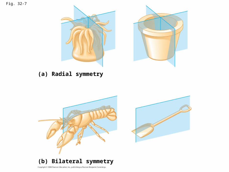
Fig. 32-7
(a) Radial symmetry
(b) Bilateral symmetry
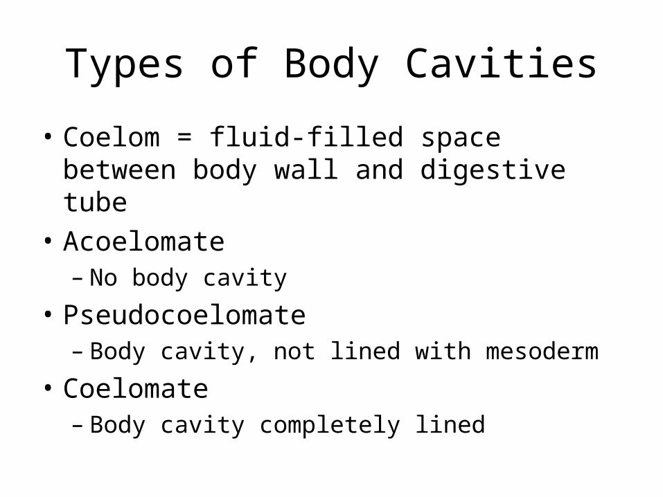
Types of Body Cavities
• Coelom = fluid-filled space between body wall and digestive tube
• Acoelomate– No body cavity
• Pseudocoelomate– Body cavity, not lined with mesoderm
• Coelomate – Body cavity completely lined
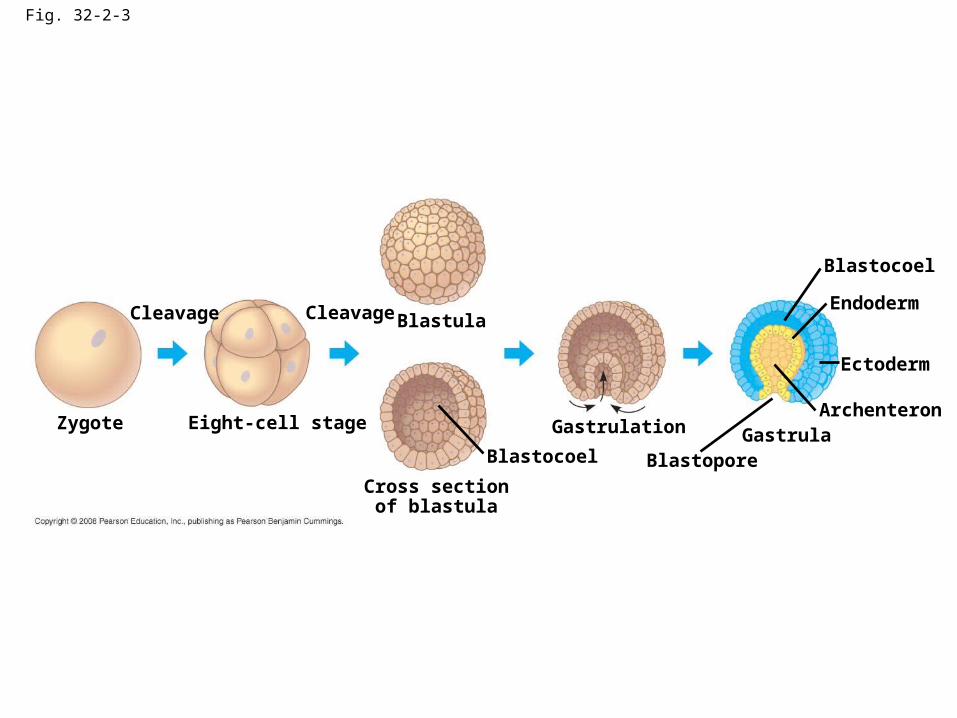
Fig. 32-2-3
Zygote
Cleavage
Eight-cell stage
Cleavage Blastula
Cross sectionof blastula
Blastocoel
Gastrulation
BlastoporeGastrula
Archenteron
Ectoderm
Endoderm
Blastocoel
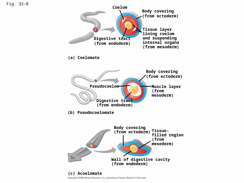
Fig. 32-8Coelom
Body covering(from ectoderm)
Digestive tract(from endoderm)
Tissue layerlining coelomand suspendinginternal organs(from mesoderm)
(a) Coelomate
Body covering(from ectoderm)
Pseudocoelom
Digestive tract(from endoderm)
Muscle layer(frommesoderm)
(b) Pseudocoelomate
Body covering(from ectoderm) Tissue-
filled region(frommesoderm)
Wall of digestive cavity(from endoderm)
(c) Acoelomate
2 Main Groups of Coelomates
• Protostomes– “first, the mouth”– Mollusks, annelids, arthropods
• Deuterostomes – “second, the mouth”
• Echinoderms, chordates
Protostomes vs. DeuterostomesCleavage
• Protostomes– Spiral
• Deuterostomes– radial
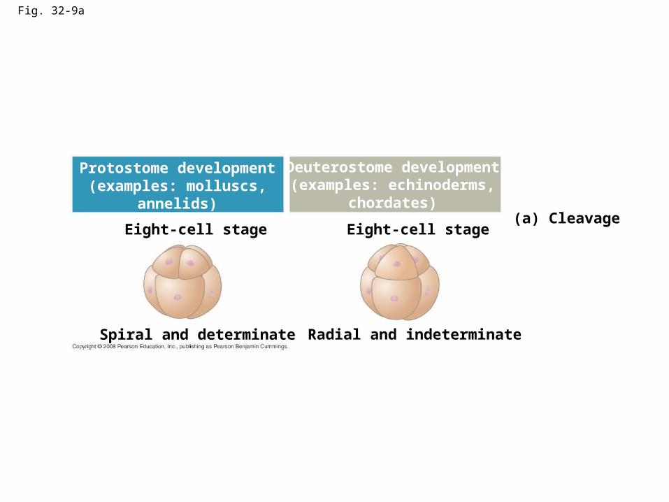
Fig. 32-9a
Eight-cell stage Eight-cell stage(a) Cleavage
Spiral and determinate Radial and indeterminate
Protostome development(examples: molluscs,
annelids)
Deuterostome development(examples: echinoderms,
chordates)
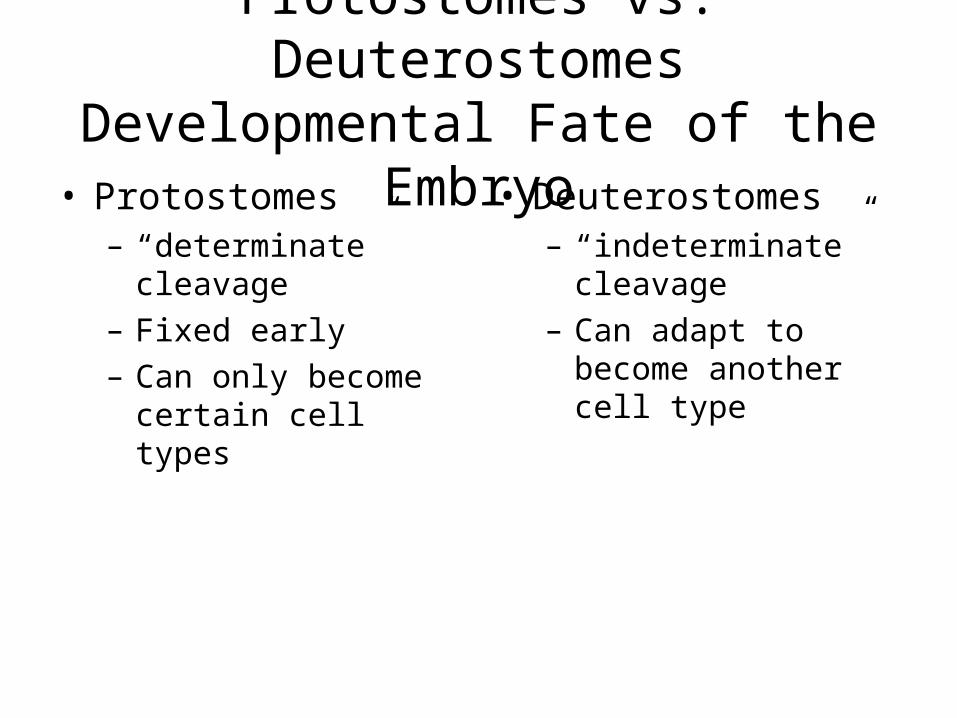
Protostomes vs. DeuterostomesDevelopmental Fate of the Embryo
• Protostomes– “determinate”
cleavage– Fixed early– Can only become
certain cell types
• Deuterostomes– “indeterminate”
cleavage– Can adapt to become
another cell type
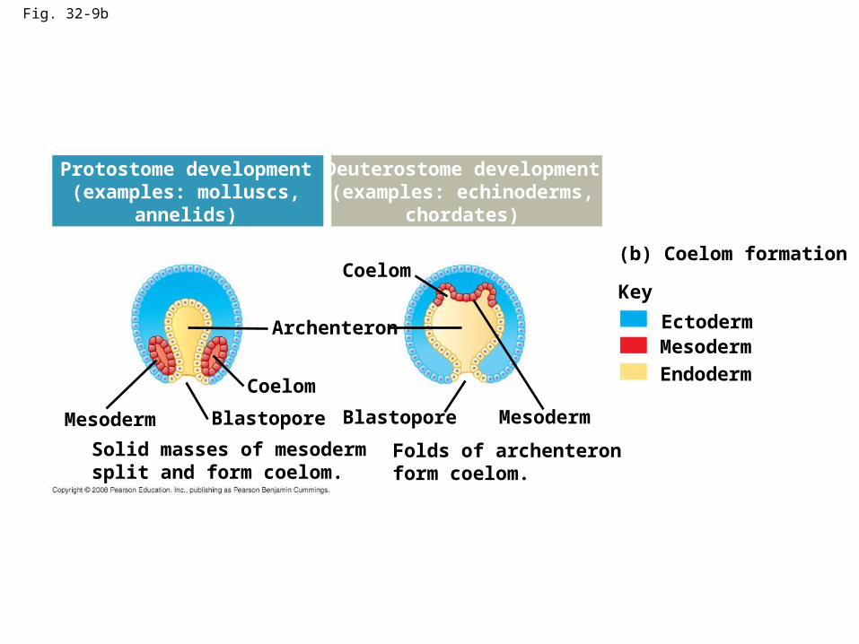
Fig. 32-9b
Coelom
Protostome development(examples: molluscs,
annelids)
Deuterostome development(examples: echinoderms,
chordates)
(b) Coelom formation
Key
EctodermMesoderm
Endoderm
MesodermMesoderm
Coelom
Archenteron
Blastopore Blastopore
Solid masses of mesodermsplit and form coelom.
Folds of archenteronform coelom.
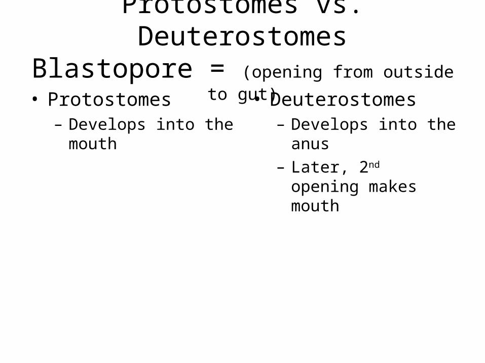
Protostomes vs. DeuterostomesBlastopore = (opening from outside to gut)
• Protostomes– Develops into the
mouth
• Deuterostomes– Develops into the anus– Later, 2nd opening
makes mouth
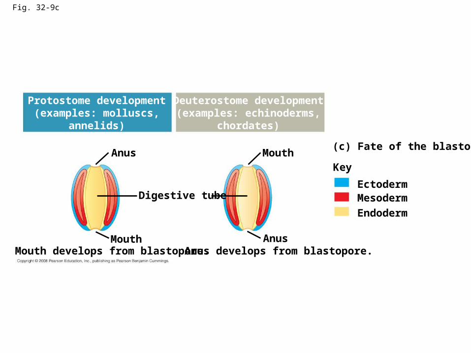
Fig. 32-9c
Anus
Protostome development(examples: molluscs,
annelids)
Deuterostome development(examples: echinoderms,
chordates)
Anus
Mouth
Mouth
Digestive tube
(c) Fate of the blastopore
Key
EctodermMesoderm
Endoderm
Mouth develops from blastopore. Anus develops from blastopore.
Sponges – Phylum Porifera
• “to have pores”
• Bodies – tiny holes
• Marine
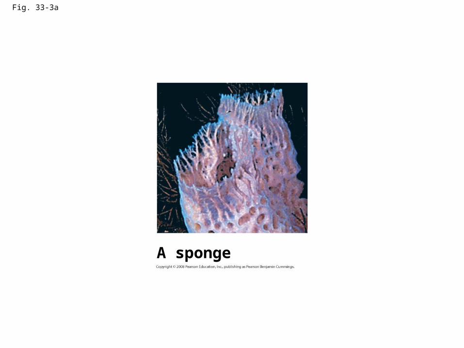
Fig. 33-3a
A sponge
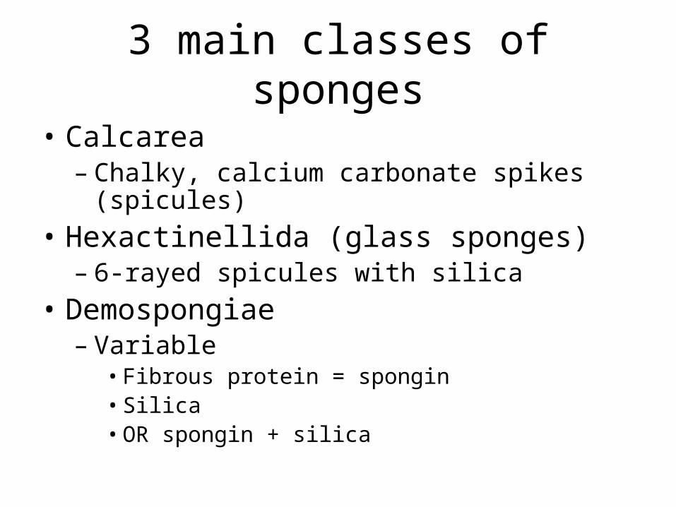
3 main classes of sponges
• Calcarea– Chalky, calcium carbonate spikes (spicules)
• Hexactinellida (glass sponges)– 6-rayed spicules with silica
• Demospongiae– Variable
• Fibrous protein = spongin• Silica• OR spongin + silica
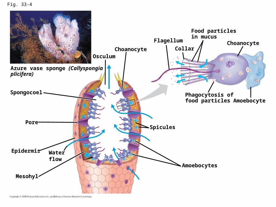
Fig. 33-4
Azure vase sponge (Callyspongiaplicifera)
Spongocoel
Osculum
Pore
Epidermis Waterflow
Mesohyl
Choanocyte
Flagellum
Collar
Food particlesin mucus
Choanocyte
AmoebocytePhagocytosis offood particles
Spicules
Amoebocytes
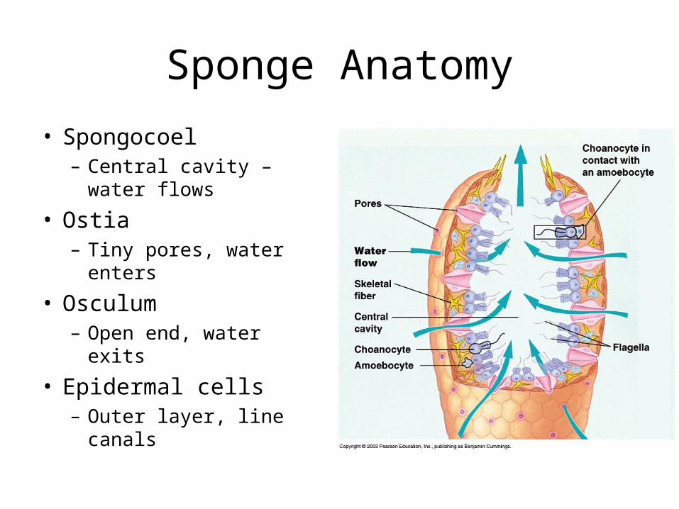
Sponge Anatomy
• Spongocoel– Central cavity – water
flows
• Ostia– Tiny pores, water enters
• Osculum– Open end, water exits
• Epidermal cells– Outer layer, line canals
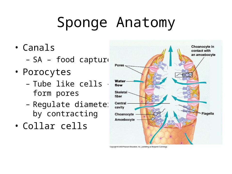
Sponge Anatomy
• Canals– SA – food capture
• Porocytes– Tube like cells – form
pores– Regulate diameter by
contracting
• Collar cells
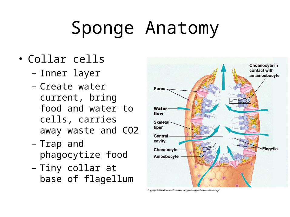
Sponge Anatomy
• Collar cells– Inner layer– Create water current,
bring food and water to cells, carries away waste and CO2
– Trap and phagocytize food
– Tiny collar at base of flagellum
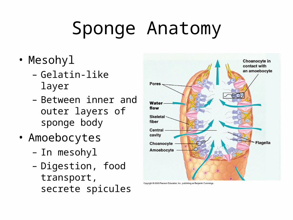
Sponge Anatomy
• Mesohyl– Gelatin-like layer– Between inner and
outer layers of sponge body
• Amoebocytes – In mesohyl– Digestion, food
transport, secrete spicules
Sponge Feeding
• Suspension feeders– Trap + eat whatever food the water brings– Water circulates in body– Food trapped on sticky collars of choanocytes– Food digested in collar or amoeboid cell – Undigested – out to water through osculum
Gas exchange/Excretion
• Diffusion – in/out of individual cells
Response to Stimuli
• No special nerve cells – can’t react as a whole
• Individual cells can respond
Reproduction of sponges
• Asexual– Fragment or bud
• Sexual – Hermaphrodite – egg + sperm– Some amoeboid cells become sperm, some eggs– Eggs/sperm made at different times cross fertilize– Sperm released into water, taken in by other sponges
of same species– Fertilization and early dev. In mesohyl– Embryo moves to spongocoel, leaves with water– Swims, attaches to solid object sessile
Cnidarians – Phylum Cnidaria
• marine
• Solitary
• colonies
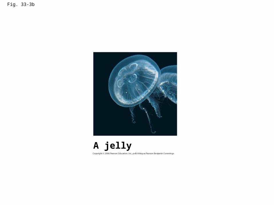
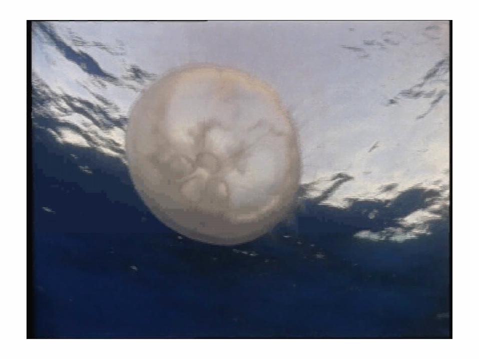
Fig. 33-3b
A jelly
3 classes of Cnidarians:
• Hydrozoa– Hydras, hydroids– Polyp dominant
• Scyphozoa– Jellyfish– Medusa dominant
• Anthozoa – Sea anemones, corals– No medusa
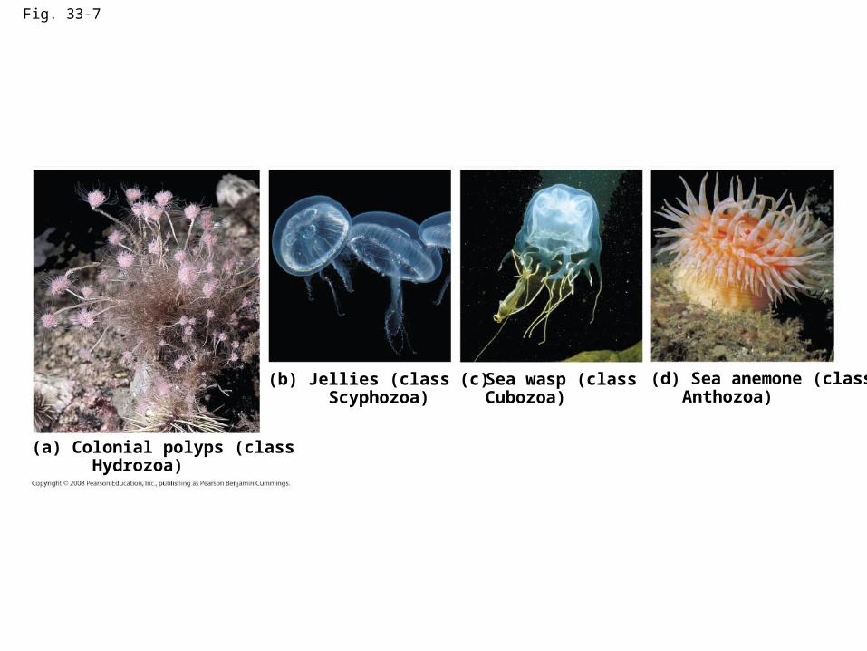
Fig. 33-7
(a) Colonial polyps (class Hydrozoa)
(b) Jellies (class Scyphozoa)
Sea wasp (classCubozoa)
(d) Sea anemone (class Anthozoa)
(c)
Body of Cnidarians
• Radial symmetry
• Hollow sac w/ mouth + surrounding tentacles at 1 end
• Mouth leads to GV cavity (digestive)
• Mouth – ingests food, expels waste
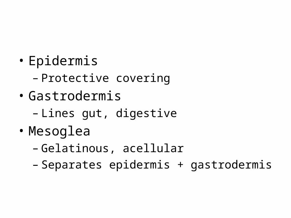
• Epidermis– Protective covering
• Gastrodermis– Lines gut, digestive
• Mesoglea– Gelatinous, acellular– Separates epidermis + gastrodermis
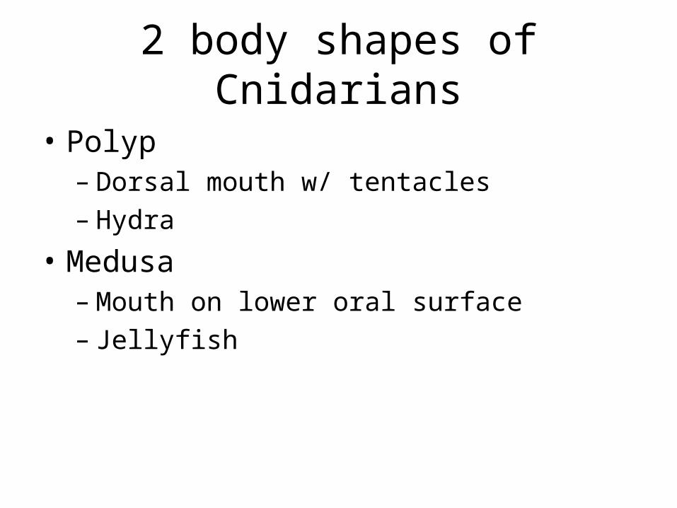
2 body shapes of Cnidarians
• Polyp– Dorsal mouth w/ tentacles– Hydra
• Medusa – Mouth on lower oral surface– Jellyfish
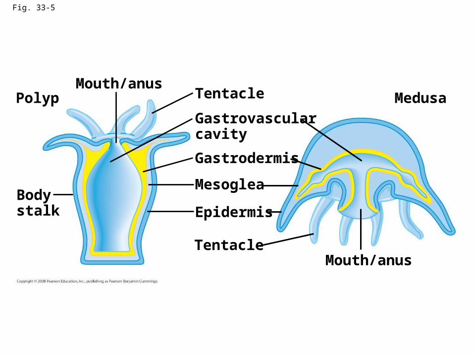
Fig. 33-5
PolypMouth/anus
Bodystalk
Tentacle
Gastrovascularcavity
Gastrodermis
Mesoglea
Epidermis
TentacleMouth/anus
Medusa
Response in Cnidarians
• Nerve nets– Nerve cells that connect sensory cells in body
wall to contractile + gland cells– Cells contacted, entire body responds –
crunches in
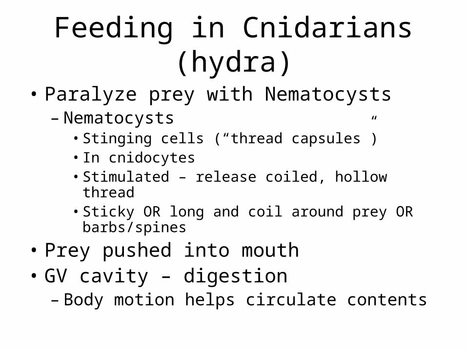
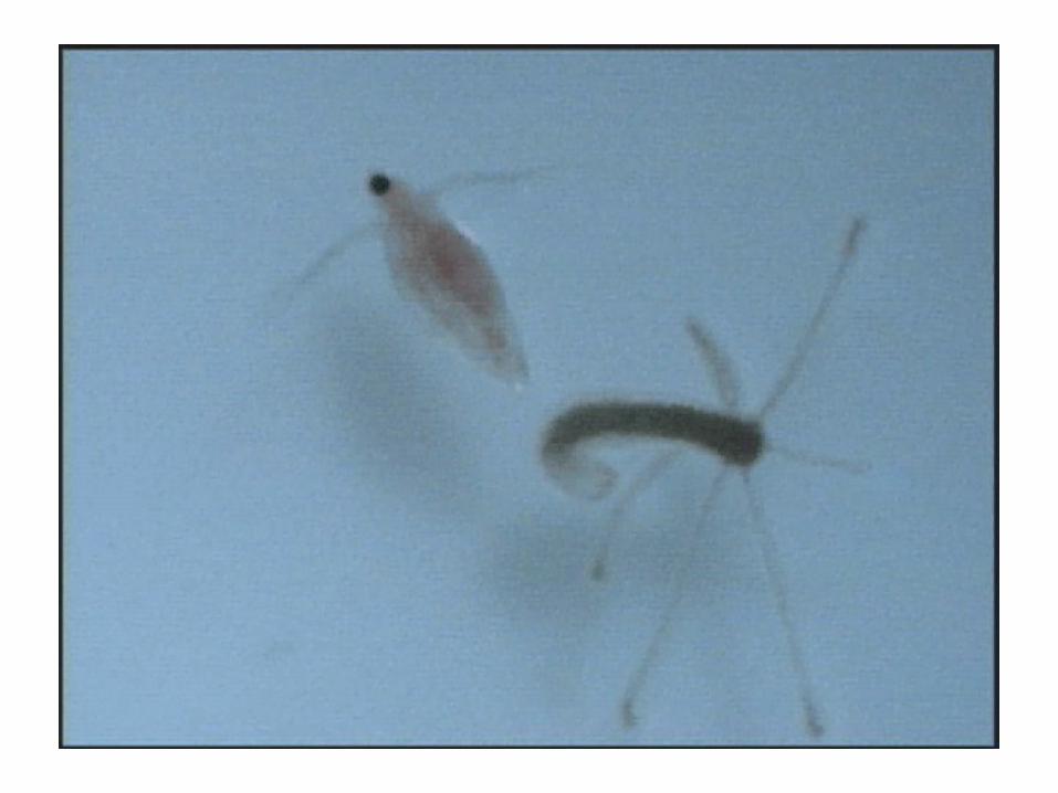
Feeding in Cnidarians (hydra)
• Paralyze prey with Nematocysts – Nematocysts
• Stinging cells (“thread capsules”)• In cnidocytes• Stimulated – release coiled, hollow thread• Sticky OR long and coil around prey OR
barbs/spines
• Prey pushed into mouth• GV cavity – digestion
– Body motion helps circulate contents
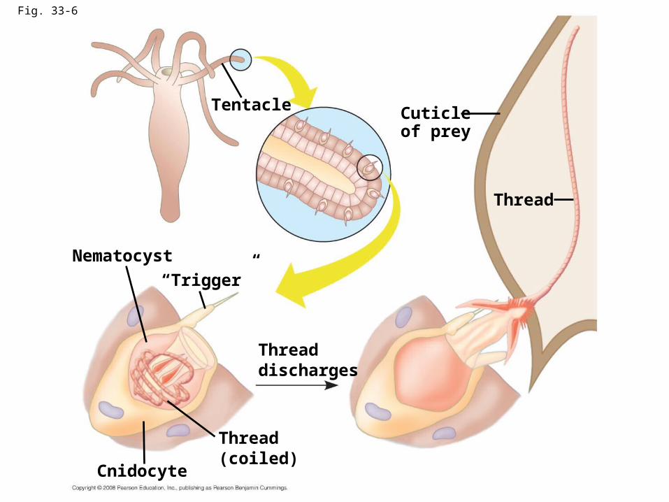
Fig. 33-6
Tentacle
Nematocyst
“Trigger”
Cuticleof prey
Threaddischarges
Thread(coiled)
Cnidocyte
Thread
Gas exchange/Excretion
• Diffusion– No cell far from surface
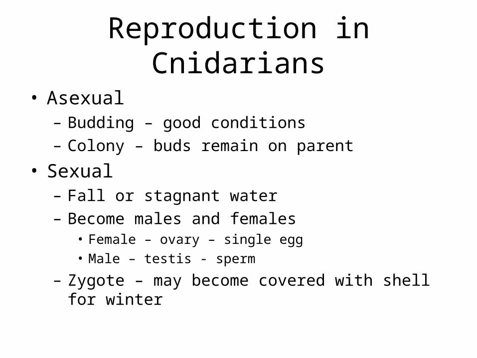
Reproduction in Cnidarians
• Asexual– Budding – good conditions– Colony – buds remain on parent
• Sexual– Fall or stagnant water– Become males and females
• Female – ovary – single egg• Male – testis - sperm
– Zygote – may become covered with shell for winter
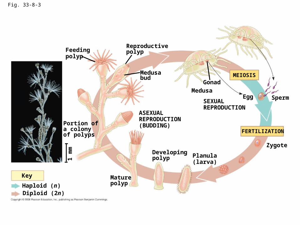
Fig. 33-8-3
Feedingpolyp
Reproductivepolyp
Medusabud
Medusa
ASEXUALREPRODUCTION(BUDDING)Portion of
a colonyof polyps
1 m
m
Key
Haploid (n)Diploid (2n)
Gonad
SEXUALREPRODUCTION
MEIOSIS
FERTILIZATION
Egg Sperm
Zygote
Planula(larva)
Developingpolyp
Maturepolyp
Comb Jellies – Phylum Ctenophora
• Marine• Luminescent• 8 rows cilia (comb)• 2 tentacles – no nematocysts – adhesive glue
cells• Radial symmetry• 2 cells layers w/ mesoglea• Mouth – food in; 2 anal pores – waste out (other
end)
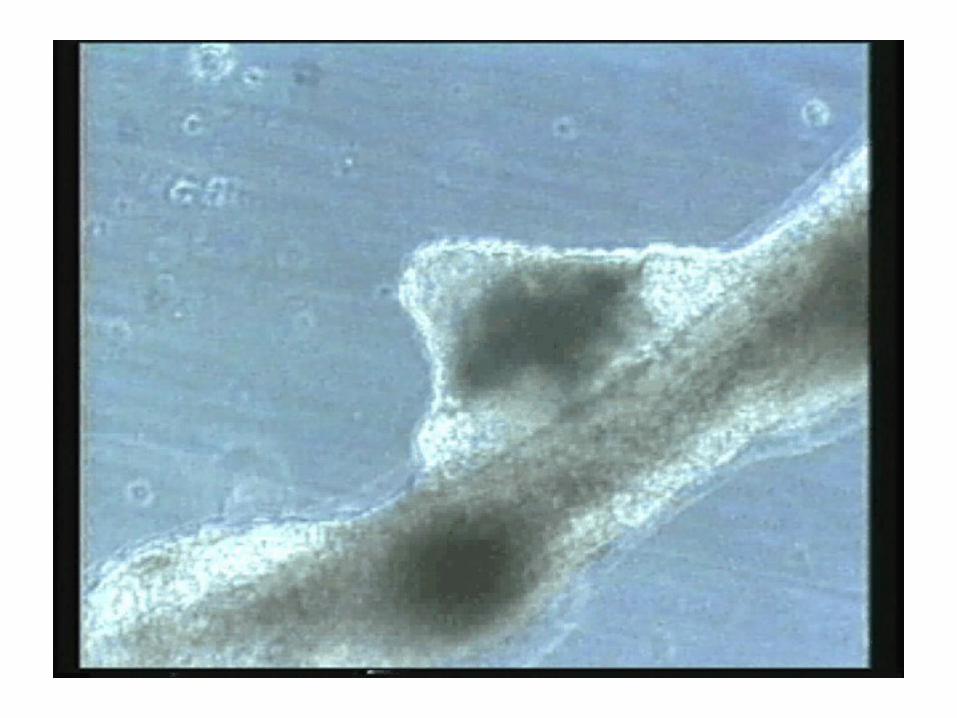
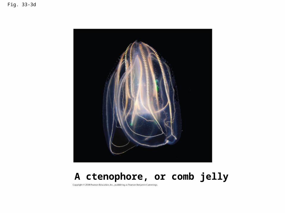
Fig. 33-3d
A ctenophore, or comb jelly
Flatworms – Phylum Platyhelminthes
• Flat, elongated, acoelomate
• Bilateral symmetry
• Cephalization– “head” at anterior – moves forward; eyespots
• 3 germ layers – – ectoderm, mesoderm, endoderm
• Muscular pharynx– Takes in food – 1 opening mouth
Flatworms cont.
• Nervous system– Simple brain = 2 mass nerve tissue = ganglia
– connect to 2 nerve cords
• Protonephridia– Osmoregulation, waste disposal
• Complex reproductive organs
• No organs for circulation, gas exchange– Diffusion through body wall
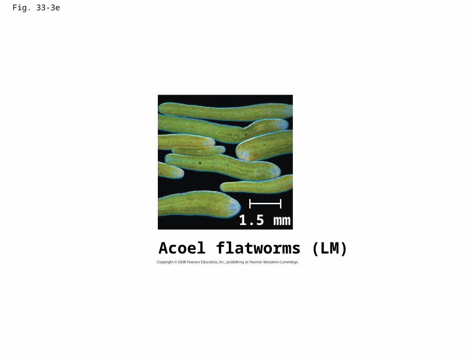
Fig. 33-3e
Acoel flatworms (LM)
1.5 mm
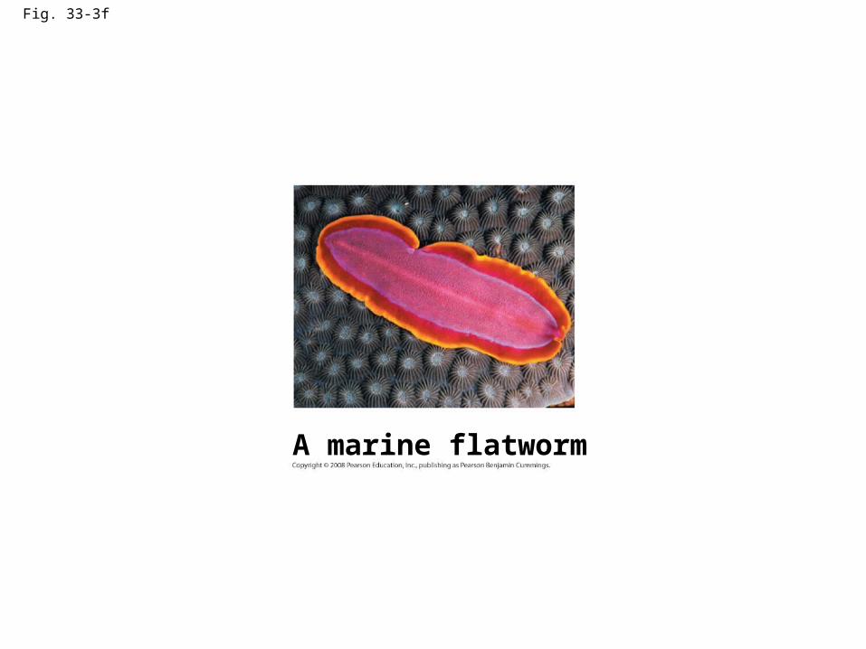
Fig. 33-3f
A marine flatworm
3 classes of Flatworms
• 1. Turbellaria– Free-living– Planarians – pond
• Crossed eyes• Auricles (“ears”) – locate food• Carnivore – mouth, pharynx, GV cavity• Reproduction
– asexual – splits in 2– Sexual – hermaphrodite – cross-fertilization
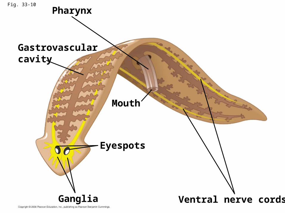
Fig. 33-10
Pharynx
Gastrovascularcavity
Mouth
Eyespots
Ganglia Ventral nerve cords
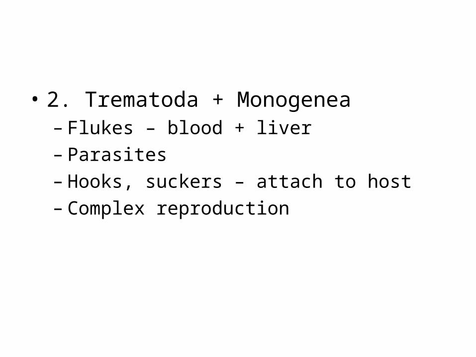
• 2. Trematoda + Monogenea– Flukes – blood + liver– Parasites– Hooks, suckers – attach to host– Complex reproduction
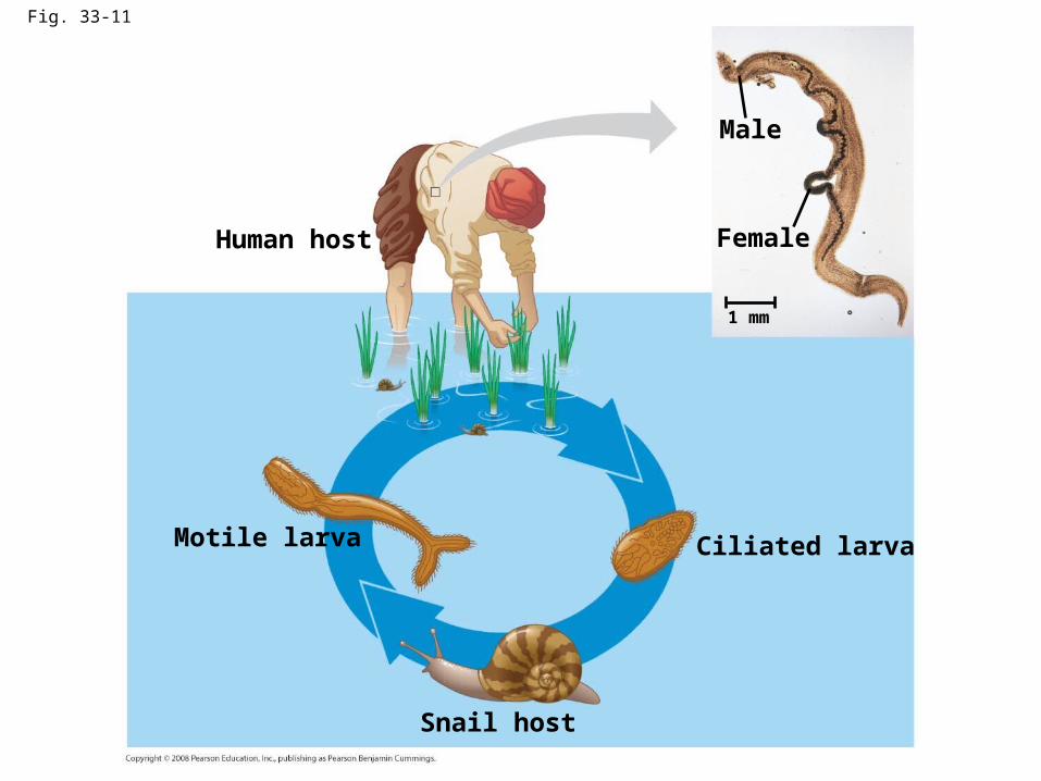
Fig. 33-11
Human host
Motile larva
Snail host
Ciliated larva
Male
Female
1 mm
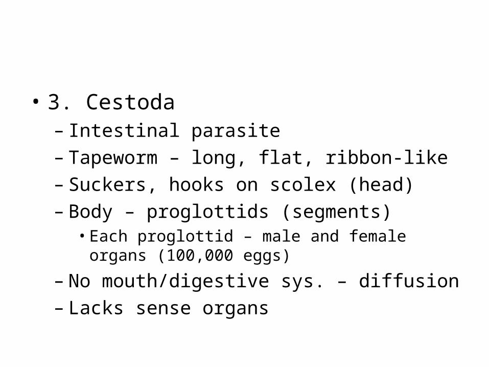
• 3. Cestoda– Intestinal parasite– Tapeworm – long, flat, ribbon-like – Suckers, hooks on scolex (head)– Body – proglottids (segments)
• Each proglottid – male and female organs (100,000 eggs)
– No mouth/digestive sys. – diffusion– Lacks sense organs
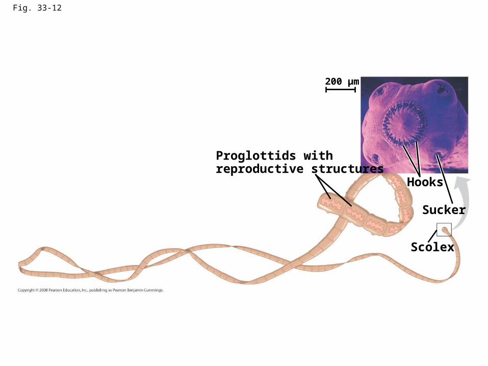
Fig. 33-12
Proglottids withreproductive structures
Hooks
Sucker
Scolex
200 µm