Embed Size (px)
Citation preview

Copyright © 2010 Pearson Education, Inc.
CHAPTER 3: CELLS

Copyright © 2010 Pearson Education, Inc.
CELL THEORY
Copyright © 2010 Pearson Education, Inc.
CELL DIVERSITY

Copyright © 2010 Pearson Education, Inc.
Figure 3.1 Cell diversity.
Fibroblasts
Erythrocytes
Epithelial cells
(d) Cell that
fights disease
Nerve cell
Fat cell
Sperm
(a) Cells that connect body parts,
form linings, or transport gases
(c) Cell that stores
nutrients
(b) Cells that move organs and
body parts
(e) Cell that gathers information
and control body functions
(f) Cell of reproduction
Skeletal
Muscle
cell
Smooth muscle cells
Macrophage

Copyright © 2010 Pearson Education, Inc.
The concept that a cell has a unique shape
which allows it to perform a unique function
is an example of…
1) Homeostasis
2) Complementarity of structure
and function
3) Negative feedback

Copyright © 2010 Pearson Education, Inc.
The smallest unit which has all the properties
of life is…
1) a cell
2) a tissue
3) an organism
4) a protein

Copyright © 2010 Pearson Education, Inc.
GENERALIZED CELL

Copyright © 2010 Pearson Education, Inc.
Figure 3.2 Structure of the generalized cell.
Nucleus
Organelles Cytoskeleton
Cytosol
Cytoplasm
Plasma
membrane

Copyright © 2010 Pearson Education, Inc.
PLASMA MEMBRANE STRUCTURE

Copyright © 2010 Pearson Education, Inc.
Figure 3.3 Structure of the plasma membrane according to the fluid mosaic model.
Integral
proteins
Extracellular fluid
(watery environment)
Cytoplasm
(watery environment)
Polar head of
phospholipid
molecule Glycolipid
Cholesterol
Peripheral
proteins Bimolecular
lipid layer
containing
proteins
Inward-facing
layer of
phospholipids
Outward-
facing
layer of
phospholipids
Carbohydrate
of glycocalyx
Glycoprotein
Filament of cytoskeleton
Nonpolar tail of phospholipid molecule

Copyright © 2010 Pearson Education, Inc.
Figure 3.4a Membrane proteins perform many tasks.
(a) Transport
Signal
Receptor
(b) Receptors for signal transduction

Copyright © 2010 Pearson Education, Inc.
Figure 3.4c Membrane proteins perform many tasks.
(c) Attachment to the
cytoskeleton and
extracellular matrix (ECM)
Enzymes
(d) Enzymatic activity

Copyright © 2010 Pearson Education, Inc.
Figure 3.4e Membrane proteins perform many tasks.
CAMs
(e) Intercellular joining
Glycoprotein
(f) Cell-cell recognition

Copyright © 2010 Pearson Education, Inc.
EXTRACELLULAR JUNCTIONS
Copyright © 2010 Pearson Education, Inc.
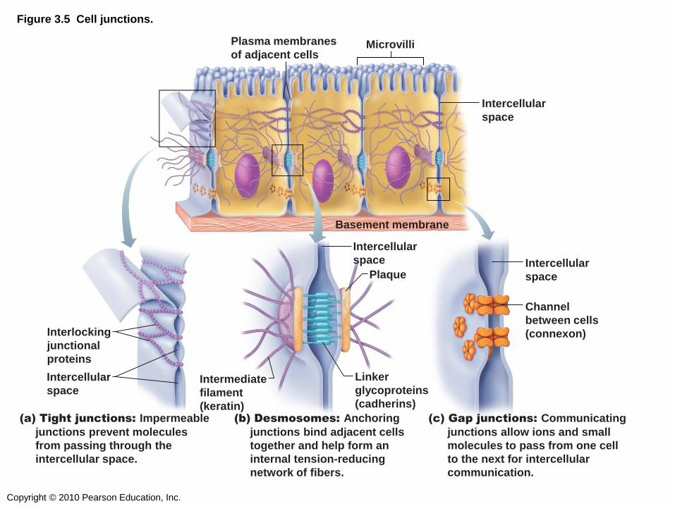
Figure 3.5 Cell junctions.
Interlocking
junctional
proteins
Intercellular
space
Intercellular
space
Plasma membranes
of adjacent cells Microvilli
Intercellular
space
Basement membrane
Plaque
Linker
glycoproteins
(cadherins)
Intermediate
filament
(keratin)
Intercellular
space
Channel
between cells
(connexon)
(a) Tight junctions: Impermeable
junctions prevent molecules
from passing through the
intercellular space.
(b) Desmosomes: Anchoring
junctions bind adjacent cells
together and help form an
internal tension-reducing
network of fibers.
(c) Gap junctions: Communicating
junctions allow ions and small
molecules to pass from one cell
to the next for intercellular
communication.

Copyright © 2010 Pearson Education, Inc.
Phospholipids interact with the intracellular
fluid and extracellular fluid via their…
1) Glycerol
2) Polar head group
3) Hydrophobic tails
4) Cholesterol
5) Proteins
Copyright © 2010 Pearson Education, Inc.
The “hydrophobic core” of the membrane is
formed by which of the following?
1) Glycerol in triglycerides
2) Polar head groups in phospholipids
3) Nonpolar tails in phospholipids
4) Cholesterol
5) Proteins
6) Carbohydrates
7) RNA
Copyright © 2010 Pearson Education, Inc.
Integral proteins (embedded in the membrane)
can have all EXCEPT which of these functions?
1) Joining adjacent cells together
2) Catalyzing reactions
3) Making new proteins
4) Transporting ions into the cell
5) Anchoring the cell in place

Copyright © 2010 Pearson Education, Inc.
PLASMA MEMBRANE TRANSPORT

Copyright © 2010 Pearson Education, Inc.
PASSIVE TRANSPORT:
DIFFUSION & OSMOSIS

Copyright © 2010 Pearson Education, Inc.
Figure 3.6 Diffusion.

Copyright © 2010 Pearson Education, Inc.
Figure 3.7 Diffusion through the plasma membrane.
Extracellular fluid
Lipid-
soluble
solutes
Cytoplasm
Lipid-insoluble
solutes (such as
sugars or amino
acids)
Small lipid-
insoluble
solutes
Water
molecules
Lipid
billayer
Aquaporin
(a) Simple diffusion of
fat-soluble molecules
directly through the
phospholipid bilayer
(b) Carrier-mediated facilitated
diffusion via a protein carrier
specific for one chemical; binding of
substrate causes shape change in
transport protein
(c) Channel-mediated
facilitated diffusion through a channel protein; mostly ions selected on basis of size and charge
(d) Osmosis, diffusion
of a solvent such as
water through a
specific channel protein
(aquaporin) or through
the lipid bilayer
Copyright © 2010 Pearson Education, Inc.
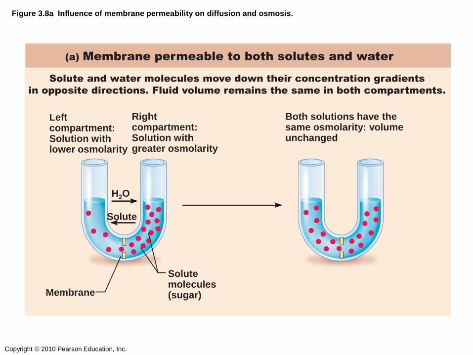
(a) Membrane permeable to both solutes and water
Solute and water molecules move down their concentration gradients
in opposite directions. Fluid volume remains the same in both compartments.
Left compartment: Solution with lower osmolarity
Right compartment: Solution with greater osmolarity
Membrane
H2O
Solute
Solute molecules (sugar)
Both solutions have the same osmolarity: volume unchanged
Figure 3.8a Influence of membrane permeability on diffusion and osmosis.
Copyright © 2010 Pearson Education, Inc.
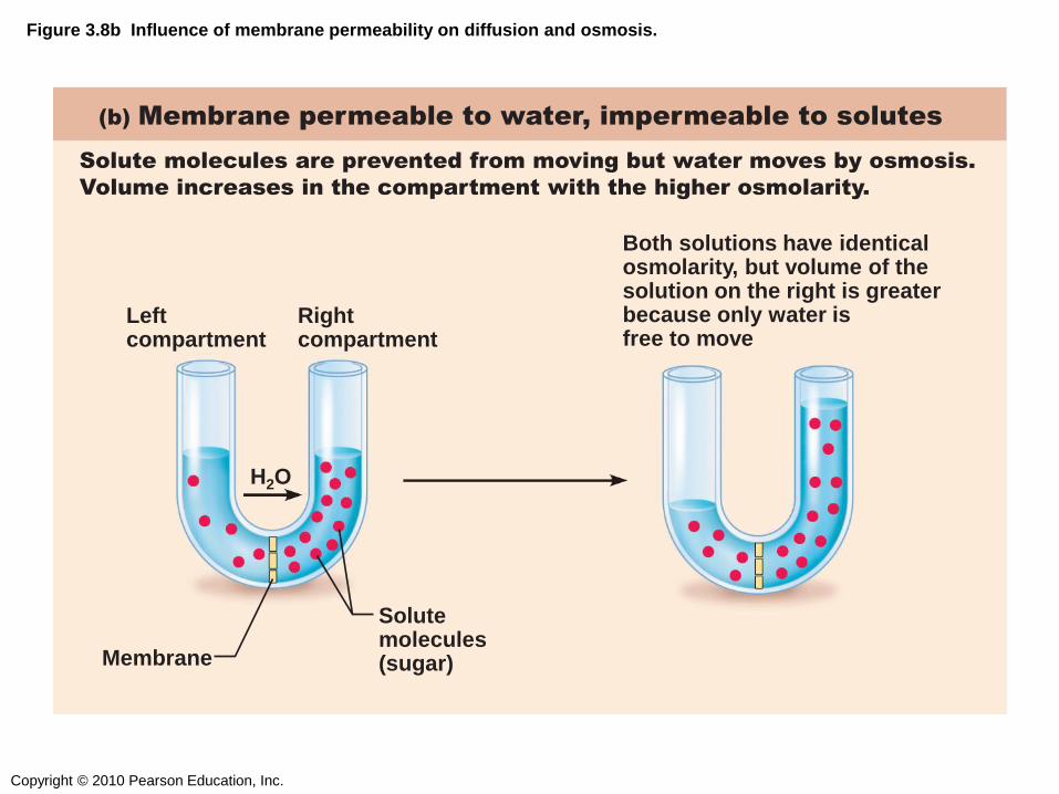
(b) Membrane permeable to water, impermeable to solutes
Both solutions have identical osmolarity, but volume of the solution on the right is greater because only water is free to move
Solute molecules are prevented from moving but water moves by osmosis.
Volume increases in the compartment with the higher osmolarity.
Left compartment
Right compartment
Membrane
Solute molecules (sugar)
H2O
Figure 3.8b Influence of membrane permeability on diffusion and osmosis.

Copyright © 2010 Pearson Education, Inc.
Figure 3.9 The effect of solutions of varying tonicities on living red blood cells.
Cells retain their normal size and
shape in isotonic solutions (same solute/water concentration as inside
cells; water moves in and out).
Cells lose water by osmosis and
shrink in a hypertonic solution
(contains a higher concentration
of solutes than are present inside
the cells).
(a) Isotonic solutions (b) Hypertonic solutions (c) Hypotonic solutions
Cells take on water by osmosis until
they become bloated and burst (lyse) in a hypotonic solution (contains a
lower concentration of solutes than are present in cells).
Copyright © 2010 Pearson Education, Inc.
ACTIVE TRANSPORT:
PUMPS

Copyright © 2010 Pearson Education, Inc.
Figure 3.10 Primary Active Transport: The Na+-K+ Pump
Extracellular fluid
6 K+ is released from the pump protein
and Na+ sites are ready to bind Na+ again.
The cycle repeats.
2 Binding of Na+ promotes
phosphorylation of the protein by ATP.
1 Cytoplasmic Na+ binds to pump protein.
Na+
K+ released
ATP-binding site Na+ bound
Cytoplasm
ATP ADP
P
K+
5 K+ binding triggers release of the
phosphate. Pump protein returns to its
original conformation.
3 Phosphorylation causes the protein to
change shape, expelling Na+ to the outside.
4 Extracellular K+ binds to pump protein.
Na+ released
P
K+
P
Pi
Na+–K+ pump
K+ bound
McGraw-Hill Animation

Copyright © 2010 Pearson Education, Inc.
Figure 3.11 Secondary active transport.
1 2 The ATP-driven Na+-K+ pump
stores energy by creating a
steep concentration gradient for
Na+ entry into the cell.
As Na+ diffuses back across the
membrane through a membrane
cotransporter protein, it drives glucose
against its concentration gradient
into the cell. (ECF = extracellular fluid)
Na+-glucose
symport
transporter
loading
glucose from
ECF
Na+-glucose
symport transporter
releasing glucose
into the cytoplasm
Glucose
Na+-K+
pump
Cytoplasm
Extracellular fluid

Copyright © 2010 Pearson Education, Inc.
Which of the following is an active process?
1) Oxygen moving from blood into cells
2) Glucose moving into cells from plasma
3) Carbon dioxide moving from cells to blood
4) Moving sodium ions out of the cell

Copyright © 2010 Pearson Education, Inc.
VESICULAR TRANSPORT
Copyright © 2010 Pearson Education, Inc.
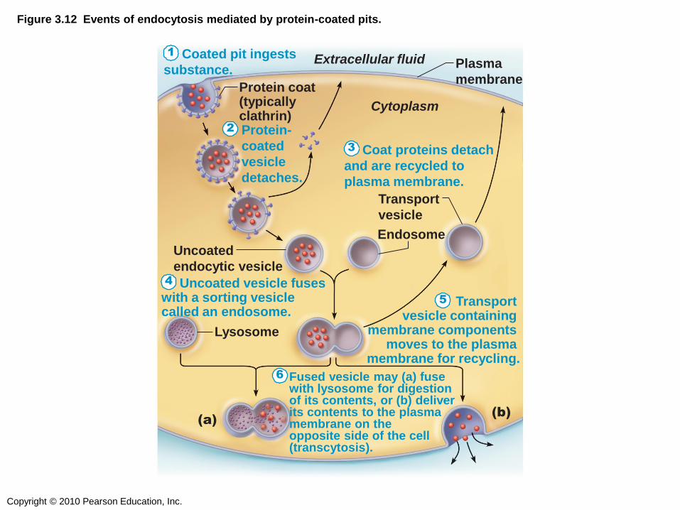
Figure 3.12 Events of endocytosis mediated by protein-coated pits.
Coated pit ingests
substance.
Protein-
coated
vesicle
detaches.
Coat proteins detach
and are recycled to
plasma membrane.
Uncoated vesicle fuses with a sorting vesicle called an endosome.
Transport vesicle containing
membrane components moves to the plasma
membrane for recycling.
Fused vesicle may (a) fuse with lysosome for digestion of its contents, or (b) deliver its contents to the plasma membrane on the opposite side of the cell (transcytosis).
Protein coat (typically clathrin)
Extracellular fluid Plasma
membrane
Endosome
Lysosome
Transport
vesicle
(b) (a)
Uncoated
endocytic vesicle
Cytoplasm
1
2
3
4
5
6

Copyright © 2010 Pearson Education, Inc.
Phagosome
(a) Phagocytosis
Figure 3.13a Comparison of three types of endocytosis.
(b) Pinocytosis
(c) Receptor-mediated
endocytosis

Copyright © 2010 Pearson Education, Inc.
Figure 3.14b Exocytosis.
Copyright © 2010 Pearson Education, Inc.
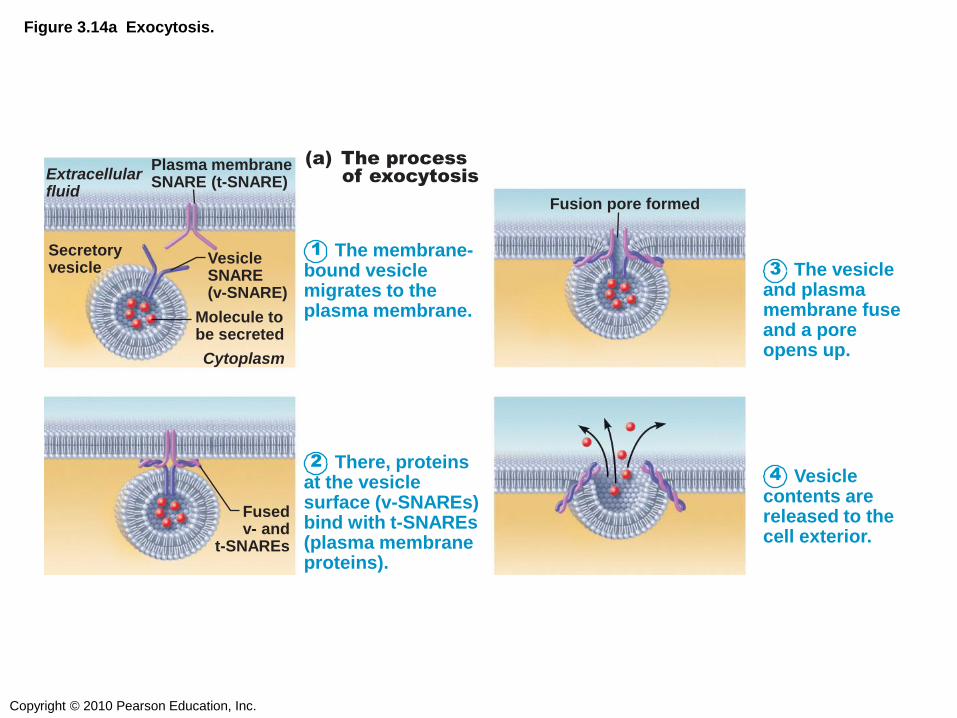
Figure 3.14a Exocytosis.
1 The membrane- bound vesicle migrates to the plasma membrane.
2 There, proteins at the vesicle surface (v-SNAREs) bind with t-SNAREs (plasma membrane proteins).
(a) The process
of exocytosis Extracellular fluid
Plasma membrane SNARE (t-SNARE)
Secretory vesicle
Vesicle SNARE (v-SNARE)
Molecule to be secreted
Cytoplasm
Fused v- and
t-SNAREs
3 The vesicle and plasma membrane fuse and a pore opens up.
4 Vesicle contents are released to the cell exterior.
Fusion pore formed

Copyright © 2010 Pearson Education, Inc.
Which of the following occurs when an immune
cell “eats” pieces of foreign material?
1) Receptor-mediated endocytosis
2) Phagocytosis
3) Endocytosis
4) Exocytosis

Copyright © 2010 Pearson Education, Inc.
CYTOPLASM

Copyright © 2010 Pearson Education, Inc.
Figure 3.2 Structure of the generalized cell.
Secretion being released from cell by exocytosis
Peroxisome
Ribosomes
Rough endoplasmic reticulum
Nucleus
Nuclear envelope Chromatin
Golgi apparatus
Nucleolus
Smooth endoplasmic reticulum
Cytosol
Lysosome
Mitochondrion
Centrioles
Centrosome matrix
Cytoskeletal elements • Microtubule • Intermediate filaments
Plasma membrane

Copyright © 2010 Pearson Education, Inc.
ORGANELLES

Copyright © 2010 Pearson Education, Inc.
Figure 3.17 Mitochondrion.
Enzymes
Matrix
Cristae
Mitochondrial DNA
Ribosome
Outer mitochondrial membrane
Inner mitochondrial membrane
(b)
(a)
(c)

Copyright © 2010 Pearson Education, Inc.
Figure 3.18 The endoplasmic reticulum.
Nuclear
envelope
Ribosomes
Rough ER
Smooth ER
(a) Diagrammatic view of smooth
and rough ER (b) Electron micrograph of
smooth and rough ER
(10,000x)

Copyright © 2010 Pearson Education, Inc.
Figure 3.19 Golgi apparatus.
Cis face— “receiving” side of Golgi apparatus
Secretory
vesicle
(a) Many vesicles in the process of pinching
off from the membranous Golgi apparatus.
(b) Electron micrograph of the Golgi
apparatus (90,000x)
Transport vesicle from the Golgi apparatus
Transport vesicle from trans face
Trans face— “shipping” side of Golgi apparatus
New vesicles forming
New vesicles forming
Cisternae
Transport vesicle from rough ER
Golgi apparatus

Copyright © 2010 Pearson Education, Inc.
Figure 3.20 The sequence of events from protein synthesis on the rough ER to the final distribution of those proteins.
Protein- containing vesicles pinch off rough ER and migrate to fuse with membranes of Golgi apparatus.
Proteins are modified within the Golgi compartments.
Proteins are then packaged within different vesicle types, depending on their ultimate destination.
Plasma mem- brane
Secretion by exocytosis
Vesicle becomes lysosome
Golgi
apparatus
Rough ER ER membrane
Phagosome
Proteins in cisterna
Pathway B:
Vesicle membrane
to be incorporated into plasma
membrane Pathway A:
Vesicle contents destined for exocytosis Extracellular fluid
Secretory
vesicle
Pathway C:
Lysosome containing
acid hydrolase enzymes
1
3
2

Copyright © 2010 Pearson Education, Inc.
Figure 3.21 Electron micrograph of a cell containing lysosomes (12,000x).
Lysosomes
Light areas are regions where
materials are being digested.

Copyright © 2010 Pearson Education, Inc.
What is the function of the Golgi apparatus?
1) To make proteins
2) To convert glucose into ATP
3) To finish, package, and sort proteins
4) To fold proteins

Copyright © 2010 Pearson Education, Inc.
CYTOSKELETON

Copyright © 2010 Pearson Education, Inc.
Figure 3.23 Cytoskeletal elements support the cell and help to generate movement.
Strands made of spherical protein
subunits called actins
Tough, insoluble protein fibers
constructed like woven ropes
(a) Microfilaments (b) Intermediate filaments (c) Microtubules
Hollow tubes of spherical protein
subunits called tubulins
Actin subunit
7 nm 10 nm 25 nm
Fibrous subunits
Tubulin subunits
Microfilaments form the blue network surrounding the pink nucleus in this photo.
Intermediate filaments form the purple batlike network in this photo.
Microtubules appear as gold networks surrounding the cells’ pink nuclei in this photo.
Copyright © 2010 Pearson Education, Inc.
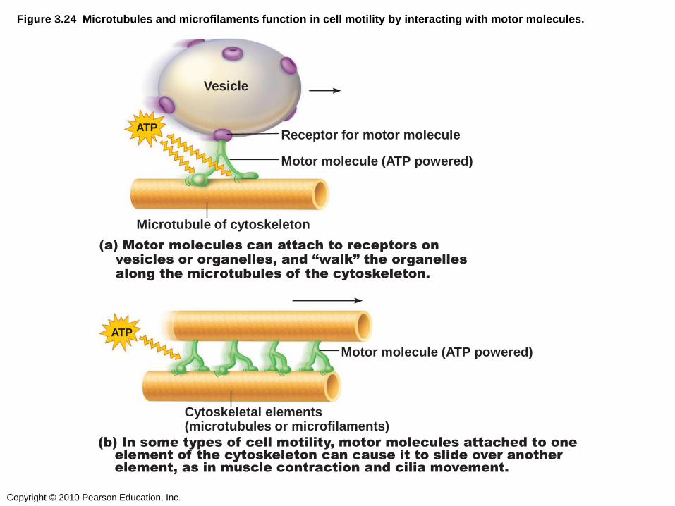
Figure 3.24 Microtubules and microfilaments function in cell motility by interacting with motor molecules.
Cytoskeletal elements (microtubules or microfilaments)
Motor molecule (ATP powered)
ATP
(b) In some types of cell motility, motor molecules attached to one
element of the cytoskeleton can cause it to slide over another element, as in muscle contraction and cilia movement.
ATP
Vesicle
(a) Motor molecules can attach to receptors on
vesicles or organelles, and “walk” the organelles
along the microtubules of the cytoskeleton.
Motor molecule (ATP powered)
Microtubule of cytoskeleton
Receptor for motor molecule
Copyright © 2010 Pearson Education, Inc.
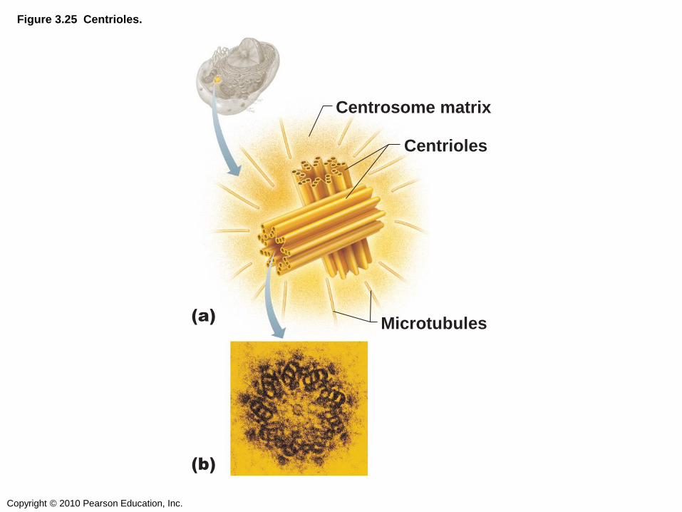
Figure 3.25 Centrioles.
Centrosome matrix
(b)
(a)
Centrioles
Microtubules

Copyright © 2010 Pearson Education, Inc.
CELLULAR EXTENSIONS

Copyright © 2010 Pearson Education, Inc.
Figure 3.26 Structure of a cilium.
Plasma
membrane
Outer microtubule
doublet
Dynein arms
Central
microtubule
Radial spoke
Radial spoke
TEM
TEM
Triplet
Basal body
(centriole)
Cilium
Microtubules
Plasma
membrane
Basal body
Cross-linking
proteins inside
outer doublets
Cross-linking
proteins inside
outer doublets
A longitudinal section of a cilium shows microtubules running the length of the structure.
The doublets also have attached motor proteins, the dynein arms.
The outer microtubule doublets and the two central microtubules are held together by cross-linking proteins and radial spokes.
A cross section through the basal body. The nine outer doublets of a cilium extend into a basal body where each doublet joins another microtubule to form a ring of nine triplets.
A cross section through the cilium shows the “9 + 2” arrangement of microtubules.
TEM

Copyright © 2010 Pearson Education, Inc.
Figure 3.28 Microvilli.
Microvillus
Actin
filaments
Terminal
web

Copyright © 2010 Pearson Education, Inc.
Which of these is NOT a function
of microtubules?
1) To help cilia move
2) To help chromosomes move during mitosis
3) To help carry cargo around the cell
4) To make muscle cells contract

Copyright © 2010 Pearson Education, Inc.
THE NUCLEUS
Copyright © 2010 Pearson Education, Inc.
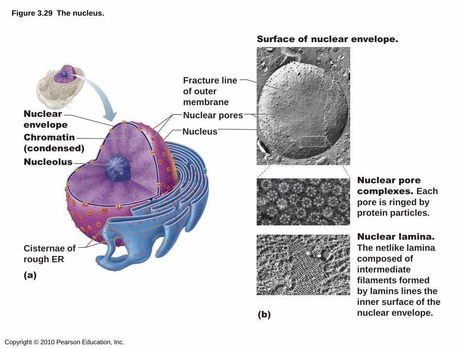
Figure 3.29 The nucleus.
Chromatin
(condensed)
Nuclear
envelope Nucleus
Nuclear pores
Fracture line
of outer
membrane
Nuclear pore
complexes. Each
pore is ringed by protein particles.
Surface of nuclear envelope.
Nuclear lamina.
The netlike lamina
composed of
intermediate
filaments formed
by lamins lines the
inner surface of the
nuclear envelope.
Nucleolus
Cisternae of
rough ER
(a)
(b)
Copyright © 2010 Pearson Education, Inc.
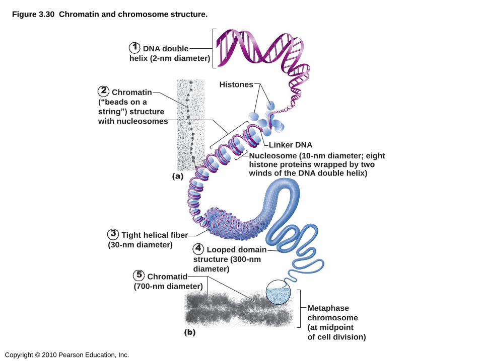
Figure 3.30 Chromatin and chromosome structure.
Metaphase
chromosome
(at midpoint
of cell division)
Nucleosome (10-nm diameter; eight histone proteins wrapped by two winds of the DNA double helix)
Linker DNA
Histones
(a)
(b)
1 DNA double
helix (2-nm diameter)
2 Chromatin
(“beads on a
string”) structure
with nucleosomes
3 Tight helical fiber
(30-nm diameter)
5 Chromatid
(700-nm diameter)
4 Looped domain
structure (300-nm
diameter)

Copyright © 2010 Pearson Education, Inc.
The “beads” on a string of chromatin, made
f DNA and histone proteins, are…
1) linkers
2) chromosomes
3) chromatids
4) nucleosomes

Copyright © 2010 Pearson Education, Inc.
CELL GROWTH & REPRODUCTION
Copyright © 2010 Pearson Education, Inc.
CELL CYCLE

Copyright © 2010 Pearson Education, Inc.
Figure 3.31 The cell cycle.
G1
Growth
S
Growth and DNA
synthesis G2
Growth and final
preparations for
division M
G2 checkpoint
G1 checkpoint
(restriction point)

Copyright © 2010 Pearson Education, Inc.
MITOSIS

Copyright © 2010 Pearson Education, Inc.
Figure 3.33 Mitosis (1 of 2)
Centrosomes
(each has 2
centrioles)
Early
mitotic
spindle
Spindle pole
Kinetochore Kinetochore
microtubule
Polar microtubule
Nucleolus
Interphase Early Prophase Late Prophase
Centromere
Plasma
membrane Fragments
of nuclear
envelope Aster
Nuclear
envelope
Chromosome
consisting of two
sister chromatids
Chromatin
Interphase Early Prophase Late Prophase

Copyright © 2010 Pearson Education, Inc.
Figure 3.33 Mitosis (2 of 2)
Contractile
ring at
cleavage
furrow
Nuclear
envelope
forming
Nucleolus forming
Spindle
Metaphase
plate
Metaphase Anaphase Telophase
Daughter
chromosomes
Metaphase Anaphase Telophase and
Cytokinesis

Copyright © 2010 Pearson Education, Inc.
When DNA is folded into relatively loose, long
strands during interphase, this is called…
1) chromatin
2) chromosomes
3) centromeres
4) nucleosomes

Copyright © 2010 Pearson Education, Inc.
Sister chromatids are separated into two
identical distinct chromosomes during…
1) anaphase
2) metaphase
3) prophase
4) telophase

Copyright © 2010 Pearson Education, Inc.
PROTEIN SYNTHESIS:
THE CENTRAL DOGMA
Copyright © 2010 Pearson Education, Inc.
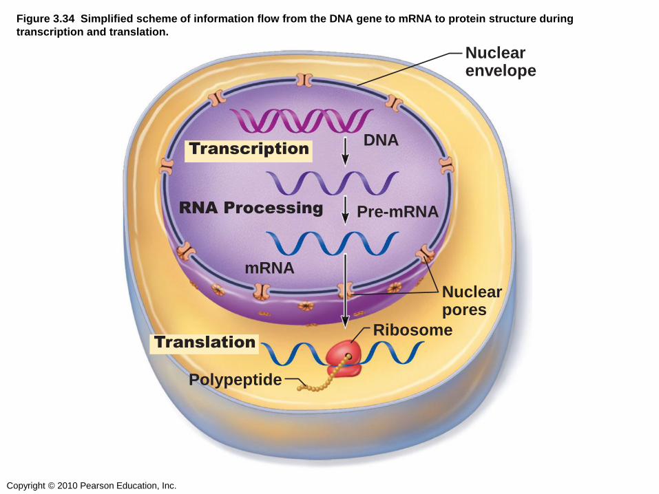
Figure 3.34 Simplified scheme of information flow from the DNA gene to mRNA to protein structure during
transcription and translation.
Nuclear pores
mRNA
Pre-mRNA RNA Processing
Transcription
Translation
DNA
Nuclear envelope
Ribosome
Polypeptide

Copyright © 2010 Pearson Education, Inc.
EXTRACELLULAR FLUID &
THE EXTRACELLULAR MATRIX

Copyright © 2010 Pearson Education, Inc.
DEVELOPMENTAL ASPECTS OF CELLS
Copyright © 2010 Pearson Education, Inc.
The fluid and proteins surrounding a cell are
called the …
1) Intracellular fluid
2) Extracellular fluid
3) Extracellular matrix
4) Intercellular matrix
Copyright © 2010 Pearson Education, Inc.
A cell which retains the ability to divide over
and over throughout life is called a …
1) Stem cell
2) Mature cell
3) Differentiated cell
4) Embryonic cell