Embed Size (px)
Citation preview


Catechol Pyrazolinones as Trypanocidals:
Fragment-Based Design, Synthesis and
Pharmacological Evaluation of Nanomolar
Inhibitors of Trypanosomal Phosphodiesterase B1
Chapter 3
Kristina M Orrling, Chimed Jansen, Xuan Lan Vu, Vreni Balmer, Patrick Bregy, Anitha
Shanmugham, Paul England, Paul Cos, Louis Maes, Emily Adams, Erika van den
Bogaart, Eric Chatelain, Jean-Robert Ioset, Andrea van de Stolpe, Stèphanie Zorg, Johan
Veerman, Thomas Seebeck, Geert Jan Sterk, Iwan de Esch, Rob Leurs
Published as:
Orrling, et al, J. Med. Chem. 2012, 55 (20), 8745-8756

86
87
CH
AP
TE
R 3
Abstract
Trypanosomal phosphodiesterases B1 and B2 (TbrPDEB1 and TbrPDEB2) play an
important role in the life cycle of Trypanosoma brucei, the causative parasite of human
African trypanosomiasis (HAT), also known as African sleeping sickness. We used
homology modelling and docking studies to guide fragment growing into the parasite-
specific P-pocket in the enzyme binding site. The resulting catechol pyrazolinones act as
potent TbrPDEB1 inhibitors with IC50 values down to 49 nM. The compounds also block
parasite proliferation (e.g. VUF13525 (20b): T. brucei rhodesiense IC50 = 60 nM, T. brucei
brucei IC50 = 520 nM, T. cruzi = 7.6 µM), inducing a typical multiple nuclei- and kinetoplast
phenotype without being generally cytotoxic. The mode of action of 20b was investigated
with recombinantly engineered trypanosomes expressing a cAMP-sensitive FRET sensor,
confirming a dose-response related increase of intracellular cAMP levels in trypanosomes.
Our findings further validate the TbrPDEB family as antitrypanosomal target.
88
3.1 Introduction
Human African trypanosomiasis (HAT), commonly known as African sleeping sickness, is
an extremely deadly disease; without treatment, all patients entering second stage HAT
following the spread of the parasite to the CNS will eventually die.1 Millions of people are
living in the endemic areas in rural central Africa, where poverty and poor access to accurate
healthcare hamper the chances of survival of infected patients.2, 3 Even if the number of
reported cases has dropped to its lowest level in 50 years, epidemics, increased political
turbulence, and resistance emergence can quickly alter this trend.3, 4 HAT is caused by the
kinetoplastid protozoa Trypanosoma brucei and is transmitted by infected tsetse flies
(Glossina ssp.). Two subspecies of T. brucei are infectious to humans, T. b. gambiense and
T. b. rhodesiense. The course of the disease, symptoms and appropriate treatment depend
on the etiological subspecies.2 Despite the high medical need, current treatment options are
limited and suffer from suboptimal dosage regimens and/or severe toxicity.2 During the
second stage of the disease, the only choices are the arsenic-containing drug melarsoprol
and the ornithine decarboxylase inhibitor eflornithine, used as monotherapy or in
combination with nifurtimox (NECT).5 However, eflornithine is not effective against T. b.
rhodesiense. Consequently, public-private initiatives such as the Drugs for Neglected
Disease initiative investigate new approaches to target HAT.6
The mapping of the T. brucei genome revealed the presence of five trypanosomal cyclic
nucleotide phosphodiesterases (PDEs).7 These enzymes play an important role in the
regulation of cyclic nucleotide levels by catalysing the hydrolysis of the phosphodiester
bond of cAMP and/or cGMP. In humans, 11 main families of PDEs can be distinguished,
and several of the human PDE enzymes have been extensively explored as molecular targets
for a diverse set of diseases conditions, e.g. COPD, erectile dysfunction, pulmonary arterial
hypertension, asthma, psoriasis, type 2 diabetes, depression, cognitive disorders, and
psychosis. Although the exact role of cAMP in the lifecycle of the trypanosomes is not fully
understood,8 two trypanosomal PDEs have been found to play a pivotal role in T. brucei
proliferation.9, 10 Knocking down of both TbrPDEB1 and TbrPDEB2 simultaneously leads
89
CH
AP
TE
R 3
to an arrest of parasite cell division, lysis of the parasites, and to elimination of the infection
in vivo in the infected mouse model.11 Additional studies on a hit from a high throughput
screening campaign, PPS54019 (1), pharmacologically validated TbrPDEB1 and
TbrPDEB2 as anti-trypanosomal targets.12 In fact, 1 was once synthesised as potent human
PDE4 inhibitor.13 The benefits of extracting acquired knowledge from very advanced drug
discovery programs targeting human proteins for use in neglected diseases drug discovery
cannot be overestimated. Scarcity in the drug research pipeline can thus be compensated
with better pharmacological predictability and an early understanding of possible adverse
effects. A recent investigation shows that the hPDE4 inhibitor piclamilast (2) and analogues
also inhibit the TbrPDEB1 and TbrPDEB2 (Figure 1).12, 14 Inhibition of TbrPDEB1 has
furthermore been observed with analogues of tadalafil (3)15 and sildenafil (4),16 both hPDE5
inhibitors, however at high micromolar concentrations.
Figure 1. Known TbrPDE inhibitors, 1,12 2,14 3,15 and 416.
90
These findings indicate that in the area of neglected infectious diseases, basing the drug
discovery efforts on lead compounds originally developed for human PDE targets can be
quite a successful strategy. Such an approach proved not only successful in the discovery
of the first TbrPDEB1 inhibitors 1–4, but also the recent discovery of antiplasmodial
compounds based on the PDE5 inhibitor tadalafil.17
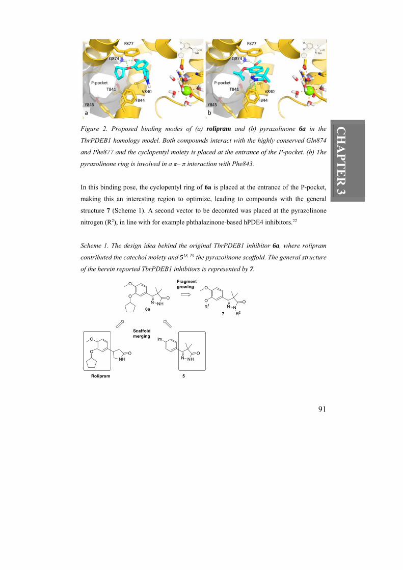
Following up on chemotypes related to HTS hit 1 catechol pyrazolinone 6a (TbrPDEB1
inhibition with IC50 = 12 µM) was discovered. This fragment contains several structural
features of known human PDE inhibitors and can be considered a merger of the known
PDE4 inhibitor rolipram (i.e., catechol moiety connected to a cyclopentyl group, also
present in 2) and of published cardiotonic hPDE3 inhibitors 518, 19 and hPDE4 inhibitors in
the patent literature20 (i.e., the pyrazolinone moiety) as outlined in Scheme 1. Following
these fragment-based drug design considerations, we investigated the putative binding
mode of these compounds by constructing a homology model of TbrPDEB1 based on the
x-ray structure (PDB id 2R8Q)21 of the Leishmania major orthologue LmjPDEB1 (66%
sequence identity). Interestingly, the so-called P-pocket, a unique sub-pocket that extends
from the invariant glutamine (Gln874) through the protein to the solvent in the ligand-
binding site of the LmjPDEB1 proved to be conserved in our model of TbrPDEB1.
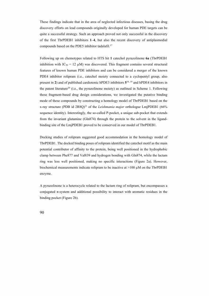
Docking studies of rolipram suggested good accommodation in the homology model of
TbrPDEB1. The docked binding poses of rolipram identified the catechol motif as the main
potential contributor of affinity to the protein, being well positioned in the hydrophobic
clamp between Phe877 and Val839 and hydrogen bonding with Gln874, while the lactam
ring was less well positioned, making no specific interactions (Figure 2a). However,
biochemical measurements indicate rolipram to be inactive at >100 µM on the TbrPDEB1
enzyme.
A pyrazolinone is a heterocycle related to the lactam ring of rolipram, but encompasses a
conjugated π-system and additional possibility to interact with aromatic residues in the
binding pocket (Figure 2b).
91
CH
AP
TE
R 3
Figure 2. Proposed binding modes of (a) rolipram and (b) pyrazolinone 6a in the
TbrPDEB1 homology model. Both compounds interact with the highly conserved Gln874
and Phe877 and the cyclopentyl moiety is placed at the entrance of the P-pocket. (b) The
pyrazolinone ring is involved in a π– π interaction with Phe843.
In this binding pose, the cyclopentyl ring of 6a is placed at the entrance of the P-pocket,
making this an interesting region to optimize, leading to compounds with the general
structure 7 (Scheme 1). A second vector to be decorated was placed at the pyrazolinone
nitrogen (R2), in line with for example phthalazinone-based hPDE4 inhibitors.22
Scheme 1. The design idea behind the original TbrPDEB1 inhibitor 6a, where rolipram
contributed the catechol moiety and 518, 19 the pyrazolinone scaffold. The general structure
of the herein reported TbrPDEB1 inhibitors is represented by 7.
O
O
NHO
Im
N NHO
O
N NHOO
O
N NOO
R1
R27
Rolipram 5
6a
Fragmentgrowing
Scaffoldmerging
92
3.2 Results and Discussion
Homology model of TbrPDEB1. The homology model of TbrPDEB1 was constructed
based on the TbrPDEB1 sequence published by Zoraghi and Seebeck (Uniprot: Q8WQX9)9
and the structure of LmjPDB1 (2R8Q),21 the only orthologue of TbrPDEB1 crystallized to
date. The B chain of the LmjPDEB1 structure was used to construct the homology model,
since the B-factors in the region of the catalytic site is lower in the B chain. The residues
on the surface of the catalytic site show a very high sequence homology with only one
V836I substitution. The P-pocket region has two substitutions N881M and R885K, both on
the far side of the P-pocket from the invariant glutamine (Gln874). The overall sequence
identity of the catalytic domains is 66%.
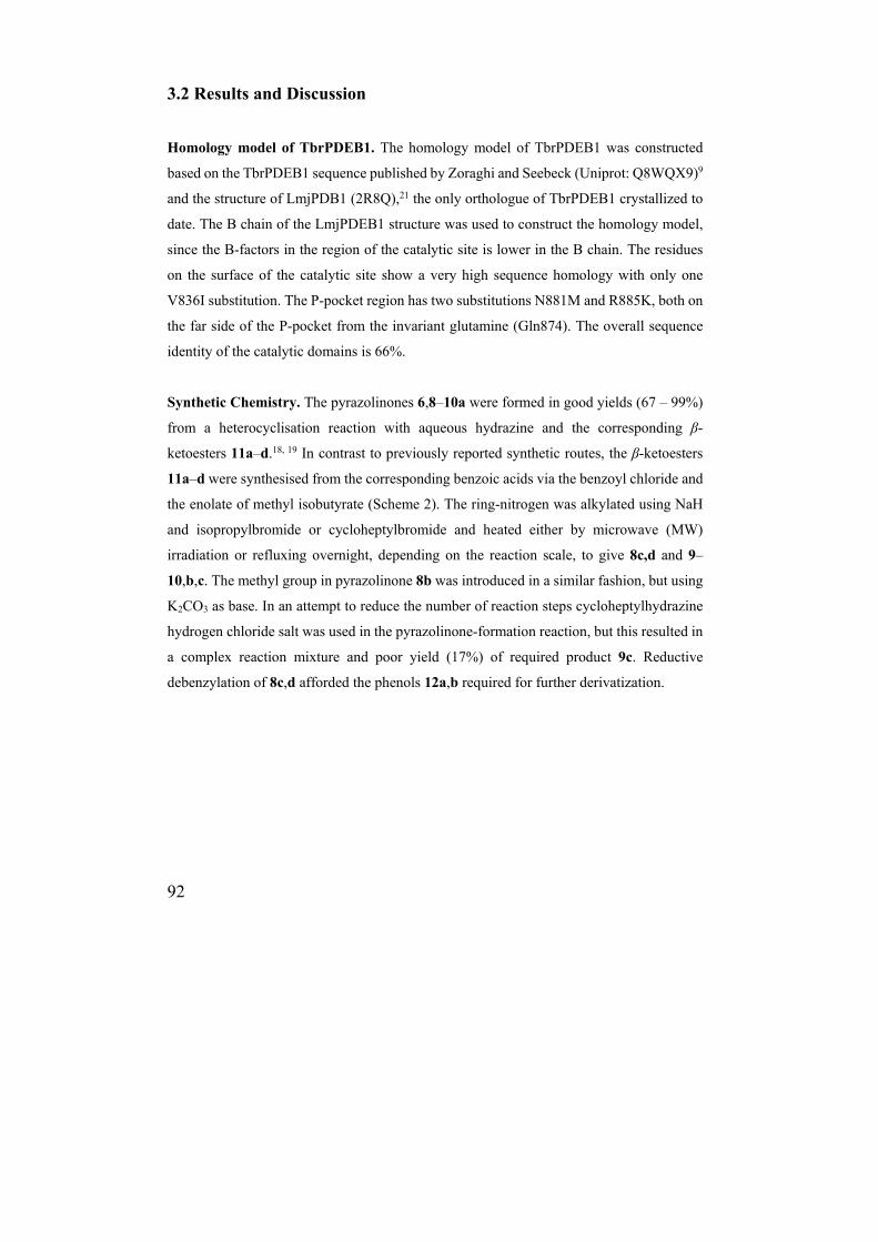
Synthetic Chemistry. The pyrazolinones 6,8–10a were formed in good yields (67 – 99%)
from a heterocyclisation reaction with aqueous hydrazine and the corresponding β-
ketoesters 11a–d.18, 19 In contrast to previously reported synthetic routes, the β-ketoesters
11a–d were synthesised from the corresponding benzoic acids via the benzoyl chloride and
the enolate of methyl isobutyrate (Scheme 2). The ring-nitrogen was alkylated using NaH
and isopropylbromide or cycloheptylbromide and heated either by microwave (MW)
irradiation or refluxing overnight, depending on the reaction scale, to give 8c,d and 9–
10,b,c. The methyl group in pyrazolinone 8b was introduced in a similar fashion, but using
K2CO3 as base. In an attempt to reduce the number of reaction steps cycloheptylhydrazine
hydrogen chloride salt was used in the pyrazolinone-formation reaction, but this resulted in
a complex reaction mixture and poor yield (17%) of required product 9c. Reductive
debenzylation of 8c,d afforded the phenols 12a,b required for further derivatization.
93
CH
AP
TE
R 3
Scheme 2. Synthetic route to pyrazolinonesa.
a Reagents: (a) (COCl)2, DMF, CH2Cl2, 1-18 h or SOCl2, rt, 1-2 h; (b) methyl isobutanoate,
LDA, THF, –45 °C, 30 min, then added to acyl chloride, –55 °C to rt, 1–18 h; (c) N2H4,
EtOH, reflux, 18 h; (d) 8b: K2CO3, DMF, MW (145 °C, 20 min), then MeI, MW (145 °C, 20
min); 8c,d, 9–10b,c: R2Br, NaH, DMF/MeCN, MW (120 °C, 40 min); (e) Pd/C, NH4CHO2,
MeOH:CH2Cl2 (2:1), 40 °C, 18 h.
A diverse set of benzyl derivatives was introduced on the phenols 12a,b with either of two
MW-assisted nucleophilic substitution protocols to afford inhibitors 13–20a,b (Scheme 3,
Table 2) using substituted bromides or chlorides, where chlorides normally needed longer
reaction times for full conversion. For the transformation of nitriles 19a,b into the
corresponding tetrazoles 20a,b a novel expedient MW-assisted protocol was developed
(Scheme 3). Compared to conventional refluxing overnight, the yield was improved from
19% to 62–75% and reaction time was reduced to one hour.
94
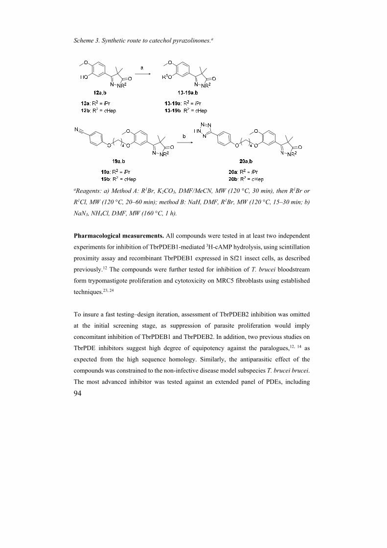
Scheme 3. Synthetic route to catechol pyrazolinones.a
aReagents: a) Method A: R1Br, K2CO3, DMF/MeCN, MW (120 °C, 30 min), then R1Br or
R1Cl, MW (120 °C, 20–60 min); method B: NaH, DMF, R1Br, MW (120 °C, 15–30 min; b)
NaN3, NH4Cl, DMF, MW (160 °C, 1 h).
Pharmacological measurements. All compounds were tested in at least two independent
experiments for inhibition of TbrPDEB1-mediated 3H-cAMP hydrolysis, using scintillation
proximity assay and recombinant TbrPDEB1 expressed in Sf21 insect cells, as described
previously.12 The compounds were further tested for inhibition of T. brucei bloodstream
form trypomastigote proliferation and cytotoxicity on MRC5 fibroblasts using established
techniques.23, 24
To insure a fast testing–design iteration, assessment of TbrPDEB2 inhibition was omitted
at the initial screening stage, as suppression of parasite proliferation would imply
concomitant inhibition of TbrPDEB1 and TbrPDEB2. In addition, two previous studies on
TbrPDE inhibitors suggest high degree of equipotency against the paralogues,12, 14 as
expected from the high sequence homology. Similarly, the antiparasitic effect of the
compounds was constrained to the non-infective disease model subspecies T. brucei brucei.
The most advanced inhibitor was tested against an extended panel of PDEs, including
95
CH
AP
TE
R 3
TbrPDEB2, and trypanosome species. The inhibition of the isolated catalytic domain of
TbrPDEB1 was determined using the PDELight™ HTS cAMP phosphodiesterase kit.25, 26
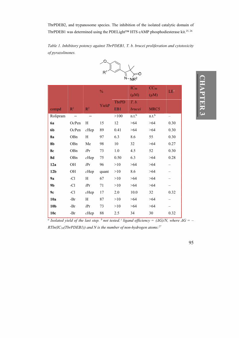
Table 1. Inhibitory potency against TbrPDEB1, T. b. brucei proliferation and cytotoxicity
of pyrazolinones.
% IC50
(µM)
CC50
(µM) LEc
compd R1 R2 Yielda
TbrPD
EB1
T. b.
brucei MRC5
Rolipram >100 n.t.b n.t.b –
6a OcPen H 15 12 >64 >64 0.30
6b OcPen cHep 89 0.41 >64 >64 0.30
8a OBn H 97 6.3 8.6 55 0.30
8b OBn Me 98 10 32 >64 0.27
8c OBn iPr 73 1.0 4.5 52 0.30
8d OBn cHep 75 0.50 6.3 >64 0.28
12a OH iPr 96 >10 >64 >64 –
12b OH cHep quant >10 8.6 >64 –
9a -Cl H 67 >10 >64 >64 –
9b -Cl iPr 71 >10 >64 >64 –
9c -Cl cHep 17 2.0 10.0 32 0.32
10a -Br H 87 >10 >64 >64 –
10b -Br iPr 73 >10 >64 >64 –
10c -Br cHep 88 2.5 34 30 0.32
a Isolated yield of the last step. b not tested. c ligand efficiency = (ΔG)/N, where ΔG = –
RTln(IC50(TbrPDEB1)) and N is the number of non-hydrogen atoms.27
96
3.3 Fragment growing.
Compared to hit structure 6a, the benzyl analogue 8a was slightly more potent (IC50 = 6.3
µM, Table 1), possibly attributed to a π – π interaction between the phenyl ring of 8a and
Phe843 in the binding pocket. By introducing aliphatic substituents (R2) on the nitrogen
atom in the pyrazolinone ring the potency could be further increased in a size-dependent
manner (R2 = H ≈ Me < iPr < cHep) and submicromolar activities were observed for the
cycloheptyl derivatives (e.g. 8d IC50 = 0.50 µM). According to our docking studies, this
gain appears to result from the hydrophobic interaction with Phe843 and slightly shifted
positioning of the pyrazolinone ring strengthening the π – π interactions with Phe843
(Figure 3a). Removal of the catechol ether (12a,b) or replacing it with a chloride or bromide
(9,10a–c) was not tolerated, although measurable potencies were observed for the two
cycloheptyl derivatives 9,10c. An analysis of the ligand efficiencies (LEs)27 of this series
indicates that with the 24-fold improvement in IC50 values the LE values remain constant.
Whereas the cyclopentyl derivatives 6a,b were moderately active in the TbrPDEB1 enzyme
assay, no anti-proliferative effect was observed in the parasite assay (Table 1). In contrast,
the benzyloxy derivatives 8a,c,d, all inhibited parasite proliferation with IC50 values < 10
µM, without being cytotoxic in MRC5 cells in this concentration range. The difference in
antiproliferative efficacy might be attributed to differential parasite cell membrane
permeability.
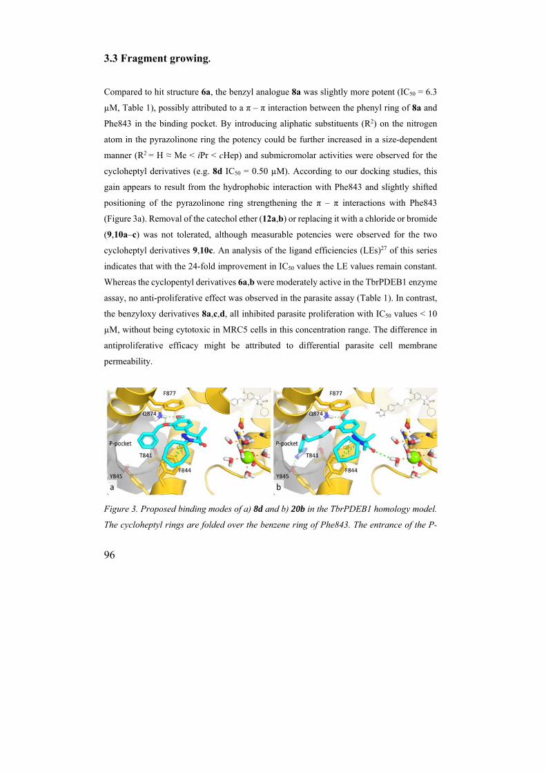
Figure 3. Proposed binding modes of a) 8d and b) 20b in the TbrPDEB1 homology model.
The cycloheptyl rings are folded over the benzene ring of Phe843. The entrance of the P-
97
CH
AP
TE
R 3
pocket is occupied by a) the benzyl moiety of 8d and b) the aliphatic tether of 20b of the
tetrazole, which interacts with Tyr844 further into the P-pocket.
The observed antitrypanosomal efficacy in combination with expedient synthetic access to
analogues led us to proceed with the benzyl-catechol chemotype. In the docking poses, the
benzene ring of these compounds is accommodated at the entrance of the P-pocket
suggesting that it should be possible to grow the inhibitors further into this pocket from the
4-position, which is directed towards the P-pocket channel (Figure 3a). It was for example
hypothesized that Thr854 in the P-pocket could be targeted by H-bond acceptors. We
therefore synthesized the nitriles 13a,b and anisoles 14a,b providing opposite electronic
influences on the aromatic ring. In addition, methoxy derivatives with a two-carbon spacer
(15a,b), as well as napthyl-substituted analogues 16a,b were synthesized to monitor the size
of the P-pocket. As the pyrazolinones with R2 = isopropyl provided better LEs (0.30) and
ligand–lipophilicity efficiencies (LLEs),28 while good potencies were observed for R2 =
cycloheptyl in the first series, both analogues were synthesized and evaluated. As can be
seen in Table 2, all analogues were active TbrPDEB1 inhibitors with IC50 values in the
range of the parent compounds 8c,d. In fact, naphthyl 16b provided a further 2-fold increase
in IC50 value.
The phenyl of 8c,d was also replaced with ortho-pyridine (17a,b) and 5-methylisoxazol
(18a,b) rings in order to potentially pick up an additional hydrogen bond with the second
hydrogen of the Gln874 nitrogen, uniquely accessed from the P-pocket. However, this
approach did not result in an increase in activity (Table 2). Interestingly, as in the previously
identified TbrPDEB1 inhibitor 1, a butyloxy spacer could be incorporated to tether the
benzonitriles (19a,b) without major loss of activity; in fact, the cycloheptyl derivative 19b
was even more potent than 13b with an IC50 value of 0.16 µM (Table 2).
98
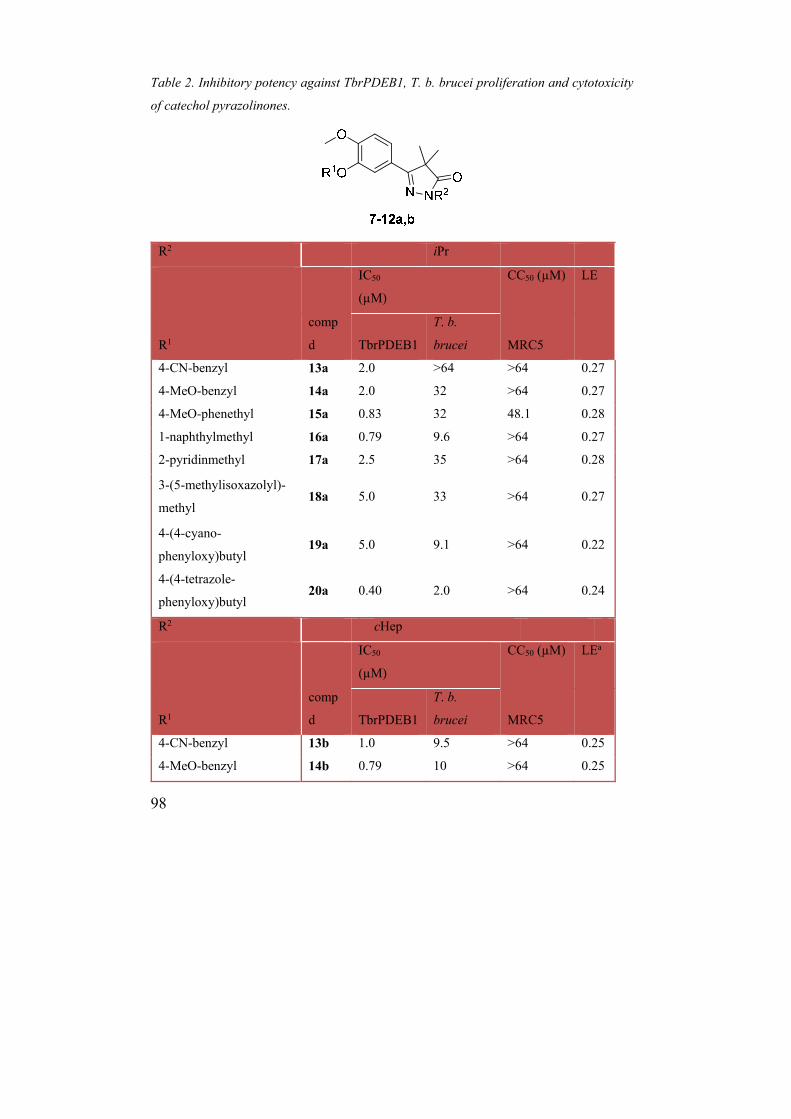
Table 2. Inhibitory potency against TbrPDEB1, T. b. brucei proliferation and cytotoxicity
of catechol pyrazolinones.
R2 iPr
IC50
(µM)
CC50 (µM) LE
R1
comp
d TbrPDEB1
T. b.
brucei MRC5
4-CN-benzyl 13a 2.0 >64 >64 0.27
4-MeO-benzyl 14a 2.0 32 >64 0.27
4-MeO-phenethyl 15a 0.83 32 48.1 0.28
1-naphthylmethyl 16a 0.79 9.6 >64 0.27
2-pyridinmethyl 17a 2.5 35 >64 0.28
3-(5-methylisoxazolyl)-
methyl 18a 5.0 33 >64 0.27
4-(4-cyano-
phenyloxy)butyl 19a 5.0 9.1 >64 0.22
4-(4-tetrazole-
phenyloxy)butyl 20a 0.40 2.0 >64 0.24
R2 cHep
IC50
(µM)
CC50 (µM) LEa
R1
comp
d TbrPDEB1
T. b.
brucei MRC5
4-CN-benzyl 13b 1.0 9.5 >64 0.25
4-MeO-benzyl 14b 0.79 10 >64 0.25
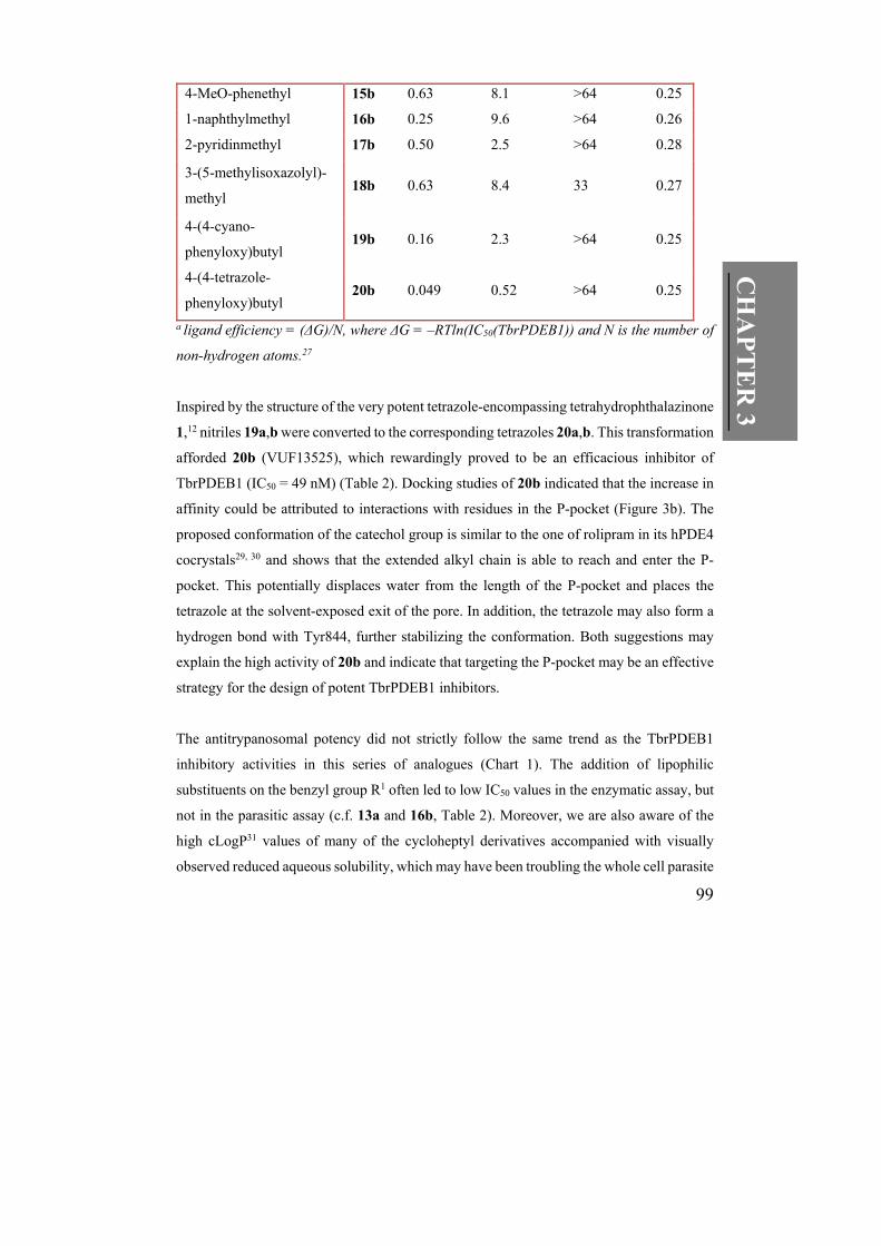
99
CH
AP
TE
R 3
4-MeO-phenethyl 15b 0.63 8.1 >64 0.25
1-naphthylmethyl 16b 0.25 9.6 >64 0.26
2-pyridinmethyl 17b 0.50 2.5 >64 0.28
3-(5-methylisoxazolyl)-
methyl 18b 0.63 8.4 33 0.27
4-(4-cyano-
phenyloxy)butyl 19b 0.16 2.3 >64 0.25
4-(4-tetrazole-
phenyloxy)butyl 20b 0.049 0.52 >64 0.25
a ligand efficiency = (ΔG)/N, where ΔG = –RTln(IC50(TbrPDEB1)) and N is the number of
non-hydrogen atoms.27
Inspired by the structure of the very potent tetrazole-encompassing tetrahydrophthalazinone
1,12 nitriles 19a,b were converted to the corresponding tetrazoles 20a,b. This transformation
afforded 20b (VUF13525), which rewardingly proved to be an efficacious inhibitor of
TbrPDEB1 (IC50 = 49 nM) (Table 2). Docking studies of 20b indicated that the increase in
affinity could be attributed to interactions with residues in the P-pocket (Figure 3b). The
proposed conformation of the catechol group is similar to the one of rolipram in its hPDE4
cocrystals29, 30 and shows that the extended alkyl chain is able to reach and enter the P-
pocket. This potentially displaces water from the length of the P-pocket and places the
tetrazole at the solvent-exposed exit of the pore. In addition, the tetrazole may also form a
hydrogen bond with Tyr844, further stabilizing the conformation. Both suggestions may
explain the high activity of 20b and indicate that targeting the P-pocket may be an effective
strategy for the design of potent TbrPDEB1 inhibitors.
The antitrypanosomal potency did not strictly follow the same trend as the TbrPDEB1
inhibitory activities in this series of analogues (Chart 1). The addition of lipophilic
substituents on the benzyl group R1 often led to low IC50 values in the enzymatic assay, but
not in the parasitic assay (c.f. 13a and 16b, Table 2). Moreover, we are also aware of the
high cLogP31 values of many of the cycloheptyl derivatives accompanied with visually
observed reduced aqueous solubility, which may have been troubling the whole cell parasite
100
assays to some extent. Yet, the most potent TbrPDEB1 inhibitor 20b concentration-
dependently reduced proliferation of T. b. brucei in the cellular assay at submicromolar
concentrations (IC50 = 520 nM, Table 2). Compound 20b was therefore analysed in a
broader set of assays (Table 3). As could be expected on the basis of the high homology
(88% sequence identity) between the catalytic domains of the TbrPDEB1 and B2
isoenzymes, 20b also acted as a potent TbrPDEB2 inhibitor (IC50 = 72 nM). Similarly, the
inhibition of the isolated TbrPDEB1 catalytic domain using the PDE light assay25, 26
remained invariant (IC50 = 55 nM), suggesting that 20b is not binding to any allosteric sites
such as the GAF domains. To evaluate the selectivity across the PDE families, 20b was also
tested against a panel of other PDE enzymes (Table 3). The L. major orthologue LmjPDEB1
was inhibited with a slightly lower potency (IC50 = 0.13 µM). On the other hand 20b was
more potent to inhibit representative isoenzymes of PDE4A–D families (IC50 at 0.0012 –
0.010 µM). The relatively low bias for hPDE4 inhibition (4 – 35-fold), compared to, for
example, piclamilast 2 (>1000-fold), might be attributed to the proposed interactions with
the parasite-specific P-pocket. Nevertheless, the data indicates that the binding mode of the
compounds in TbrPDEB1 and hPDE4 still needs careful considerations. Evidently, the
flexibility of the ligands and/or the hPDE4 protein allows accommodation in the binding
site. Considering that the clinical development of hPDE4 inhibitors has been accompanied
by emesis and adverse gastrointestinal and pro-inflammatory side effects,32, 33 the level of
hPDE4 inhibition must be considered. The selectivity ratio needed for a safe PDE-targeting
trypanocidal is difficult to predict at this stage. The narrow therapeutic window of hPDE4
inhibitors that have advanced to clinical trials indicates that at least equipotency is preferred
and most likely future lead optimization efforts should focus on a 10–100-fold selectivity
for the parasite PDE.
101
CH
AP
TE
R 3
Chart 1. Correlation between inhibition of TbrPDEB1 and inhibition of T. b. b.
proliferation.a
Interestingly, besides being a good inhibitor of T. b. brucei proliferation, 20b was also an
excellent inhibitor of T. b. rhodesiense (IC50 = 60 nM, Table 3), the causative agent of acute
East-African HAT, for which the only treatment is the arsenic-containing drug melarsoprol,
associated with fatal encephalopathy in 5-10% patients.34 Moreover, 20b also showed some
activity against T. cruzi, the causative agent of Chagas disease.35 Pyrazolinone 20b was
well-tolerated by the human fibroblast cell line (CC50 > 64 µM) and murine macrophages,
providing a sensitivity index of at least 250-fold for T. b. rhodesiense, i.e. well within the
conventional thresholds.36
102
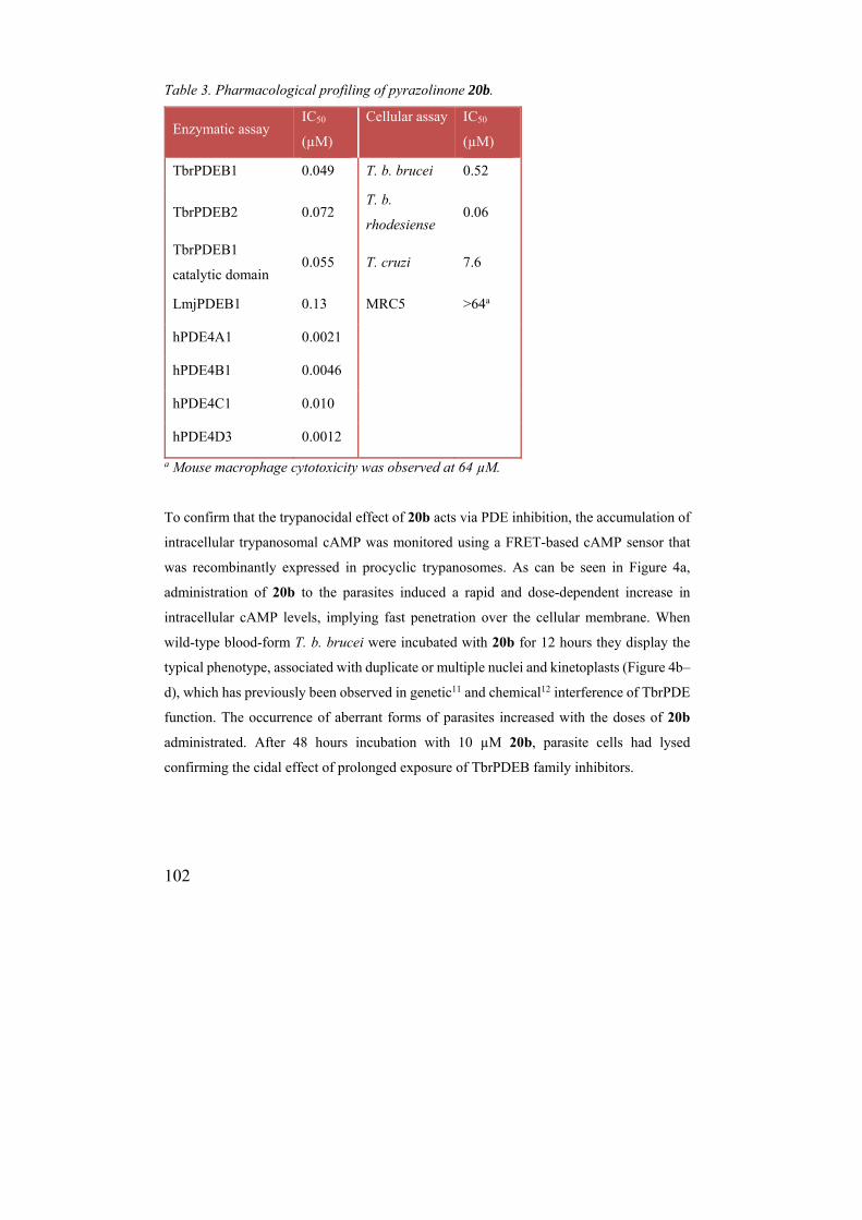
Table 3. Pharmacological profiling of pyrazolinone 20b.
Enzymatic assay IC50
(µM)
Cellular assay IC50
(µM)
TbrPDEB1 0.049 T. b. brucei 0.52
TbrPDEB2 0.072 T. b.
rhodesiense 0.06
TbrPDEB1
catalytic domain 0.055 T. cruzi 7.6
LmjPDEB1 0.13 MRC5 >64a
hPDE4A1 0.0021
hPDE4B1 0.0046
hPDE4C1 0.010
hPDE4D3 0.0012
a Mouse macrophage cytotoxicity was observed at 64 µM.
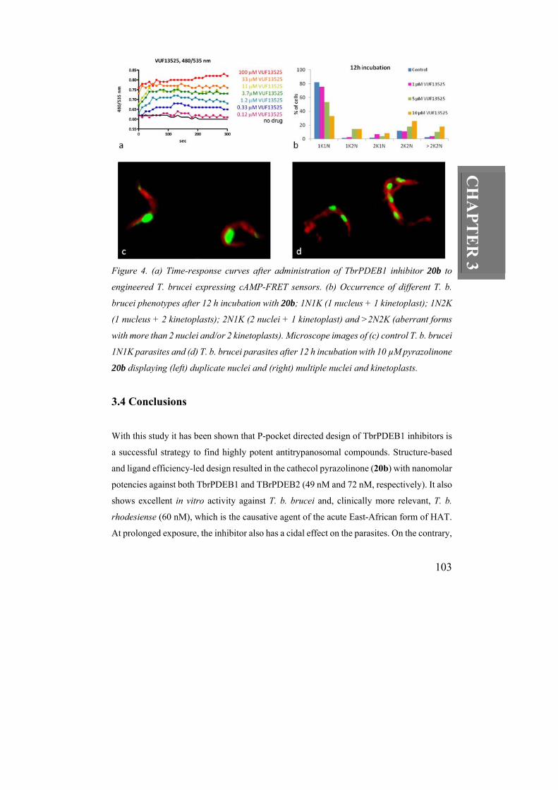
To confirm that the trypanocidal effect of 20b acts via PDE inhibition, the accumulation of
intracellular trypanosomal cAMP was monitored using a FRET-based cAMP sensor that
was recombinantly expressed in procyclic trypanosomes. As can be seen in Figure 4a,
administration of 20b to the parasites induced a rapid and dose-dependent increase in
intracellular cAMP levels, implying fast penetration over the cellular membrane. When
wild-type blood-form T. b. brucei were incubated with 20b for 12 hours they display the
typical phenotype, associated with duplicate or multiple nuclei and kinetoplasts (Figure 4b–
d), which has previously been observed in genetic11 and chemical12 interference of TbrPDE
function. The occurrence of aberrant forms of parasites increased with the doses of 20b
administrated. After 48 hours incubation with 10 µM 20b, parasite cells had lysed
confirming the cidal effect of prolonged exposure of TbrPDEB family inhibitors.
103
CH
AP
TE
R 3 Figure 4. (a) Time-response curves after administration of TbrPDEB1 inhibitor 20b to
engineered T. brucei expressing cAMP-FRET sensors. (b) Occurrence of different T. b.
brucei phenotypes after 12 h incubation with 20b; 1N1K (1 nucleus + 1 kinetoplast); 1N2K
(1 nucleus + 2 kinetoplasts); 2N1K (2 nuclei + 1 kinetoplast) and >2N2K (aberrant forms
with more than 2 nuclei and/or 2 kinetoplasts). Microscope images of (c) control T. b. brucei
1N1K parasites and (d) T. b. brucei parasites after 12 h incubation with 10 µM pyrazolinone
20b displaying (left) duplicate nuclei and (right) multiple nuclei and kinetoplasts.
3.4 Conclusions
With this study it has been shown that P-pocket directed design of TbrPDEB1 inhibitors is
a successful strategy to find highly potent antitrypanosomal compounds. Structure-based
and ligand efficiency-led design resulted in the cathecol pyrazolinone (20b) with nanomolar
potencies against both TbrPDEB1 and TBrPDEB2 (49 nM and 72 nM, respectively). It also
shows excellent in vitro activity against T. b. brucei and, clinically more relevant, T. b.
rhodesiense (60 nM), which is the causative agent of the acute East-African form of HAT.
At prolonged exposure, the inhibitor also has a cidal effect on the parasites. On the contrary,
104
mammalian cell lines seem to tolerate the catechol pyrazolinone chemotype very well;
fibroblast toxicity was only observed for a handful of compounds.
For the first time, the increase in cAMP levels in the parasites upon administration of PDE
inhibitor could be followed in real-time using a FRET-based cAMP sensor expressed in
engineered trypanosomes. Being aware of the many challenges that remain, such as
selectivity over hPDE4, blood-brain barrier penetration, acceptable pharmacokinetic profile
and metabolic stability, we still believe that the promising pharmacological profile of 20b
brings new hope to the prospect of developing a less toxic alternative to melarsoprol.
3.5 Experimental Section
Homology model of TbrPDEB1. The homology model was constructed using the
TbrPDEB1 sequence published by Zoraghi and Seebeck (Uniprot: Q8WQX9).9 The closest
homologue of TbrPDEB1 crystallized to date is Leishmania major PDEB1, the structure is
published as 2R8Q on the RCSB Protein Data Bank. Aligning the sequences using ClustalW
2.137 gave a sequence identity of 66%. The residues on the surface of the catalytic site show
an even higher homology with only one substitution (V836I). The P-pocket region has two
substitutions, N881M and R885K, both on the far side of the P-pocket from the invariant
glutamine (G874).
The homology model was built using the Molecular Operating Environment (MOE)
2010.10 software package.38 The B chain of the LmjPDEB1 structure was used to construct
the homology model, since the B chain showed lower B factors in the region of the catalytic
site. The LmjPDEB1 structure was opened in MOE and all solvent and metal atoms were
removed. The TbrPDEB1 sequence was added and the sequence alignment from ClustalW
2.1 applied. Those residues which were identical in both sequences had their atoms tethered
at 200 in the 2R8Q.B model. The Amber99 forcefield and born solvation model were used
during the homology model construction. There were 10 main-chain and 1 side-chain
models formed, the intermediate models were refined to an RMS of 0.1 and the final model
was refined once more to an RMS of 0.5. Models were protonated using the Protonate 3D
105
CH
AP
TE
R 3
function prior to refinement and scored using the GB/VI scoring function. The final model
had the metal ions and the six metal-coordinating water molecules included directly from
the LmjPDEB1 model, with the hydrogen atoms minimized following inclusion.
The full-length amino acid sequences of the TbrPDEB1 and TbrPDEB2 (Uniprot: Q38F42)
catalytic domains were aligned using ClustalW 2.1 giving a sequence identity of 75%.
Restricting the sequence alignment to the 334 residues of the catalytic domain increased the
sequence identity to 88%.
Inhibitor docking. The structures of the ligands were prepared in MOE and exported as an
SDF database. A single low energy conformation was generated for each structure using
Omega 2.4.3, OpenEye Scientific Software.39 The mol2 file generated by Omega was used
as input for GOLD.40 The compounds were docked into the homology model of TbrPDEB1
described above, which was used as prepared. GOLD uses a genetic algorithm to efficiently
explore the conformational space of the ligand in the protein pocket. The parameters used
in GOLD include, autoscaling of operations per ligand (dependent on the number of
rotatable bonds), PLP scoring function, water included with spin, metal ions included, and
the generation of 30 solutions per ligand with an RMS tolerance of 1.5 Å. Results were
analyzed using MOE and binding poses were rendered in PyMOL.41
Synthetic Chemistry.
General Information. Chemicals and reagents were purchased from commercial suppliers
and were used without further purification. Microwave reactions were performed with
Biotage Initiator single mode cavity, producing controlled irradiation at 2450 MHz in sealed
reaction vials (capable of withholding elevated pressure) and with magnetic stirring. For
column chromatography commercially available Silica Gel 60 (particle size 0.040–0.063
mm) was used. Gradient flash column chromatography purification was performed on
Biotage Isolera with prepacked silica (KP-sil) cartridges supplied by Biotage and
ethylacetate in n-heptane as eluent, unless otherwise stated. Analytical thin layer
chromatography was performed using glass sheets precoated with Silica Gel 60 F254 and
visualization of components was made by UV (254 nm), I2 and/or Pancaldi’s solution
106
followed by heating. Analytical HPLC–MS was performed on a Shimadzu LC-20AD liquid
chromatography pump system with a Shimadzu LCMS-2010 liquid chromatography mass
spectrometer equipped with Xbridge (C18) 5 μm column (50 mm × 4.6 mm), using
acetonitrile in 0.10% aqueous formic acid and a 5% – 90% gradient over 4.5 min followed
by 2 min isocratic 90% mobile phase. 1H NMR and 13C NMR spectra were measured on a
Bruker 200, 400 or 500 at 250 MHz, 400 MHz or 500 MHz, respectively. The chemical
shifts for 1H NMR and 13C NMR were referenced to TMS via the CDCl3 solvent signal (1H
NMR at 7.26 ppm and 13C NMR at 77.16 ppm). Exact molecular masses (HRMS) were
determined on Bruker micrOTOF-Q mass spectrometer equipped with an electrospray ion
source using caffeine as reference. The purity for all compounds was established to be ≥95%
by HP-LC, 1H NMR and 13C NMR, unless otherwise stated. Acyl chloride precursors of β-
keto esters 11a–d42-45 are known compounds. The synthetic preparation and spectroscopic
data of catechol pyrazolinone 8a has previously been published in a patent.20
Experimental details and spectroscopic data for precursors 11a,b.
Methyl 3-(3-(cyclopentyloxy)-4-methoxyphenyl)-2,2-dimethyl-3-oxopropanoate (11a).
3-(cyclopentyloxy)-4-methoxybenzoic acid (5.0 g, 21.1 mmol) was suspended in 25 mL dry
DCM and DMF (3 drops) was added. Oxalyl chloride (2.78 ml, 31.7 mmol) was added
dropwise under vigorous stirring and N2 atmosphere. The reaction mixture was stirred at
room temperature for 1.5 h. The reaction was monitored with TLC of small aliquots of the
reaction mixture, which were diluted with MeOH and heated with a heat-gun. When full
conversion was observed, the volatiles were evaporated in vacuo to give 3-
(cyclopentyloxy)-4-methoxybenzoyl chloride as a yellow crystalline solid, which was used
as such in the subsequent step without any further purification.
Lithium diisopropyl amide (13 mL, 1.8 M in THF/Hex/ether, 1.1 equiv) was added to a dry
3-neck flask with 25 mL freshly distilled THF and cooled to -45 °C and methyl isobutyrate
(3.64 ml, 31.7 mmol, 1.5 equiv) was added dropwise. The mixture was stirred for 30 min at
-40 °C. Freshly prepared 3-(cyclopentyloxy)-4-methoxybenzoyl chloride (approx. 21
mmol) was dissolved in dry THF (25 mL) and added dropwise during 30 min at -50 – -40
°C. The reaction mixture was stirred for another 1 h before the cooling source was removed

107
CH
AP
TE
R 3
and the stirring continued at rt overnight. The reaction mixture was acidified with 3 M HCl
(aq), diluted with EtOAc (10 mL) and the aqueous layer was extracted with EtOAc (2 x 10
mL). The combined organic layers were washed with sat NaHCO3 and brine, dried over
MgSO4 and reduced in vacuo. The crude product was purified with flash chromatography
to give the title compound as a pale syrup (1.1 g, 15%): 1H NMR (500 MHz, CDCl3) δ
(ppm) 7.50 – 7.41 (m, 2H), 6.83 (d, J = 9.08 Hz, 1H), 4.82 – 4.73 (m, 1H), 3.83 (s, 3H),
3.64 (s, 3H), 2.07 – 1.94 (m, 2H), 1.94 – 1.75 (m, 4H), 1.72 – 1.59 (m, 2H), 1.54 (s, 6H); 13C NMR (126 MHz, CDCl3) δ (ppm) 196.14, 176.13, 153.94, 147.59, 127.85, 122.77,
114.49, 110.64, 80.55, 56.18, 53.09, 52.66, 32.93, 24.33, 24.31; LC–MS–ESI+ found 321
[M+H]+.
Methyl 3-(3-(benzyloxy)-4-methoxyphenyl)-2,2-dimethyl-3-oxopropanoate (11b).
Following the procedure to 11b using 3-(benzyloxy)-4-methoxybenzoic acid on a 148
mmol scale first gave 3-(benzyloxy)-4-methoxybenzoyl chloride as a yellow semi-solid
(41.5 g, quant., >95% purity according to 1H NMR); 1H NMR (250 MHz, CDCl3) δ (ppm)
7.78 (dd, J = 8.45, 1.94 Hz, 1H), 7.66 (d, J = 1.90 Hz, 1H), 7.48 (d, J = 7.26 Hz, 2H), 7.43
– 7.28 (m, 3H), 6.94 (d, J = 8.52 Hz, 1H), 5.30 (s, 1H), 5.19 (s, 3H), 3.95 (s, 3H)) and the
title compound as a yellow syrup (59 g, quant., 90% purity): 1H NMR (250 MHz, CDCl3)
δ (ppm) 7.55 – 7.44 (m, 4H), 7.43 – 7.26 (m, 3H), 6.88 (d, J = 8.4 Hz, 1H), 5.18 (s, 2H),
3.94 (s, 3H), 3.61 (s, 3H), 1.51 (s, 6H); 13C NMR (63 MHz, CDCl3) δ (ppm) 195.81,
175.85, 153.58, 147.94, 136.54, 128.63, 128.05, 127.85, 127.49, 123.30, 113.78, 110.59,
70.85, 56.06, 52.97, 52.47, 24.13; LC–MS–ESI+ found 343 [M+H]+.
Experimental details and spectroscopic data for pyrazolinones.
3-(3-(Cyclopentyloxy)-4-methoxyphenyl)-4,4-dimethyl-1H-pyrazol-5(4H)-one (6a). To
a reaction tube with 11a (0.851 g, 2.66 mmol), dissolved in ethanol (5.5 mL, 0.5 M reaction
molarity), aqueous hydrazine hydrate (80%, 1.4 mL, 8 equiv) was added. The reaction
mixture was stirred at 50 °C overnight. The reaction mixture was cooled on ice and the
precipitate was filtered off over a glass filter, washed with cold ethanol and dried at 40 °C
under vacuum to give the title compound as a solid foam (0.72 g, 90%): 1H NMR (400
MHz, CDCl3) δ (ppm) 9.43 (s, 1H), 7.42 (d, J = 1.93 Hz, 1H), 7.27 (dd, J = 8.35, 2.06 Hz,
108
1H), 6.86 (d, J = 8.46 Hz, 1H), 4.92 – 4.69 (m, 1H), 3.87 (s, 3H), 2.04 – 1.74 (m, 6H), 1.71
– 1.55 (m, 2H), 1.51 (s, 6H); 13C NMR (101 MHz, CDCl3) δ (ppm) 181.71, 163.35, 151.93,
148.11, 123.93, 119.36, 112.34, 111.41, 80.68, 56.13, 47.40, 32.93, 24.21, 22.85; LC–MS–
ESI+ found 303, [M+H]+; HRMS–ESI+: [M+H]+ calcd for C17H23N2O3: 303.1703 found
303.1698.
1-Cycloheptyl-3-(3-(cyclopentyloxy)-4-methoxyphenyl)-4,4-dimethyl-1H-pyrazol-
5(4H)-one (6b). Sodium hydride (52 mg, 1.3 mmol, 2 equiv.) was added to a chilled
suspension of 6a (195 mg, 0.65 mmol) in DMF/MeCN (2:1) under stirring and N2
atmosphere. The reaction mixture was stirred for 15 min before bromocycloheptane (0.31
ml, 2.3 mmol, 3.5 equiv.) was added. The reaction mixture was heated at 120 °C overnight.
The volatiles were evaporated in vacuo. The residue was diluted with EtOAc (50 mL) and
washed with brined (3 x 15 mL), dried over Na2SO4, filtered and reduced in vacuo. The
residue was purified via flash chromatography to give title compound as a pale solid (230
mg, 89 % yield): 1H NMR (400 MHz, CDCl3) δ (ppm) 7.39 (d, J = 1.90 Hz, 1H), 7.28 (dd,
J = 8.47, 1.90 Hz, 1H), 6.85 (d, J = 8.47 Hz, 1H), 4.91 – 4.72 (m, 1H), 4.37 – 4.18 (m, 1H),
3.86 (s, 3H), 2.03 – 1.72 (m, 12H), 1.72 – 1.47 (m, 8H), 1.43 (s, 6H); 13C NMR (126 MHz,
CDCl3) δ (ppm) 177.50, 161.48, 151.59, 147.82, 124.10, 119.35, 112.55, 111.35, 80.69,
56.07, 54.57, 48.58, 33.43, 32.95, 28.33, 24.81, 24.27, 22.86; LC–MS–ESI+: m/z 399,
[M+H]+; HRMS–ESI+: [M+H]+ calcd for C24H35N2O3: 399.2642 found 399.2624.
3-(3-(Benzyloxy)-4-methoxyphenyl)-4,4-dimethyl-1H-pyrazol-5(4H)-one (8a).
Following the procedure to 6a using 11b on a 0.15 mol scale gave the title compound as a
yellow solid (47.36 g. 97%): 1H NMR (400 MHz, CDCl3) δ (ppm) 9.67 (br s, 1H), 7.44
(appar. d, J = 7.36 Hz, 2H), 7.41 (d, J = 1.93 Hz, 1H), 7.39 – 7.26 (m, 4H), 6.89 (d, J = 8.48
Hz, 1H), 5.18 (s, 2H), 3.91 (s, 3H), 1.41 (s, 6H); 13C NMR (101 MHz, CDCl3) δ (ppm)
181.72, 163.11, 151.49, 148.35, 136.85, 128.68, 128.05, 127.39, 123.88, 119.99, 111.78,
111.38, 71.16, 56.07, 47.32, 22.62; LC–MS–ESI+: m/z 325, [M+H]+; HRMS–ESI+: [M+H]+
calcd for C19H21N2O3: 325.1547 found 325.1531.
109
CH
AP
TE
R 3
3-(3-(Benzyloxy)-4-methoxyphenyl)-1-isopropyl-4,4-dimethyl-1H-pyrazol-5(4H)-one
(8c). Following the procedure to 6b using isopropylbromide and 8a on a 2.46 mmol scale
gave the title compound as a white solid (706 mg, 78%): 1H NMR (400 MHz, CDCl3) δ
(ppm) 7.49 (d, J = 7.3 Hz, 2H), 7.44 (d, J = 1.9 Hz, 1H), 7.34 (m, 4H), 6.91 (d, J = 8.5 Hz,
1H), 5.22 (s, 2H), 4.50 (hept, J = 6.7 Hz, 1H), 3.93 (s, 3H), 1.38 (s, 6H), 1.37 (d, J = 7.0
Hz, 6H); 13C NMR (101 MHz, CDCl3) δ (ppm) 177.85, 161.28, 151.30, 148.28, 137.00,
128.66, 128.05, 127.47, 124.11, 119.92, 112.17, 111.38, 71.36, 56.06, 48.73, 45.26, 22.67,
20.82; LC–MS–ESI+: m/z 367, [M+H]+; HRMS–ESI+: [M+H]+ calcd for C22H27N2O3:
367.2016 found 367.1998.
3-(3-(Benzyloxy)-4-methoxyphenyl)-1-cycloheptyl-4,4-dimethyl-1H-pyrazol-5(4H)-
one (8d). Following the procedure to 6b on a 1.59 mmol scale using 8a gave the title
compound as a white solid (544 mg, 81% yield): 1H NMR (400 MHz, CDCl3) δ (ppm) 7.49
– 7.43 (m, 2H), 7.41 (d, J = 2.00 Hz, 1H), 7.38 – 7.33 (m, 2H), 7.32 – 7.26 (m, 2H), 6.87
(d, J = 8.49 Hz, 1H), 5.20 (s, 2H), 4.36 – 4.16 (m, 1H), 3.90 (s, 3H), 1.98 – 1.74 (m, 6H),
1.67 – 1.47 (m, 6H), 1.34 (s, 6H); 13C NMR (101 MHz, CDCl3) δ (ppm) 177.41, 161.16,
151.21, 148.23, 136.98, 128.61, 127.99, 127.38, 124.09, 119.83, 112.10, 111.32, 71.31,
56.02, 54.47, 48.39, 33.39, 28.34, 24.73, 22.62; LC–MS–ESI+: m/z 421, [M+H]+; HRMS–
ESI+: [M+H]+ calcd for C26H33N2O3: 421.2486 found 421.2467.
3-(3-Hydroxy-4-methoxyphenyl)-1-isopropyl-4,4-dimethyl-1H-pyrazol-5(4H)-one
(12a). Ammonium formate (10 g, 159 mmol) and 10% Pd/C (50 mg, 0.047 mmol) were
added to benzylether 8c (12 g, 32.7 mmol) dissolved in MeOH/CH2Cl2 (2:1, 200 mL). The
reaction mixture was stirred for 5 h at 40 °C. The reaction mixture was filtered through a
cake of celite and the volatiles were evaporated. The residue was diluted with EtOAc,
washed with brine (2 x 200 mL), dried over Na2SO4, filtered and reduced in vacuo with
CH2Cl2 as co-solvent to give the title compound as an off-white solid (8.73 g, 96% yield): 1H NMR (400 MHz, CDCl3) δ 7.44 (d, J = 2.0 Hz, 1H), 7.29 (dd, J = 8.4, 2.0 Hz, 1H), 6.86
(d, J = 8.5 Hz, 1H), 5.97 (s, 1H), 4.49 (hept, J = 6.6 Hz, 1H), 3.92 (s, 3H), 1.44 (s, 6H), 1.34
(d, J = 6.7 Hz, 6H); 13C NMR (101 MHz, CDCl3) δ (ppm) 178.06, 161.60, 148.22, 145.99,
110
124.91, 118.78, 112.61, 110.62, 56.10, 49.00, 45.37, 22.74, 20.85; LC–MS–ESI+: m/z 277,
[M+H]+; HRMS–ESI+: [M+H]+ calcd for C15H21N2O3: 277.1547 found 277.1543.
1-Cycloheptyl-3-(3-hydroxy-4-methoxyphenyl)-4,4-dimethyl-1H-pyrazol-5(4H)-one
(12b). Following the procedure of 12a using 8d on a 1.32 mmol scale afforded the title
compound as a pale oil (1.35g, quantitative yield, 99.8% purity): 1H NMR (400 MHz,
CDCl3) δ (ppm) 7.46 (d, J = 2.05 Hz, 1H), 7.29 (dd, J = 8.43, 2.08 Hz, 1H), 6.87 (d, J =
8.49 Hz, 1H), 6.30 (br s, 1H), 4.36 – 4.22 (m, 1H), 3.92 (s, 3H), 2.03 – 1.74 (m, 6H), 1.70
– 1.47 (m, 6H), 1.45 (s, 6H); 13C NMR (101 MHz, CDCl3) δ 177.62, 161.58, 148.30,
146.00, 124.72, 118.64, 112.63, 110.63, 56.00, 54.58, 48.66, 33.37, 28.25, 24.72, 22.61;
LC–MS–ESI+: m/z 331, [M+H]+; HRMS–ESI+: [M+H]+ calcd for C19H27N2O3: 331.2016
found 331.1997.
General procedure to O-alkylations A. Corresponding phenol 12a or 12b was added to a
MW reaction tube with K2CO3 (3.0 equiv) and MeCN/DMF (1:1, 0.2 M final reaction
molarity). It was MW irradiated for 30 min at 120 °C. After releasing the CO2-gas from the
MW tube, the corresponding bromide or chloride (2 equiv.) was added via a needle through
the septum. The reaction mixture was again MW irradiated for 20 min at 120 °C. In case of
low conversion, 1 more equivalent of halide was added and the reaction mixture was
reirradiated for 40 min. The volatiles were evaporated in vacuo. The residue was dissolved
in EtOAc and the organic layer was washed with brine and brine with sat NH4Cl 1:1. The
organic phase was dried over Na2SO4, filtered, and reduced in vacuo. The crude product
was purified using gradient flash column chromatography.
General procedure to O-alkylations B. Sodium hydride (60% dispersed in oil, 2–5 equiv.)
was added to corresponding phenol 12a or 12b dissolved in DMF (0.14 M reaction
molarity) under N2 atmosphere. The mixture was stirred for 5 – 15 min prior to addition of
corresponding bromide (1.5 equiv). The reaction mixture was microwave irradiated at 120
°C for 15–30 min and then worked-up as in general procedure A.
111
CH
AP
TE
R 3
4-(4-(5-(1-Isopropyl-4,4-dimethyl-5-oxo-4,5-dihydro-1H-pyrazol-3-yl)-2-
methoxyphenoxy)butoxy)benzonitrile (19a). Following the general procedure A on a
1.12 mmol scale gave the title compound as an off-white solid (201 mg, 40%): 1H NMR
(400 MHz, CDCl3) δ (ppm) 7.59 – 7.50 (m, 2H), 7.45 (d, J = 1.96 Hz, 1H), 7.29 – 7.23 (m,
1H), 6.96 – 6.88 (m, 2H), 6.85 (d, J = 8.46 Hz, 1H), 4.48 (hept, J = 6.70 Hz, 1H), 4.13 (dt,
J = 14.51, 5.74 Hz, 4H), 3.85 (s, 3H), 2.08 – 2.00 (m, 4H), 1.44 (s, 6H), 1.35 (d, J = 6.72
Hz, 6H); 13C NMR (101 MHz, CDCl3) δ 177.95, 162.44, 161.51, 151.17, 148.76, 134.08,
124.27, 119.78, 119.39, 115.33, 111.13, 110.63, 103.90, 68.71, 68.13, 56.07, 48.94, 45.40,
26.14, 25.91, 22.97, 20.93; LC–MS–ESI+: m/z 450, [M+H]+; HRMS–ESI+: [M+H]+ calcd
for C26H32N3O4: 450.2387 found 450.2366.
4-(4-(5-(1-Cycloheptyl-4,4-dimethyl-5-oxo-4,5-dihydro-1H-pyrazol-3-yl)-2-
methoxyphenoxy)butoxy)benzonitrile (19b). Following the general procedure B on a
0.58 mmol scale gave the title compound as an off-white solid (166 mg, 57%): 1H NMR
(400 MHz, CDCl3) δ (ppm) 8.11 (appar. d, J = 8.85 Hz, 2H), 7.42 (d, J = 1.98 Hz, 1H), 7.32
(dd, J = 8.43, 1.99 Hz, 1H), 7.01 (appar. d, J = 8.88 Hz, 2H), 6.88 (d, J = 8.53 Hz, 1H), 4.35
– 4.26 (m, 1H), 4.18 (t, J = 5.97 Hz, 2H), 4.13 (t, J = 5.75 Hz, 2H), 3.89 (s, 3H), 2.11 – 1.94
(m, 6H), 1.94 – 1.84 (m, 2H), 1.84 – 1.73 (m, 2H), 1.69 – 1.50 (m, 6H), 1.49 (s, 6H); 13C
NMR (101 MHz, CDCl3) δ (ppm) 178.14, 175.72, 162.46, 161.57, 151.31, 148.69, 129.25,
123.92, 119.98, 116.45, 115.28, 111.31, 110.81, 68.88, 68.02, 56.12, 55.25, 49.24, 33.44,
28.21, 26.19, 26.14, 24.89, 22.88; LC–MS–ESI+: m/z 504, [M+H]+; HRMS–ESI+: [M+H]+
calcd for C30H38N3O4: 504.2857 found 504.2840.
3-(3-(4-(4-(2H-tetrazole-5-yl)phenoxy)butoxy)-4-methoxyphenyl)-1-isopropyl-4,4-
dimethyl-1H-pyrazol-5(4H)-one (20a). A mixture of compound 19a (141 mg, 0.31 mmol),
sodium azide (203 mg, 3.1 mmol) and NH4Cl (167 mg, 3.1 mmol) in DMF (6 mL) was MW
irradiated at 160 °C for 1 h. The reaction mixture was cooled to RT, diluted with EtOAc,
washed twice with brine acidified with HCl (aq). The first aqueous layer was extracted with
EtOAc (2 x 5 mL). The combined organic layers were further washed with brine acidified
with HCl (aq), reduced, and the crude product was purified with flash column
chromatography (eluent: MeOH in CH2Cl2 with 0.5% AcOH) to afford the title compound
112
as an off-white solid (115 mg, 75%); 1H NMR (500 MHz, CDCl3) δ (ppm) 8.11 (appar. d,
J = 8.76 Hz, 2H), 7.44 (d, J = 1.71 Hz, 1H), 7.33 (dd, J = 8.42, 1.78 Hz, 1H), 7.02 (appar.
d, J = 8.79 Hz, 2H), 6.89 (d, J = 8.49 Hz, 1H), 4.54 (hept, J = 6.70 Hz, 1H), 4.19 (t, J = 6.06
Hz, 2H), 4.14 (t, J = 5.77 Hz, 2H), 3.89 (s, 3H), 2.07 (dt, J = 12.48, 6.49 Hz, 4H), 1.49 (d,
J = 9.55 Hz, 6H), 1.39 (d, J = 6.70 Hz, 6H).); MS-ESI+ (m/z 493, M+H+); 13C NMR (126
MHz, CDCl3) δ (ppm)178.53, 162.43, 161.56, 156.09, 151.29, 148.70, 129.25, 123.82,
119.92, 116.34, 115.26, 111.14, 110.61, 68.82, 67.94, 56.08, 49.50, 45.83, 26.17, 26.06,
22.90, 20.87; LC–MS–ESI+: m/z 493, [M+H]+; HRMS–ESI+: [M+H]+ calcd for
C26H33N6O4: 493.2558 found 493.2545.
3-(3-(4-(4-(2H-tetrazole-5-yl)phenoxy)butoxy)-4-methoxyphenyl)-1-cycloheptyl-4,4-
dimethyl-1H-pyrazol-5(4H)-one (20b). Following the procedure to compound 20a using
nitrile 19b (100 mg, 0.20 mmol) to afford the title compound as an off-white solid (95 mg,
62%): 1H NMR (500 MHz, CDCl3) δ (ppm) 1H NMR (500 MHz, CDCl3) δ (ppm) 8.14 (d,
J = 8.87 Hz, 2H), 7.44 (d, J = 2.05 Hz, 1H), 7.30 (dd, J = 8.46, 2.04 Hz, 1H), 7.01 (d, J =
8.92 Hz, 2H), 6.88 (d, J = 8.53 Hz, 1H), 4.36 – 4.25 (m, 1H), 4.18 (t, J = 6.00 Hz, 2H), 4.13
(t, J = 5.94 Hz, 2H), 3.88 (s, 3H), 2.12 – 1.93 (m, 6H), 1.93 – 1.85 (m, 2H), 1.82 – 1.73 (m,
2H), 1.67 – 1.49 (m, 6H), 1.49 (s, 6H); 13C NMR (126 MHz, CDCl3) δ (ppm) 178.14,
162.48, 161.56, 156.14, 151.29, 148.68, 129.27, 123.82, 119.93, 116.36, 115.25, 111.17,
110.66, 68.83, 67.94, 56.09, 55.26, 49.25, 33.41, 28.16, 26.18, 26.05, 24.85, 22.87; LC–
MS–ESI+: m/z 547, [M+H]+; HRMS–ESI+: [M+H]+ calcd for C30H39N6O4: 547.3027 found
547.3015.
Pharmacological evaluation.
Expression of recombinant TbrPDEB1 and TbrPDEB2. Full length hPDE4A1, B1, C1
and D3 and TbrPDEB1 were expressed in Sf21 insect cells and full length TbrPDEB2 was
expressed in yeast, as previously described.12 His-tagged catalytic domains of TbrPDEB1
were expressed in E. coli BL21 codon plus cells and column purified as recommended in
the manual, “The Qiaexpressionist” (Qiagen). PDEs were dissolved in 10 mM HEPES
buffer (pH 7.4) containing 100 mM NaCl, 5 mM MgCl2.
113
CH
AP
TE
R 3
Determination of PDE Activity. PDE activities were determined according to two
different methods throughout the study.
Full length PDEs. The standard scintillation proximity assay (SPA) for determination of
PDE activities was used exactly following the procedure as recently outlined by De Koning
et al.,12 (14) to analyze effects of test compounds on the enzymatic activities of TbrPDEB1,
TbrPDEB2, LmjPDEB1, human PDE4A1, 4B1, 4C1 and 4D3. In these assay, the cAMP
substrate concentration was 0.5 µM. Briefly, PDE activity of supernatants of sonicated
PDE-overexpressing Sf21 cells was determined in at least duplicates by published
procedures.12, 46, 47 Enzyme concentrations were always adjusted so that <20% of substrate
was consumed. Blank values (measured in the presence of denatured protein) were always
<2% of total radioactivity.
Catalytic Domain TbrDEB1. The determination of TbrPDEB1 catalytic domain activity
was performed with PDELight™ HTS cAMP phosphodiesterase kit25, 26 in a reaction
volume of 50 µL comprising 1% DMSO in a 96 well-format. PDELight assay is a non-
radioactive, bioluminescent detection system for measuring the activity of PDEs developed
by Lonza Rockland, Inc. This assay was used in the determination of IC50 values for
inhibitors against catalytic domain of TbrPDEB1. This assay offers a continuous readout of
luminescence based on the following reactions: PDEs hydrolyse cAMP to AMP; AMP is
then converted directly to ATP by “AMP-DR”, Lonza’s proprietary AMP detection reagent;
luciferase is then utilized to catalyze the formation of light from luceferin and the newly
formed ATP. Luciferase catalysis step also regenerates AMP and therefore the light output
is linear with the rate of AMP production by the PDE. The reaction uses cAMP in the
concentration of 0.5 µM. From the rate of increase in light intensity over a period of time,
the reaction rate is calculated. The reaction output light was monitored at 25 °C for 20
minutes using Victor2 yielding a specific activity of about 2500U/min in the given
conditions. Dose-response curves were generated by plotting the percent of inhibition of
PDE against the inhibitor concentrations. Nonlinear regression analysis was used to
calculate IC50 values from each dose-response curve by using GraphPad Prism (version
5.01).
114
In vitro susceptibility testing of trypanosomes and MRC5 fibroblasts. T. b. brucei
(Squib-427 strain, suramin-sensitive) or T. b. rhodesiense (STIB-900 strain)
trypomastigotes were cultured at 37 °C and 5% CO2 in Hirumi-9 medium,48 supplemented
with 10% fetal calf serum (FCS). Compound stock solutions were prepared in 100% DMSO
at 20 mM or mg/ml. The compounds were serially pre-diluted (2-fold or 4-fold) in DMSO
followed by a further (intermediate) dilution in demineralized water to assure a final in-test
DMSO concentration of <1%.
Assays were performed by adding 1.5×10⁴ trypomastigotes/well containing the pre-diluted
compounds. After 72 h incubation, parasite growth was assessed fluorimetrically by adding
resazurin24 for 24 h at 37 °C. Fluorescence was measured using a GENios Tecan fluorimeter
(excitation 530 nm, emission 590 nm).
For cytotoxicity evaluation, human fetal lung fibroblast were cultivated in MEM,
supplemented with L-glutamine (20 mM), 16.5 mM sodium hydrogen carbonate and 5%
FCS at 37 °C and 5% CO2. For the assay, 104 MRC-5 cells/well were seeded onto the test
plates containing the pre-diluted compounds and incubated at 37 °C and 5% CO2 for 72 h.
Cell viability was determined after addition of resazurin. Fluorescence was measured using
a GENios Tecan fluorimeter (excitation 530 nm, emission 590 nm).23, 49
Phenotype determination. T. b. brucei trypomastigotes (1x10⁶ parasites/mL) were
incubated with 20b (1, 5, or 10 µM or vehicle) for 12 hours. Thereafter, parasites were
harvested and washed once with saline. Culture aliquots were spread onto glass slides. After
drying, they were fixed for 15 min with 2% formaldehyde and stained for 30 min at room
temperature with Syto-9 (Invitrogen) 250 nM and Nyle Red (Sigma) 50 mg/ml. Slides were
analyzed using an Olympus BX41 microscope. For each sample, >300 cells were analyzed.
Cells were manually scored and assigned to the following categories: 1N1K (1 nucleus + 1
kinetoplast); 1N2K (1 nucleus + 2 kinetoplasts); 2N1K (2 nuclei + 1 kinetoplast) and
>2N2K (aberrant forms with more than 2 nuclei and/or 2 kinetoplasts).

115
CH
AP
TE
R 3
FRET cAMP sensor construct.
Expression of cAMP sensors in trypanosomes. Nikolaev et al.50, 51 had developed a series
of FRET-based cAMP sensors to analyze the spatial and temporal regulation of cAMP
signalling in living cells. They are based on a YFP and a CFP fluorescent protein linked to
various cAMP-binding domains. For use in trypanosomes, a sensor variant utilizing the
cAMP binding domain of EPAC 152 (KD(cAMP) ~1 µM cAMP) was selected. For expression
in procycylic trypanosomes, the EPAC 1 sensor was amplified from the corresponding
pcDNA3 vector (generously provided by V. Nikolaev) using the following primers:
cAMPsensor-fo (5’-TACTCGAGCTATAGGGAGACCC-AAGCTT-3’; underlined XhoI
restriction site) and cAMPsensor-re (5’-TAGGATCCTA-GGTGACACTATAGATAG-3’;
underlined BamHI restriction site). The PCR product was cloned into the pGAPRONEΔLII-
mcs vector (pGΔLII-mcs).53 The final construct was linearized with SpeI and transfected
into the procyclic T. brucei strain 42754 for stable integration into the EP procyclin locus.
Transfectants were selected by adding neomycin (1µg/mL) into the medium. Expression of
the EPAC 1 sensor was verified by FACS analysis and by immunofluorescence.
FRET analysis. Procyclic trypanosomes were cultured in SDM79 medium containing 5 %
fetal calf serum.55 For FRET analysis, cells were collected by centrifugation from
logarithmically growing culture (50 mL), suspended in 15 ml Hanks balanced salt solution
(HBSS, GIBCO), centrifuged for 10 min at 4000 rpm and suspended to a final cell density
of 5-6 x 10⁷/mL in fresh HBSS buffer. Drugs were dissolved in dimethylsulfoxide (DMSO),
and appropriate dilution series in DMSO were prepared. Drugs were spotted (1 µL per well)
into black, flat-bottom 96-well plates, followed by the addition of 100 µL / well of HBSS
and extensive mixing. Plates were allowed to equilibrate at 27 oC in the thermostatted
chamber of a Gemini Spectramax fluorescence reader. Immediately before FRET recording
was started, 100 µL of cell suspension was added to each well and plates were vibrated for
5 sec. Fluorescence was then measured using an excitation wavelength of 436 nm, while
recording the appropriate emissions at 480 and 535 nm, with cutoff filters set at 475 and
530 nm, respectively. After finishing each series, cell motility was checked microscopically.
As a positive control, a dilution series of the PDE inhibitor PPS5041912 was included in
each experiment.
116
Acknowledgements
This research was performed under the framework of the Top Institute Pharma project
“Phosphodiesterase inhibitors for Neglected Tropical Diseases“ with partners the VU
University Amsterdam, University of Bern, The Royal Tropical Institute, Netherlands,
Mercachem BV, Nycomed: a Takeda Company, IOTA Pharmaceuticals Ltd, the Drugs for
Neglected Diseases initiative (DNDi) and TI Pharma. Shaïsta Bakhtali, Hans Custers, and
An Matheeussen are acknowledged for excellent technical assistance and Hermann Tenor
for invaluable input to the manuscript and the project in general. DNDi is grateful to its
donors, public and private, who have provided funding to DNDi since its inception in 2003.
With the support of these donors, DNDi is well on its way to achieving the objectives of
building a robust pipeline and delivering 11 to 13 new treatments by 2018. A full list of
DNDi’s donors can be found at http://www.dndi.org/index.php/donors.html?ids=8. For the
work described in this paper, DNDi allocated non-earmarked funding (Department for
International Development (DFID)/ United Kingdom, Dutch Ministry of Foreign Affairs
(DGIS) / The Netherlands, Federal Ministry of Education and Research (BMBF) /
Germany, Spanish Agency for International Development Cooperation (AECID) / Spain,
Swiss Agency for Development and Cooperation (SDC) / Switzerland, Médecins Sans
Frontières (Doctors without Borders) / International. The donors had no role in study design,
data collection and analysis, decision to publish, or preparation of the manuscript.
Abbreviations Used
CFP cyan fluorescent protein
cHep cycloheptyl
cPen cyclopentyl
FRET fluorescence resonance energy transfer
HAT human African trypanosomiasis
hPDE human phosphodiesterase
iPr isopropyl
K kinetoplast
LLE ligand-lipophilicity efficiency
117
CH
AP
TE
R 3
LmjPDEB1 Leishmania major phosphodiesterase B1
MOE Molecular Operating Environment
MW microwave
N nucleus
NECT nifurtimox-eflornithine combination therapy
TbrPDEB1 Trypanosoma brucei phosphodiesterase B1
TbrPDEB2 Trypanosoma brucei phosphodiesterase B2
YFP Yellow fluorescent protein
References
1. Kennedy, P. G. The continuing problem of human African trypanosomiasis (sleeping sickness). Ann. Neurol. 2008, 64, 116-126. 2. Brun, R.; Blum, J.; Chappuis, F.; Burri, C. Human African trypanosomiasis. Lancet 2010, 375, 148-159. 3. Simarro, P. P.; Diarra, A.; Postigo, J. A. R.; Franco, J. R.; Jannin, J. G. The Human African Trypanosomiasis Control and Surveillance Programme of the World Health Organization 2000-2009: The Way Forward. PLoS Negl. Trop. Dis. 2011, 5, e1007. 4. Fevre, E. M.; Wissmann, B. V.; Welburn, S. C.; Lutumba, P. The burden of human African trypanosomiasis. PLoS Negl. Trop. Dis. 2008, 2, e333. 5. Yun, O.; Priotto, G.; Tong, J.; Flevaud, L.; Chappuis, F. NECT Is Next: Implementing the New Drug Combination Therapy for Trypanosoma brucei gambiense Sleeping Sickness. PLoS Negl. Trop. Dis. 2010, 4, e720. 6. Ioset, J. R.; Chang, S. Drugs for Neglected Diseases initiative model of drug development for neglected diseases: current status and future challenges. Future Med. Chem. 2011, 3, 1361-1371. 7. Berriman, M.; Ghedin, E.; Hertz-Fowler, C.; Blandin, G.; Renauld, H.; Bartholomeu, D. C.; Lennard, N. J.; Caler, E.; Hamlin, N. E.; Haas, B.; Bohme, U.; Hannick, L.; Aslett, M. A.; Shallom, J.; Marcello, L.; Hou, L.; Wickstead, B.; Alsmark, U. C.; Arrowsmith, C.; Atkin, R. J.; Barron, A. J.; Bringaud, F.; Brooks, K.; Carrington, M.; Cherevach, I.; Chillingworth, T. J.; Churcher, C.; Clark, L. N.; Corton, C. H.; Cronin, A.; Davies, R. M.; Doggett, J.; Djikeng, A.; Feldblyum, T.; Field, M. C.; Fraser, A.; Goodhead, I.; Hance, Z.; Harper, D.; Harris, B. R.; Hauser, H.; Hostetler, J.; Ivens, A.; Jagels, K.; Johnson, D.; Johnson, J.; Jones, K.; Kerhornou, A. X.; Koo, H.; Larke, N.; Landfear, S.; Larkin, C.; Leech, V.; Line, A.; Lord, A.; Macleod, A.; Mooney, P. J.; Moule, S.; Martin, D. M.; Morgan, G. W.; Mungall, K.; Norbertczak, H.; Ormond, D.; Pai, G.; Peacock, C. S.; Peterson, J.; Quail, M. A.; Rabbinowitsch, E.; Rajandream, M. A.; Reitter, C.; Salzberg, S. L.; Sanders, M.; Schobel, S.; Sharp, S.; Simmonds, M.; Simpson, A. J.; Tallon, L.; Turner, C. M.; Tait, A.; Tivey, A. R.; Van Aken, S.; Walker, D.; Wanless, D.; Wang, S.; White, B.; White, O.; Whitehead, S.; Woodward, J.; Wortman, J.; Adams, M. D.; Embley, T. M.; Gull,
118
K.; Ullu, E.; Barry, J. D.; Fairlamb, A. H.; Opperdoes, F.; Barrell, B. G.; Donelson, J. E.; Hall, N.; Fraser, C. M.; Melville, S. E.; El-Sayed, N. M. The genome of the African trypanosome Trypanosoma brucei. Science 2005, 309, 416-422. 8. Gould, M. K.; de Koning, H. P. Cyclic-nucleotide signalling in protozoa. FEMS Microbiol. Rev. 2011, 35, 515-541. 9. Zoraghi, R.; Seebeck, T. The cAMP-specific phosphodiesterase TbPDE2C is an essential enzyme in bloodstream form Trypanosoma brucei. Proc. Natl. Acad. Sci. U.S.A. 2002, 99, 4343-4348. 10. Seebeck, T.; Sterk, G. J.; Ke, H. M. Phosphodiesterase inhibitors as a new generation of antiprotozoan drugs: exploiting the benefit of enzymes that are highly conserved between host and parasite. Future Med. Chem. 2011, 3, 1289-1306. 11. Oberholzer, M.; Marti, G.; Baresic, M.; Kunz, S.; Hemphill, A.; Seebeck, T. The Trypanosoma brucei cAMP phosphodiesterases TbrPDEB1 and TbrPDEB2: flagellar enzymes that are essential for parasite virulence. FASEB J. 2007, 21, 720-731. 12. de Koning, H. P.; Gould, M. K.; Sterk, G. J.; Tenor, H.; Kunz, S.; Luginbuehl, E.; Seebeck, T. Pharmacological Validation of Trypanosoma brucei Phosphodiesterases as Novel Drug Targets. J. Infect. Dis. 2012, 206, 229-237. 13. van der Mey, M.; Hatzelmann, A.; van Klink, G. P. M.; van der Laan, I. J.; Sterk, G. J.; Thibaut, U.; Ulrich, W. R.; Timmerman, H. Novel selective PDE4 inhibitors. 2. Synthesis and structure-activity relationships of 4-aryl-substituted cis-tetra- and cis-hexahydrophthalazinones. J. Med. Chem. 2001, 44, 2523-2535. 14. Bland, N. D.; Wang, C. H.; Tallman, C.; Gustafson, A. E.; Wang, Z.; Ashton, T. D.; Ochiana, S. O.; McAllister, G.; Cotter, K.; Fang, A. P.; Gechijian, L.; Garceau, N.; Gangurde, R.; Ortenberg, R.; Ondrechen, M. J.; Campbell, R. K.; Pollastri, M. P. Pharmacological Validation of Trypanosoma brucei Phosphodiesterases B1 and B2 as Druggable Targets for African Sleeping Sickness. J. Med. Chem. 2011, 54, 8188-8194. 15. Ochiana, S. O.; Gustafson, A.; Bland, N. D.; Wang, C.; Russo, M. J.; Campbell, R. K.; Pollastri, M. P. Synthesis and evaluation of human phosphodiesterases (PDE) 5 inhibitor analogs as trypanosomal PDE inhibitors. Part 2. Tadalafil analogs. Bioorg. Med. Chem. Lett. 22, 2582-2584. 16. Wang, C.; Ashton, T. D.; Gustafson, A.; Bland, N. D.; Ochiana, S. O.; Campbell, R. K.; Pollastri, M. P. Synthesis and evaluation of human phosphodiesterases (PDE) 5 inhibitor analogs as trypanosomal PDE inhibitors. Part 1. Sildenafil analogs. Bioorg. Med. Chem. Lett. 22, 2579-2581. 17. Beghyn, T. B.; Charton, J.; Leroux, F.; Laconde, G.; Bourin, A.; Cos, P.; Maes, L.; Deprez, B. Drug to Genome to Drug: Discovery of New Antiplasmodial Compounds. J. Med. Chem. 2011, 54, 3222-3240. 18. Sircar, I.; Weishaar, R. E.; Kobylarz, D.; Moos, W. H.; Bristol, J. A. Cardiotonic agents. 7. Inhibition of separated forms of cyclic nucleotide phosphodiesterase from guinea pig cardiac muscle by 4,5-dihydro-6-[4-(1H-imidazol-1-yl)phenyl]-3(2H)-pyridazinones and related compounds. Structure-activity relationships and correlation with in vivo positive inotropic activity. J. Med. Chem. 1987, 30, 1955-1962. 19. Edmondson, S. D.; Mastracchio, A.; He, J.; Chung, C. C.; Forrest, M. J.; Hofsess, S.; MacIntyre, E.; Metzger, J.; O'Connor, N.; Patel, K.; Tong, X.; Tota, M. R.; Van der Ploeg, L. H.; Varnerin, J. P.; Fisher, M. H.; Wyvratt, M. J.; Weber, A. E.; Parmee, E. R. Benzyl vinylogous amide substituted aryldihydropyridazinones and
119
CH
AP
TE
R 3
aryldimethylpyrazolones as potent and selective PDE3B inhibitors. Bioorg. Med. Chem. Lett. 2003, 13, 3983-3987. 20. NycomedGmbh; Schlemminger, I.; Schmidt, B.; Flockerzi, D.; Tenor, H.; Zitt, C.; Hatzelmann, A.; Marx, D.; Braun, C.; Kuelzer, R.; Heuser, A.; Kley, H.-P.; Sterk, G. J. Novel Pyrazolone-Derivatives and their Use as PDE4 Inhibitors. 2010/05/20 2010. 21. Wang, H.; Yan, Z.; Geng, J.; Kunz, S.; Seebeck, T.; Ke, H. Crystal structure of the Leishmania major phosphodiesterase LmjPDEB1 and insight into the design of the parasite-selective inhibitors. Mol. Microbiol. 2007, 66, 1029-1038. 22. van der Mey, M.; Boss, H.; Hatzelmann, A.; van der Laan, I. J.; Sterk, G. J.; Timmerman, H. Novel selective PDE4 inhibitors. 3. In vivo antiinflammatory activity of a new series of N-substituted cis-tetra- and cis-hexahydrophthalazinones. J. Med. Chem. 2002, 45, 2520-2525. 23. Cos, P.; Vlietinck, A. J.; Vanden Berghe, D.; Maes, L. Anti-infective potential of natural products: How to develop a stronger in vitro 'proof-of-concept'. J. Ethnopharmacol. 2006, 106, 290-302. 24. Räz, B.; Iten, M.; Grether-Bühler, Y.; Kaminsky, R.; Brun, R. The Alamar Blue® assay to determine drug sensitivity of African trypanosomes (T.b. rhodesiense and T.b. gambiense) in vitro. Acta Trop. 1997, 68, 139-147. 25. Conti, M. Phosphodiesterases and cyclic nucleotide signaling in endocrine cells. Mol. Endocrinol. 2000, 14, 1317-1327. 26. Conti, M.; Richter, W.; Mehats, C.; Livera, G.; Park, J. Y.; Jin, C. Cyclic AMP-specific PDE4 phosphodiesterases as critical components of cyclic AMP signaling. J. Biol. Chem. 2003, 278, 5493-5496. 27. Hopkins, A. L.; Groom, C. R.; Alex, A. Ligand efficiency: a useful metric for lead selection. Drug Discov. Today 2004, 9, 430-431. 28. Leeson, P. D.; Springthorpe, B. The Influence of Drug-Like Concepts on Decision-Making in Medicinal Chemistry. Nat. Rev. Drug Discov. 2007, 6, 881-890. 29. Xu, R. X.; Rocque, W. J.; Lambert, M. H.; Vanderwall, D. E.; Luther, M. A.; Nolte, R. T. Crystal structures of the catalytic domain of phosphodiesterase 4B complexed with AMP, 8-Br-AMP, and rolipram. J. Mol. Biol. 2004, 337, 355-365. 30. Zhang, K. Y. J.; Card, G. L.; Suzuki, Y.; Artis, D. R.; Fong, D.; Gillette, S.; Hsieh, D.; Neiman, J.; West, B. L.; Zhang, C.; Milburn, M. V.; Kim, S.-H.; Schlessinger, J.; Bollag, G. A Glutamine Switch Mechanism for Nucleotide Selectivity by Phosphodiesterases. Mol. Cell 2004, 15, 279-286. 31. Marvin, Version 5.6.0.4; ChemAxon: Budapest, Hungary, 2011. 32. Calverley, P. M. A.; Sanchez-Torill, F.; McIvor, A.; Teichmann, P.; Bredenbroeker, D.; Fabbri, L. M. Effect of 1-year treatment with roflumilast in severe chronic obstructive pulmonary disease. Am. J. Respir. Crit. Care Med. 2007, 176, 154-161. 33. Rennard, S.; Knobil, K.; Rabe, K. F.; Morris, A.; Schachter, N.; Locantore, N.; Canonica, W. G.; Zhu, Y. J.; Barnhart, F. The Efficacy and Safety of Cilomilast in COPD. Drugs 2008, 68, 3-57. 34. Geldern, T. v.; Harhay, M. O.; Scandale, I.; Don, R. Kinetoplastid Parasites. In Third World Diseases, 7 ed.; Elliott, R. L., Ed. Springer Verlag: Berlin Heidelberg, 2011; Vol. 7, pp 181-242. 35. Andrade, L. O.; Andrews, N. W. The Trypanosoma cruzi-host-cell interplay: location, invasion, retention. Nat. Rev. Microbiol. 2005, 3, 819-823.
120
36. Nwaka, S.; Ramirez, B.; Brun, R.; Maes, L.; Douglas, F.; Ridley, R. Advancing drug innovation for neglected diseases-criteria for lead progression. PLoS Negl. Trop. Dis. 2009, 3, e440. 37. Larkin, M. A.; Blackshields, G.; Brown, N. P.; Chenna, R.; McGettigan, P. A.; McWilliam, H.; Valentin, F.; Wallace, I. M.; Wilm, A.; Lopez, R.; Thompson, J. D.; Gibson, T. J.; Higgins, D. G. Clustal W and Clustal X version 2.0. Bioinformatics 2007, 23, 2947-2948. 38. Molecular Operating Environment, Version 2010.10; Chemical Computing Group (CCG): Montreal, Canada, 2010. 39. Boström, J.; Greenwood, J. R.; Gottfries, J. Assessing the performance of OMEGA with respect to retrieving bioactive conformations. J. Mol. Graphics Modell. 2003, 21, 449-462. 40. Jones, G.; Willett, P.; Glen, R. C. Molecular recognition of receptor sites using a genetic algorithm with a description of desolvation. J. Mol. Biol. 1995, 245, 43-53. 41. The PyMOL Molecular Graphics System, Version 1.3; Schrödinger Inc.: New York, NY, U.S.A., 2010. 42. Ashton, M. J.; Cook, D. C.; Fenton, G.; Karlsson, J. A.; Palfreyman, M. N.; Raeburn, D.; Ratcliffe, A. J.; Souness, J. E.; Thurairatnam, S.; Vicker, N. Selective type IV phosphodiesterase inhibitors as antiasthmatic agents. The syntheses and biological activities of 3-(cyclopentyloxy)-4-methoxybenzamides and analogues. J. Med. Chem. 1994, 37, 1696-1703. 43. Wang, M.; Gao, M.; Mock, B. H.; Miller, K. D.; Sledge, G. W.; Hutchins, G. D.; Zheng, Q. H. Synthesis of carbon-11 labeled fluorinated 2-arylbenzothiazoles as novel potential PET cancer imaging agents. Biorg. Med. Chem. 2006, 14, 8599-8607. 44. Mehta, N. B.; Musso, D. L. Design and Synthesis of a Hapten for the Radioimmunoassay of Bupropion. J. Pharm. Sci. 1986, 75, 410-412. 45. Schäfer, W.; Uhlig, G.; Zaschke, H.; Demus, D.; Diele, S.; Kresse, H.; Ernst, S.; Wedler, W. New Naphthalene Derivatives with Liquid-Crystalline Glassy States. Mol. Cryst. Liq. Cryst. Sci. Technol. 1990, 191, 269-276. 46. Thompson, W. J.; Brooker, G.; Appleman, M. M. Assay of cyclic nucleotide phosphodiesterases with radioactive substrates. Methods Enzymol. 1974, 38, 205-212. 47. Bauer, A. C.; Schwabe, U. An improved assay of cyclic 3',5'-nucleotide phosphodiesterases with QAE-Sephadex columns. Naunyn. Schmiedebergs Arch. Pharmacol. 1980, 311, 193-198. 48. Hirumi, H.; Hirumi, K. Continuous Cultivation of Trypanosoma-Brucei Blood Stream Forms in a Medium Containing a Low Concentration of Serum-Protein without Feeder Cell-Layers. J. Parasitol. 1989, 75, 985-989. 49. Berg, M.; Kohl, L.; Van der Veken, P.; Joossens, J.; Al-Salabi, M. I.; Castagna, V.; Giannese, F.; Cos, P.; Versees, W.; Steyaert, J.; Grellier, P.; Haemers, A.; Degano, M.; Maes, L.; de Koning, H. P.; Augustyns, K. Evaluation of Nucleoside Hydrolase Inhibitors for Treatment of African Trypanosomiasis. Antimicrob. Agents Chemother. 2010, 54, 1900-1908. 50. Nikolaev, V. O.; Bünemann, M.; Hein, L.; Hannawacker, A.; Lohse, M. J. Novel single chain cAMP sensors for receptor-induced signal propagation. J. Biol. Chem. 2004, 279, 37215-37218. 51. Nikolaev, V. O.; Bünemann, M.; Schmitteckert, E.; Lohse, M. J.; Engelhardt, S. Cyclic AMP Imaging in Adult Cardiac Myocytes Reveals Far-Reaching β1-Adrenergic but
121
CH
AP
TE
R 3
Locally Confined β2-Adrenergic Receptor–Mediated Signaling. Circul. Res. 2006, 99, 1084-1091. 52. Gloerich, M.; Bos, J. L. Epac: Defining a New Mechanism for cAMP Action. Annu. Rev. Pharmacool. Toxicol. 2010, 50, 355-375. 53. Furger, A.; Jungi, T. W.; Salomone, J. Y.; Weynants, V.; Roditi, I. Stable expression of biologically active recombinant bovine interleukin-4 in Trypanosoma brucei. FEBS Lett. 2001, 508, 90-94. 54. Cross, G. A. M.; Manning, J. C. Cultivation of Trypanosoma brucei sspp. in semi-defined media. Parasitology 1973, 67, 315-331. 55. Brun, R.; Schonenberger, M. Cultivation and In Vitro Cloning of Procyclic Culture Forms of Trypanosoma brucei in a Semi-Defined Medium. Acta Trop. 1979, 36, 289-292.


![Ultraviolet Radiation Induction of Ornithine …...[CANCER RESEARCH 50, 2631-2635, May 1, 1990] Ultraviolet Radiation Induction of Ornithine Decarboxylase in Rat Keratinocytes1 Cheryl](https://img.pdfslide.net/doc/110x75/5f96afeee057bb0804298361/ultraviolet-radiation-induction-of-ornithine-cancer-research-50-2631-2635.jpg)


![t e c h n ol gy Journal of Biotechnology & Biomaterials · argF proB kgd) for L-ornithine production, which could produce 4.62 g/L of L-ornithine [13]. The level of L-ornithine production](https://img.pdfslide.net/doc/110x75/5e22e2c1220ab9163b5a39e7/t-e-c-h-n-ol-gy-journal-of-biotechnology-biomaterials-argf-prob-kgd-for-l-ornithine.jpg)