Embed Size (px)
Citation preview

Chapter 3The Chromosomal Basis
of Heredity

Chromosomes
• The chromosome complement = the complete set of chromosomes of plants and animals
• The nucleus of each somatic cell contains a fixed number of chromosomes typical of the particular species
• The number of chromosomes vary tremendously among species and have little relationship to the complexity of the organism

• The chromosomes in the nuclei of somatic cells are usually present in pairs. For example, the 46 chromosomes of human being consist of 23 pairs
• Cells with nuclei of this sort, containing two similar sets of chromosomes, are called diploid
Chromosomes

Chromosomes• The germ cells, or gametes, are haploid and
contain only one set of chromosomes, consisting of one member of each of the pairs
• The haploid gametes unite in fertilization to produce the diploid state of somatic cell
• The chromosomes are present in pairs because one chromosome of each pair derives from the maternal parent of the organism and the other from its paternal parent

Mitosis• Mitosis is a precise process of nuclear division that
ensures that each of two daughter cells receives a diploid complement of chromosomes identical with the diploid complement of the parent cell
• Mitosis is usually accompanied by cytokinesis, the process in which the cell itself divides to yield two daughter cells

Cell Cycle• In a cell that is not undergoing mitosis, the chromosomes
are invisible with a light microscope. This stage of the cell cycle is called interphase
• DNA in the chromosomes is replicated during a period of interphase called S = DNA synthesis
• Before and after S, there are periods, called G1 and G2, respectively
• These three interphase are followed by mitosis, M

Figure 03.02: The cell cycle of a typical mammalian cell growing in tissue culture.

Stages of Mitosis• Prophase is marked by the condensation of chromosomes.
Each chromosome is already longitudinally double, consisting of two subunits called chromatids
• Each pair of chromatids is the product of the duplication of one chromosome in the S period of interphase
• The chromatids in a pair are held together at a specific region of the chromosome called the centromere.

Prophase of Haemanthus
© Andrew S. Bajer - Research Projects

Stages of Mitosis• At the beginning of metaphase, the mitotic spindle forms
• The spindle is a bipolar structure arching between the centrosomes that consists of microtubules
• The spindle fibers attach to each chromosome in the region of the centromere called the kinetochore
• The chromosomes move toward the center of the cell until all the kinetochores lie on an imaginary plane equidistant from the spindle poles = the metaphase plate

Metaphase of Haemanthus
© Andrew S. Bajer - Research Projects

• In anaphase, the centromeres divide longitudinally, and the two sister chromatids of each chromosome move toward opposite poles of the spindle
• Once the centromeres divide, each sister chromatid is regarded as a separate chromosome in its own right.
Stages of Mitosis

Anaphase of Haemanthus
© Andrew S. Bajer - Research Projects

• In telophase, a nuclear envelope forms around each compact group of chromosomes, nucleoli are formed, and the spindle disappears
• The chromosomes undergo decondensation until they are no longer visible as discrete entities
• The two daughter nuclei assume a typical interphase appearance
• The cytoplasm of the cell divides in two
Stages of Mitosis

Telophase of Haemanthus
© Andrew S. Bajer - Research Projects

Figure 03.03: Chromosome behavior during mitosis in an organism with two pairs of chromosome.

Meiosis
• Meiosis is a mode of cell division in which cells are created that contain only one member of each pair of chromosomes
• Meiosis consists of two successive nuclear divisions
• Meiosis results in four daughter cells, each genetically different and each containing one haploid set of chromosomes

Figure 03.04: Behavior of a single pair of homologous chromosomes in meiosis

• Meiosis is a more complex and considerably longer process than mitosis and usually requires days or even weeks
• In animals, meiosis takes place in specific cells called meiocytes: the oocytes form egg cells and the spermatocytes form sperm cells
• In the females of animals and plants, only one of the four products develops into a functional cell (the other three disintegrate)
Meiosis

Figure 03.05: The life cycle of a typical animal.

• In plants, the products of meiosis form spores, which undergo one or more mitotic divisions to produce a haploid gametophyte organism
• The gametophyte produces gametes by mitotic division of a haploid nucleus
• Fusion of haploid gametes creates a diploid zygote that develops into the sporophyte plant, which undergoes meiosis to produce spores and so restarts the cycle
Meiosis
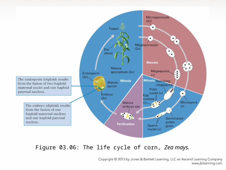
Figure 03.06: The life cycle of corn, Zea mays.

Outline of Meiosis• Prior to the first nuclear division, the members of
each pair of chromosomes become closely associated along their length
• The chromosomes that pair with each other are said to be homologous chromosomes
• Each member of a pair of homologs consists of a duplex of two sister chromatids joined at the centromere. The pairing of the homologous chromosomes, therefore, produces a four-stranded structure

• At the time of pairing, the homologs can exchange genes that results in chromosomes that consist of segments from one homolog intermixed with segments from the other
• In the first nuclear division, the homologous chromosomes are separated from each other, one member of each pair going to opposite poles of the spindle
• Two nuclei are formed, each containing a haploid set of duplex chromosomes
Outline of Meiosis

• The second nuclear division resembles a mitotic division, but there is no DNA replication
• At metaphase, the chromosomes align on the metaphase plate, and at anaphase, the chromatids are separated into opposite daughter nuclei
• The net effect of the two divisions is the creation of four haploid nuclei, each containing the equivalent of a single sister chromatid from each pair of homologous chromosomes
Outline of Meiosis

Meiosis I• The first meiotic division—reductional division,
reduces the chromosome number by half
• Prophase I is the longest stage and is commonly divided into five substages: leptotene, zygotene, pachytene, diplotene, and diakinesis
These are descriptive terms that indicate the appearance of the chromosomes at each substage

Meiosis: Prophase I• Leptotene – the chromosomes first become
visible as long, thread-like structures
• Zygotene – synapsis of homologous chromosomes = bivalent
• Pachytene – crossing-over between homologs
Parts A, B, and C courtesy of Marta Walters and Santa Barbara Botanic Gardens, Santa Barbara, California. Part D courtesy of Herbert Stern. Used with permission.

Figure 03.09AB: Bivalent consisting of a pair of homologous chromosomes.

Meiosis: Prophase I• Diplotene – chromosome repulsion, however, they
remain held together by cross-connections resulting from crossing-over. Each cross-connection, called a chiasma, is formed by a breakage and rejoining between nonsister chromatids
• Diakinesis – maximum chromosome contraction

Meiosis: Metaphase I
• As each bivalent moves onto the metaphase plate, its centromeres are oriented at random with respect to the poles of the spindle
• Genes on different chromosomes undergo independent assortment because nonhomologous chromosomes align at random in metaphase I
• Metaphase I – the bivalents positioned with the centromeres of the two homologs on opposite sides of the metaphase plate

Figure 03.11AB: Independent assortment of genes on nonhomologous chromosomes.

• Anaphase I – homologous chromosomes, each composed of two chromatids joined at an undivided centromere, separate from one another and move to opposite poles of the spindle
Meiosis: Anaphase I
• The physical separation of homologous chromosomes in anaphase I is the physical basis of Mendel’s principle of segregation

• Telophase I – a haploid set of chromosomes consisting of one homolog from each bivalent is located near each pole of the spindle
• The spindle breaks down, the chromosomes enter the second meiotic division after only a limited uncoiling
Meiosis: Telophase I
• Chromosome replication never takes place
between the two divisions

• The second meiotic division (meiosis II) is called the equational division because the chromosome number remains the same in each cell before and after the second division
• In some species, the chromosomes pass directly from telophase I to prophase II without loss of condensation
• After a short prophase II and the formation of second-division spindles, the centromeres of the chromosomes in each nucleus become aligned on the central plane of the spindle at metaphase II
Meiosis II

• In anaphase II, the centromeres divide and the chromatids of each chromosome move to opposite poles of the spindle
• Once the centromere has split at anaphase II, each chromatid is considered a separate chromosome
• Telophase II is a transition to the interphase condition of the chromosomes in the four haploid nuclei, accompanied by division of the cytoplasm.
Meiosis II

Meiosis vs. Mitosis• Meiosis produces four haploid cells: each
contains one copy of each pair of homologous chromosomes which are usually not genetically identical because of crossing-over associated with the formation of chiasmata during prophase of the first division
• Mitosis produces two diploid cells that contain both members of each pair of homologous chromosomes which are genetically identical

Chromosome Structure
• Eukaryotic chromosomes are highly coiled stable complexes of DNA and protein called chromatin
• Each eukaryotic chromosome contains a single DNA molecule of enormous length
• Some of the proteins present in chromatin determine chromosome structure and the changes in structure during the cell cycle
• Other chromatin proteins appear to have important roles in regulating chromosome functions

Figure 03.13A: Separations of chromosomes of yeast
Part A © 1988 Bio-Rad Laboratories, Inc.; permission to reproduce and to publish this image has been granted by Bio-Rad Laboratories, Inc. solely for the purpose of this publication. Any additional use outside of this textbook would require written permissioin.

Chromatin Structure
• The nucleosome is the basic structural unit of chromatin
• Each nucleosome is composed of a core particle, ~55 base pairs of DNA called linker DNA that links adjacent core particles and one molecule of histone H1 that binds to the core particle and to the linker DNA
• Histones are small proteins that are highly conserved among different organisms

Chromatin Structure• Each core particle consists of an octamere of pairs each
of histone H2A, H2B, H3, and H4; a segment of DNA containing about 145 base pairs
Figure 03.15A: Organization of nucleosomes.

Figure 03.15B: Organization of nucleosomes.

Chromatin Structure
• In the nucleus of a nondividing cell, chromatin fibers form discrete chromosome territories
• Chromosome territories are correlated with gene densities
• Territories of chromosome domains that are relatively gene rich tend to be located toward the interior of the nucleus
Figure 03.18: Chromosome territories formed by 30-nm chromatin fibers within the
nucleus of a nondividing cell.
Figure courtesy of Tobias A. Knoch, Erasmus MC, Rotterdam, and Kirchhoff-Institute for Physics, Ruperto-Carola University, Heidelberg

• Nucleosomes coil to form higher order DNA structure called the 30- nm chromatin fiber
Figure 03.19: Condensation of DNA and chromatin to form a metaphase chromosome.

Chromatin Structure• The spaces between the chromatin domains form a
network of channels large enough to allow passage of the molecular machinery for replication, transcription, and RNA processing
• Replication takes place in small discrete regions that exhibit a reproducible temporal and spatial pattern, and transcription takes place in a few hundred discrete locations
• The metaphase chromosome is a hierarchy of coiled coils

Chromatin Structure• Compact and heavily stained regions of chromatin
are known as heterochromatin, which mainly consists of highly repeated noncoding DNA sequences—satellite DNA
• The rest of the chromatin, which becomes visible only after chromosome condensation in mitosis or meiosis, is called euchromatin
• The number of genes located in heterochromatin is small relative to the number in euchromatin

Part A courtesy of T.C. Hsu, Ph.D., and used with permission of Sen Pathak, Ph.D., Anderson Cancer Center, University of Texas.
Figure 03.21A: Metaphase chromosomes of the ground squirrel.
Figure 03.21B: An interpretive drawing of metaphase chromosomes of the ground
squirrel.

Chromosome Structure
• The centromere is essential for chromosome segregation
• The centromere is a specific region of the eukaryotic chromosome. It serves as a central component of the kinetochore the complex of DNA and proteins to which the spindle fibers attach and move the chromosomes in both mitosis and meiosis

Figure 03.22: A yeast centromere.
Adapted from K. S. Bloom, M. Fitzgerald-Hayes, and J. Carbon, Cold Spring Harb. Symp. Quant. Biol. 47 (1982): 1175.

• The telomere is essential for the stability of the chromosome tips
• Due to the nature of DNA replication, chromosomes require special mechanism to restore DNA in telomeres in each cycle of replication
• The mechanism relies on an enzyme called telomerase
Chromosome Structure

Figure 03.25: The function of telomerase.

Figure 03.26: Telomere formation in Tetrahymena.

Figure 03.27: Chromosomal sex determination

Chromosomes and Heredity• Chromosome Theory of Heredity: Genes are
located in chromosomes
• Early evidence that genes are located on chromosomes was found by Thomas Hunt Morgan in 1910
• Morgan’s studied inheritance patterns in Drosophila melanogaster and found that in some cases reciprocal crosses yield different results

Morgan’s Fruit Fly Experiments• Morgan realized that it might happen if the alleles
for some genes were present in the X chromosome
• The X chromosome is transmitted in a different pattern by males and females, and the Y chromosome does not contain alleles homologous to genes on the X chromosome

Figure 03.29: A chromosomal interpretation of results obtained in F1 and F2 progenies in crosses of Drosophila.

Nondisjunction• Experimental proof of the chromosome theory of heredity
came from nondisjunction
• Nondisjunction = chromosomes fail to separate (disjoin) and move to opposite poles of the division spindle, results in loss or gain of a chromosome
• Calvin Bridges demonstrated that exceptional behavior of chromosomes is precisely paralleled by exceptional inheritance of their genes

Figure 03.32: The results of meiotic nondisjunction of the X chromosomes in a female Drosophila.

• Special chromosomes determine sex in many organisms• X and Y chromosomes = sex chromosomes, which are
non-identical but share some genes• In most organisms, the Y chromosome carries few genes
other than those related to male determination• X-linked genes are inherited according to sex• Hemophilia is a classic example of human X-linked
inheritance
X-Linked Inheritance

Figure 03.30: Genetic transmission of hemophilia A

• In many organisms, the male is the heterogametic sex • Males produce two different types of gametes: one
containing X and another Y chromosome• Females have two X chromosomes and produce only
X-bearing gametes• In some organisms (birds, butterflies, and some
reptiles), females are heterogametic
X-Linked Inheritance

Figure 03.31: Sex determination in birds: matched and unmatched sex chromosomes

Data analysis• Genetic data analysis makes use of probability and
statistics• Progeny of crosses are predicted by the binomial
probability• If the probability of possibility A is p and the
probability of the alternative possibility B is q, then the probability that, in n trials, A is realized s times and B is realized t times is
n!psqts!t!

Chi-Square Analysis• The test of goodness of fit = test analyzes whether
observed data agree with theoretical expectation
• A conventional measure of goodness of fit is a value called chi-square, c2
• c2 = ∑(observed – expected)2 / expected
• A value of c2 = 0 means that the observed numbers fit the expected numbers perfectly

Chi-Square Analysis• Probability P that a worse fit (or one equally bad)
would be obtained by chance, assuming that the genetic hypothesis is true
• The critical values of P are conventionally chosen as 0.05 (the 5 percent level) and 0.01 (the 1 percent level)
• Statistically significant refers to the magnitude of the difference between the observed and the expected numbers

Chi-Square Analysis
• To determine the P value corresponding to a calculated c2 we need the number of degrees of freedom of the particular chi-square test
• The number of degrees of freedom equals the number of classes of data minus 1

Figure 03.34: Graphs for interpreting goodness of fit to genetic predictions using the chi-square test.