Embed Size (px)
Citation preview

Chapter 3
The Molecules of Life

Biology and Society: Got Lactose?
• Lactose is the main sugar found in milk.
• Lactose intolerance is the inability to properly digest lactose.• Instead of lactose being broken down and absorbed
in the small intestine, lactose is broken down by bacteria in the large intestine, producing gas and discomfort.

ORGANIC COMPOUNDS
• A cell is mostly water.
• The rest of the cell consists mainly of carbon-based molecules.
• Carbon forms large, complex, and diverse molecules necessary for life’s functions.
• Organic compounds are carbon-based molecules.

Carbon Chemistry
Carbon is a versatile atom.• It has four electrons in an outer shell that holds
eight electrons.
• Carbon can share its electrons with other atoms to form up to four covalent bonds.

• The simplest organic compounds are hydrocarbons, which contain only carbon and hydrogen atoms.
• The simplest hydrocarbon is methane, a single carbon atom bonded to four hydrogen atoms.
Carbon Chemistry

• Larger hydrocarbons form fuels for engines.
• Hydrocarbons of fat molecules are important fuels for our bodies.
Carbon Chemistry
C8H18
CH3(CH2)16CO2H

• Each type of organic molecule has a unique three-dimensional shape.
• The shapes of organic molecules relate to their functions.
• The groups of atoms that usually participate in chemical reactions are called functional groups. Two common examples are
• hydroxyl groups (-OH) and
• carboxyl groups (-COOH).
Carbon Chemistry

Giant Molecules from Smaller Building Blocks
• On a molecular scale, many of life’s molecules are gigantic, earning the name
macromolecules.• Three categories of macromolecules are• carbohydrates
• proteins
• nucleic acids
• Lipids are technically not macromolecules

• Most macromolecules are polymers.
• Polymers are made by stringing together many smaller molecules called monomers.
Giant Molecules from Smaller Building Blocks

H2O
OH H
A dehydration reaction• links two monomers together and
• removes a molecule of water

• Organisms also have to break down macromolecules.
• Digestion breaks down macromolecules to make monomers available to your cells.
Giant Molecules from Smaller Building Blocks

• Hydrolysis
– breaks bonds between monomers,
– adds a molecule of water, and
– reverses the dehydration reaction.
Giant Molecules from Smaller Building Blocks

LARGE BIOLOGICAL MOLECULES
• There are four categories of large biological molecules:• carbohydrates
• lipids
• proteins
• nucleic acids

Carbohydrates
• Carbohydrates include sugars and polymers of sugar.• small sugar molecules in energy drinks
• long starch molecules in spaghetti and French fries.
• molecules made up of C, H, and O in a 1:2:1 ratio

Carbohydrates• In animals
• a primary source of dietary energy
• raw material for manufacturing other kinds of organic compounds.
• used for structural purposes
• In plants,
• carbohydrates serve as a building material for much of the plant body.

Carbohydrates
3 kinds of carbohydrates
• Monosaccharides
• Disaccharides
• Polysaccharides

Monosaccharides
• Monosaccharides
• simple sugars that cannot be broken down by hydrolysis into smaller sugars
• the monomers of carbohydrates.
• Common examples
• glucose in sports drinks
• fructose found in fruit.

• Monosaccharides are the main fuels for cellular work.
• In water, many monosaccharides form rings.
Monosaccharides

Monosaccharides
• Both glucose and fructose are found in honey.
• Glucose and fructose are isomers, molecules that have the same molecular formula but different structures.
• Isomers are like anagrams – heart and earth

Disaccharides
Disaccharide• a double sugar
• constructed from two monosaccharides
• formed by a dehydration reaction.
(C12H22O11)3

Figure 3.7a
Galactose
H2O
OH H
Glucose
Lactose

• Disaccharides include• lactose in milk
• maltose in beer, malted milk shakes, and malted milk ball candy
• sucrose in table sugar
Disaccharides

• Sucrose
• the main carbohydrate in plant sap
• rarely used as a sweetener in processed foods in the United States.
• High-fructose corn syrup
• made by a commercial process that converts natural glucose in corn syrup to much sweeter fructose.
Disaccharides

• The United States is one of the world’s leading markets for sweeteners.
• The average American consumes
• about 45 kg of sugar (about 100 lb) per year,
• mainly as sucrose and high-fructose corn syrup.
Disaccharides

Polysaccharides
• Polysaccharides
• complex carbohydrates
• made of long chains of sugar units—polymers of monosaccharides.


Starch
• is a familiar example of a polysaccharide
• is used by plant cells to store energy
• consists of long strings of glucose monomers.
• Potatoes and grains are major sources of starch in our diet.
Polysaccharides

Glycogen• used by animals cells to store energy
• converted to glucose when it is needed.
Polysaccharides

Cellulose• is the most abundant organic compound on Earth
• forms cable-like fibrils in the walls that enclose plant cells
• cannot be broken apart by most animals.
Polysaccharides


• Monosaccharides and disaccharides dissolve readily in water.
• Cellulose does not dissolve in water, but water does stick to it!!!
• Almost all carbohydrates are hydrophilic, or “water-loving,” adhering water to their surface.
Polysaccharides


Lipids
• Lipids• consists of two categories; fats and steroids
• made up of mostly C and H atoms, some O atoms
• neither macromolecules nor polymers
• Hydrophobic = unable to mix with water.
Fats
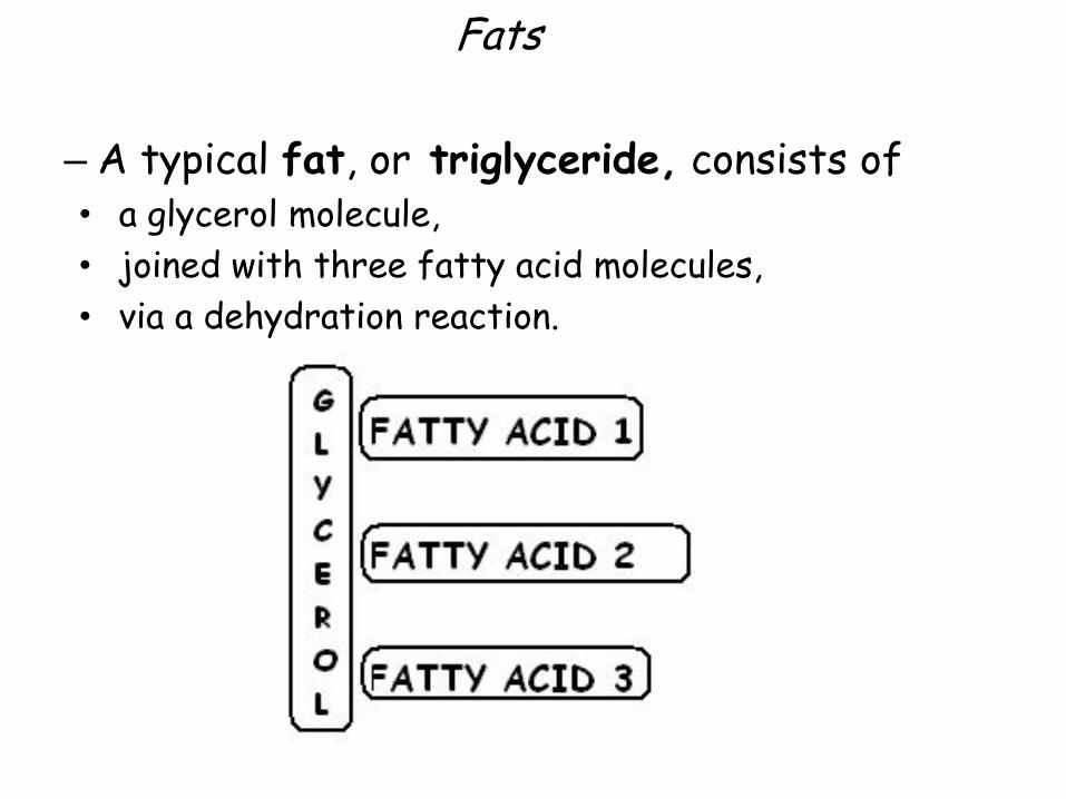
– A typical fat, or triglyceride, consists of• a glycerol molecule,
• joined with three fatty acid molecules,
• via a dehydration reaction.

Figure 3.11a
Fatty acid
H2O
HOH
Glycerol
(a) A dehydration reaction linking a fatty acid to glycerol

Figure 3.11
Fatty acid
H2O
HOH
Glycerol
(a) A dehydration reaction linking a fatty acid to glycerol
(b) A fat molecule with a glycerol “head” and three
energy-rich hydrocarbon fatty acid “tails”

Figure 3.11b
(b) A fat molecule with a glycerol “head” and three
energy-rich hydrocarbon fatty acid “tails”

– Fats perform essential functions in the human body including
• energy storage – more energy in fat than in carbs
• cushioning
• insulation
• plasma membrane of cells – phospholipid bilayer
Fats

– If the carbon skeleton of a fatty acid• has fewer than the maximum number of hydrogens,
it is unsaturated;
• if it has the maximum number of hydrogens, it is saturated.
– A saturated fat has• no double bonds
• all three of its fatty acids saturated.
Fats

Fats

Figure 3.12a
Saturated Fats

Figure 3.12b
Omega-3 fatsTrans fatsPlant oils
Margarine
Unsaturated Fats

– Most animal fats are saturated
• have a high proportion of saturated fatty acids
• can easily stack, tending to be solid at room temperature
• contribute to atherosclerosis, in which lipid-containing plaques build up along the inside walls of blood vessels.
Fats

– Most plant and fish oils tend to be• high in unsaturated fatty acids
• liquid at room temperature.
Fats

• Hydrogenation adds hydrogen atoms to fatty acid tails
• converts unsaturated fats to saturated fats
• makes liquid fats solid at room temperature
• creates trans fat, a type of unsaturated fat that is particularly bad for your health
Fats
trans fat levels of less than 0.5 grams per serving can be listed as 0 grams trans fat on the food label.

Steroids
– Steroids are very different from fats in structure and function.
• The carbon skeleton is bent to form four fused rings.
• Steroids vary in the functional groups attached to this set of rings, and these chemical variations affect their function.

Steroids
– Cholesterol is• a key component of
cell membranes
• the “base steroid” from which your body produces other steroids, such as estrogen and testosterone, which are chemical messengers.

Figure 3.13
Cholesterol can be convertedby the body to
Testosterone A type of estrogen

Proteins
Proteins• composed of C, H, N, O and S.
• are polymers constructed from amino acid monomers
• account for more than 50% of the dry weight of most cells
• perform most of the tasks required for life
• form enzymes, chemicals that change the rate of a chemical reaction without being changed in the process.
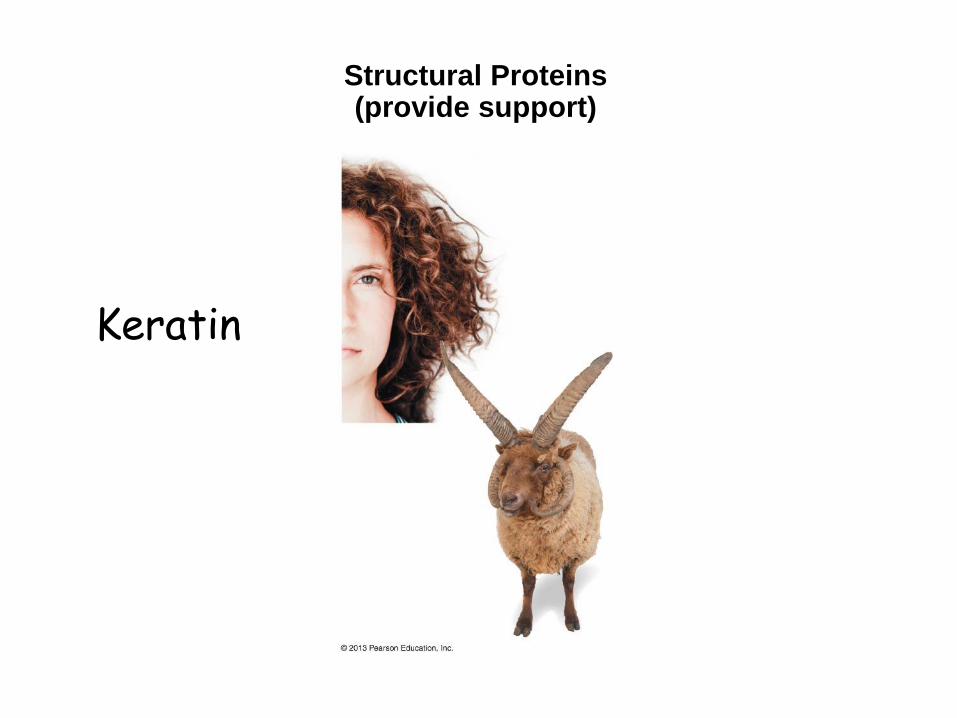
Structural Proteins(provide support)
Keratin

Storage Proteins(provide amino
acids for growth)

ContractileProteins
(help movement)
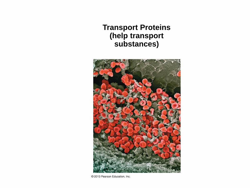
Transport Proteins(help transport
substances)
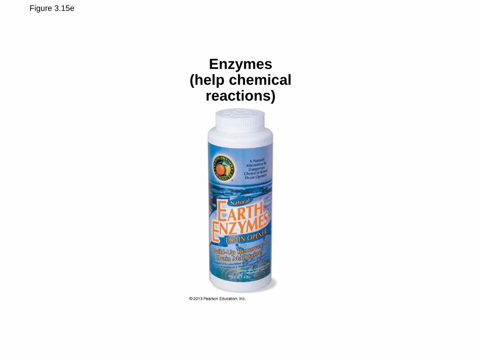
Figure 3.15e
Enzymes(help chemical
reactions)
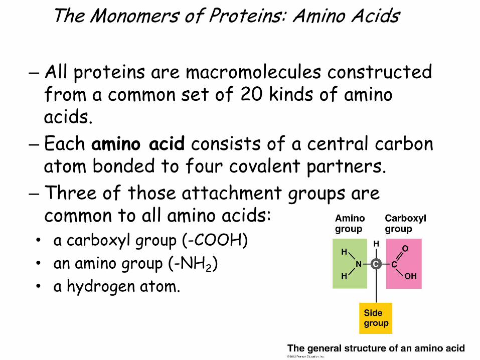
The Monomers of Proteins: Amino Acids
– All proteins are macromolecules constructed from a common set of 20 kinds of amino acids.
– Each amino acid consists of a central carbon atom bonded to four covalent partners.
– Three of those attachment groups are common to all amino acids:
• a carboxyl group (-COOH)
• an amino group (-NH2)
• a hydrogen atom.

Figure 3.16a
Aminogroup
Carboxylgroup
Sidegroup
The general structure of an amino acid

Figure 3.16b
Hydrophobicside group
Hydrophilicside group
Leucine Serine

Proteins as Polymers
• Cells link amino acids together• by dehydration reactions
• forming peptide bonds
• creating long chains of amino acids called polypeptides.

Figure 3.17-1Carboxyl Amino
OH H

Figure 3.17-2
Dehydration reaction
Carboxyl Amino
Peptide bond
H2O
OH H

• Your body has tens of thousands of different kinds of protein each with a different function!!!!
• Proteins differ in their arrangement of amino acids.
• The specific sequence of amino acids in a protein is its primary structure.
Proteins as Polymers

Amino acid
35
1 5
30
45
65
40
15
70
10
25
50 55
20
75
60
8580
95 90
115
100
105
110
120125
129
Primary Structure

• A slight change in the primary structure of a protein affects its ability to function.
• The substitution of one amino acid for another in hemoglobin causes sickle-cell disease, an inherited blood disorder.
Proteins as Polymers

Normal red blood cell
Sickled red blood cell Sickle-cell hemoglobin
Normal hemoglobin
1 2 3 45
6 7. . . 146
1 2 3 4 56 7. . . 146
SE
MS
EM
Leu
Leu
The sickle-cell disease occurs when the sixth amino acid, glutamic
acid, is replaced by valine to change its structure and function; as such,
sickle cell anemia is also known as E6V. Valine is hydrophobic,
causing the haemoglobin to collapse in on itself occasionally. The
structure is not changed otherwise. When enough haemoglobin
collapses in on itself the red blood cells become sickle-shaped.

Protein Shape
• A functional protein consists of• one or more polypeptide chains
• precisely twisted, folded, and coiled into a molecule of unique shape.

Protein Shape
– Proteins consisting of one polypeptide have three levels of structure.
– Proteins consisting of more than one polypeptide chain have a fourth level, quaternary structure.

Figure 3.20-1
(a) Primarystructure

Figure 3.20-2
(a) Primarystructure
(b) Secondary structure
Aminoacids
Pleated sheet
Alpha helix
Hydrogenbond

Figure 3.20-3
(a) Primarystructure
(b) Secondary structure
Aminoacids
Pleated sheet
Alpha helix
(c) Tertiarystructure
Polypeptide
Hydrogenbond

Figure 3.20-4
(a) Primarystructure
(b) Secondary structure
Aminoacids
Pleated sheet
Alpha helix
(c) Tertiarystructure
Polypeptide
(d) Quaternarystructure
A protein withfour polypeptidesubunits
Hydrogenbond

– A protein’s three-dimensional shape• typically recognizes and binds to another molecule
• enables the protein to carry out its specific function in a cell.
Protein Shape

What Determines Protein Shape?
– A protein’s shape is sensitive to the surrounding environment.
– An unfavorable change in temperature and/or pH can cause denaturation of a protein, in which it unravels and loses its shape.
– High fevers (above 104F) in humans can cause some proteins to denature.

– Misfolded proteins are associated with• Alzheimer’s disease
• mad cow disease
• Parkinson’s disease
• Sickle Cell Anemia
What Determines Protein Shape?

Nucleic Acids
– Nucleic acids are macromolecules that • store information,
• provide the directions for building proteins,
• include DNA and RNA– Deoxyribonucleic acid
– Ribonucleic acid

– DNA resides in cells in long fibers called chromosomes.
– A gene is a specific stretch of DNA that programs the amino acid sequence of a polypeptide.
– The chemical code of DNA must be translated from “nucleic acid language” to “protein language.”
Nucleic Acids

Gene
DNA
RNA
Protein
Amino acid
Nucleic
acids

– Nucleic acids are polymers made from monomers called nucleotides.
– Each nucleotide has three parts:1. a five-carbon sugar,
2. a phosphate group, and
3. a nitrogen-containing base.
Nucleic Acids
Phosphate group
Nitrogen base
5-carbon sugar

Figure 3.23a
Nitrogenous base(A, G, C, or T)
Thymine (T)
Phosphategroup
Sugar(deoxyribose)
(a) Atomic structure

Figure 3.24
Adenine (A) Guanine (G)
Thymine (T) Cytosine (C)
Adenine (A) Guanine (G) Thymine (T) Cytosine (C)
Space-filling model of DNA
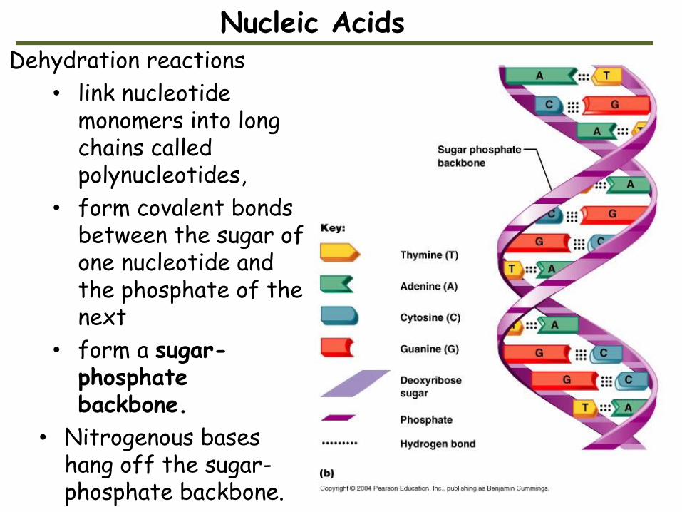
Dehydration reactions
• link nucleotide monomers into long chains called polynucleotides,
• form covalent bonds between the sugar of one nucleotide and the phosphate of the next
• form a sugar-phosphate backbone.
• Nitrogenous bases hang off the sugar-phosphate backbone.
Nucleic Acids


Figure 3.25
Sugar-phosphatebackbone
NucleotideBasepair
Hydrogenbond
Bases
(a) DNA strand(polynucleotide)
(b) Double helix(two polynucleotide strands)
T
G
C
T
G
A
T
G
C
A
C
T
A
A
A
T
A T
AT
G


Nucleic Acids
• Two strands of DNA join together to form a double helix.
• Bases along one DNA strand hydrogen-bond to bases along the other strand.
• The functional groups hanging off the base determine which bases pair up:
– A only pairs with T and
– G can only pair with C.

– RNA, ribonucleic acid, is different from DNA.• RNA uses the sugar ribose instead of deoxyribose
• RNA uses the base uracil (U) instead of thymine (T).
• RNA is usually single-stranded, but DNA usually exists as a double helix.
Nucleic Acids

Figure 3.26
Phosphate
group
Nitrogenous base
(A, G, C, or U)
Uracil (U)
Sugar (ribose)

ENZYMES are PROTEINS!!
• Metabolism is the total of all chemical reactions in an organism.
• Most metabolic reactions require the assistance of enzymes, proteins that speed up chemical reactions.
• All living cells contain thousands of different enzymes, each promoting a different chemical reaction.

Activation Energy
• Activation energy is the energy needed to get a reaction started.
• It works by• activating the reactants• triggering a chemical reaction.• Enzymes reduce the amount of activation energy
required to break bonds of reactant molecules.
Small input needed to
trigger a large
output.

Figure 5.7a
(a) Without enzyme
Reactant
Products
Activation
energy barrier
En
erg
y

Figure 5.7b
(b) With enzyme
Reactant
Products
Activation
energy barrier
reduced by
enzyme
Enzyme
En
erg
y

Induced Fit
• An enzyme is very selective in the reaction it catalyzes (a catalyst is a substance that speeds up the rate of reaction)
• Each enzyme recognizes a substrate, a specific reactant molecule.
– The active site fits to the substrate, and the enzyme changes shape slightly.
– This interaction is called induced fit because the entry of the substrate induces the enzyme to change shape slightly.

• Enzymes can function over and over again, a key characteristic of enzymes.
• Many enzymes are named for their substrates, but with an –ase ending. Example = lactase.
Induced Fit

Figure 5.9-1
Active site
Enzyme
(sucrase)
Ready for
substrate
1

Figure 5.9-2
Active site
Enzyme
(sucrase)
Ready for
substrate
Substrate (sucrose)
Substrate
binding
1
2
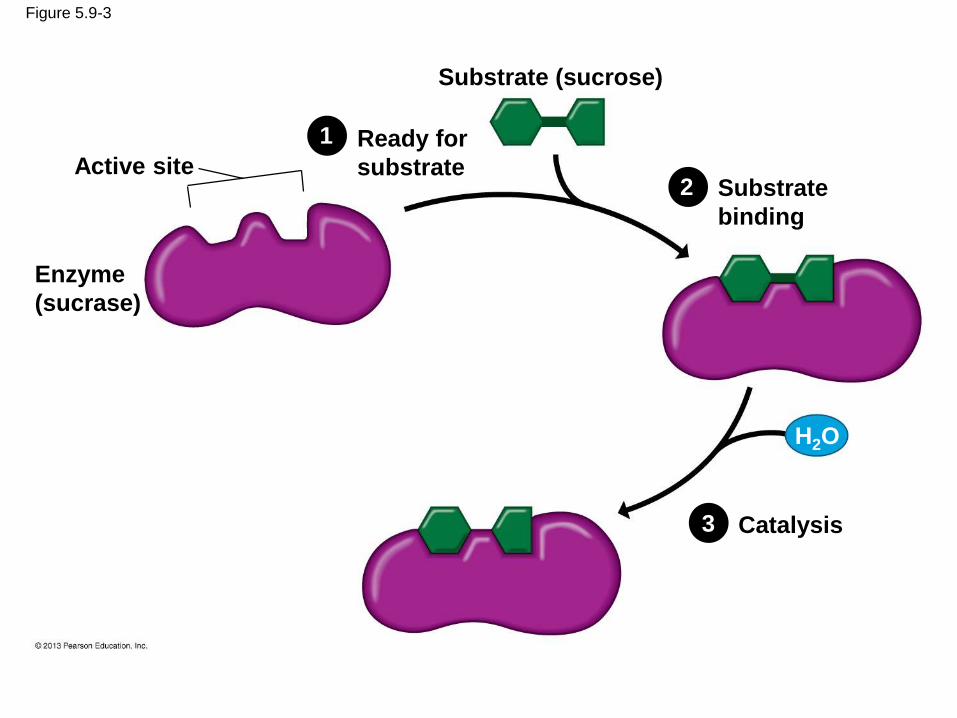
Figure 5.9-3
Active site
Enzyme
(sucrase)
Ready for
substrate
Substrate (sucrose)
Substrate
binding
Catalysis
H2O
1
2
3

Figure 5.9-4
Active site
Enzyme
(sucrase)
Ready for
substrate
Substrate (sucrose)
Substrate
binding
Catalysis
H2O
Fructose
Glucose
Product
released
4
1
2
3

Enzyme Inhibitors
• Enzyme inhibitors can prevent metabolic reactions by binding
– to the active site
– near the active site, resulting in changes to the enzyme’s shape so that the active site no longer accepts the substrate.

Figure 5.10a
(a) Enzyme and substrate binding normally
Substrate
Enzyme
Active site

Figure 5.10b
(b) Enzyme inhibition by a substrate imposter
Substrate
Active site
Inhibitor
Enzyme

Figure 5.10c
(c) Inhibition of an enzyme by a molecule
that causes the active site to change shape
SubstrateActive site
Inhibitor
Enzyme

• Some products of a reaction may inhibit the enzyme required for its production.
– This is called feedback regulation.
– It prevents the cell from wasting resources.
• Many beneficial drugs work by inhibiting enzymes.
– Penicillin blocks the active site of an enzyme that bacteria use in making cell walls.
– Ibuprofen inhibits an enzyme involved in sending pain signals.
– Many cancer drugs inhibit enzymes that promote cell division.
Enzyme Inhibitors

The Process of Science: Does Lactose Intolerance Have a Genetic Basis?
– Observation: Most lactose-intolerant people have a normal version of the lactase gene.
– Question: What is the genetic basis for lactose intolerance?
– Hypothesis: Lactose-intolerant people have a mutation but not within the lactase gene.
© 2013 Pearson Education, Inc.

– Prediction: A mutation would be found near the lactase gene.
– Experiment: Genes of 196 lactose-intolerant people were examined.
– Results: Researchers found a 100% correlation between lactose intolerance and a nucleotide at a site approximately 14,000 nucleotides away from the lactase gene.
The Process of Science:
Does Lactose Intolerance Have a Genetic Basis?
© 2013 Pearson Education, Inc.

Figure 3.27
DNA
Human
cell Chromosome 2 Section
ofchromosome 2
Lactase gene
C or T

Evolution Connection: The Evolution of Lactose Intolerance in Humans
– Most people are lactose-intolerant as adults.
– Lactose intolerance is found in
• 80% of African Americans and Native Americans,
• 90% of Asian Americans, but
• only 10% of Americans of northern European descent.
© 2013 Pearson Education, Inc.

– Lactose tolerance appears to have evolved in northern European cultures that relied upon dairy products.
– Ethnic groups in East Africa that rely upon dairy products are also lactose tolerant but due to different mutations.
Evolution Connection:
The Evolution of Lactose Intolerance in Humans
© 2013 Pearson Education, Inc.

Figure 3.28