Embed Size (px)
Citation preview
Trichothecene Mycotoxins
655
Chapter 34
TRICHOTHECENE MYCOTOXINS
ROBERT W. WANNEMACHER, JR., P H.D.*; AND STANLEY L. WIENER, M.D.†
INTRODUCTION
HISTORY AND MILITARY SIGNIFICANCEUse in Biological WarfareThe Yellow Rain ControversyWeaponization
DESCRIPTION OF THE AGENTOccurrence in NatureChemical and Physical Properties
TOXICOLOGY AND TOXICOKINETICSMechanism of ActionMetabolism
CLINICAL DISEASEAcute EffectsChronic Toxicity
DIAGNOSISBattlefield DiagnosisConfirmatory Procedures
MEDICAL MANAGEMENTIndividual and UnitSpecific or Supportive TherapyProphylaxis
SUMMARY
*Assistant Chief, Toxinology Division, U.S. Army Medical Research Institute of Infectious Diseases, Fort Detrick, Frederick, Maryland 21702-5011†Colonel, Medical Corps, U.S. Army Reserve; Professor of Medicine and Chief, Section of General Internal Medicine, Department of Medicine,University of Illinois College of Medicine, 840 Wood Street, Chicago, Illinois 60612
Medical Aspects of Chemical and Biological Warfare
656
INTRODUCTION
mental conditions. The distinguishing chemicalfeature of trichothecenes is the presence of atrichothecene ring, which contains an olefinic bondat C-9, 10; and an epoxide group at C-12, 12.5 Alltrichothecenes are mycotoxins, but not all mycotox-ins are trichothecenes. This family of mycotoxinscauses multiorgan effects including emesis and di-arrhea, weight loss, nervous disorders, cardiovas-cular alterations, immunodepression, hemostaticderangements, skin toxicity, decreased reproductivecapacity, and bone marrow damage.4,6
In this chapter, we will concentrate on T-2 myco-toxin, a highly toxic trichothecene that, togetherwith some closely related compounds, has been thecausative agent of a number of illnesses in humansand domestic animals. 1,2,4 During the 1970s and1980s, the trichothecene mycotoxins gained somenotoriety as putative biological warfare agentswhen they were implicated in “yellow rain” attacksin Southeast Asia.7–11
Mycotoxins, by-products of fungal metabolism,have been implicated as causative agents of ad-verse health effects in humans and animals thathave consumed fungus-infected agricultural prod-ucts.1,2 Consequently, fungi that produce mycotox-ins, as well as the mycotoxins themselves, are po-tential problems from both public health andeconomic perspectives. The fungi are a vast assem-blage of living organisms, but mycotoxin pro-duction is most commonly associated with theterrestrial filamentous fungi called the molds. 3
Various genera of toxigenic fungi are capableof producing such diverse mycotoxins as the afla-toxins, rubratoxins, ochratoxins, fumonisins, andtrichothecenes.1,2
The trichothecenes are a very large family ofchemically related toxins produced by various spe-cies of Fusarium, Myrotecium, Trichoderma, Cepha-losporium, Verticimonosporium, and Stachybotrys.4
They are markedly stable under different environ-
HISTORY AND MILITARY SIGNIFICANCE
Fungi that produce trichothecenes are plantpathogens and invade various agricultural productsand plants. Since Fusarium and other related fungiinfect important foodstuff, they have been associ-ated worldwide with intoxication of humans andanimals. Thus, these fungi have potential as bio-logical weapons.
Use in Biological Warfare
From 1974 to 1981, toxic agents were used by theSoviet Union and its client states in such Cold Warsites as Afghanistan, Laos, and Kampuchea (Cam-bodia). Aerosol-and-droplet clouds were producedby delivery systems in the Soviet arsenal such asaircraft spray tanks, aircraft-launched rockets,bombs (exploding cylinders), canisters, a Soviethand-held weapon (DH-10), and booby traps. Air-craft used for delivery included L-19s, AN-2s, T-28s,T-41s, MiG-21s (in Laos) and Soviet MI-24 helicop-ters (in Afghanistan and Laos).
Attacks in Laos (1975–1981) were directed againstHmong villagers and resistance forces who opposedthe Lao People’s Liberation Army and the NorthVietnamese. In Kampuchea, North Vietnamesetroops used 60-mm mortar shells; 120-mm shells;107-mm rockets; M-79 grenade launchers contain-ing chemicals; and chemical rockets, bombs, and
sprays delivered by T-28 aircraft (1979–1981) againstKhmer Rouge troops. The chemical munitions weresupplied by the Soviets and delivered by NorthVietnamese or Laotian pilots. In Afghanistan, thechemical weapons were delivered by Soviet or Af-ghan pilots against Mujahidin guerrillas (1979–1981). Lethality of the attacks is documented by aminimum of 6,310 deaths in Laos (from 226 attacks);981 deaths in Kampuchea (from 124 attacks); and3,042 deaths in Afghanistan (from 47 attacks). 7
Trichothecenes appear to have been used in someof these attacks.
The air attacks in Laos have been described as“yellow rain” and consisted of a shower of sticky,yellow liquid that sounded like rain as it fell fromthe sky. Other accounts described a yellow cloudof dust or powder, a mist, smoke, or insect spray–like material. Liquid agent rapidly dried to apowder. In Laos, 50% to 81%7 of attacks involvedmaterial associated with a yellow pigment. Otherattacks were associated with red, green, white, orbrown smoke or vapor. More than 80%7 of attackswere delivered by air-to-surface rockets; the remain-der, from aircraft spray tanks or bombs. Intelligenceinformation and some of the victims’ descriptionsof symptoms raised the possibility that chemicalwarfare agents such as phosgene, sarin, soman,mustards, CS, phosgene oxime, or BZ may also
Trichothecene Mycotoxins
657
have been used. These agents may have been usedin mixtures or alone, and with or without thetrichothecenes.
Unconfirmed reports have implicated the useof trichothecenes in the 1964 Egyptian (or Rus-sian) attacks on Yemeni Royalists in Yemen12 and incombination with mustards during chemical war-fare attacks in the Iran–Iraq War (1983–1984).13 Ac-cording to European sources, Soviet–Cuban forcesin Cuba are said to have been equipped with myco-toxins, and a Cuban agent is said to have died of ahemorrhagic syndrome induced by a mycotoxinagent.14
The Yellow Rain Controversy
Actual biological warfare use of trichothecenesin Southeast Asia and Afghanistan is strongly sup-ported by the epidemiological and intelligence as-sessments and trichothecene assays, although re-ports in the open literature have discounted thiscontention. An article written by L. R. Ember, 15
published in 1984 in Chemical Engineering News, isthe most exhaustive and authoritative account ofthe controversy surrounding the use of tricho-thecene mycotoxins in Southeast Asia during the1970s.
The United States government, its allies, andjournalists exhaustively studied the possibility thatyellow rain attacks had occurred, based on evi-dence 7,14,15 such as the following:
• interviews of Hmong survivors of and eye-witnesses to lethal yellow rain attacks inLaos, who provided consistent descriptionsof the episodes;
• interrogations of a defecting Laotian AirForce officer and North Vietnamese groundtroops, who corroborated the descriptionsof attacks and admitted using the chemi-cals;
• interrogations of prisoners of war, who ad-mitted being involved in attacks where un-conventional weapons were used (ie, inAfghanistan);
• laboratory confirmations of Soviet use ofchemical agents, and
• the presence of Soviet-manufactured chemi-cal agents and Soviet technicians in Laos.
The evidence supports the contention that tricho-thecene mycotoxins were used as biological war-fare agents in Southeast Asia and Afghanistan bythe former Soviet Union and its surrogates. The
Russians have not recently denied such use but havedeclined to discuss the subject.
In addition to the evidence stated above, elevatedlevels and naturally rare mixtures of trichothe-cene toxins were recovered from the surfaces ofplants, fragments of plastic, and rocks in areas at-tacked9,11,15,16; and were detected in the blood ofattack survivors and the tissues of a dead casu-alty.10,15 Control samples that were taken (a) froman environment that had not been attacked, andduring another season of the year,15 and (b) fromHmong who had never been exposed to an attackwere consistently negative.
The evidence that trichothecenes were used inSoutheast Asia has been challenged: questions havebeen raised about the interview methodology usedby U.S. Army physicians and U.S. State Departmentpersonnel in Hmong refugee camps in Thailand toobtain descriptions of the attacks. Some inconsis-tencies of specific individuals’ stories were demon-strated, but the frequency of unreliable informationhas not been reported and is unlikely to be largeenough to discredit all witnesses.15 Symptom de-scriptions are generally consistent with knowntrichothecene effects.
The paucity of positive evidence of the presenceof trichothecenes (5 positive environmental and 20positive biomedical samples) has been used to chal-lenge the belief that biological warfare attacks oc-curred, since only 10% of samples were positive.However, 32% of samples from victims were posi-tive, a value too high for natural causes (eg, foodcontamination) to be used as an explanation, since98% of controls in nonattack areas of Thailand werenegative.17 The 2% of samples that were positivecould represent either a nonspecific result or low-prevalence food contamination. The paucity andtype of control samples have also been questioned.
Some experts18–21 have claimed that yellow rainwas not a biological warfare attack at all, but thatthe yellow residue was caused by showers and de-posits of bee feces—the result of massive beeswarming and cleansing–defecation flights oversome areas of Southeast Asia. The presence of pol-len in bee feces and some samples has not onlyadded confusion18 but is also the supporting evi-dence used by the skeptics. It is important to re-member that persons caught in a shower of bee fe-ces do not get sick and die. Although bee flightshave occurred before and since 1982, reports of at-tacks of yellow rain and death in Asia have not.
Then what explains the symptoms consistentwith trichothecene effects in the casualties, and thepollen and bee feces in some of the yellow spots on
Medical Aspects of Chemical and Biological Warfare
658
vegetation in the area? Bee feces do not containtrichothecenes, yet pollen and trichothecenes with-out mold are found together in some samples fromattack areas. The most likely explanation is thatduring biological warfare attacks, dispersedtrichothecenes landed in pollen-containing areas.
French scientists have reported the simultaneoussynthesis of three trichothecene toxins by Fusariumgrowing on corn, but actual production of thesetoxins by Fusarium species in Southeast Asia has notbeen demonstrated, presumably because of highenvironmental temperature (ie, toxin productionusually increases at low temperatures). Whetheror not Fusarium toxin is produced in the high-moun-tain temperate regions of Laos inhabited by theHmong remains unanswered. The presence of toxinon leaves without accompanying mold also is un-explained by critics of the trichothecene weaponhypothesis. In vivo studies have demonstrated thatF semitectum var semitectum will grow on leaves inSoutheast Asia, but have not shown that it will pro-duce toxin in vivo.15
In support of the weapon hypothesis are the posi-tive trichothecene analyses performed by two lead-ing researchers9,10 in the detection of trichothecenes;the Defense Research Establishment, Ottawa,Canada11,22; and the U.S. Army Chemical Researchand Development Center, Edgewood, Maryland.23
Negative results of analyses of biomedical and en-vironmental samples from Southeast Asia havecome from Porton Down Laboratory in England,17,24
but according to the British, such results do not ex-clude sampling problems, including delay insample collection after an attack, as a cause of thenegative results.15
Proponents have been accused of analyzingsamples that were purposely contaminated withtoxin, either after collection or during the analysis.Other methodological criticisms include poor recov-ery (< 10% of one sample spiked with T-2 toxin);low precision of quantitative data when analyzingtwo portions of the same leaf; and lack of well-docu-mented, confirming, replicate analyses in Mirocha’sor a similarly equipped second laboratory.15 Thepresence of polyethylene glycol in the sample ana-lyzed by Rosen9 also indicates that the trichothecenemixture detected was manufactured, not natural.
Many experts in the intelligence community,16
academia, 8,9 the U.S. Department of State, 7 andthe authors of this chapter believe that tricho-thecenes were used as biological weapons in South-east Asia and Afghanistan. However, a weaponcontaining trichothecenes was not found in South-
east Asia, and the Soviets have not declared anystockpiles of trichothecenes among their chemicalor biological weapons. Thus, it has not been pos-sible for the United States to prove unequivocallythat trichothecene mycotoxins were used as biologi-cal weapons.
Weaponization
Trichothecene mycotoxins can be delivered asdusts, droplets, aerosols, or smoke from aircraft,rockets, missiles, artillery, mines, or portable spray-ers. Because of their antipersonnel properties, easeof large-scale production, and apparent proven de-livery by various aerial dispersal systems, thetrichothecene mycotoxins (especially T-2 toxin)have an excellent potential for weaponization.
When delivered at low doses, trichothecene my-cotoxins cause skin, eye, and gastrointestinal prob-lems. In nanogram amounts,4,25 they (T-2 toxin, inparticular) cause severe skin irritation (erythema,edema, and necrosis).4,6 Skin vesication has beenobserved in a number of humans exposed to yel-low rain attacks.4,14,15 T-2 toxin is about 400-foldmore potent (50 ng vs 20 µg) than mustard in pro-ducing skin injury.26 Lower-microgram quantitiesof trichothecene mycotoxins cause severe eye irri-tation, corneal damage, and impaired vision.4,16,26,27
Emesis and diarrhea have been observed at amountsthat are one fifth to one tenth the lethal doses oftrichothecene mycotoxins.26
Depending on the species of experimental ani-mal tested and the exposure procedure,28,29 the le-thality of T-2 toxin by aerosol exposure can be 10-to 50-fold greater than when injected parenterally.30
With larger doses in humans, aerosolized tricho-thecenes may produce death within minutes tohours.7,14,15 The term LCt50 (the concentration • timethat is lethal to 50% of the exposed population) isused to describe exposure to vapors and aerosols;milligrams • minutes per cubic meter is the con-ventional unit of measurement. LCt50 and its rela-tion to LD50 (the dose that is lethal to 50% of theexposed population) are discussed in detail inChapter 5, Nerve Agents, and will not be furtherexplicated here.
The toxicity of T-2 toxin by the inhalational routeof exposure (LCt50 range: 200–5,800 mg•min/m3)28–30
is similar to that observed for mustards or Lewisite(LC t50 range: 1,500–1,800 mg•min/m3). 31 How-ever, the lethality of T-2 toxin by the dermal route(LD50 range: 2–12 mg/kg6) is higher than that forliquid Lewisite (LD 50 : approximately 30 mg/
Trichothecene Mycotoxins
659
kg31(p39)) or liquid mustards (LD50: approximately100 mg/kg 31(p32)). Therefore, the trichothecenemycotoxins are considered to be primarily blisteragents that, at lower exposure concentrations, cancause severe skin and eye irritation, and at largerdoses can produce considerable incapacitation anddeath within minutes to hours.
By solid substrate fermentation, T-2 toxin can beproduced at approximately 9 g/kg of substrate,with a yield of 2 to 3 g of crystalline product.32 Sev-eral of the trichothecene mycotoxins have been pro-duced in liquid culture at medium yields and largevolumes of culture for extraction. 33 Thus, usingexisting state-of-the-art fermentation processes that
were developed for brewing and antibiotics, itwould be fairly simple to produce ton quantities ofa number of the trichothecene mycotoxins.
In Southeast Asia, most of the yellow rain attackswere delivered by aircraft or helicopter spray,bombs, and air-to-surface rockets. The attacks weredescribed as a shower of sticky liquid, a yellowcloud of dust or powder, or a mist (like an insectspray).7,15 The delivery of the trichothecene myco-toxins was similar in many aspects to the spraying ofpesticides on agricultural crops. This would resultin a very low-efficiency respiratory aerosol (1–5 µmparticles)34 but a highly effective droplet aerosol thatcould cause severe skin and eye irritation.
DESCRIPTION OF THE AGENT
Occurrence in Nature
Potentially hazardous concentrations of thetrichothecene mycotoxins can occur naturally inmoldy grains, cereals, and agricultural products.4,35
Toxigenic species of Fusarium occur worldwide inhabitats as diverse as deserts, tidal salt flats, andalpine mountain regions.35 For example, a food-related disease has been recorded in Russia fromtime to time, probably since the 19th century. 36 Overthe period 1942 through 1947, more than 10% of thepopulation in Orenburg, near Siberia, were fatallyaffected by overwintered millet, wheat, and bar-ley.4,36 The syndrome was officially named alimen-tary toxic aleukia (ATA). Extensive investigationsin Russia indicated that a toxin from Fusarium spe-cies of fungi was the causative agent of ATA.36,37
Subsequently, it was demonstrated that T-2 toxin, apotent trichothecene mycotoxin, was the likelyagent.37
Stachybotryotoxicosis has been reported amongfarm workers in Russia, Yugoslavia, and Hun-gary.38,39 This disease is caused by the presence ofa mold, Stachybotrys atra (S alternans), on the hayfed to domestic animals. A macrocyclic tricho-thecene (satratoxin) produced by the Stachybotrysspecies of the mold may be in part responsible forthis toxicosis.40 The only literature citation onapparent human cases of stachybotryotoxicosis inthe United States occurred in people living in awater-damaged house with a heavy infestation ofS atra.41
Russian scientists have reported a case of “cot-ton lung disease,” which was brought about by theinhalation of cotton dust that was contaminatedwith Dendrochium toxicum. This fungus is consid-
ered to be synonymous with Myrothecium verrucaria(a natural producer of the verrucarin class oftrichothecenes).42
The “red mold disease” of wheat and barleyin Japan is prevalent in the region that faces thePacific Ocean. 4 Toxic trichothecenes, includ-ing nivalenol, deoxynivalenol, and monoacetyl-nivalenol (fusarenon-X) from Fusarium nivale ,can be isolated from moldy grains. In the sub-urbs of Tokyo, an illness similar to “red molddisease” was described in an outbreak of a food-borne disease, as a result of the consumptionof Fusarium-infected rice. 35 Ingestion of moldygrains that are contaminated with trichotheceneshas been associated with mycotoxicosis in domes-tic farm animals.4
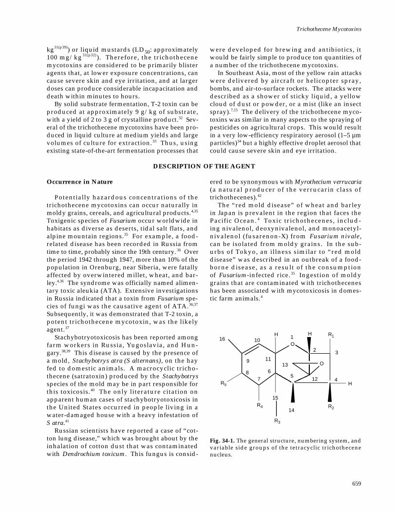
Fig. 34-1. The general structure, numbering system, andvariable side groups of the tetracyclic trichothecenenucleus.
O
O
H
R2
R1
H
16 1
4
3
12
2
5
R3
13
14
6
R4
15
78
9
10H
11
R5
Medical Aspects of Chemical and Biological Warfare
660
Chemical and Physical Properties
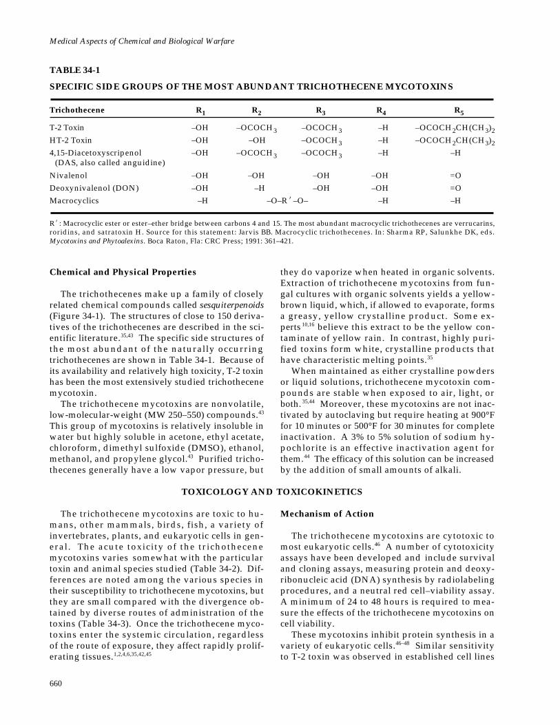
The trichothecenes make up a family of closelyrelated chemical compounds called sesquiterpenoids(Figure 34-1). The structures of close to 150 deriva-tives of the trichothecenes are described in the sci-entific literature.35,43 The specific side structures ofthe most abundant of the naturally occurringtrichothecenes are shown in Table 34-1. Because ofits availability and relatively high toxicity, T-2 toxinhas been the most extensively studied trichothecenemycotoxin.
The trichothecene mycotoxins are nonvolatile,low-molecular-weight (MW 250–550) compounds.43
This group of mycotoxins is relatively insoluble inwater but highly soluble in acetone, ethyl acetate,chloroform, dimethyl sulfoxide (DMSO), ethanol,methanol, and propylene glycol.43 Purified tricho-thecenes generally have a low vapor pressure, but
TABLE 34-1
SPECIFIC SIDE GROUPS OF THE MOST ABUNDANT TRICHOTHECENE MYCOTOXINS
Trichothecene R1 R2 R3 R4 R5
T-2 Toxin –OH –OCOCH3 –OCOCH3 –H –OCOCH2CH(CH3)2
HT-2 Toxin –OH –OH –OCOCH3 –H –OCOCH2CH(CH3)2
4,15-Diacetoxyscripenol –OH –OCOCH3 –OCOCH3 –H –H(DAS, also called anguidine)
Nivalenol –OH –OH –OH –OH =O
Deoxynivalenol (DON) –OH –H –OH –OH =O
Macrocyclics –H –O–R’–O– –H –H
R’: Macrocyclic ester or ester–ether bridge between carbons 4 and 15. The most abundant macrocyclic trichothecenes are verrucarins,roridins, and satratoxin H. Source for this statement: Jarvis BB. Macrocyclic trichothecenes. In: Sharma RP, Salunkhe DK, eds.Mycotoxins and Phytoalexins. Boca Raton, Fla: CRC Press; 1991: 361–421.
they do vaporize when heated in organic solvents.Extraction of trichothecene mycotoxins from fun-gal cultures with organic solvents yields a yellow-brown liquid, which, if allowed to evaporate, formsa greasy, yellow crystalline product. Some ex-perts 10,16 believe this extract to be the yellow con-taminate of yellow rain. In contrast, highly puri-fied toxins form white, crystalline products thathave characteristic melting points.35
When maintained as either crystalline powdersor liquid solutions, trichothecene mycotoxin com-pounds are stable when exposed to air, light, orboth. 35,44 Moreover, these mycotoxins are not inac-tivated by autoclaving but require heating at 900°Ffor 10 minutes or 500°F for 30 minutes for completeinactivation. A 3% to 5% solution of sodium hy-pochlorite is an effective inactivation agent forthem.44 The efficacy of this solution can be increasedby the addition of small amounts of alkali.
TOXICOLOGY AND TOXICOKINETICS
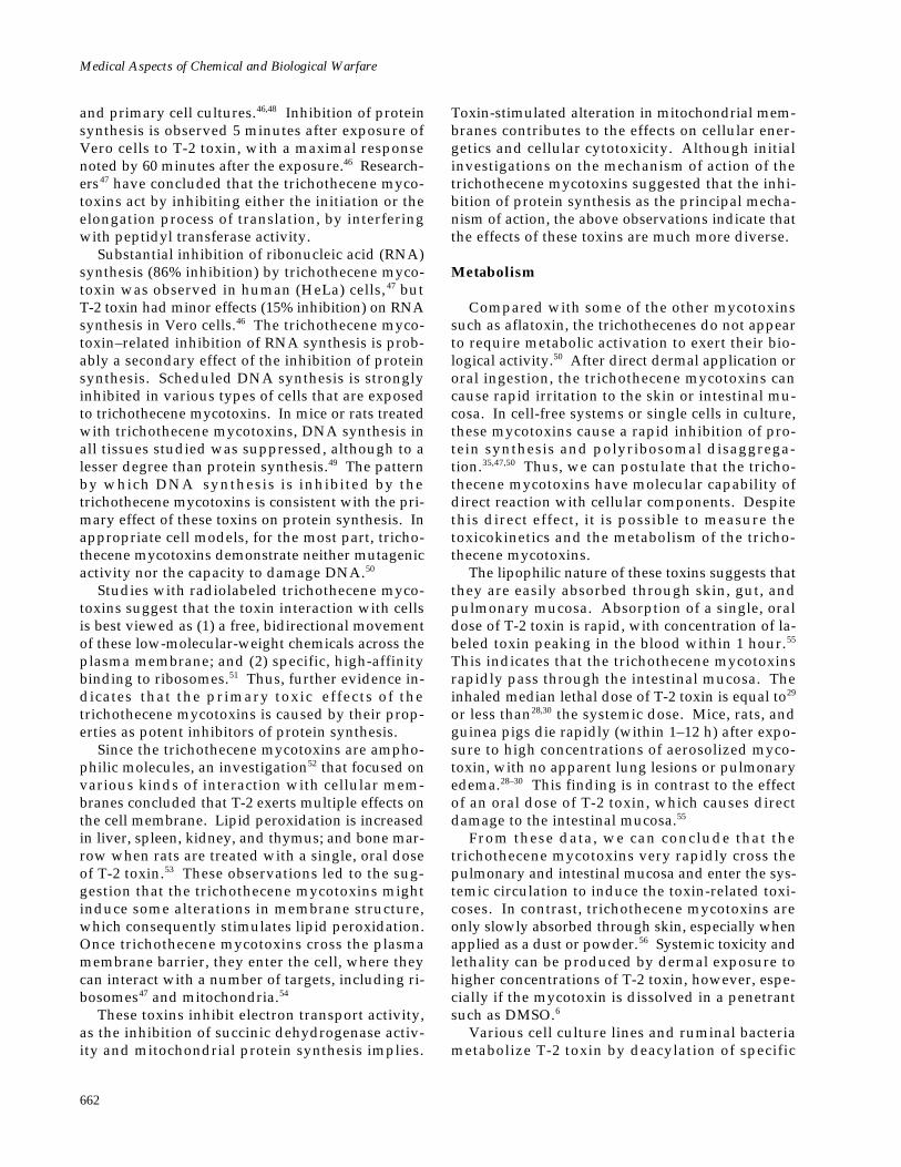
The trichothecene mycotoxins are toxic to hu-mans, other mammals, birds, fish, a variety ofinvertebrates, plants, and eukaryotic cells in gen-eral. The acute toxicity of the trichothecenemycotoxins varies somewhat with the particulartoxin and animal species studied (Table 34-2). Dif-ferences are noted among the various species intheir susceptibility to trichothecene mycotoxins, butthey are small compared with the divergence ob-tained by diverse routes of administration of thetoxins (Table 34-3). Once the trichothecene myco-toxins enter the systemic circulation, regardlessof the route of exposure, they affect rapidly prolif-erating tissues.1,2,4,6,35,42,45
Mechanism of Action
The trichothecene mycotoxins are cytotoxic tomost eukaryotic cells.46 A number of cytotoxicityassays have been developed and include survivaland cloning assays, measuring protein and deoxy-ribonucleic acid (DNA) synthesis by radiolabelingprocedures, and a neutral red cell–viability assay.A minimum of 24 to 48 hours is required to mea-sure the effects of the trichothecene mycotoxins oncell viability.
These mycotoxins inhibit protein synthesis in avariety of eukaryotic cells.46–48 Similar sensitivityto T-2 toxin was observed in established cell lines
Trichothecene Mycotoxins
661
TABLE 34-3
COMPARATIVE TOXICITY OF T-2 TOXIN BY VARIOUS ROUTES OF ADMINISTRATION
Mammals Tested
Mouse Rat Guinea Pig Rabbit Cat Pig Monkey
Route of Administration T-2 Toxin LD 50 (mg/kg)
Intravenous 4.2–7.3 0.7–1.2 1.0–2.0 — — 1.2 —
Intraperitoneal 5.2–9.1 1.3–2.6 — — — — —
Subcutaneous 2.1–3.3 0.6–2.0 1.0–2.0 — < 0.5 — —
Intramuscular — 0.5–0.9 1.0 1.1 — — 0.8
Intragastric 9.6–10.5 2.3–5.2 3.1–5.3 — — — —
Intranasal — 0.6 — — — — —
Intratracheal 0.16 0.1 — — — — —
Inhalational 0.24 0.05 0.6–2.0 — — — —
Intracerebral — 0.01 — — — — —
Dermal in DMSO 6.6 4.3 2.2 10 — — > 8.0
Dermal in Methanol — > 380 > 80 — — — —
DMSO: dimethyl sulfoxide—: Not determinedData sources: (1) Ueno Y. Trichothecene mycotoxins: Mycology, chemistry, and toxicology. Adv Nut Res . 1989;3:301–353. (2)Wannemacher RW Jr, Bunner DL, Neufeld HA. Toxicity of trichothecenes and other related mycotoxins in laboratory animals. In:Smith JE, Henderson RS, eds. Mycotoxins and Animal Foods. Boca Raton, Fla: CRC Press; 1991: 499–552. (3) Sharma RP, Kim Y-W.Trichothecenes. In: Sharma RP, Salunkhe DK, eds. Mycotoxins and Phytoalexins. Boca Raton, Fla: CRC Press; 1991: 339–359.
TABLE 34-2
RELATIVE ACUTE PARENTERAL TOXICITY OF THE MOST ABUNDANT TRICHOTHECENEMYCOTOXINS
Mammals Tested
Mouse Rat Guinea Pig Rabbit Cat Dog Pig Monkey
Trichothecenes Tested LD50 (mg/kg)
T-2 Toxin 5.2 (IV) 0.9 (IV) 1.0 (IV) 1.0 (IM) < 0.5 (SC) — 1.2 (IV) 0.8 (IM)
HT-2 Toxin 9.0 (IP) — — — — — — —
4,15-Diacetoxy-scripenol (DAS) 12.0 (IV) 1.3 (IV) — 1.0 (IV) — 1.1 (IV) 0.38 (IV) —
Nivalenol 6.3 (IV) — — — — — — —
Deoxynivalenol (DON) 43 (SC) — — — — — — —
Verrucarin A 1.5 (IV) 0.8 (IV) — 0.54 (IV) — — — —
Roridin A 1.0 (IV) — — — — — — —
Satratoxin H 1.0 (IP) — — — — — — —
Routes of administration of the mycotoxin: IV: intravenous; IM: intramuscular; SC: subcutaneous; IP: intraperitoneal—: Not determinedData sources: (1) Ueno Y. Trichothecene mycotoxins: Mycology, chemistry, and toxicology. Adv Nut Res . 1989;3:301–353. (2)Wannemacher RW Jr, Bunner DL, Neufeld HA. Toxicity of trichothecenes and other related mycotoxins in laboratory animals. In:Smith JE, Henderson RS, eds. Mycotoxins and Animal Foods. Boca Raton, Fla: CRC Press; 1991: 499–552. (3) Sharma RP, Kim Y-W.Trichothecenes. In: Sharma RP, Salunkhe DK, eds. Mycotoxins and Phytoalexins. Boca Raton, Fla: CRC Press; 1991: 339–359. (4) JarvisBB. Macrocyclic trichothecenes. In: Sharma RP, Salunkhe DK, eds. Mycotoxins and Phytoalexins . Boca Raton, Fla: CRC Press; 1991:361–421.
Medical Aspects of Chemical and Biological Warfare
662
and primary cell cultures.46,48 Inhibition of proteinsynthesis is observed 5 minutes after exposure ofVero cells to T-2 toxin, with a maximal responsenoted by 60 minutes after the exposure.46 Research-ers47 have concluded that the trichothecene myco-toxins act by inhibiting either the initiation or theelongation process of translation, by interferingwith peptidyl transferase activity.
Substantial inhibition of ribonucleic acid (RNA)synthesis (86% inhibition) by trichothecene myco-toxin was observed in human (HeLa) cells, 47 butT-2 toxin had minor effects (15% inhibition) on RNAsynthesis in Vero cells.46 The trichothecene myco-toxin–related inhibition of RNA synthesis is prob-ably a secondary effect of the inhibition of proteinsynthesis. Scheduled DNA synthesis is stronglyinhibited in various types of cells that are exposedto trichothecene mycotoxins. In mice or rats treatedwith trichothecene mycotoxins, DNA synthesis inall tissues studied was suppressed, although to alesser degree than protein synthesis.49 The patternby which DNA synthesis is inhibited by thetrichothecene mycotoxins is consistent with the pri-mary effect of these toxins on protein synthesis. Inappropriate cell models, for the most part, tricho-thecene mycotoxins demonstrate neither mutagenicactivity nor the capacity to damage DNA.50
Studies with radiolabeled trichothecene myco-toxins suggest that the toxin interaction with cellsis best viewed as (1) a free, bidirectional movementof these low-molecular-weight chemicals across theplasma membrane; and (2) specific, high-affinitybinding to ribosomes.51 Thus, further evidence in-dicates that the primary toxic effects of thetrichothecene mycotoxins is caused by their prop-erties as potent inhibitors of protein synthesis.
Since the trichothecene mycotoxins are ampho-philic molecules, an investigation52 that focused onvarious kinds of interaction with cellular mem-branes concluded that T-2 exerts multiple effects onthe cell membrane. Lipid peroxidation is increasedin liver, spleen, kidney, and thymus; and bone mar-row when rats are treated with a single, oral doseof T-2 toxin.53 These observations led to the sug-gestion that the trichothecene mycotoxins mightinduce some alterations in membrane structure,which consequently stimulates lipid peroxidation.Once trichothecene mycotoxins cross the plasmamembrane barrier, they enter the cell, where theycan interact with a number of targets, including ri-bosomes47 and mitochondria.54
These toxins inhibit electron transport activity,as the inhibition of succinic dehydrogenase activ-ity and mitochondrial protein synthesis implies.
Toxin-stimulated alteration in mitochondrial mem-branes contributes to the effects on cellular ener-getics and cellular cytotoxicity. Although initialinvestigations on the mechanism of action of thetrichothecene mycotoxins suggested that the inhi-bition of protein synthesis as the principal mecha-nism of action, the above observations indicate thatthe effects of these toxins are much more diverse.
Metabolism
Compared with some of the other mycotoxinssuch as aflatoxin, the trichothecenes do not appearto require metabolic activation to exert their bio-logical activity.50 After direct dermal application ororal ingestion, the trichothecene mycotoxins cancause rapid irritation to the skin or intestinal mu-cosa. In cell-free systems or single cells in culture,these mycotoxins cause a rapid inhibition of pro-tein synthesis and polyribosomal disaggrega-tion.35,47,50 Thus, we can postulate that the tricho-thecene mycotoxins have molecular capability ofdirect reaction with cellular components. Despitethis direct effect, it is possible to measure thetoxicokinetics and the metabolism of the tricho-thecene mycotoxins.
The lipophilic nature of these toxins suggests thatthey are easily absorbed through skin, gut, andpulmonary mucosa. Absorption of a single, oraldose of T-2 toxin is rapid, with concentration of la-beled toxin peaking in the blood within 1 hour.55
This indicates that the trichothecene mycotoxinsrapidly pass through the intestinal mucosa. Theinhaled median lethal dose of T-2 toxin is equal to29
or less than28,30 the systemic dose. Mice, rats, andguinea pigs die rapidly (within 1–12 h) after expo-sure to high concentrations of aerosolized myco-toxin, with no apparent lung lesions or pulmonaryedema.28–30 This finding is in contrast to the effectof an oral dose of T-2 toxin, which causes directdamage to the intestinal mucosa.55
From these data, we can conclude that thetrichothecene mycotoxins very rapidly cross thepulmonary and intestinal mucosa and enter the sys-temic circulation to induce the toxin-related toxi-coses. In contrast, trichothecene mycotoxins areonly slowly absorbed through skin, especially whenapplied as a dust or powder.56 Systemic toxicity andlethality can be produced by dermal exposure tohigher concentrations of T-2 toxin, however, espe-cially if the mycotoxin is dissolved in a penetrantsuch as DMSO.6
Various cell culture lines and ruminal bacteriametabolize T-2 toxin by deacylation of specific
Trichothecene Mycotoxins
663
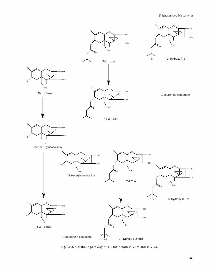
Fig. 34-2. Metabolic pathway of T-2 toxin both in vitro and in vivo.
Glucuronide conjugate
Glucuronide conjugate
O
OAc
OH
O
O
OOAc
T-2 Toxin
O
OAc
OH
O
O
O
OH
OAc
3'-Hydroxy T-2
O
OAc
OH
HO
O
OAc
Neosolaniol
O
OH
OH
O
O
OOAc
HT-2 Toxin
O
OAc
OH
HO
O
OH
15-Deacetylneosolaniol
O
OH
OH
HO
O
OAc
4-Deacetylneosolanide
O
OH
OH
O
O
OOH
T-2 Triol
O
OH
OH
O
O
O
OH
OAc
3'-Hydroxy HT -2O
OH
OH
HO
O
OH
T-2 Tetraol
O
OH
OH
O
O
O
OH
OH
3'-Hydroxy T-2 triol
Medical Aspects of Chemical and Biological Warfare
664
deepoxidylation (ie, removal of the oxygen from theepoxide ring at the C-12, 13 position to yield a car-bon–carbon double bond) and oxidization of the C-3' and C-4' positions on the isovaleryl side chainsof T-2 toxin and HT-2 toxin, a metabolite (Figure34-2).57–59 A number of different cell types containthe metabolic processes necessary to metabolizetrichothecene mycotoxins.
Pharmacokinetic studies60,61 have demonstratedT-2 toxin in the plasma of animals that were admin-istered this mycotoxin both intravascularly and byaerosol. As plasma concentrations of the parenttrichothecene mycotoxin decrease, the deacylatedand hydroxylated metabolites and their glucu-ronide conjugates rapidly appear and disappearfrom circulation. From these various observations,we can conclude that the pharmacokinetics of thetrichothecene mycotoxins are functions of the rateof absorption into the general circulation, metabo-lism, tissue distribution, and excretion.
Tissue-distribution studies55 suggest that the liveris the major organ for metabolism of the tricho-thecene mycotoxins. The bile and the gastrointes-tinal tract contained large amounts of radioactivityafter intravascular, intramuscular, oral, or dermaladministration of radiolabeled T-2 toxin. Althoughthe liver is the major organ for the metabolism ofthe trichothecene mycotoxins, other tissues such asthe intestine are capable of metabolic alteration ofthese toxins. After an intravenous dose of T-2 toxin,95% of the total radioactivity was excreted in theurine and feces, in a ratio of 3 to 1.61 The majorityof the excreted products were either metabolites orglucuronide conjugates of the metabolites.
Regardless of the route of administration or thespecies of animal tested, the trichothecene mycotox-ins were rapidly metabolized and excreted in urine
and feces. The route of exposure to the toxins andthe species can, however, influence the pattern ofmetabolites that are excreted in the urine. Thedeacetylated and hydroxylated metabolites appearto be present in most of the species that have beenevaluated to date.
A microsomal, nonspecific carboxylesterase [EC3.1.1.1] from liver selectively hydrolyses the C-4acetyl group of T-2 toxin to yield HT-2 toxin.62 Inaddition to hepatic microsomes, the trichothecene-specific carboxylesterase activity has been detectedin brain, kidney, spleen, intestine, white blood cells,and erythrocytes. These findings emphasize theimportance of carboxylesterase in detoxifying thetrichothecene mycotoxins. A hepatic cytochrome,P-450, is responsible for catalyzing the hydroxyla-tion of the C-3' and C-4' positions on the isovalerylside chain of the T-2 and HT-2 toxins.59 When oxy-gen is removed from the epoxide group of atrichothecene mycotoxin to yield the carbon–carbonbond, deepoxy metabolites are formed. The de-epoxy metabolites are essentially nontoxic.58 Thislatter observation indicates that epoxide reductionis a single-step detoxification reaction for trichothe-cene mycotoxins.
Four hours after swine received intravenous tri-tium-labeled T-2 toxin, glucuronide conjugates rep-resented 63% of the metabolic residues in urine, and77% in bile.63 The formation of glucuronide conju-gates generally results in the elimination of toxico-logical activity of xenobiotics, which in certain spe-cies could represent a major route of detoxificationof trichothecene mycotoxins.
In summary, then, very little of the parenttrichothecene mycotoxin is excreted intact. Rather,elimination by detoxification of the toxin is the re-sult of extensive and rapid biotransformation.
CLINICAL DISEASE
The degree of illness in an individual exposed totrichothecene mycotoxins could be affected by anumber of factors, including the nutritional statusof the host, liver damage, intestinal infections, routeof toxin administration, and stress.
The pathological effects and clinical signs formany toxic materials can vary with the route andtype (acute, single dose vs chronic, subacute doses)of exposure. For the trichothecene mycotoxins,however, a number of the toxic responses are simi-lar, regardless of the route of exposure. As we dis-cussed earlier in this chapter, once they enter thesystemic circulation, trichothecene mycotoxins af-fect rapidly proliferating tissue regardless of the
route of exposure.In contrast, the symptoms and clinical signs of
trichothecene intoxication can vary depending onwhether the exposure is acute or chronic. Acuteexposure to trichothecene mycotoxins used as biologi-cal warfare agents is the major concern for militarymedicine, but for continuity and historical impli-cations, chronic intoxication will also be addressedin this chapter.
Acute Effects
Acute oral, parenteral, dermal, or aerosol expo-sures to trichothecene mycotoxins produce gastric
Trichothecene Mycotoxins
665
and intestinal lesions. Hematopoietic and immu-nosuppressive effects are radiomimetic. Centralnervous system toxicity causes anorexia, lassitude,and nausea; suppression of reproductive organfunction; and acute vascular effects leading to hy-potension and shock. While a number of toxic ef-fect are common to different routes of exposure,route-specific effects have been observed in animalmodels. Examples of local, route-specific effectsinclude the following:
• dermal exposure: local cutaneous necrosisand inflammation6;
• oral exposure: lesions to the upper gas-trointestinal tract64; and
• ocular exposure: corneal injury.6
In Southeast Asia during the 1970s, symptomsbegan within minutes after an exploding munition(air-to-surface rocket, aerial bomb, cylinder) causeda yellow, oily, droplet mist to fall on individualswithin 100 m of the explosion site. The falling drop-let rain was inhaled, swallowed, and collected onskin and clothing; contaminated the terrain andfood and water supply; and caused humans andanimals to become acutely ill and to die after a vari-able period.7 Massive cutaneous contact was preva-lent when the sources of exposure were sprays orcoarse mists that were used deliberately to contami-nate humans and the environment. Although thesuspected trichothecene mycotoxin attacks inSoutheast Asia would have involved multipleroutes of exposure, we can postulate that the skinwould have been the major site for deposition of aaerosol spray or coarse mist.
Early symptoms and signs included severe nau-sea, vomiting, burning superficial skin discomfort,lethargy, weakness, dizziness, and loss of coordi-nation. Within minutes to hours, diarrhea—at firstwatery brown and later grossly bloody—began.During the first 3 to 12 hours, dyspnea, coughing,sore mouth, bleeding gums, epistaxis, hematemesis,abdominal pain, and central chest pain could oc-cur. The exposed cutaneous areas could becomered, tender, swollen, painful, or pruritic, in any com-bination. Small or large vesicles and bullae mightform; and petechiae, ecchymoses, and black, leath-ery areas of necrosis might appear. After death, thenecrotic areas might slough easily when the corpsewas moved.
Marked anorexia and dehydration were frequent.Dying patients became hypothermic and hypoten-sive, and developed tachycardia. A bloody ooze fromthe nares and mouth and an associated hematochezia
occurred in severely poisoned individuals. Deathcould occur within minutes, hours, or days, and wasoften preceded by tremors, seizures, and coma, inany combination.
The most common symptoms in both SoutheastAsia and Afghanistan included vomiting (71%);diarrhea (53%); skin irritation, burning, and itch-ing (44%); rash or blisters (33%); bleeding (52%);and dyspnea (48%).7,15,27 All of the symptoms list-ed could be attributed to trichothecene mycotoxintoxicity.
Dermal Exposure
Similar cutaneous irritations have been observedin numerous accidental and experimental settings:
• Individuals who were exposed to hay orhay dust contaminated with trichothecene-producing molds developed severe cutane-ous irritations.38
• In working up large batches of fungal cul-tures from trichothecene-producing organ-isms, laboratory personnel suffered facialinflammation followed by desquamation ofthe skin and considerable local irritation.65
• When trichothecene mycotoxins of rela-tively low toxicity (crotocin and tricho-tecin) were applied to the volar surface ofhuman forearm or to the human head, red-dening and irritation occurred within a fewhours of exposure, and was followed byinflammation or scrabbling that healed in1 to 2 weeks.66
• The hands of two laboratory workers wereexposed to crude ethyl acetate extracts con-taining T-2 toxin (approximately 200 µg/mL) when the extract accidently got insidetheir plastic gloves. 66 Even though theworkers thoroughly washed their handswith a mild detergent within 2 minutes af-ter contact, they experienced severe cuta-neous irritations.
These observations provide evidence that whenhuman skin is exposed in vivo to small amounts oftrichothecene mycotoxins, severe cutaneous irrita-tions develop and can last 1 to 2 weeks after acuteexposure.
A number of animal models have been usedto assess local and systemic toxicity and lethalityfrom skin exposure to trichothecenes.6 In a dermalstudy that used a mouse model, necrosis in the skinwas present by 6 hours after dermal application
Medical Aspects of Chemical and Biological Warfare
666
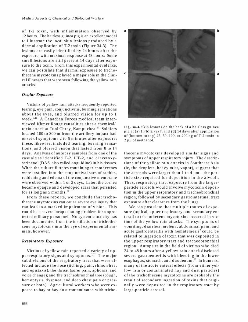
Fig. 34-3. Skin lesions on the back of a hairless guineapig at (a) 1, (b) 2, (c) 7, and (d) 14 days after applicationof (bottom to top) 25, 50, 100, or 200 ng of T-2 toxin in2 µL of methanol.
of T-2 toxin, with inflammation observed by12 hours. The hairless guinea pig is an excellent modelto illustrate the local skin lesions produced by adermal application of T-2 toxin (Figure 34-3). Thelesions are easily identified by 24 hours after theexposure, with maximal response at 48 hours. Somesmall lesions are still present 14 days after expo-sure to the toxin. From this experimental evidence,we can postulate that dermal exposure to tricho-thecene mycotoxins played a major role in the clini-cal illnesses that were seen following the yellow rainattacks.
Ocular Exposure
Victims of yellow rain attacks frequently reportedtearing, eye pain, conjunctivitis, burning sensationsabout the eyes, and blurred vision for up to 1week.7,16 A Canadian Forces medical team inter-viewed Khmer Rouge causalities after a chemical/toxin attack at Tuol Chrey, Kampuchea.27 Soldierslocated 100 to 300 m from the artillery impact hadonset of symptoms 2 to 5 minutes after exposure;these, likewise, included tearing, burning sensa-tions, and blurred vision that lasted from 8 to 14days. Analysis of autopsy samples from one of thecasualties identified T-2, HT-2, and diacetoxy-scripenol (DAS, also called anguidine) in his tissues.When the culture filtrates containing trichotheceneswere instilled into the conjunctival sacs of rabbits,reddening and edema of the conjunctive membranewere observed within 1 or 2 days. Later, the corneabecame opaque and developed scars that persistedfor as long as 5 months.67
From these reports, we conclude that tricho-thecene mycotoxins can cause severe eye injury thatcan lead to a marked impairment of vision. Thiscould be a severe incapacitating problem for unpro-tected military personnel. No systemic toxicity hasbeen documented from the instillation of trichothe-cene mycotoxins into the eye of experimental ani-mals, however.
Respiratory Exposure
Victims of yellow rain reported a variety of up-per respiratory signs and symptoms.7,27 The majorsubdivisions of the respiratory tract that were af-fected include the nose (itching, pain, rhinorrhea,and epistaxis); the throat (sore/pain, aphonia, andvoice change); and the tracheobronchial tree (cough,hemoptysis, dyspnea, and deep chest pain or pres-sure or both). Agricultural workers who were ex-posed to hay or hay dust contaminated with tricho-
thecene mycotoxins developed similar signs andsymptoms of upper respiratory injury. The descrip-tions of the yellow rain attacks in Southeast Asia(ie, the droplets, heavy mist, vapor), suggest thatthe aerosols were larger than 1 to 4 µm—the par-ticle size required for deposition in the alveoli.Thus, respiratory tract exposure from the larger-particle aerosols would involve mycotoxin deposi-tion in the upper respiratory and tracheobronchialregion, followed by secondary gastrointestinal tractexposure after clearance from the lungs.
We can postulate that multiple routes of expo-sure (topical, upper respiratory, and secondary en-teral) to trichothecene mycotoxins occurred in vic-tims of the yellow rain attacks. The symptoms ofvomiting, diarrhea, melena, abdominal pain, andacute gastroenteritis with hematemesis7 could berelated to ingestion of toxin that was deposited inthe upper respiratory tract and tracheobronchialregion. Autopsies in the field of victims who died24 to 48 hours after a yellow rain attack disclosedsevere gastroenteritis with bleeding in the loweresophagus, stomach, and duodenum.27 In humans,many of the acute enteral effects (from either yel-low rain or contaminated hay and dust particles)of the trichothecene mycotoxins are probably theresult of secondary ingestion of toxins that origi-nally were deposited in the respiratory tract bylarge-particle aerosol.
a b
c d
Trichothecene Mycotoxins
667
Chronic Toxicity
Chronic exposure to subacute doses of trichothe-cene mycotoxins is not thought to be an effect ofbiological warfare. This type of exposure, however,was responsible for ATA toxicosis in humans andmycotoxicosis in domestic animals. In addition,chronic toxicity has been iatrogenically induced whenrepeated subacute doses of a trichothecene mycotoxinwere administrated intravenously to cancer patientsas a chemotherapy for colon adenocarcinoma.
Alimentary Toxic Aleukia Toxicosis
The clinical course of ATA is divided into fourstages. The first stage develops immediately or sev-eral days after consumption of grain products thatare contaminated with trichothecene mycotoxins.Inflammation of the gastric and intestinal mucosacauses vomiting, diarrhea, and abdominal pain. Inmost cases, excessive salivation, headache, dizzi-ness, weakness, fatigue, and tachycardia accom-pany this stage, and fever and sweating may alsobe present.36
The disease progress to the second stage—the leu-kopenic or latent stage—which is characterized byleukopenia, granulopenia, and progressive lympho-cytosis. When the ingestion of the toxin-contami-nated food is not interrupted or if large doses areconsumed, the next stage develops.36
The third stage is characterized by the appearanceof a bright red, or dark cherry-red, petechial rashon the skin of the chest and other areas of the body.At first, the petechiae are localized in small areas,but they then spread and become more numerous.
In the most severe cases, intensive ulceration andgangrenous processes develop in the larynx, lead-ing to aphonia and death by strangulation. At thesame time, affected individuals have severe hem-orrhagic diathesis of the nasal, oral, gastric, andintestinal mucosa.36
As the necrotic lesions heal and the body tem-perature falls, the fourth stage—the recovery stage—begins. During this period, exposed patients aresusceptible to various secondary infections, includ-ing pneumonia. Convalescence is prolonged andcan last for several weeks. Usually, 2 months ormore are required for the blood-forming capacityof the bone marrow to return to normal.36
Cancer Chemotherapy
The inhibitory effect of trichothecene mycotox-ins on rapidly dividing cells was the basis for theirevaluation as antitumor chemotherapy drugs dur-ing the late 1970s and early 1980s.68 Phase I andphase II clinical evaluations of DAS (anguidine) inpatients with cancer disclosed significant toxicitywith intravenous doses 3.0 mg/m2 (0.077 mg/kg)daily for 5 days, particularly in patients withhepatic metastases. The signs and symptoms in-cluded nausea, vomiting, diarrhea, burning ery-thema, confusion, ataxia, chills, fever, hypotension,and hair loss. 69,70 Antitumor activity of thetrichothecenes was minimal or absent in the patientstreated with DAS. Because of the marked toxicityof the drug, the life-threatening hypotensive effects,and the poor tolerance by patients, the evaluationof trichothecenes as chemotherapeutic drugs wasdiscontinued.
DIAGNOSIS
Battlefield Diagnosis
In the absence of a biological detector or a par-ticular characteristic of the aerosol (such as coloror odor), diagnosis of an attack with trichothecenewould depend on clinical observations of casual-ties and identification of the toxins in biological orenvironmental samples. This would involve a com-bined effort between the medical and chemical unitsin the field. The early signs and symptoms of anaerosol exposure to trichothecene mycotoxinswould depend on particle size and toxin concen-tration. For a large-particle aerosol (particles > 10µm, found in mist, fog, and dust; similar to thatused in Southeast Asia), the signs and symptomswould include rhinorrhea, sore throat, blurred vi-
sion, vomiting, diarrhea, skin irritation (burningand itching), and dyspnea. Early (0–8 h) signs andsymptoms from a deep-respiratory aerosol exposure(from aerosol particles in the 1- to 4-µm range) havenot been fully evaluated but could include vomit-ing, diarrhea, skin irritation, and blurred vision.
Later signs and symptoms (8–24 h) would prob-ably be similar (except for the degree of skin rashand blisters) for both large-particle and deep-respiratory aerosol exposure to trichothecene my-cotoxins. They could include continued nausea andvomiting, diarrhea, burning erythema, skin rashand blisters, confusion, ataxia, chills, fever, hy-potension, and bleeding.
Nonspecific changes in serum chemistry andhematology occurred in monkeys exposed to an
Medical Aspects of Chemical and Biological Warfare
668
acute dose of T-2 toxin. Alterations in serum chem-istry included elevations in serum creatinine, serumenzymes (especially creatine kinase), potassium,phosphorous, and serum amino acids; and, due todecreased coagulation factors, elevations in pro-thrombin time and partial thromboplastin time. Aninitial rise in the absolute number of neutrophilsand lymphocytes may occur within hours, followedby a decrease in lymphocyte counts by 48 hours.Survival beyond several days may be associatedwith a fall in all blood cellular elements.6 Althoughit is likely that these acute changes will also be seenin humans, careful clinical observations of humanvictims of acute trichothecene mycotoxicosis havenot been reported to date. In patients with chronictoxicity (ie, ALA) resulting from repeated ingestionof contaminated bread, pancytopenia is an impor-tant part of the clinical picture.36
In the yellow rain attacks in Southeast Asia, di-agnosis of the causative agent was difficult and in-volved ruling out the presence of conventionalchemical warfare agents. Contamination of the en-vironment and clothing by nerve and blisteringagents would be absent, and these were, in fact, notdetectable in such samples from Southeast Asia.Sarin, soman, or other nerve agents could be missedunless thickened soman or VX was used.
The following events should suggest to medicalofficers that a biological warfare attack withtrichothecene mycotoxins has occurred:
• clinical findings that match the symptomslisted above;
• high attack and fatality rates;• all types of dead animals; and• onset of symptoms after a yellow rain or
red, green, or white smoke or vapor attack.
At present, we do not have a fieldable identifi-cation kit for any of the trichothecene mycotoxins.Several commercial immunoassay kits are marketedfor the detection of trichothecene mycotoxins (T-2toxin, deoxynivalenol, and their metabolites) ingrain extracts or culture filtrates of Fusarium spe-cies.71,72 These kits have not been evaluated againstbiomedical samples that contain typical concentra-tions of the mycotoxins, however. Screening testsfor presumptive identification of trichothecenemycotoxins in the biomedical samples would prob-ably involve bioassays, thin-layer chromatography,or immunological assays, in any combination. At anational laboratory, confirmatory methodologywould involve the use of various combinations ofgas chromatography, high-performance liquid chro-
matography, mass spectrometry, and nuclear mag-netic resonance spectrometry.
In areas that have experienced a yellow rain at-tack, environmental assays have been in the rangeof 1 to 150 parts per million (ppm) and bloodsamples in the range of 1 to 296 parts per billion(ppb).8–10,16,22 In the laboratory, at 10 and 50 min-utes after an intramuscular exposure to 0.4 mg/kgof T-2 toxin in the dog, plasma concentrations of T-2 toxin were 150 and 25 ppb, and for HT-2 toxinwere 50 and 75 ppb, respectively. 60 Thus, anyscreening procedure for trichothecene mycotoxinsin biomedical samples must have detection limitsof 1 to 100 ppb. Most of the analytical proceduresrequire extraction and cleanup treatment to removeinterfering substances.73
Screening tests for the trichothecene mycotoxinsare generally simple and rapid but, with the excep-tion of the immunochemical methods, are nonspe-cific. A number of bioassay systems have been usedfor the identification of trichothecene mycotoxins.73
Although most of these systems are very simple,they are not specific, their sensitivity is generallyrelatively low compared to other methods, and theyrequire that the laboratory maintain vertebrates,invertebrates, plants, or cell cultures. Thin-layerchromatography (TLC) is one of the simplest andearliest analytical methods developed for myco-toxin analysis. Detection limits for trichothecenemycotoxins by TLC is 0.2 to 5 ppm (0.2 to 5 µg/mL). Therefore, extracts from biomedical sampleswould have to be concentrated 10- to 1,000-fold toscreen for trichothecene mycotoxins.
To overcome the difficulties encountered with thebioassays and TLC methods, immunoassays usingspecific polyclonal and monoclonal antibodies havebeen developed for most of the major trichothecenemycotoxins and their metabolites.73 These antibod-ies have been used to produce simple, sensitive, andspecific radioimmunoassays (RIAs) and enzyme-linked immunosorbent assays (ELISAs) for themycotoxins. In the presence of the sample matrix,the lower detection limits for identification oftrichothecene mycotoxins by RIA is about 2 to 5ppb73 and by ELISA, 1 ppb.74 We conclude that im-munoassays are useful tools for screening biomedi-cal samples for evidence of a biological warfare at-tack with trichothecene mycotoxins.
Confirmatory Procedures
Gas-liquid chromatography (GLC) is one of themost commonly used methods for the identificationof the trichothecene mycotoxins in both agricultural
Trichothecene Mycotoxins
669
products and biomedical samples.75 Before GLCanalysis, the polar groups in mycotoxin moleculesmust first be converted to their esters or ethers.Extensive treatment to clean up the sample is requiredbefore derivatization and subsequent analysis can beperformed. By the most sensitive procedures, thedetection limit for trichothecene mycotoxins is 10 ppb.If the analysis is on a sample that contains an un-known toxic material, such as those from the yel-low rain attacks, then the GLC method can onlyprovide presumptive evidence of a trichothecenemycotoxin exposure. Confirmation will require theidentification with more definitive physicochemi-cal procedures.
Mass spectrometry (MS) is the physicochemicalmethod of choice for characterizing, identifying,and confirming the presence of trichothecenemycotoxins. 76,77 Picogram quantities of tricho-thecene mycotoxins are readily detectable by MSmethods. In some cases, extensive cleanup stepsare unnecessary.
The combination of GLC and MS techniques(GLC–MS) has proven to be a more-specific methodfor identifying mycotoxins than is GLC alone.76,77
As a result, the GLC–MS method has become thestandard for identifying trichothecene mycotoxinsin agricultural products as well as in biomedicalsamples. As little as 1 ppb of T-2 toxin can be iden-tified without extensive cleanup.76 One major draw-back of this methodology is the time-consumingderivatization step that trichothecene mycotoxinidentification by GLC–MS requires. A high-perfor-mance liquid chromatography–mass spectrometry(HPLC–MS) procedure was described in 1991 andprovides a specific and reliable method for the iden-tification of trichothecene mycotoxins withoutderivatization.78 The HPLC–MS procedure achievessensitivity at the 0.1-ppb level. This technology willrequire further evaluation and development, but itappears to be a promising approach for the rapidconfirmation of trichothecene mycotoxins in a bio-medical sample.
MEDICAL MANAGEMENT
Individual and Unit
The immediate use of protective clothing andmask at the first sign of a yellow rain–like attackshould protect an individual from the lethal effectsof this mycotoxin. The mask can be applied in lessthan 9 seconds and can be worn at first sighting ofan incoming rocket or enemy aircraft. Contami-nated battle dress uniforms (BDUs) should be re-moved before protective clothing is donned. Sincethe area covered with agent is likely to be small,another helpful tactic is to leave the area after tak-ing samples to document the attack. Vulnerabilityis increased by lack of protective clothing, mask, ortraining (as was demonstrated in Laos) or by a sur-prise biological warfare attack (such as a night oran undetected attack). A lightweight face mask,outfitted with filters that block the penetration ofaerosol particles 3 to 4 µm or larger, should pro-vide respiratory protection against yellow rain.Only 1% or 2% of aerosolized T-2 toxin penetratednuclear, biological, chemical protective covers(NBC–PC).79 Regular BDUs would offer some pro-tection, but the degree would be functions of theage and condition of the fabric, and the type of en-vironmental conditions.
Two topical skin protectants (TPS1 and TSP2) arein advanced development for protection againstchemical warfare agents. When applied to the skinof rabbits 60 minutes before exposure to 50 µg of T-
2 toxin, both topical skin protectants completelyprotected the rabbits from the dermal irritating ef-fects of this mycotoxin for at least 6 hours.80
As soon as individuals or units suspect that theyhave been exposed to a mycotoxin attack, theyshould remove their BDUs, wash their contami-nated skin with soap and water, and then rinse withwater. Washing the contaminated area of the skinwithin 4 to 6 hours after exposure to T-2 toxin re-moved 80% to 98% of the toxin and prevented der-mal lesions and death in experimental animals.25
Contaminated BDUs as well as wash waste frompersonnel decontamination should be exposed tohousehold bleach (5% sodium hypochlorite) for 6hours or more to inactivate any residue mycotoxin.
Two skin decontamination kits, the M258A1 andthe M238A1, have been designed for the removaland detoxification of chemical warfare agents. TheM258A1 kit is the currently fielded standard. Whenevaluated against trichothecene mycotoxins, how-ever, the M238A1 kit effectively removed T-2 toxinfrom the skin of rats but did not detoxify this bio-logical warfare agent.81 Several of the componentsof the M258A1 kit are themselves highly toxic, caus-tic compounds that caused dermal irritation andlethality in rats and rabbits.82
A second-generation skin decontamination kit,the XM291, has been developed, and contains anXE-555 resin material as the active component. Thisskin decontamination kit is efficacious against most
Medical Aspects of Chemical and Biological Warfare
670
chemical warfare agents and presents no serioushuman factor or human safety problems. The XE-556 resin, a similar but different formulation, waseffective in the physical removal of T-2 toxin fromthe skin of rabbits and guinea pigs.83 The forego-ing observations suggest that the skin decontami-nation kits that were designed specifically for re-moval and detoxification of chemical warfare agentscould also afford a significant degree of protectionthrough the physical removal of mycotoxins fromthe skin of exposed individuals.
Specific or Supportive Therapy
No specific therapy for trichothecene-inducedmycotoxicosis is known or is presently under ex-perimental evaluation. Several therapeutic ap-proaches have been evaluated in animal models. Itis perhaps significant, however, that although ex-perimental procedures for treatment of systemicexposure have been successful in reducing mortal-ity in animal models, they have not been tested inprimates. Thus, these treatments are not availablefor field use for humans exposed to trichothecenemycotoxins.
Individuals exposed to a yellow rain–like attackshould be treated with standardized clinical toxi-cology and emergency medicine practices for inges-tion of toxic compounds. After an aerosol expo-sure to a yellow rain–like attack, mycotoxins willbe trapped in the nose, throat, and upper respira-tory tract. The particles will be returned by ciliaryaction to be swallowed, resulting in a significantoral exposure. Superactive charcoal has a very highmaximal binding capacity (0.48 mg of T-2 toxin per1 mg of charcoal), and treatment either immediatelyor 1 hour after oral or parenteral exposure to T-2toxin significantly improves the survival of mice.84
Superactivated charcoal with magnesium sulfate isstocked in the chemical and biological warfare kitsof U.S. Army field hospitals.
Symptomatic measures for the treatment of ex-posure to trichothecene mycotoxins are modeled af-ter the care of casualties of mustard poisoning.85 Irri-gation of the eyes with large volumes of isotonicsaline may assist in the mechanical removal of tricho-thecene mycotoxins, but would have limited usefultherapeutic effects. After the skin has been decon-taminated, some erythema may appear, accompa-nied by burning and itching. Most casualties whoseskin has been treated with soap and water within 12hours of exposure will have mild dermal effects; theseshould be relieved by calamine and other lotion orcream, such as 0.25% camphor and methanol.
Limited data are available on the respiratory ef-fects of inhaled trichothecene mycotoxins, althoughacute pulmonary edema is one of the serious, oftenlethal consequences of a yellow rain attack.16,27 Oneof the major symptoms following the yellow rainattacks was an upper respiratory irritation (sorethroat, hoarseness, nonproductive cough), 7,16,27
which can be relieved by steam inhalation, codeine,or another substance to suppress the cough, andother simple measures.85 A casualty who developssevere respiratory symptoms should be under thecare of a physician skilled in respiratory care.
The early use of high doses of systemic gluco-corticosteriods increases survival time by decreas-ing the primary injury and the shocklike state thatfollows exposure to trichothecene mycotoxins.86 Aselective platelet activating factor antagonist, BN52021, can prolong the survival of rats exposed to alethal intravenous dose of T-2 toxin.87 This findingsuggests that platelet activating factor is an impor-tant mediator of T-2 toxicosis. Dosing before andafter the exposure with diphenhydramine (anantihistaminic agent) or naloxone (an opioid an-tagonist) prolonged the survival times of mice ex-posed subcutaneously or topically with lethal dosesof T-2 toxin.88
We can postulate that a number of bioregulatorsare the mediators of the shocklike state of tricho-thecene mycotoxicosis. Methylthiazolidine-4-car-boxylate increased hepatic glutathione content andenhanced the survival of mice after an acute intra-peritoneal exposure to T-2 toxin.89 The protectiveeffects of this drug may be the result of increaseddetoxification and excretion of the glucuronide con-jugate of T-2 toxin. A general therapeutic protocolthat included combinations of metoclopramide, ac-tivated charcoal, magnesium sulfate, dexametha-sone, sodium phosphate (which had very little ef-fect), sodium bicarbonate, and normal saline as thetherapeutic agents was evaluated in swine given anintravenous LD50 dose of T-2 toxin.90 All treatmentgroups showed improved survival times when com-pared with the nontreated T-2 controls.
Prophylaxis
The mycotoxins are low-molecular-weight com-pounds that must be conjugated to a carrier pro-tein to produce an effective antigen.73 When T-2toxin is conjugated to a protein, it develops rela-tively low antibody titers and is still a marked skinirritant.91 This would preclude mycotoxins’ useas immunogens in the production of protective im-munity. To circumvent such problems, a deoxy-
Trichothecene Mycotoxins
671
verrucarol (DOVE)–protein conjugate was used toimmunize rabbits.92 Antibody titers to DOVE de-veloped rapidly after immunization, but they werehighly specific for DOVE rather than a commontrichothecene backbone.92
Another approach was to develop antibody-based vaccines (anti-idiotype) against T-2 toxin.Protective monoclonal antitoxin antibodies werefirst generated and then used to induce specificmonoclonal anti-idiotype antibodies. When micewere immunized with specific monoclonal anti-idiotype antibodies, they developed neutralizingantibodies and were protected against challengewith a lethal dose of T-2 toxin.93 Thus, it would befeasible to develop a despeciated monoclonal anti-idiotype antibody that could be a vaccine candidateagainst T-2 toxin.
Several monoclonal antibodies against T-2 toxinwill protect against the T-2–induced cytotoxicityin various cell lines.94,95 When a monoclonal anti-
body against T-2 toxin (15H6) was given to rats(250 mg/kg) 30 minutes before or 15 minutes aftera lethal dose of mycotoxin, it conferred 100% sur-vival.94 Thus, monoclonal antibodies do have someprophylactic and therapeutic value against T-2toxicosis, but very large quantities are required forprotection.
Prophylactic induction of enzymes involved inthe conjugation of xenobiotics reduced or preventedthe acute toxic effects of T-2 toxin in the rat, whileinhibition of these enzymes resulted in a higher tox-icity for this trichothecene.96 Pretreatment with fla-vonoids,97 ascorbic acid,98 vitamin E,99 selenium,100
or chemoprotective compounds such as emetine101
that block trichothecene–cell association all reduceacute toxicity of these mycotoxins. However, noneof these chemoprotective treatments have under-gone extensive efficacy studies to evaluate theirability to protect against an aerosol or dermal ex-posure to trichothecene mycotoxins.
SUMMARY
Trichothecene mycotoxins are noted for theirmarked stability under different environmental con-ditions. On a weight-for-weight basis, they are lesstoxic than other toxins such as ricin, botulinum, andstaphylococcal enterotoxin B, but trichothecenemycotoxins are proven lethal agents in warfare.Symptoms include vomiting, pain, weakness, diz-ziness, ataxia, anorexia, diarrhea, bleeding, skinredness, blistering, and gangrene, as well as shockand rapid death. Sensitive immunoassays andchemical procedures are available for the identifi-
cation of trichothecene mycotoxins in biologicalsamples, but no detection kits have been fielded.
Prevention of exposure is the only current de-fense, with a protective mask and clothing wornwhen under attack. Previous successful lethal at-tacks have always occurred against unprotected ci-vilians and soldiers. Skin decontamination withwater and soap can be used effectively up to 6 hoursafter exposure. Experimental treatments for sys-temic toxicity are being investigated, but no therapyis available for humans.
REFERENCES
1. Ciegler A. Mycotoxins: Occurrence, chemistry, biological activity. Lloydia. 1975;38(1):21–35.
2. Ciegler A, Bennett JW. Mycotoxins and mycotoxicoses. Bioscience. 1980;30(8):512–515.
3. Moss MO. Mycotoxins of Aspergillus and other filamentous fungi. J Appl Bacteriol . 1989;67(symposiumsuppl):69S–81S.
4. Ueno Y. Trichothecene mycotoxins: Mycology, chemistry, and toxicology. Adv Nutr Res. 1989;3:301–353.
5. Godtfredsen WO, Grove JF, Tamm Ch. Trichothecenes. Hev Chim Acta. 1967;50:1666–1668.
6. Wannemacher RW Jr, Bunner DL, Neufeld HA. Toxicity of trichothecenes and other related mycotoxins inlaboratory animals. In: Smith JE, Henderson RS, eds. Mycotoxins and Animal Foods. Boca Raton, Fla: CRC Press;1991: 499–552.
7. Haig AM Jr. Chemical Warfare in Southeast Asia and Afghanistan. Washington, DC: US Government PrintingOffice; March 22, 1982. Report to the Congress.
Medical Aspects of Chemical and Biological Warfare
672
8. Mirocha CJ. Hazards of scientific investigation: Analysis of samples implicated in biological warfare. Journal ofToxicology-Toxin Reviews. 1982;1(1):199–203.
9. Rosen RT, Rosen JD. Presence of four Fusarium mycotoxins and synthetic material in “yellow rain”: Evidencefor the use of chemical weapons in Laos. Biomed Mass Spectrom. 1982;9(10):443–450.
10. Mirocha CJ, Pawlosky RA, Chatterjee K, Watson S, Hayes W. Analysis for Fusarium toxins in various samplesimplicated in biological warfare in Southeast Asia. J Assoc Off Anal Chem. 1983;66(6):1485–1499.
11. Greenhalgh R, Miller JD, Neish GA, Schiefer HB. Toxigenic potential of some Fusarium isolates from SoutheastAsia. Appl Environ Microbiol. 1985;50(2):550–552.
12. Ricaud D. Les Recherche de Défense Contre les Armés Biologique et Chimiques. Paris, France: ÉcolePolytechique; 1983. ISBN 2–7170–0738–5.
13. Ember LR, Sorenson WG, Lewis DM. Charges of toxic arms use by Iraq escalate. Chemical and Engineering News.1984;62(12):16–18.
14. Seagrave S. Yellow Rain: A Journey Through the Terror of Chemical Warfare. New York, NY: M Evans; 1981.
15. Ember LR. Yellow rain. Chemical and Engineering News. 1984;62(2):8–34.
16. Watson SA, Mirocha CJ, Hayes AW. Analysis for trichothecenes in samples from Southeast Asia associatedwith “Yellow Rain.” Fundam Appl Toxicol. 1984;4(5):700–717.
17. Marshall E. Yellow rain evidence slowly whittled away. Science. 1986;233(4759):18–19.
18. Marshall E. Bugs in the yellow rain theory. Science. 1983;220(4604):1356–1358.
19. Nowicke JW, Meselson M. Yellow rain—A palynological analysis. Nature . 1984;309(5965):205–207.
20. Seeley TD, Nowicke JW, Meselson M, Guillemin J, Akratanakul P. Yellow rain. Sci Am. 1985;253(3):128–137.
21. Dashek WV, Mayfield JE, Llewellyn GC, O’Rear CE, Bata A. Trichothecenes and yellow rain: Possible biologi-cal warfare agents. Bioessays. 1986;4(1):27–30.
22. Yellow rain report. NBC Defense Technology International. 1986;1(2):11–12.
23. Marshall E. The apology of yellow rain. Science. 1983;221(4608):242.
24. Yellow rain: British analyses find no toxin. Nature . 1986;321(6069):459. News.
25. Wannemacher RW, Bunner DL, Pace JG, Neufeld HA, Brennecke LH, Dinterman RE. Dermal toxicity of T-2toxin in guinea pigs, rats, and cynomolgus monkeys. In: Lacey J, ed. Trichothecenes and Other Mycotoxins .Chichester, England: John Wiley & Sons Ltd; 1985: 423–432.
26. Bunner DL, Upshall DG, Bhatti AR. Toxicology data on T-2 toxin. In: Report of Focus Officers Meeting onMycotoxin Toxicity, September 23–24, 1985. Suffield, Alta, Canada: Defense Research Establishment at Suffield;1985.
27. Stahl CJ, Green CC, Farnum JB. The incident at Tuol Chrey: Pathological and toxicological examination of acasualty after chemical attack. J Forensic Sci. 1985;30(2):317–337.
28. Creasia DA, Thurman JD, Wannemacher RW Jr, Bunner DL. Acute inhalation toxicity of T-2 mycotoxin in therat and guinea pig. Fundam Appl Toxicol. 1990;14(1):54–59.
Trichothecene Mycotoxins
673
29. Marrs TC, Edginton JA, Price PN, Upshall DG. Acute toxicity of T2 mycotoxin to the guinea-pig by inhalationand subcutaneous routes. Br J Exp Path. 1986;67(2):259–268.
30. Creasia DA, Thurman JD, Jones LJ, et al. Acute inhalation toxicity of T-2 mycotoxin in mice. Fundam ApplToxicol . 1987;8(2):230–235.
31. US Department of Defense. Potential Military Chemical/Biological Agents and Compounds. Washington, DC: Head-quarters, Departments of the Army, Navy, and Air Force; 1990. Field Manual 3-9, Air Force Regulation 355-7,NAVFAC P-467.
32. Burmeister HR. T-2 toxin production by Fusarium tricinctum on solid substrate. Appl Microbiol . 1971;21(4):739–742.
33. Miller JD, Taylor A, Greenhalgh R. Production of deoxynivalenol and related compounds in liquid culture byFusarium graminearum. Can J Microbiol. 1983;29(9):1171–1178.
34. Spertzel RO, Wannemacher RW Jr, Patrick WC, Linden CD, Franz DR. Technical Ramifications of Inclusion ofToxins in the Chemical Weapons Convention (CWC). Alexandria, Va: Defense Nuclear Agency; 1993. DNA Techni-cal Report 92–116.
35. Committee on Protection Against Mycotoxins, Board on Toxicology and Environmental Health Hazards, Com-mission on Life Sciences, National Research Council. Protection Against Trichothecene Mycotoxins. Washington,DC: National Academy Press; 1983.
36. Joffe AZ. Alimentary toxic aleukia. In: Kadis S, Ciegler A, Ajl SJ, eds. Microbiol Toxins. Vol 7. In: Algal and FungalToxins. New York, NY: Academic Press; 1971: 139–189.
37. Yagen B, Joffe AZ, Horn P, Mor N, Lutsky II. Toxins from a strain involved in ATA. In: Rodericks JV, HesseltineCW, Mehlman MA, eds. Mycotoxins in Human and Animal Health. Park Forest South, Ill: Pathotox Publishers;1977: 329–336.
38. Forgacs J. Stachybotryotoxicosis. In: Kadis S, Ciegler A, Ajl SJ, eds. Microbial Toxins. Vol 8. New York, NY:Academic Press; 1972: 95–128.
39. Hintikka E-L. Stachybotryotoxicosis as a veterinary problem. In: Rodricks JV, Hesseltine CW, Mehlman MA,eds. Mycotoxins in Human Health. Park Forest South, Ill: Pathotox Publishers; 1977: 277–284.
40. Eppley RM. Chemistry of stachybotryotoxicosis. In: Rodericks JV, Hesseltine CW, Mehlman MA, eds. Mycotox-ins in Human and Animal Health. Park Forest South, Ill: Pathotox Publishers; 1977: 285–293.
41. Croft WA, Jarvis BB, Yatawara CS. Airborne outbreak of trichothecene toxicosis. Atmos Environ. 1986;20(3):549–552.
42. Jarvis BB. Macrocyclic trichothecenes. In: Sharma RP, Salunkhe DK, eds. Mycotoxins and Phytoalexins. BocaRaton, Fla: CRC Press; 1991: 361–421.
43. Cole RJ, Cox RH. The trichothecenes. In: Cole RJ, Cox RH. Handbook of Toxic Fungal Metabolites. New York, NY:Academic Press; 1981: 152–263.
44. Wannemacher RW Jr, Bunner DL, Dinterman RE. Inactivation of low molecular weight agents of biologicalorigin. In: Proceedings for the Symposium on Agents of Biological Origins. Aberdeen Proving Ground, Md: USArmy Chemical Research Development and Engineering Center; 1989.
45. Sharma RP, Kim Y-W. Trichothecenes. In: Sharma RP, Salunkhe DK, eds. Mycotoxins and Phytoalexins. BocaRaton, Fla: CRC Press; 1991: 339–359.
46. Thompson WL, Wannemacher RW Jr. Detection and quantitation of T-2 mycotoxin with a simplified proteinsynthesis inhibition assay. Appl Environ Microbiol. 1984;48(6):1176–1180.
Medical Aspects of Chemical and Biological Warfare
674
47. McLaughlin CS, Vaughan MH, Campbell IM, Wei CM, Stafford ME, Hansen BS. Inhibition of protein synthesisby trichothecenes. In: Rodericks JV, Hesseltine CW, Mehlman MA, eds. Mycotoxins in Human and Animal Health.Park Forest South, Ill: Pathotox Publishers; 1977: 263–275.
48. Yoshizawa T, Morooka N. Trichothecenes from mold infested cereals in Japan. In: Rodericks JV, Hesseltine CW,Mehlman MA, eds. Mycotoxins in Human and Animal Health . Park Forest South, Ill: Pathotox Publishers; 1977:309–321.
49. Thompson WL, Wannemacher RW Jr. In vivo effects of T-2 mycotoxin on synthesis of proteins and DNA in rattissues. Toxicol Appl Pharmacol. 1990;105(3):482–491.
50. Busby WF Jr, Wogan GN. Trichothecenes. In: Shank RC, ed. Mycotoxins and N-Nitroso Compounds: Environmen-tal Risks. Vol 2. Boca Raton, Fla: CRC Press; 1981: 29–41.
51. Middlebrook JL, Leatherman DL. Specific association of T-2 toxin with mammalian cells. Biochem Pharmacol.1989;38(18):3093–3102.
52. Bunner DL, Morris ER. Alteration of multiple cell membrane functions in L–6 myoblasts by T-2 toxin: Animportant mechanism of action. Toxicol Appl Pharmacol. 1988;92(1):113–121.
53. Suneja SK, Wagle DS, Ram GC. Effect of oral administration of T-2 toxin on glutathione shuttle enzymes, mi-crosomal reductase and lipid peroxidation in rat liver. Toxicon . 1989;27(9):995–1001.
54. Pace JG, Watts MR, Canterbury WJ. T-2 mycotoxin inhibits mitochondrial protein synthesis. Toxicon . 1988;26(1):77–85.
55. Matsumoto H, Ito T, Ueno Y. Toxicological approaches to the metabolites of fusaria, XII: Fate and distributionof T-2 toxin in mice. Japan Journal of Experimental Medicine. 1978;48(5):393–399.
56. Kemppainen BW, Riley RT. Penetration of [3H]T-2 toxin through excised human and guinea-pig skin duringexposure to [3H]T-2 toxin adsorbed to corn dust. Food Chem Toxicol. 1984;22(11):893–896.
57. Westlake K, Mackie RI, Dutton MF. T-2 toxin metabolism by ruminal bacteria and its effect on their growth.Appl Environ Microbiol. 1987;53(3):587–592.
58. Swanson SP, Helaszek C, Buck WB, Rood HDJ, Haschek WM. The role of intestinal microflora in the metabo-lism of trichothecene mycotoxins. Food Chem Toxicol. 1988;26(10):823–830.
59. Yoshizawa T, Sakamoto T, Okamkoto K. In vitro formation of 3'-hydroxy T-2 and 3'-hydroxy HT-2 toxinsfrom T-2 toxin by liver homogenates from mice and monkeys. Appl Environ Microbiol . 1984;47(1):130–134.
60. Yagen B, Bialer M. Metabolism and pharmacokinetics of T-2 toxin and related trichothecenes. Drug Metab Rev.1993;25(3):281–323.
61. Wannemacher RW Jr, Pace JG. Medical defense against biological warfare: Exploratory immunotherapy stud-ies on toxins of potential BW threat. In: US Army Medical Research Institute of Infectious Diseases Annual Report1987 . Fort Detrick, Frederick, Md: USAMRIID; 1987: 129–135.
62. Johnsen H, Odden E, Lie O, Johnsen BA, Fonnum F. Metabolism of T-2 toxin by rat liver carboxylesterase.Biochem Pharmacol. 1986;35(9):1469–1473.
63. Corley RA, Swanson SP, Buck WB. Glucuronide conjugates of T-2 toxin and metabolites in swine bile andurine. J Agric Food Chem. 1985;33(6):1085–1089.
64. Hoerr FJ, Carlton WW, Tuite J, Vesonder RF, Rohwedder WK, Szigett G. Experimental trichothecene mycotoxico-sis produced in broiler chickens by Fusarium sporotrichiella var sporotrichioides . Avian Pathology. 1982;11(3):385–405.
65. Bamburg JR, Marasas WFO, Riggs NV, Smalley EB, Strong FM. Toxic spiroepoxy compounds from fusaria andother hyphomycetes. Biotechnol Bioeng. 1968;10(4):445–455.
Trichothecene Mycotoxins
675
66. Bamburg JR, Strong FM. 12,13-Epoxytrichothecenes. In: Kadis S, Ciegler A, Ajl SJ, eds. Microbial Toxins. Vol 7.In: Algal and Fungal Toxins. New York, NY: Academic Press; 1971: 207–292.
67. Mortimer PH, Campbell J, Di Menna ME, White EP. Experimental myrotheciotoxicosis and poisoning in rumi-nants by verrucarin A and roridin A. Res Vet Sci. 1971;12(6):508–515.
68. Claridge CA, Schmitz H, Bradner WT. Antitumor activity of some microbial and chemical transformation prod-ucts of anguidine (4,15-diacetoxyscirpene–3-ol). Cancer Chemother Pharmacol. 1979;2(3):181–182.
69. Goodwin W, Hass CD, Fabian C, Heller-Bettinger I, Hoogstraten B. Phase I evaluation of anguidine (diace-toxyscirpenol, NSC–141537). Cancer. 1978;4(1):23–26.
70. Murphy WK, Burgress MA, Valdivieso M, Livingston RB, Bodey GP, Freireich EJ. Phase I clinical evaluation ofanguidine. Cancer Treat Rep. 1978;62(10):1497–1502.
71. Chiba J, Kawamura O, Kajii H, Ohtani K, Nagayama S, Ueno Y. A sensitive enzyme-linked immunosorbentassay for detection of T-2 toxin with monoclonal antibodies. Food Addit Contam. 1988;5(4):629–639.
72. Hart LP, Pestka JJ, Gendloff EH. Method and test kit for detecting a trichothecene using novel monoclonalantibodies. US patent 4772551, September 20 1988. Off Gaz US Pat Trademark Off Pat. 1988;1094:1518.
73. Chu FS. Detection and determination of mycotoxins. In: Sharma RP, Salunkhe DK, eds. Mycotoxins and Phyto-alexins. Boca Raton, Fla: CRC Press; 1991: 33–79.
74. Fan TSL, Zhang GS, Chu FS. An indirect enzyme-linked immunosorbent assay for T-2 toxin in biological flu-ids. Journal of Food Protection. 1984;47(12):964–967.
75. Romer TR. Chromatographic techniques for mycotoxins. In: Lawrence JF, ed. Food Constituents and Food Resi-dues: Their Chromatographic Determination. New York, NY: Marcel Dekker; 1984: 393–415.
76. Mirocha CJ, Panthre SV, Pawlosky RJ, Hewetson DW. Mass spectra of selected trichothecenes. In: Cole RJ, ed.Modern Methods in the Analysis and Structure Elucidation of Mycotoxins. New York, NY: Academic Press; 1986:353–392.
77. Vesonder RF, Rohwedder WK. Gas chromatographic-mass spectrometric analysis of mycotoxins. In: Cole RJ, ed.Modern Methods in the Analysis and Structure Elucidation of Mycotoxins. New York, NY: Academic Press; 1986: 335–352.
78. Kostiainen R, Matsuura K, Nojima K. Identification of trichothecenes by frit-fast atom bombardment liquidchromatography–high-resolution mass spectrometry. J Chromatogr. 1991;538(12):323–330.
79. Lowe RC, Roberts CE, Martin DD. International Material Evaluation (IME) of Nuclear, Biological, Chemical Protec-tive Covers (NBC-PC), Ultra-Ply (Japan). Final Report, Phase II. Dugway, Utah: US Army Dugway Proving Ground.Memorandum to US Army Material Command, Chemical Research and Development Center, 13 April 1989.US Army Test and Evaluation Command Project 8-ES-825-PCS-004.
80. Wannemacher RW Jr. Evaluation of Two Topical Skin Protectants (TSP1 & TSP2) in the Rabbit Model. Fort Detrick,Frederick, Md: US Army Medical Research Institute of Infectious Disease. Memorandum to US Army MedicalMaterial Development Activity, 19 July 1994.
81. Wannemacher RW Jr, Bunner DL. Evaluation of the Ability of Various Agents to Decontaminate Skin of Rats Exposedto T-2 Toxin . Fort Detrick, Frederick, Md: US Army Medical Research Institute of Infectious Disease. Memoran-dum to US Army Armament, Munitions, and Chemical Command, Chemical Research and Development Com-mand, 10 May 1983.
82. Jederberg WW, Fruin JT. Primary Dermal Irritation Potential of Components of the M256A–1 Decontamination Kit(Study 8). San Francisco, Calif: Letterman Army Institute of Research; 1981. LAIR Technical Report 82–27TN.
83. Wannemacher RW Jr, Bunner DL. Screening AMBERGARD XE–556 Resin Blend as a Candidate Decontaminating
Medical Aspects of Chemical and Biological Warfare
676
Material for Removing T-2 Mycotoxin From Exposed Skin of Guinea Pigs and Rabbits. Fort Detrick, Frederick, Md:US Army Medical Research Institute of Infectious Diseases. Memorandum to US Army Medical Material De-velopment Activity, 10 January 1987.
84. Fricke RF, Jorge J. Assessment of efficacy of activated charcoal for treatment of acute T-2 toxin poisoning. JToxicol Clin Toxicol. 1990;28(4):421–431.
85. Sidell FR, Hurst CG. Clinical considerations in mustard poisoning. In: Somani SM, ed. Chemical Warfare Agents.New York, NY: Academic Press; 1992: 51–66.
86. Shohami E, Wisotsky B, Kempski O, Feuerstein G. Therapeutic effect of dexamethasone in T-2 toxicosis. PharmacolRes . 1987;4(6):527–530.
87. Feuerstein G, Leader P, Siren AL, Braquet P. Protective effect of a PAF-acether antagonist BN–52021 in tricho-thecene toxicosis. Toxicol Lett. 1987;38(3):271–274.
88. Ryu J, Shiraki N, Ueno Y. Effects of drugs and metabolic inhibitors on the acute toxicity of T-2 toxin in mice.Toxicon . 1987;25(7):743–750.
89. Fricke RF, Jorge J. Methylthiazolidine-4-carboxylate for treatment of acute T-2 toxin exposure. J Appl Toxicol .1991;11(2):135–140.
90. Poppenga RH, Lundeen GR, Beasley VR, Buck WB. The assessment of a general therapeutic protocol for thetreatment of acute T-2 toxicosis in swine. Vet Hum Toxicol. 1987;29(3):237–239.
91. Chu FS. Immunoassays for mycotoxins. In: Cole RJ, ed. Modern Methods in the Analysis and Structural Elucida-tion of Mycotoxins. New York, NY: Academic Press; 1986: 207–237.
92. Chu FS, Zhang GS, Williams MD, Jarvis BB. Production and characterization of antibody against deoxy-verrucarol. Appl Environ Microbiol. 1984;48(4):781–784.
93. Chanh TC, Siwak EB, Hewetson JF. Anti-idiotype–based vaccines against biological toxins. Toxicol Appl Pharmacol .1991;108(2):183–193.
94. Feuerstein G, Powell JA, Knower AT, Hunter KW. Monoclonal antibodies to T-2 toxin: In vitro neutralization ofprotein synthesis inhibition and protection of rats against lethal toxemia. J Clin Invest . 1985;76(6):2134–2138.
95. Chanh TC, Hewetson JF. Structure/function studies of T-2 mycotoxin with a monoclonal antibody. Immuno-pharmacology. 1991;21(2):83–90.
96. Kravchenko LV, Avreneva LI, Tutelian VA. Lowering the content of SH-glutathione and glutathione transferaseactivity in the liver as a factor in increasing the toxicity of T-2 toxin. Vopr Med Khim. 1983;29(5):135–137. Trans-lated from Russian.
97. Markham RJ, Erhardt NP, Di Ninno VL, Penman D, Bhatti AR. Flavonoids protect against T-2 mycotoxins bothin vitro and in vivo. J Gen Microbiol. 1987;133(6):1589–1592.
98. Masood A, Ranjan KS. Cumulative effect of vitamin C and T-2 toxin on clinical abnormalities in guinea pigs(Cavea cavea). Biomed Lett. 1994;49(195):213–217.
99. Kravchenko LV, Kranauskas AE, Dzhaparidze LM, Avreneva LI, Spirichev VB. Effect of different supplies ofvitamin E on biochemical changes in T-2 mycotoxicosis in rats. Vopr Med Khim. 1986;32(6):99–103. Translatedfrom Russian.
100. Tutelyan VA, Kravchenko LV, Kuzmina EE, Avrenieva LI, Kumpulainen JT. Dietary selenium protects againstacute toxicity of T-2 toxin in rats. Food Addit Contam. 1990;7(6):821–827.
101. Leatherman DL, Middlebrook JL. Effect of emetine on T-2 toxin-induced inhibition of protein synthesis inmammalian cells. J Pharmacol Exp Ther. 1993;266(2):741–748.