Embed Size (px)
Citation preview
Chapter - 4
Fungal Extracellular Pigments
' chapter- 4 ===^==^== TungaC e^raceHuCar pigments
4. Fungal Extracellular Pigments
4.1 Introduction
The worldwide demand for food and textile colourants is rapidly
increasing and they are important determinants for acceptability from the public. For
imparting pleasing and attractive colours to food and textile, many natural colours have
been in use since ancient times (Francis, 1987). There are many plant materials that can
be used for dyeing yams and materials like roots, bark, leaves, berries, seeds, twigs,
branches, tubers, and nut hulls, each capable of producing a range of colours with various
mordants. In addition, when properly applied, natural dyes are fast, resisting was fading
due to exposure to sunlight (David, 1980).
Many companies have decided to utilize natural pigments mainly from
plant and animal sources. However, these additives have numerous drawbacks such as
instability and low water solubility, and are often not available throughout the year when
compared to microbial pigments are of industrial interest (Jiang et al., 2005; Gunasekaran
and Poomiammal, 2008; Mendez et al., 2011).
Several attempts have so far been made to evaluate the techno-economic
feasibility of today's alternative dye crops. Among the species examined, common
madder {Rubia tinctorum L), woad {Isatis tinctoria L) and weld {Reseda luteola L)
proved to be quite interesting sources of red (alizarin), indigo (indigotin) and yellow
(luteolin) dyes respectively, either for their agronomic characteristics or for their dyeing
properties (Marotti, 1997). In fact, all three dyes were extensively exploited until the
commercial success of their synthetic analogues (Ball, 2002). The main disadvantage of
these natural dyes lies in the order of magnitude of their extraction yield factors. To
132
•• Cfio-pter- 4 = = = = ^ = = ^ = ^ = <FungaCej(traceffu(ar pigments
overcome this limitation, it was suggested to exploit the potentiality of other biological
sources such as fungi (both moulds and yeasts), bacteria, algae and plant cell cultures,
since appropriate selection, mutation or genetic engineering techniques are likely to
improve the pigment production yields significantly with respect to wild organisms
(Santis et al.,2005).
Mushrooms have been regarded as popular folk or effective medicines
used to treat various human diseases, such as hepatics, hypertension and hyper
cholesterolemia, and their biochemical potential and their adaptation to extreme life
conditions in liquid media have been exploited to produce useful substances such as
antibiotics, enzymes, organic acids and pigments (Cho et ai, 2002). Monascus red
pigments are of polyketide origin and are used commercially in the orient as non-toxic
colourants for colouring rice wine, "Koji", soybean, cheese and red meat. The red
pigments have attracted worldwide commercial interest, but little information is available
on the production of this pigment by other microbial sources. It has been reported that
Penicillium may be the potential candidate to produce polyketide structure compounds
(Juzlova et al, 1996; Jiang et al, 2005).
Characteristic pigments are produced by a wide variety of fungi. Species
of Drechslera produced hydroxyanthraquinones (e.g., Helminthosporin - maroon,
brown), catenarin (red), cynodontin (bronze) and tritisporin (red brown). A red-violet
pigment was isolated from Rissula vinosa. Dark brown or black pigments occur widely in
fungi. Except for reactions to wounding, melanogenesis in fungi is restricted to certain
developmental stages in special structures such as chlamydospores or microsclerotia
(Thielaviopsis verticillium), conidia {Aspergillus niger), and hyaline mycellium. In the
Dematiaceae, both hyphae and conidia were heavily pigmented (Alternaria, Curvularia,
Drechslera) (MargaVith, 1992).
133
•• Cfiapter- 4 ••• TungaC ep^traceOkCar pigments
Natural colourants are considered to be safer than synthetic ones and their
applications in foods, cosmetics and pharmaceuticals are growing t'apidly. There are a
number of natural pigments, but only a few are available in sufficient quantities for
industrial production. Production of pigments from microorganisms is advantageous over
other sources because microorganisms can grow rapidly which may lead to a high
productivity of the product (Lauro, 1991; Kim et al., 1999). In food industries they are
used as additives, colour intensifiers, antioxidants, etc., textile industries used for textile
dyeing and also in the manufacturing of cloths with antimicrobial properties for example
anthraquinones. Pigments come in a wide variety of colours and some are water-soluble.
For these reasons, many of these compounds have been produced, isolated, and
characterized (Duran et al., 2002).
Polyketides are synthesized by the catalytic action of the multifunction
enzyme polyketide synthases (PKSs). Polyketides can be classified on the basis of their
biosynthesis indicating the number of "C2-units" that have contributed to the polyketide
chain and according to the type of cyclization the precursor has undergone. The terms
triketide, tetraketide, pentaketide, etc., denote compounds derived from three, four or five
"C2-units" respectively (Turner, 1971). Fungal polyketide pigments represent tetraketides
to octaketides and some of them involve mixed biosynthesis meaning that they involve
other pathways (such as amino acid, or terpenoids) in addition to the polyketide pathway.
They typically represent anthraquinones, hydroxyanthraquinones, naphthoquinones and
azaphilone structures. Role of polyketide pigments is still not specifically known but the
following information would give a fair idea about the diverse roles they could play for
their producers. Many of the polyketide pigments have an antibiotic activity; therefore,
their production confers upon the organism an advantage in its natural environment.
134
•• CHapter- 4 = = = = = ^ = = = = Tungafe^racefCuCar pigments
4.2 REVIEW OF LITERATURE
Natural products are synthesized from the plants, animals and filamentous
fungi, because they have an ecological function and are of value to the producer.
Depending on the type of compound, they serve different functions varying from a
protective action against lethal photo-oxidations (carotenoids) to protection against
environmental stress (melanins) and acting as cofactors in enzyme catalysis (flavins).
Besides providing functional diversity to the host, these pigments exhibit a unique
structural and chemical diversity with an extraordinary range of colours (Mapari et ai,
2005).
Water soluble pigments or dyes are of fungal orgin, they have great
biotechnological interest. Monascus is a typical ascomycete that produces a
cleistothecium, a closed fruiting body containing eight ascospores, but reproduces
asexually by the formation of conidiospores and a vegetative mycelium. The pigments
may appear both in the mycelium (intracellular) and in the fermentation broth
(extracellular). It was suggested earlier that microbial pigments having no obvious
function, they should be classified as secondary metabolites, but many published data
showed in a special cases that the pigments are certainly not a secondary metabolite
(Duran et al, 2002). The main components of Monascus pigments are a series of
azaphilone compounds and their N-substituents, such as monascorubramine and
rubropunctamine (red-purple), monascorubrin and rubropunctatin (orange), and monascin
and ankaflavin (yellow). Special attention has been focused only on the strains belonging
to the Monascus genus of filamentous fungi by earlier workers, but later some other
authors referred to these fungi as potent producers of natural pigments (Tseng et al.
135
: Chapter- 4 ====^===2 TungaC extracellular pigments
2000; Carvalho et al, 2003). One of the most important are Monascus pigments, which
have been used for centuries as food colours in eastern countries, and which present
potential use in meats, beverages, sauces and soups.
Production of water-soluble pigments directly by the fermentation process
offers a more acceptable alternative to the chemical semi-synthetic processes, since it
avoids the use of chemical additives in foods (Spears, 1988) and pigments produced by
an alternative route through the application of biotechnological tools, the microorganisms
like microalgae and several classes of fungi are known to produce a wide range of
excreted water-soluble pigments, but they have low productivity (Mapari et al, 2005).
Ascomycetous and hyphomycetous fungi are more suitable for
biotechnological production because they can be grown in a relatively easier way to give
high yields using existing culture techniques. Food colourants from ascomycetous fungi
have been explored with few successful attempts. Carotenoids such as B-carotene and
lycopene have been known to be produced by fungal cell factories. The successful
industrial production of B-carotene by Blakeslea trispora is the best example to be given.
Other sources are Mucor circinelloides (zygomycete fungus), and Phycomyces
blakesleeanus (Mapari et al, 2006). Pigments like catenarin, chrysophanol, cynodontin,
helminthosporin, tritisporin and erythroglaucin are produced by Eurotium spp., Fusarium
spp., Curvularia lunata and Drechslera spp. The red colourant is an extracellular
metabolite of the anthraquinone class and is claimed to be produced by a variety of
Penicillium oxalicum. Non-carotenoid pigments exhibit a broader colour range when
compared with the limited colour range of carotenoids and as these pigments are water-
soluble, they do not require chemical modification or the use of carriers and stabilizers
136
: chapter- 4 ^==^^=^==== TungaC extraceHuCar pigments
for dispersion in foods. Penicillium marneffei produces large amounts of extracellular red
pigments and one of the pigments was identified as monascorubramine, the red Monascus
pigment (Mapari et al, 2005).
Cho et al., (2002) reported that, there are other microorganisms other than
Monascus genus, which have the ability to produce pigments in high quantities, such as
those belonging to the genus Paecilomyces, producing red, yellow, and violet pigments in
quantities of up to 4.73 g/L. Microorganisms belonging to the genera Aspergillus and
Penicillium have also been studied as potential producers of natural pigments (Engstrom
et al, 1982; Suhr et al, 2002; Jiang et al, 2005; Dufosse, 2006; Zavala et al, 2007;
Rivera et al, 2008). The production o^Monascus-Wkt pigments from Penicillium strains
has recently been reported that, these pigments have a potential use in the food industry
because they are not associated with citrinin production. They are homologues of
pigments of Monascus which have similar chromophore polyketides (Mapari et al,
2008a) and of fungal strains of the species Epicoccum nigrum that produce yellow
pigments (Mapari et al, 2008b).
According to Mendez et al, (2011) during the production studies of fungal
pigments metabolically, the effects of pH and temperature are associated with changes in
the activities of proteins, so the culture conditions can control some activities such as
cellular growth, production of primary and secondary metabolites, fermentation, and the
oxidation processes of the cell. Velmurugan et al, (2010) reported that, liquid state
fermentation under different conditions of illumination, using the blue 492-455 nm,
green 577-492 nm, yellow 597-577 nm, red 780-622 nm, white and darkness for the
extracellular pigment production from the filamentous fungi. The absorption spectra of
137
•• Cfiapter- 4 - TungaC ej(tracellu(ar pigments
the pigments extracted from different coloured ligiit and darkness indicated that pigment
composition largely changes depending on the light and its conditions. Incubation in the
total darkness resulted in increased pigments production, followed by red, blue,
unscreened white light, green and yellow in extracellular pigment yield in all the isolates.
According to Ma et al, (2000) recent clinical observations now clearly
shown that, the red yeast rice has ability to lower blood-lipid levels in animal models and
in humans and this observation is partly due to the presence of cholesterol synthetase
inhibitors (HMG-CoA reductase inhibitors). To understand the health-related properties
ascribed to red yeast rice, they were undertaken complete study of the metabolites of red
yeast rice {Monascus purpureus). Their study reports that the isolation and identification
of seven monacolin analogues from red yeast rice, so due to the presence of these
compounds it may explain in part the cholesterol lowering ability associated with this
traditional Chinese food. Red yeast rice contains substances, such as a group of
antihypercholesteromic agents, including monacolin K and the hypotensic agent L-
aminobutyric acid and antibacterial compounds (Li et al., 2004). The evolution of highly
resistant bacterial strains has compromised the use of newer generations of antibiotics.
Several microorganisms like Monascus, Peacilomyces, Serratia, Cordyceps,
Streptomyces and Penicillium have the ability to produced pigments in high yield, which
have been developed and used to treat the wound infections and skin diseases caused by
the pathogens. Fungal pigments may be a better source of antimicrobial compounds than
synthetic drugs, therefore, the investigations of the antimicrobial activity of natural
products have opened new ways for drug development in the control of antibiotic
resistant pathogens (Visalakchi and Muthumary, 2009).
138
: Cfiapter- 4 = = ^ ^ = ^ ^ = ^ ^ : ^ = = = TungaC ex^raceHutar pigments
Recent studies were reported about the manufacturing of textiles coated
with antimicrobial compounds. In the textile industrial sectors, there has been increasing
interest in the manufacture of clothing products especially under garments coated with
anti-bacterial components. Now a day consumers are looking for clothing products,
which should provide greater comfort and remain fresh and odour-free in use. Clothing of
textile materials can act as carriers for microorganisms such as pathogenic or odour-
generating bacteria and moulds. Often leads to objectionable odour, dermal infection,
product deterioration, allergic responses and other related diseases, which necessitate the
development of clothing products with anti-microbial properties (Velmurugan et al,
2009).
Varieties of antimicrobial textile materials have been reported, including
the development of antibacterial nylon fibre by attaching a phosphate glass as an anti
bacterial agent. Multiple chemical surface coatings and chemical reagents have been tried
on nylon, cellulose, polypropylene and polyethylene fibres, but many of these approaches
lead to environmental and health related problems. Anthraquinone pigments and their
derivatives identified from the various species of fungus and lichens exhibit the various
interesting biological activities like antibacterial, antifungal, immunomodulatory,
teratogenic, cytotoxic and antiprotozoal activities (Duran et al., 1983; Ogihara et al,
2000; Nagia and EL-Mohamedy, 2007). During recent years, many observations have
been published with regard to the antioxidant activity of carotenoids, inhibition of
mutagenesis, enhancement of the immune response and inhibition of tumor development
(Margalith, 1992).
139
: Cfiapter- 4 ^ = : ^ = = ^ = = = = TungaC ex^raceHuCar pigments
The use of non-toxic and eco-friendly natural dyes on textiles has become
a matter of significant importance because of the increased environmental awareness in
order to avoid some hazardous synthetic dyes. In the traditional natural dyeing of textiles
an important part of red/yellow dyes was formed by extraction of anthocyanin/flavonoid
dyes from fruits and vegetables. An excellent overview of plant sources and application is
given by Schweppes, (1992). Mushrooms and lichens have a rich history as sources of
pigments for textile colouring. Mycelial extracts of some promising mushrooms such as
Chroogomplus vinicolor gives red tints, Bankera violascens gives greens and Collybia
iocephala gives blues. They have a tremendous potential for dying wool and silk fabrics
(Maldonado and Ibarra, 2005). However, such fungi are difficult to grow under lab
conditions and therefore are not suitable for large scale industrial productions. Natural
dyes can exhibit better biodegradability and generally have a higher compatibility with
the environment. Dyeing industries were also under increasing pressure to minimize the
damage to the environment. So the industries are continuously looking for cheaper, more
environment friendly routes to existing dye (Duran et al., 2002). Natural dyeing of cotton
fabric has always posed challenges, although silk is easy to dye. Several metallic salts,
and biomordants have been used by researchers. Tannins were utilized as mordants to
increase the uptake of cationic dyes onto cotton wherein cotton was first treated with
tannin extract and then with a metal salt solution prior to dyeing, so as to impart adequate
wash and light fastness to the dyed fabrics.
Mordanting is the treatment of textile fabric with metallic salts or other
complex forming agents which bind the natural mordantable dyes onto the textile fibers.
Mordanting can be achieved by either pre-mordanting, simultaneously mordanting or
140
•• CUdpter- 4 TungaCej^tracellufar pigments
post-mordanting (Samanta and Agarwal, 2009). Different types and selective mordants or
their combination can be applied on the textile fabrics to obtain varying colour or shade,
to increase the dye uptake and improve the colour fastness behaviour of any natural dye.
Synthetic colours are found technically more suitable than natural colours
and become popular because the former are known for their fastness, available in a wide
range of colours, low cost even at high concentration in low volumes (Pattnaik, 1997).
But in the world market, the number of permitted synthetic dyes had declined, since some
of them are the sources of skin cancer (occupational), disorders and allergic to man
(Francalanci et al., 2001), generates hazardous waste and green house gases during
processing and are energy intensive. The scrutiny and negative perceptions of synthetic
food pigments by the modem consumer have given rise to a strong interest in natural
colouring alternatives (Dufosse, 2006). Natural dyes or pigments pigments produced with
chemical modification are claimed to be more stable to heat, light or pH changes (Tezuka
and Kashino, 1979; Wong, 1982).
According to Kamel et al, (2009) dyeing of cotton fabrics with anionic
dyes (synthetic) such as direct and reactive dyes requires the presence of large quantities
of electrolyte to increase dye uptake, resulting in serious environmental problems. As a
result of this process, large volumes of wastewater, containing significant amounts of
dyes and chemicals are discharged from a typical cotton dye house. So he came to know
one method, which avoids this problem. It is the process of cationizing the cotton fibre by
using cationic agents, which will increase the colour strength of the dyeing process and
improves wash fastness using natural dyes.
141
: chapter- 4 TungaCexfracelTular pigments
In the present study researcher chosen this work by keeping the above all
studies in mind that, the isolation of effective extracellular pigment producing fungal
species from the forest soil, because it is hotspot of biodiversity, here diversified
microorganisms are actively involved metabolically and competing for the food and
shelter by secreting some extracellular metabolites, they are toxic to one another. So
keeping these points in view that, the researcher concentrated on the fungal pigments,
using as antimicrobial agents and natural colouring agents for the textile.
142
i Cftapter- 4 ^^===^=^^=== 'Funga[ej(trace(fuCar pigments
4.3 MATERIAL AND METHODS
4.3.1 Study area and sample collection
Forest soil sample was collected from Bhadra Wildlife sanctuary, Western
Ghats of Southern India. Organic soil sample was collected in a sterile polyethylene bags
at a depth of 5-10 cm by random mixed sampling method in the forest and brought to the
laboratory and preserved in a refrigerator for further use. Bhadra Wildlife Sanctuary is a
hot spot biological diversity in the Western Ghats, with a wide range of tree vegetation
such as dry and moist deciduous, semi-evergreen and evergreen forests (Champion and
Seth 1968).
4.3.2 Screening and isolation of pigment producing fungi
Pigment producing fungi were screened and isolated from the forest soil
samples by serially diluting and plating method. One gram of forest soil sample was
transferred to the sterile 9 ml saline solution and mixed homogenously, it was considered
as lO'dilution. Transeferred one ml aliquot of the dilution 10'' to 10" , 10"\ 10" , 10" upto
10"'', from that 10" to 10"* dilutions were selected and inoculam was transferred to the
respective plates and pour plate method was followed in triplicates, by using Czapek Dox
Agar (CDA) and Potato Dextrose Agar medium (PDA), amended with 30 mg/L of
streptomycin sulphate to control bacterial growth. The plates were incubated at 25 ± 2 °C
in an incubator for five days. Fungi displaying intense attractive bright colours into the
medium were selected and further transferred to fresh PDA and CDA medium for the
confirmation of its colour producing ability into the solid medium.
143
; CHapter- 4 ^ = = = = = = ^ = TungaC ej^traceCCuCar pigments
4.3.3 Selection and characterization of pigment producing fungi
Based on the rate of coloured component secretion into the medium with
contrast to different colours, the fungal species were grouped in to +, less; ++, moderate
and +++, good producers of extracellular pigments. These purified colonies were
transferred to 3 sets PDA slants, one set was stored at 4 "C, second set was used for
further production studies and third set was used for characterization. Characterization
was done by observing cultural and phenotypical characters under microscope and
identified with the help of standard fungal identification manuals (Domsch et al., 1980;
Subramaniyan, 1983; Oilman, 2001; Nagamani, et al, 2006).
4.3.4 Submerged fermentation (SmF)
Actively growing fungal mycelial discs of 5 mm diameter were inoculated
on to sterile 250 ml Erlenmeyer flasks containing 100 ml of Czapek Dox Broth using
sterile cork borer for the production studies. Czapek Dox Broth of pH 7.0 in
Erlenmeyer's flask (Judd and Wyeszchi, 1975). Flasks were shaken well and incubated at
25 ± 2 °C in the dark and static condition for 6 weeks without disturbing it (Nagia and
EL-Mohamedy, 2007).
4.3.5 Extraction and purification of pigments
After the incubation period of 6 weeks, the mycelium was harvested, and
the broth was filtered in a sterilized muslin cloth. Later, two volumes of 95% (v/v)
ethanol was added to exhausted culture broth according to the following procedure: (i.)
after dilution with about 60% of the solvent volume needed, the resuhing mixture was
144
' Cfiapter- 4 - 'FungaCej(traceffu(arp^ments
kept on the rotary shaker at 180 rpm at 30 °C for 30 min; (ii.) the ethanolic mixture was
centrifuged at 4000 rpm for 15 min; (iii.) once the supernatant had been recovered, the
residue was dispersed in the remaining volume of ethanol and centrifuged again at 4000
rpm for 5 min; and (iv.) the supematants were then collected and filtered through a
Whatman filter paper (47 mm) and further diluted with 95% (v/v) ethanol to a final
volumetric dilution factor of 20. Next, the absorption spectrum was observed at 300-650
nm using JENWAY-6305 spectrometer (Santis et al, 2005). The purified pigments were
concentrated in a buchi rotary evaporator and lyophilized to obtain powder (Velmurugan,
et al, 2010). The optical density (OD) was measured at 250, 300, 350, 400, 450 and 500
nm (The wavelength which refers to the absorption maxima for red pigment).
4.3.6 Biomass estimation (Dry cell weight)
The fungal biomass in the synthetic medium was filtrated through a
preweighed Whatman filter paper GF/C disc (47 mm) and washed twice with deionised
water followed by drying at 105 °C for overnight. The dried samples were cooled to
room temperature in a descicator for 2 hrs and then weighed. Biomass was determined by
gravimetric analysis. The biomass concentrations were calculated using the following
formula.
Biomass (aL ^) = y
where, Wi-weight of empty filter paper, W2-Weight of dried biomass on the filter paper
and V-volume of culture broth in litre.
145
•• Citapter- 4 ========== <Funga[e;(tracelIu(dr pigments
4.3.7 Antibacterial activity of pigments
Fungal pigments were screened against human pathogenic bacteria (both
Gram +ve & -ve) to analyse the antibacterial property, viz., Escheritia coli (MTCC723),
Staphylococcus aureus (MTCC3160), Klebsiella pneumoniae (MTCC 7028),
Pseudomonas aerogenosa (MTCC 3541), Streptococcus pyogenes (MTCC 1924),
Salmonella typhi (MTCC734), Bacillus subtilis (MTCC) and Clostridium perfingens
(MTCC). Inoculated loopful of pathogenic bacterial cultures into the test tube having 5
mL sterile Luria Bertani broth and incubated at 37 °C for 24 h. After the incubation
transferred 100 \IL\ of the inoculums of the test pathogen was spread on to Nutrient Agar
plates and spread plate method was done with a sterile swab. A well of 5 mm diameter
well was made in each comer of the plates equidistantly, using a sterile cork borer. Filter
sterilized (0.25 ^m pore size) aqueous extracts of fungal pigments with four different
concentrations like 25, 50, 100 and 200 mg/ml compared with standard antibiotic
chloromphenicol (10 mg/ml) were tested. 50 |j,l of each concentration of pigments and
standard antibiotic was transferred into their respective labelled wells and the plates were
incubated at 37 °C for 24-48 h. sterile water was used as a control. Each bacterial strain
was tested against the fungal pigments with three replications during the study. After the
incubation, the inhibition zone (minimal inhibitory concentration) around the well was
recorded and expressed in millimeter (mm).
4.3.8 Antifungal activity of pigments
Fungal pigments were screened against human pathogenic and
dermatophytic fungi to analyse the antifungal property viz., Candida albicans
146
: chapter- 4 ^ = = = = ^ = = = = TungaC ex$racellu[ar pigments
(MTCC1637), Microsporum gypsium (MTCC2819), Chrysosporium keratinophilum
fMTCC1367), Chrysosporium meridium (MTCC4608), Chrysosporium indicum
(MTCC4965) and Trichophyton rubrum (MTCC3272). Pathogenic and dermatophytic
fungal culture suspensions were prepared by transeferring 2 loopful of fungal spores into
5 mL of sterile distilled water with non-ionic detergent Tween 20 with a sterile
inoculation loop and mixed homogenously. Transferred 100 \\\ suspensions of test
pathogens on to the Potato Dextrose Agar plate and spread plate method was followed. A
well of 5 mm diameter was made in each comer of the plate equidistantly using the sterile
cork borer. Fiher sterilized (0.25 \vm pore size) aqueous extract of fungal pigments with
four different concentrations like 25, 50, 100 and 200 mg/ml compared with standard
antibiotic Fluconazole (50 mg/ml) was tested. 50 |4,1 of each concentration of pigments
and stabdard antibiotic was transferred into their respective labelled wells and the plates
were incubated at 27 ± 2 °C for 72-120 h. and sterile water was used as a control. E^ch
fungal strain was tested against the fungal pigments with three replications in this study.
After the incubation, the inhibition zone (minimal inhibitory concentration) around the
well was recorded and expressed in millimeter (mm).
4.3.9 UV and IR Spectral analysis of pigments
UV and IR spectra were done for the dried crude extracellular fungal
pigments, absorption maxima (>.max) and characteristic peaks were identified and
analyzed. UV absorption maxima peaks were measured in terms of nanometers (nm) and
IR peaks were measured in terms of wavenumbers (cm"'), (Lee and Kim, 2004).
147
: Cfiapter- 4 = ^ = ^ = = = = = = 'FungaCej(tracelTular pigments
4.3.10 Dyeing of textiles with extracted pigments
Fungal pigments were widely used as natural textile dyes instead of the
synthetic dyes. Present study was followed for the dyeing of cotton and silk fabrics using
four water soluble fungal pigments like orange red, yellow, red brown and red.
Mordanting can be done by two methods like Pre-mordanting and post mordanting
methods. Mordanting was done for the silk and cotton fabrics with different mordants
like alum (Potassium Aluminum Sulphate) and ferrous sulphate (FeS04). Dyeing of
cotton and silk fabrics were done with the different per cent of dye shades of four water
soluble extracellular fungal pigments like 1, 2, 5 and 10% respectively, according to the
method described by Lee and Kim, (2004).
Pretreatment
Degummed and bleached plain silk and cotton fabrics were purchased
from Textile Company, near Davanagere. They were further treated with 5 g/L Non-ionic
detergent (o.w.f) at a liquor ratio of 1:50, for one hour in boiling water, then thoroughly
rinsed with hot and cold water sequentially and dried at ambient temperature.
Pre-mordanting and dyeing
Silk and cotton fabrics were pre-mordanted with different mordants like
5% of alum and 10% of FeS04 (o.w.f), conventionally at 40°C for 60 min. at a liquor
ratio of 1:50, under boiling water bath shaker by maintaining pH of 4 for silk and 10 for
cotton, each of separately in stainless steel jars, along with maintainined control fabrics.
148
•• CHapter- 4 = = ^ = = ^ ^ = ^ = = (FungaC ej^tracelluCar pigments
Followed by this, fabrics were rinsed with warm and cold waters to remove uncoated
mordant and dried.
Postmordanting
Dyed and air dried silk and cotton fabrics were mordanted with 5% alum
(o.w.f) and 10% FeS04 (o.w.f), conventionally at 40 °C for 60 min. at a liquor ratio of
1:50, under boiling water bath shaker by maintaining pH of 4 for silk and 10 for cotton,
each of separately in stainless steel jars, along with maintainined control fabrics.
Followed by this, fabrics were rinsed with warm and cold waters to remove uncoated
mordant and dried.
Dyeing
Extracted and completely moisture dried fungal pigments of orange red,
yellow, red brown and red colours were used. Premordanted and pretreated fabrics were
dyed separately with different per cent of shades (o.w.f) of extracted fungal pigments like
1, 2, 4, 5 and 10% (o.w.f). Premordanted and pretreated fabrics were used for dyeing at
60 °C for 60 min. at a liquor ratio of 1:50 (owf), under boiling water bath shaker, in
comparison with the conventional method at 60 and 80 °C for 60 to 90 min. Dye bath pH
of 4 for silk and 10 for cotton was maintained to control the dye uptake. The dyed silk
and cotton samples were soaped for 30 min at 60°C, with 2 g/L Nonionic detergent,
rinsed with worm and cold water to remove the uncoated dye and air dried (Mansour,
2010; Gorgani and Taylor, 2006). Pretreated and dyed cotton and silk fabrics were used
for the post mordanting methods and premordanted and dyed silk and fabrics were ironed
and used for the measurement of dyeing efficiency.
149
•• chapter- 4 : ^ = = = ^ = ^ = ^ = TungaC ex;tracellu[ar pigments
4.3.11 Measurements
Colour strength (K/S) on fabric Reflectance measurements on the dry dyed
silk and cotton fabrics were carried out along with the control fabrics, using Cary 100
UV-Vis Spectrophotometer, giving reflectance values at wavelengths between 300 and
800 nm. From these values of reflectance Kubelka-Munk values (K/S) were calculated
according to the equation (Kubelka, 1948).
F _il-Ry _K
R~ 2R ?
where, K is the absorption coefficient, S is the scattering coefficient for a colourant at a
specific wavelength, and R is the fractional reflectance value of the dye on the substrate at
the Xmax- The K/S value at Xmax is directly proportional to the concentration of dye on the
substrate.
150
: chapter- 4 =^==^=^=^=^=== TungaCej^tracelTuCar pigments
4.4 RESULTS
Screening, isolation and characterization of effective extracellular pigment
producing fungi were done from the forest soils of Bhadra wildlife Sanctuary. The
isolated fungal species like, Paecilomyces farinosus, Emericella nidulans, Paecilomyces
sinclairii and Penicillium purpurogenum, they were producing orange red, yellow, red
brown and red coloured pigments extracellularly.
4.4.1 Production of extracellular fungal pigments
Extracellular fungal pigment production was done from the fungi in a
submerged state fermentation after the 45 days of incubation under static and dark
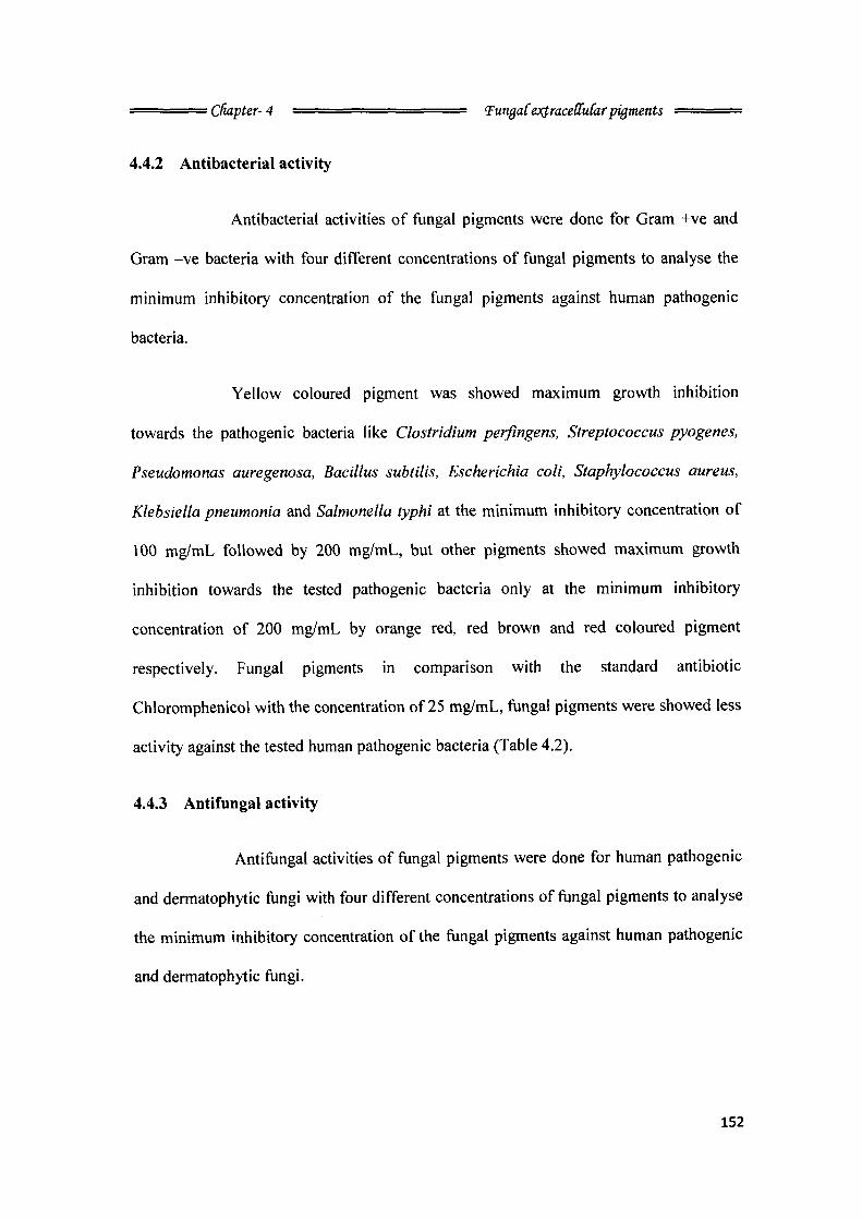
condition. Ethanol extraction, filtration and drying process gives the little quantity of
concentrated fungal pigments. Maximum yield of fungal pigment was produced by the
Peacilomyces sinclairii (5.4 g/L), followed by Penicillium purpurogenum (2.3 g/L),
Peacilomyces farinosus (1.8 g/L) and Emericella nidulans (1.5 g/L). Maximum dry cell
biomass was yielded and recovered from the fungus P. purpurogenum (8 g/L), followed
by P. farinosus (6.6 g/L), P. sinclairii (6.5 g/L) and E. nidulans (4.8 g/L). Change in the
pH of the production medium was observed by the fungus after the completion of the
incubation period of 45 days in the dark condition, which was changed the broth pH
neutral to alkaline. More alkaline pH was observed in the culture broth of P. sinclairii
(11.0), P. purpurogenum (10.0) and P. farinosus (9.5) and acidic pH was observed in the
culture broth of £. nidulans (5.5) (Table. 4.1) (Plate. 9 & 10).
151
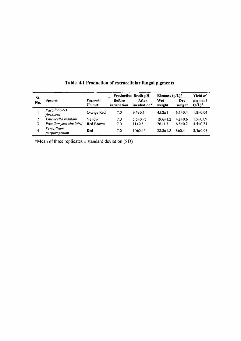
Table. 4.1 Production of extracellular fungal pigments
SI. No.
Species Pigment Colour
Production Broth pH Biomass (g/L)* Yield of SI. No.
Species Pigment Colour
Before incubation
After incubation*
Wet weight
Dry weight
pigment (g/L)*
1
2 3
4
Paecilomyces farinosus Emericella nidulans Paecilomyces sinclairii Penicillium purpurogenum
Orange Red
Yellow Red Brown
Red
7.0
7.0 7.0
7.0
9.5±0.1
5.5±0.25 11 ±0.5
10±0.45
45.8±1
19.6±1.2 26±1.5
28.8±1.8
6.6±0.4
4.8±0.6 6.5±0.2
8±0.4
1.8±0.04
1.5±0.09 5.4±0.31
2.3±0.08
''Mean of three replicates ± standard deviation (SD)
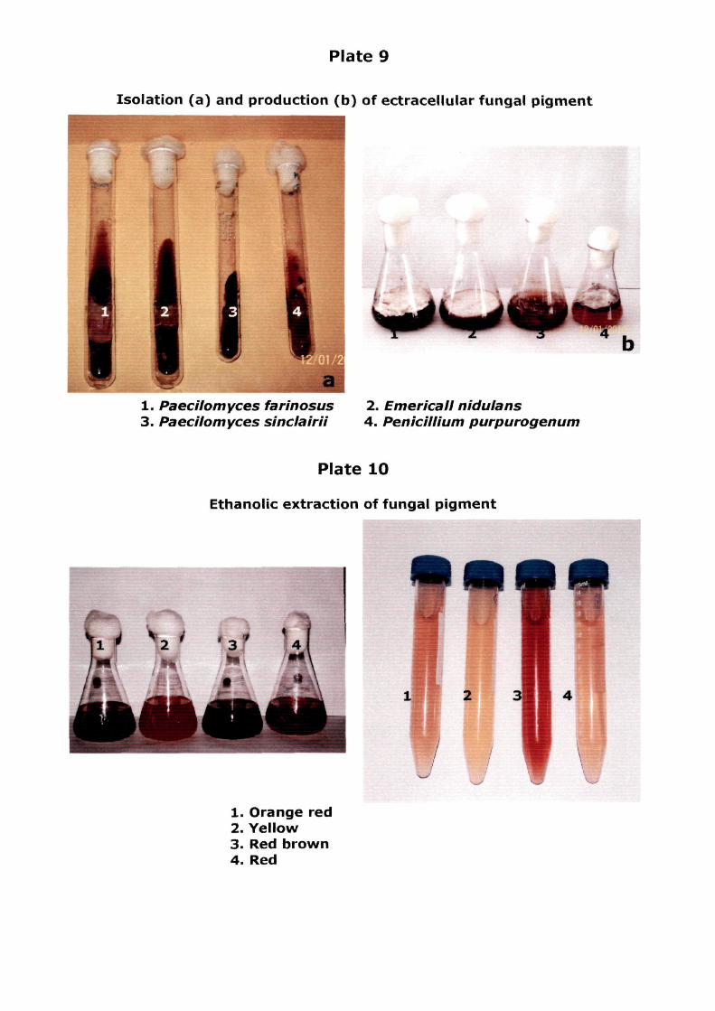
Plate 9
Isolation (a) and production (b) of ectracellular fungal pigment
1. Paecilomyces farinosus 3. Paecilomyces sinclairii
2. Emer'icall nidulans 4. PenicilUum purpurogenum
Plate 10
Ethanolic extraction of fungal pigment
1. Orange red 2. Yellow 3. Red brown 4. Red
'^^ HHp
kigj^
•• CUdpter- 4 = = ^ = ^ = = ^ = = <Fun£aCe:>(traceOM(ar pigments
4.4.2 Antibacterial activity
Antibacterial activities of fungal pigments were done for Gram +ve and
Gram -ve bacteria with four different concentrations of fungal pigments to analyse the
minimum inhibitory concentration of the fungal pigments against human pathogenic
bacteria.
Yellow coloured pigment was showed maximum growth inhibition
towards the pathogenic bacteria like Clostridium perfingem, Streptococcus pyogenes,
Pseudomonas auregenosa. Bacillus subtilis, Escherichia coli, Staphylococcus aureus,
Klebsiella pneumonia and Salmonella typhi at the minimum inhibitory concentration of
100 mg/mL followed by 200 mg/mL, but other pigments showed maximum growth
inhibition towards the tested pathogenic bacteria only at the minimum inhibitory
concentration of 200 mg/mL by orange red, red brown and red coloured pigment
respectively. Fungal pigments in comparison with the standard antibiotic
Chloromphenicol with the concentration of 25 mg/mL, fungal pigments were showed less
activity against the tested human pathogenic bacteria (Table 4.2).
4.4.3 Antifungal activity
Antifungal activities of fungal pigments were done for human pathogenic
and dermatophytic fungi with four different concentrations of fungal pigments to analyse
the minimum inhibitory concentration of the fungal pigments against human pathogenic
and dermatophytic fungi.
152
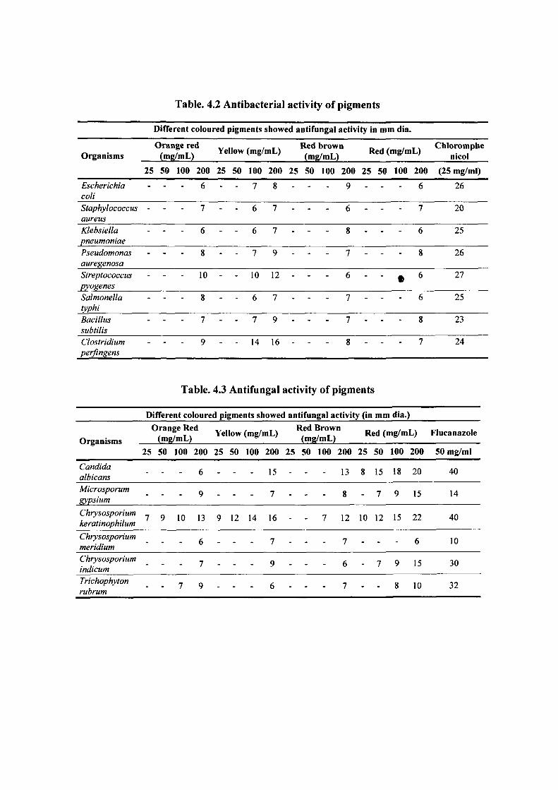
Table. 4.2 Antibacterial activity of pigments
Different coloured pigments showed antifungal activity in mm dia.
Organisms Orange red
(mg/mL) Yellow (mg/mL) Red brown (mg/mL) Red (mg/m L) Chloromphe
nicol
25 50 100 200 25 50 100 200 25 50 100 200 25 50 100 200 (25 mg/ml)
Escherichia coli
- 6 - - 7 8 - - - 9 - - - 6 26
Staphylococcus aureus
- 7 - - 6 7 - - - 6 - - - 7 20
Klebsiella pneumoniae
- 6 - - 6 7 - - - 8 - - - 6 25
Pseudomonas auregenosa
- 8 - - 7 9 - - - 7 - - - 8 26
Streptococcus pyogenes
- 10 - - 10 12 - - - 6 - - • 6 27
Salmonella typhi
- 8 - - 6 7 - - - 7 - - - 6 25
Bacillus subtilis
- 7 - - 7 9 - - - 7 - - - 8 23
Clostridium perflngens
- 9 - - 14 16 - - - 8 - - - 7 24
Table. 4.3 Antifungal activity of pigments
Different coloured pigments showed antifungal activity (in mn 1 dia.)
Organisms Orange Red
(mg/mL) Yellow , , , . Red Brown (mg/mL) , , , , ^ ^ (mg/mL) Red (mg/m iL) Flucanazole Organisms
25 50 100 200 25 50 100 200 25 50 100 200 25 50 100 200 50 mg/ml
Candida albicans - - 6 - - 15 - - 13 8 15 18 20 40
Microsporum gypsium
- - 9 - - 7 - - 8 - 7 9 15 14
Chrysosporium keratinophilum
7 9 10 13 9 12 14 16 - - 7 12 10 12 15 22 40
Chrysosporium meridium - - 6 - - 7 - - 7 - - - 6 10
Chrysosporium indicum - - 7 - - 9 - - 6 - 7 9 15 30
Trichophyton rubrum
- - 7 9 - - 6 - - 7 - - 8 10 32
• Cfiapter- 4 = = = = = = ^ = = = 'FungaCej(trace([u{ar pigments
Red coloured pigment was showed maximum growth inhibition at
minimum inhibitory concentration of 25 mg/mL followed by 50, 100 and 200 mg/mL
towards the Candida albicans and Chrysosporium keratinophilum, at 50 followed by, 100
and 200 mg/mL concentrations towards the Microsporum gypsium and Chrysosprium
indicum, at 100 followed by, 200 mg/ml concentrations towards the Trychophyton
rubrum and at 200 mg/mL concentration towards the Chrysosporium meridium. Yellow
coloured pigment was showed maximum growth inhibition at the minimum inhibitory
concentration of 25 mg/mL followed by, 50, 100 and 200 mg/mL towards the
Chrysosporium keratinophilum, at 200 mg/mL concentration towards the Candida
albicans, Chrysosprium indicum, Microsporum gypsium, Chrysosporium meridium and
Trychophyton rubrum. Orange red coloured pigment was showed maximum growth
inhibition at the minimum inhibitory concentration 25 mg/mL followed by, 50, 100 and
200 mg/mL towards the Chrysosporium keratinophilum, at 100 and 200 mg/mL
concentrations towards the Trychophyton rubrum and at 200 mg/mL concentration
towards the Microsporum gypsium, Chrysosporium indicum, Candida albicans and
Chrysosporium meridium. Red coloured pigment was showed maximum growth
inhibition at the minimum inhibitory concentration of 100 mg/mL followed by 200
mg/mL towards the Chrysosporium keratinophilum and at 200 mg/mL concentration
towards the Candida albicans, Microsporum gypsium, Chrysosporium meridium,
Trychophyton rubrum and Chrysosporium indicum. Fungal pigments in comparison with
the standard antifungal antibiotic Fluconazol with the concentration of 50 mg/mL, fungal
pigments were showed moderate activity against the tested human pathogenic and
dermatophytic fungi (Table. 4.3).
153
: chapter- 4 ^ = = = ^ = = = = = Tut^aC exJ-raceUtdar pigments
4.4.4 UV and IR spectral analysis of fungal pigments
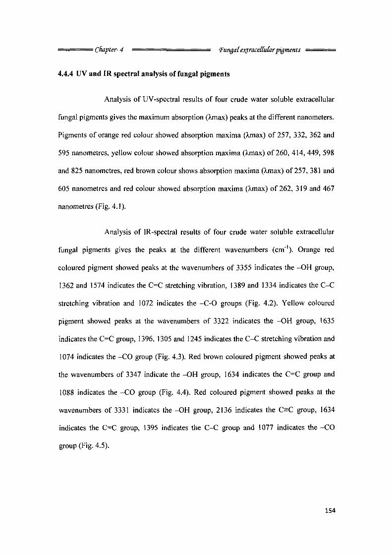
Analysis of UV-spectral results of four crude water soluble extracellular
fungal pigments gives the maximum absorption (X,max) peaks at the different nanometers.
Pigments of orange red colour showed absorption maxima (Xmax) of 257, 332, 362 and
595 nanometres, yellow colour showed absorption maxima (X.max) of 260, 414, 449, 598
and 825 nanometres, red brown colour shows absorption maxima (X,max) of 257, 381 and
605 nanometres and red colour showed absorption maxima (Xmax) of 262, 319 and 467
nanometres (Fig. 4.1).
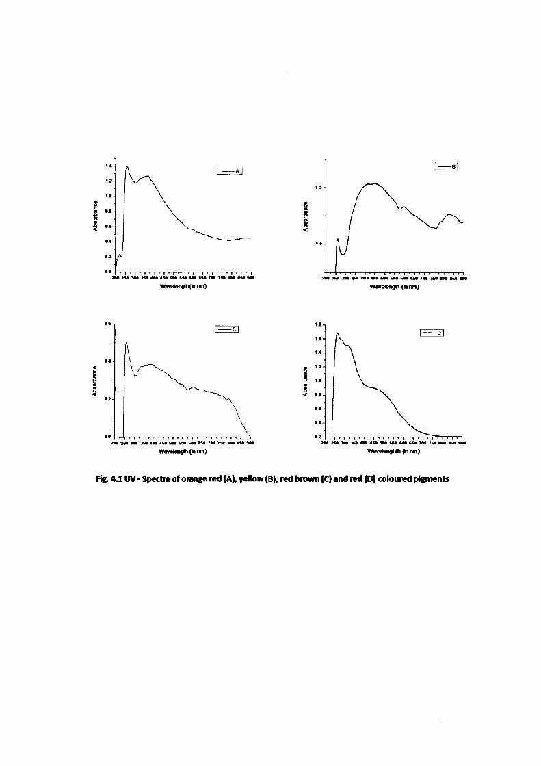
Analysis of IR-spectral results of four crude water soluble extracellular
fungal pigments gives the peaks at the different wavenumbers (cm"'). Orange red
coloured pigment showed peaks at the wavenumbers of 3355 indicates the -OH group,
1362 and 1574 indicates the C=C stretching vibration, 1389 and 1334 indicates the C-C
stretching vibration and 1072 indicates the -C-0 groups (Fig. 4.2). Yellow coloured
pigment showed peaks at the wavenumbers of 3322 indicates the -OH group, 1635
indicates the C=C group, 1396, 1305 and 1245 indicates the C-C stretching vibration and
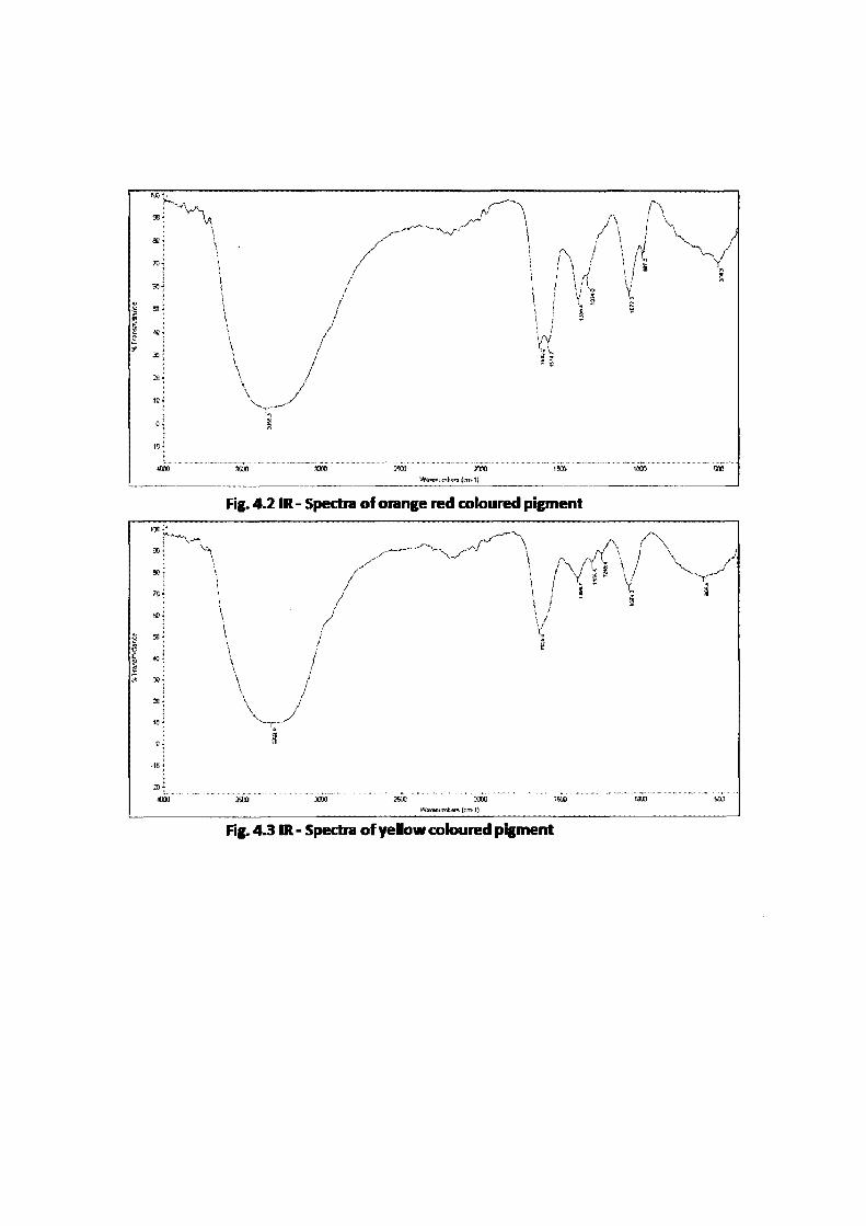
1074 indicates the -CO group (Fig. 4.3). Red brown coloured pigment showed peaks at
the wavenumbers of 3347 indicate the -OH group, 1634 indicates the C=C group and
1088 indicates the -CO group (Fig. 4.4). Red coloured pigment showed peaks at the
wavenumbers of 3331 indicates the -OH group, 2136 indicates the C=C group, 1634
indicates the C=C group, 1395 indicates the C-C group and 1077 indicates the -CO
group (Fig. 4.5).
154
WavelengltKki nm] Wavelefigti (ki nm)
I
Wavelengli C" nn)
' I ' I • I ' I ' I ' I ' I ' I ' I ' I ' I ' M I ' t 2 H » « 3 M 3 M 4 H 4 » « l t C * C M t a 7 M 7 » m m i H
WWBlengMh {nnm)
Fig. 4.1 UV- Spectra of orange red (A), yellow (B), red brown (C) and red (D) coloured pigments
*''vv 90- H . , y^~ \ ao-- \ X'" ^^^-'
\ f\ \fi \ > \ f { \ '
V /
\ /
/ /
\ f\ \fi \ > \ f { \ '
y
i se»
\ / i 1 1 '
1 1«
/ /
11) i
.10;
\ \ \
i
/
lan 3500 3a« rm 1550 laao (SIO
Fig. 4.2 IR- Spectra of orange red coloured pigment
: i / i / 1
^ \ ••• \ \ A
i ^
^ e
^ : \ /
10 ~ ^--^—^
' S H ^
20 i
«ai) 350(1 XOI 2flXI i o n 1500 1 ( H ) " s o b
Fig. 4 3 IR - Spectra of yellow coloured pigment
?0- i
- ^ ^ " ^ ' • • ^ ' \
V ^ B>4 V 1 / V ^ \ /
', \ / « ^^ ^ 50- f \ / i Y ^
JO-' V
1
i 1
\ j \' V
1
DO-' \ 1 5 f \ > i \ ! n ^-1 \ i ^
! 0 -
i,
J 1
lOOO XOQ Tum 250O MX ism loco 500
Fig. 4.4 IR - Spectra of red brown coloured pigment
pfino yam
Fig. 4.5 IR - Speara of Red coloured pigment
: CHapter- 4 ^ ^ ^ = ^ = = ^ = ^ = = 'FungaCe:)(trace(luCir pigments
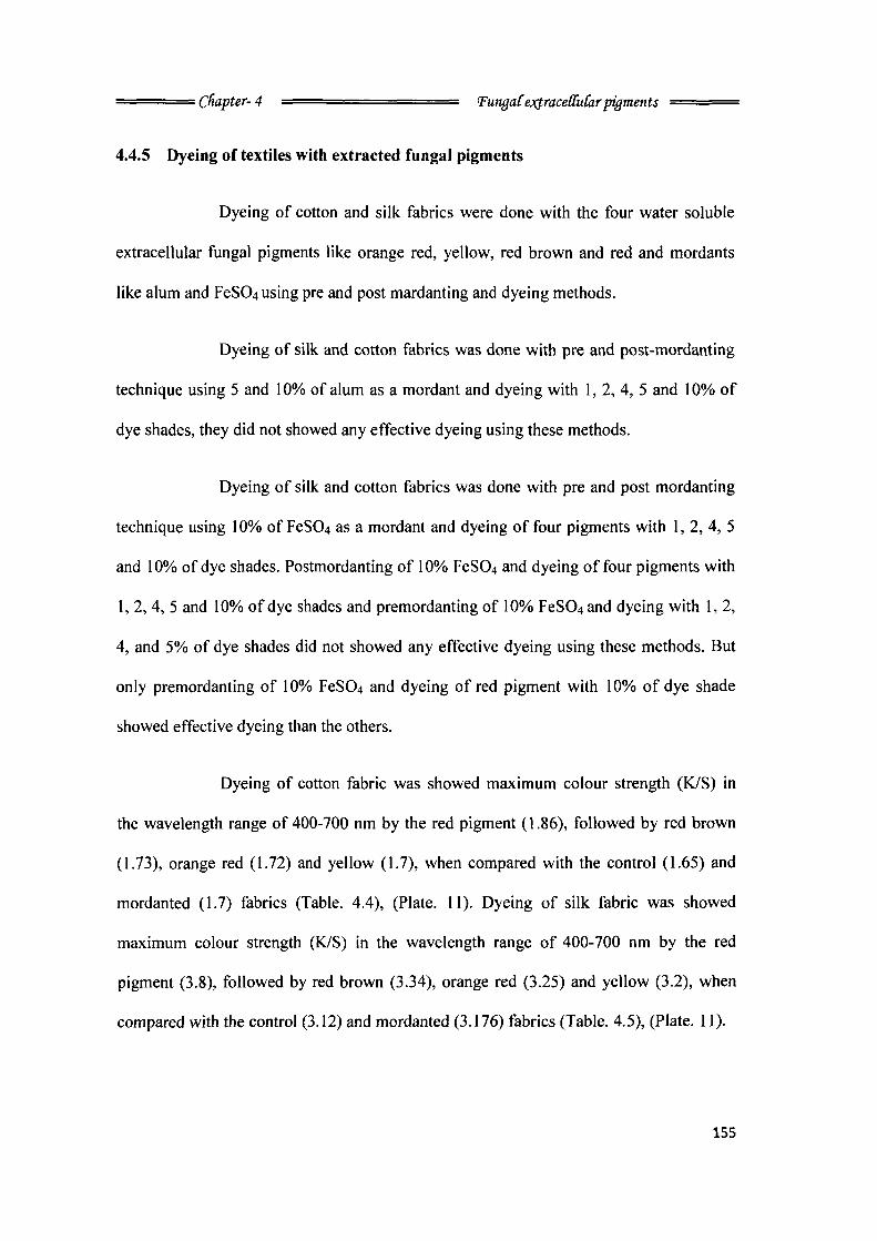
4.4,5 Dyeing of textiles with extracted fungal pigments
Dyeing of cotton and silk fabrics were done with the four water soluble
extracellular fungal pigments like orange red, yellow, red brown and red and mordants
like alum and FeS04 using pre and post mardanting and dyeing methods.
Dyeing of silk and cotton fabrics was done with pre and post-mordanting
technique using 5 and 10% of alum as a mordant and dyeing with 1, 2, 4, 5 and 10% of
dye shades, they did not showed any effective dyeing using these methods.
Dyeing of silk and cotton fabrics was done with pre and post mordanting
technique using 10% of FeS04 as a mordant and dyeing of four pigments with 1, 2, 4, 5
and 10% of dye shades. Postmordanting of 10% FeS04 and dyeing of four pigments with
1, 2, 4, 5 and 10% of dye shades and premordanting of 10% FeS04and dyeing with 1, 2,
4, and 5% of dye shades did not showed any effective dyeing using these methods. But
only premordanting of 10% FeS04 and dyeing of red pigment with 10% of dye shade
showed effective dyeing than the others.
Dyeing of cotton fabric was showed maximum colour strength (K/S) in
the wavelength range of 400-700 nm by the red pigment (1.86), followed by red brown
(1.73), orange red (1.72) and yellow (1.7), when compared with the control (1.65) and
mordanted (1.7) fabrics (Table. 4.4), (Plate. 11). Dyeing of silk fabric was showed
maximum colour strength (K/S) in the wavelength range of 400-700 nm by the red
pigment (3.8), followed by red brown (3.34), orange red (3.25) and yellow (3.2), when
compared with the control (3.12) and mordanted (3.176) fabrics (Table. 4.5), (Plate. 11).
155
Table. 4.4 Dyeing of Cotton with water soluble pigments
Pigment FeS04 (%) Dye (%) Liquor Ratio ^^Q^^Q^^^
Control - - - 1.65 Mordant 10 - 1:50 1.7 Orange Red 10 10 1:50 1.72 Yellow 10 10 1:50 1.7 Red Brown 10 10 1:50 1.73 Red 10 10 1:50 1.86
Table. 4.5 Dyeing of Silk with water soluble pigments
Pigment FeS04 (%) Dye(%) Liquor Ratio I O^Q. QQ p^,)
Control - - - 3.12 Mordant 10 - 1:50 3.176 Orange Red 10 10 1:50 3.25 Yellow 10 10 1:50 3.2 Red Brown 10 10 1:50 3.34 Red 10 10 1:50 3.8
Plate 11
Dyeing of cotton and silk fabrics with fungal pigments (Orange red. Yellow, Red brown and Red)
Pretreatment Premordanting
Dyeing
Control Test
Incubation
Control Test
Dyed silk fabric Dyed cotton fabric
: CHapter- 4 = = = = ^ = = ^ = = = TungaC ex^raceffuCar pigments
4.5 DISCUSSION
The present study shows an idea about the screening and isolation of the
extracellular pigment producing fungi and their diversity in the forest soil samples. In the
present study, an attempt has been made to identify species of pigment producing fungi
and to make use of the colour for textile and to analyse antimicrobial property and also
we can use these pigments as food colours. Four water soluble pigments producing fungal
species like Paecilomyces farinosus, Emericella nidulans, Paecilomyces sinclairii and
Penicillium purpurogenum, these isolates were secreted very intense colours of orange
red, yellow, red brown and red pigments respectively. As these four isolates consistently
registered very high quantity of pigment secretion, they were selected for detailed studies.
Microorganisms occupy a distinct place as source of natural colours. More
than bacteria, fungi display a wide range of fascinating colours, however, till recently
fungi are remained unexplored for colour production. This is perhaps due to their
association with mycotoxins and aflatoxins. The current trend in society for natural
ingredients has stimulated interest in exploring novel means and sources for the
biotechnological production of colourants. In this regard, exploring fungal chemical
diversity is a worthwhile route for the identification of novel pigments. An intelligent
screening approach for water-soluble pigments that is partly based on chemotaxonomy, it
will provide a platform for the future construction of cell factories to the production of
natural colourants. If imperative toxicological testing is carried out, fungal pigments
could be accepted by the current consumer as a food and textile colourant. Further
research using new technologies suggests a promising future for this field of study.
Fungal pigments are widely using as food colours as well as in textile and
156
• chapter- 4 = Tungat e:)(tracellutar pigments
pharmaceautical industries, because these pigments having antimicrobial, antioxidant,
anticancerous properties.
The present study the production of four water soluble pigments was done
from the selected fungal species. Maximum pigment yield was produced by
Paecilomyces sindairii than the other species and maximum dry biomass was produced
by Penicillium perpurogenum than the other isolates. The pH of the fermenting broth
could influence the pigment production and biomass yield and the quality of the pigment
(Morton and Macmilliam, 1954). Chen and Johns (1993) explored that fungal growth and
ankaflavin synthesis were favoured at low pH where as the production of other pigments
was relatively dependent of pH.
Our study showed higher biomass and pigment production at 25 ± 2 °C
under dark condition, however similar results are reported by Wong et ai, (1981). Many
scientists working with Monascus spp., they reported that, 35 °C is an optimum
temperature for the pigment production (Border and Koehler, 1980; Wong and Koehler,
1981). In addition to the effect of temperature, denaturation of the cell membranes might
also occur at higher temperatures, which affect the extraction process (Cacace and
Mazza, 2003). The pigments are heat-stable and hence, temperature of the extracting
medium need not be increased to maximize the process. Cacace and Mazza, (2003)
reported that, the higher temperatures may leads to the degradation of pigments.
Velmurugan et al, (2010) studied that, the effects of different wavelength
of light on the biomass production at the weight of the dry biomass after 7 days of
incubation. The biomass recovered from total darkness showed profuse growth giving a
157
; Cfiapter- 4 TungaC ex^raceHuCar pigments
fluffy appearance to the colonies and highly pigmented mycelia for M. purpureus and P.
purpurogenum, followed by red and blue light source obtained maximum biomass weight
in M purpureus and P. purpurogenum and flasks exposed to white unscreened, green and
yellow light showed the moderate biomass production in /. farinosa, E. nidulam and F.
verticillioides.
Paecilomyces sinclairii, belongs to the class ascomycetes, has a capability
of producing red pigment in high yields during submerged cultures (Miyake et al., 1982).
Generally, two growth forms, the filamentous and the pelleted forms can be observed in
most fungal fermentations, in which the pelleted form is usually less viscous than the
filamentous form (Berovic et al., 1991). Although fungal morphology is influenced by a
variety of factors such as medium composition, agitation intensity, and oxygen tension, it
is still difficult to relate the broth rheology to every aspect of microbial morphology
(Suijdam and Metz, 1981; Schugerl etal., 1983; Gondar etal., 1999; Cho et al., 2002).
Although traditional processes in Asia have predominantly employed solid
state fermentation methods, but liquid state fermentation or submerged fermentation is
the classical method still in use in western countries. Lin (1973) demonstrated that
Monascus spp. could be cuUivated as submerged culture and pigment yields could be
improved. Yamaguchi et al. (1973) developed a method to harvest water soluble
pigments in submerged fermentation by forming amino acids with protein as nitrogen
source. This would cause rearrangement bases with the pigment skeleton which produced
hygroscopic pigments. Lee et al. (2008) demonstrated that when incubation time
increased, pigment production was also increased.
158
: chapter- 4 'FungaC ej^traceffuCar pigments
The present study of four water soluble fungal pigments as antimicrobial
agents, they were effective against human pathogenic bacteria and fungi, which showed
good activity at the lower concentrations. Antibacterial activity of fungal yellow coloured
pigment was showed maximum growth inhibition towards the both gram -ve and gram
+ve pathogenic bacteria at minimum inhibitory concentration of 100 mg/mL, but other
pigments like orange red red brown and red pigments at 200 mg/mL concentrations
towards the tested pathogenic bacteria.
Antifungal activity of fungal red coloured pigment was showed maximum
growth inhibition towards the Candida albicans and Chrysosporium keratinophilum
fungal species at the minimum inhibitory concentration of 25 mg/mL, at 50 mg/mL
towards the Candida albicans and Chrysosporium keratinophilum, at 100 mg/mL
towards the Trychophyton rubrum and at the 200 mg/mL towards the Chrysosporium
meridium. Yellow coloured pigment was showed maximum growth inhibition at the
minimum inhibitory concentration of 25 mg/mL towards the Chrysosporium
keratinophilum and 200 mg/mL concentration towards the remaining tested pathogenic
fungal species. Orange red coloured pigment was showed maximum growth inhibition at
the minimum inhibitory concentration 25 mg/mL towards the Chrysosporium
keratinophilum, at 100 mg/mL concentration towards the Trychophyton rubrum and at
200 mg/mL concentration towards the other tested pathogenic fungi. Red coloured
pigment was showed maximum growth inhibition at the minimum inhibitory
concentration of 100 mg/mL towards the Chrysosporium keratinophilum and at 200
mg/mL concentration towards the other tested pathogenic fungal species. These results
159
• Cdapter- 4 = ^ = = = ^ = = = Tungaf e:x}racellu[ar pigments
reveal that the antimicrobial property of fungal pigments may by the anthraquinone
compounds or their derivatives.
A series of preliminary dyeing tests on different pre-mordanted cotton and
silk fabric standard specimens using ethanolic extracts containing prevailingly orange
red, yellow, red brown and red pigments from P. farinosus, E. nidulans, P. sinclaihi and
P. pupurogenum gave rise to dyed specimens with different hues depending on the
mordant used. The colourants used for dyeing purpose worked at pH 4 for silk and 10 for
cotton in the absence of glabours salt. Dyeing without salt addition was advantage and it
was similar to those reported by Kamel et ai, (2005). According to Lee and Kim, (2004)
the K/S value of dyed cotton fabrics was increased with increasing the pH of buffer
solution up to about 9, and thereafter it levelled off The K/S value of dyed silk fabrics
was not depended on the pH of buffer solution. However, the pH 9 gave the highest K/S
value for cotton and silk fabrics. The reason of the different effect of pH on K/S for
cotton and silk fabrics is not clear at the present moment. More detailed study is needed.
All et al., (2009) studies reported the effect of pre-mordanting and post-
mordanting with aluminium sulphate and ferrous sulphate under different and reported
that pre-mordanting and post-mordanting with ferrous sulfate, there was huge change in
hue and a great deal of decrease in the chroma or purity of colour. Lee and Kim, (2004)
reported in their study that, the K/S values of pre-mordanted cotton and silk fabrics were
higher than those of un-mordanted fabrics. Generally, it is known that the K/S values of
cotton fabrics dyed with natural dyes are very low. However, in his study, he also shows
that premordanted and un-mordanted cotton fabrics dyed with Cassia tora L. extract were
high K/S values. This result might be attributed to the higher affinity of anthraquinone
dye with cellulose. The K/S values of silk fabrics were higher than that of cotton fabrics.
This might be due to the higher mordanted metal ion content.
160
: CHapter- 4 r========s TungaC ex;tracellu[ar pigments
4.6 Summary
The primary objective of tliis study is to isolate and select the pigment
producing fungi for the application aspect studies, then explore the fungal pigments as a
sources for analysing antimicrobial compounds and dyeing colourants. Owing to the
conflicting reports on the safety of food and textiles during processing and dyeing, due to
the indiscriminate use of non-permitted colours, there is an urgent need to identify natural
pigment sources as safe food and textile colourants. This study critically examines the
potentials of fungi as sources for production of biocolourants.
Soil samples collected from Bhadra Wildlife Sanctuary, they are rich
sources of diversified and efficient fungal species for the production of industrially
important products. Four water soluble pigment producing fungi were isolated and
characterized. Potent pigment producing fungal isolates such as Paecilomyces farinosus,
Emericella nidulans, Paecilomyces sinclairii and Penicillium purpurogenum, they are
having the capable of producing orange red, Yellow, red brown and red coloured
pigments extracellularly, they were studied and used for production technology.
Antimicrobial and dyeing tests were done with four water soluble pigments like orange
red, yellow, red brown and red in different concentrations and evaluated. In the present
study medium pH 7.0, temperature of 25 ± 2°C, dark and static condition was used for
the production of pigments from the selected species. Maximum pigment yield was
produced by Paecilomyces sinclairii and maximum dry biomass was produced by
Penicillium perpurogenum than the other isolates.
161
•• Cfiapter- 4 ^ ^ = = = = = ^ = ^ = : TungaC extracelluCar pigments
In the present study of antibacterial activity of yellow coloured fungal
pigment showed maximum growth inhibition at the minimum inhibitory concentration
100 mg/mL towards all tested pathogenic bacteria. Antifungal activity of red coloured
fungal pigment was showed maximum growth inhibition at the minimum inhibitory
concentration of 25 mg/mL towards the Candida albicans and Chrysosporium
keratinophilum, at 50 mg/mL concentrations towards the Microsporum gypsium and
Chrysosprium indicum than the other tested pathogens.
Dyeing of cotton fabric was showed maximum colour strength (K/S) in
the wavelength range of 400-700 nm by the red pigment, followed by red brown, orange
red and yellow when compared with the control and mordanted fabrics. Dyeing of silk
fabric was showed maximum colour strength (K/S) in the wavelength range of 400-700
nm by the red pigment followed by, red brown, orange red and yellow, when compared
with the control and mordanted fabrics.
162