Embed Size (px)
Citation preview
CHAPTER 4 TISSUES - HISTOLOGY - The study of tissues. - 4 basic tissue types, each w/ subtypes:
* epithelial- covering & secretion. * connective - support & attachment. * muscle - movement (locomotion & pump). * nerve - e- transmission for info & control.
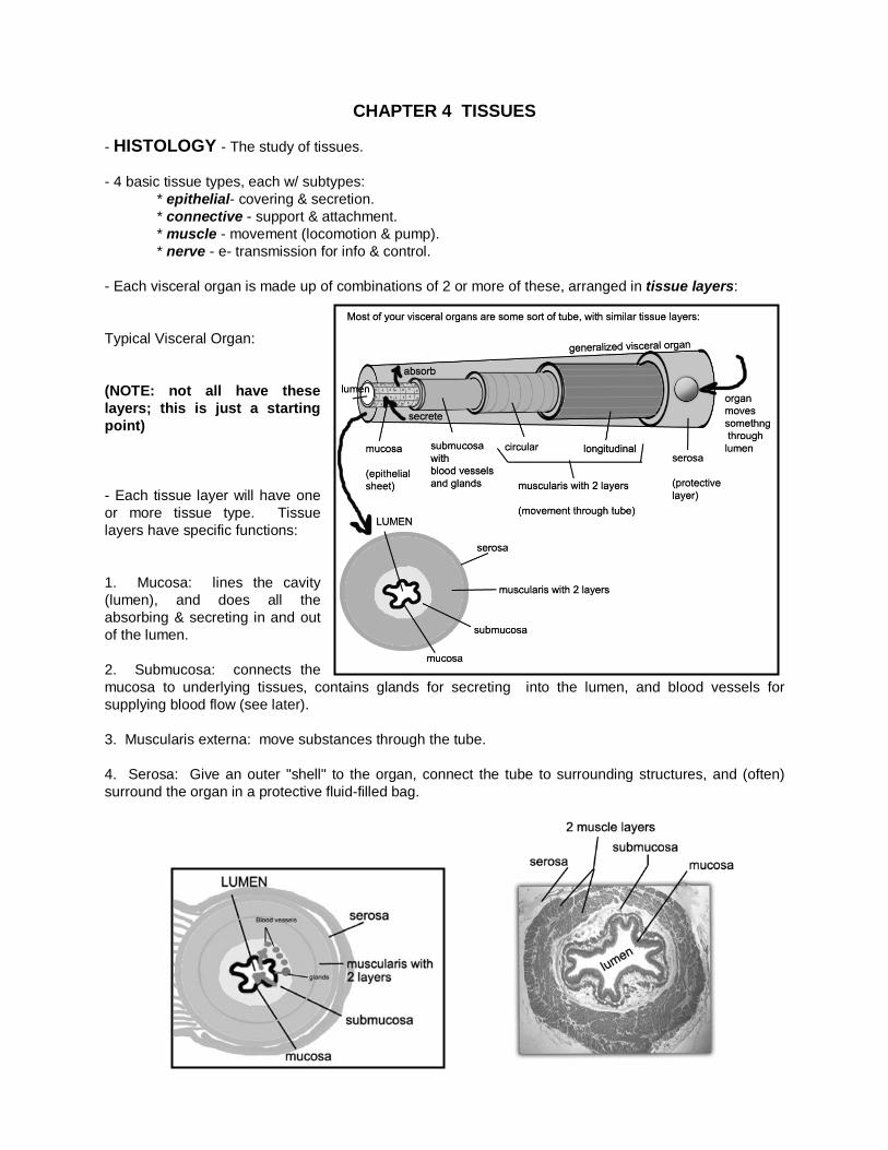
- Each visceral organ is made up of combinations of 2 or more of these, arranged in tissue layers: Typical Visceral Organ: (NOTE: not all have these layers; this is just a starting point) - Each tissue layer will have one or more tissue type. Tissue layers have specific functions: 1. Mucosa: lines the cavity (lumen), and does all the absorbing & secreting in and out of the lumen. 2. Submucosa: connects the mucosa to underlying tissues, contains glands for secreting into the lumen, and blood vessels for supplying blood flow (see later). 3. Muscularis externa: move substances through the tube. 4. Serosa: Give an outer "shell" to the organ, connect the tube to surrounding structures, and (often) surround the organ in a protective fluid-filled bag.
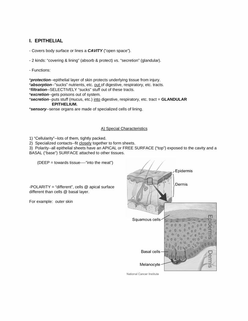
I. EPITHELIAL - Covers body surface or lines a CAVITY (“open space”). - 2 kinds: “covering & lining” (absorb & protect) vs. “secretion” (glandular). - Functions: *protection--epithelial layer of skin protects underlying tissue from injury. *absorption--”sucks” nutrients, etc. out of digestive, respiratory, etc. tracts. *filtration--SELECTIVELY “sucks” stuff out of these tracts. *excretion--gets poisons out of system. *secretion--puts stuff (mucus, etc.) into digestive, respiratory, etc. tract = GLANDULAR EPITHELIUM. *sensory--sense organs are made of specialized cells of lining.
A) Special Characteristics
1) “Cellularity”--lots of them, tightly packed. 2) Specialized contacts--fit closely together to form sheets. 3) Polarity--all epithelial sheets have an APICAL or FREE SURFACE (“top”) exposed to the cavity and a BASAL (“base”) SURFACE attached to other tissues.
(DEEP = towards tissue----”into the meat”) -POLARITY = “different”, cells @ apical surface different than cells @ basal layer. For example: outer skin

* Apical Surface, brush borders &cilia: apical layer have microvilli to increase surface area for absorption, secretion, etc. * Basal Surface & Basement Membrane: Border between epithelial layer & connective tissue made up of glycoproteins.
** Basement membrane = ACELLULAR (”without cells”) ** Acts as a SELECTIVE FILTER, and to attach to deeper layers. Sometimes, we need a thicker area (in active areas, like the small intestines), and the mucosa needs blood vessels, etc. In this case, this area expands into a LAMINA PROPRIA - loose connective tissue with blood vessels, glands, etc. Acts like a "mini-submucosa" within the mucosa ("lamina propria mucosae" means "the mucosa's own special layer").
5) Innervated (contains nerve cells) but Avascular (“without blood vessels”). Gets nutrients from blood vessels in submucosa. 6) Highly regenerative - replaced a lot. Therefore, undergoes a LOT of cell division.
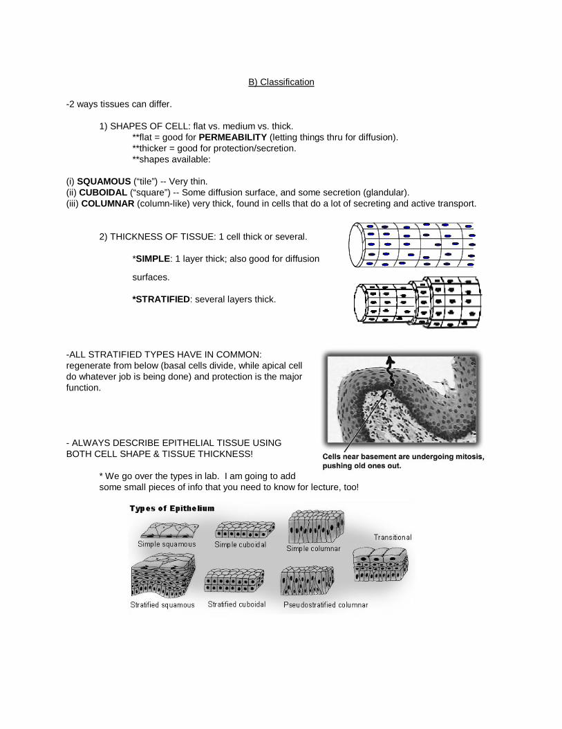
B) Classification
-2 ways tissues can differ.
1) SHAPES OF CELL: flat vs. medium vs. thick. **flat = good for PERMEABILITY (letting things thru for diffusion). **thicker = good for protection/secretion. **shapes available:
(i) SQUAMOUS (“tile”) -- Very thin. (ii) CUBOIDAL (“square”) -- Some diffusion surface, and some secretion (glandular). (iii) COLUMNAR (column-like) very thick, found in cells that do a lot of secreting and active transport. 2) THICKNESS OF TISSUE: 1 cell thick or several.
*SIMPLE: 1 layer thick; also good for diffusion
surfaces.
*STRATIFIED: several layers thick.
-ALL STRATIFIED TYPES HAVE IN COMMON: regenerate from below (basal cells divide, while apical cell do whatever job is being done) and protection is the major function.
- ALWAYS DESCRIBE EPITHELIAL TISSUE USING BOTH CELL SHAPE & TISSUE THICKNESS!
* We go over the types in lab. I am going to add some small pieces of info that you need to know for lecture, too!
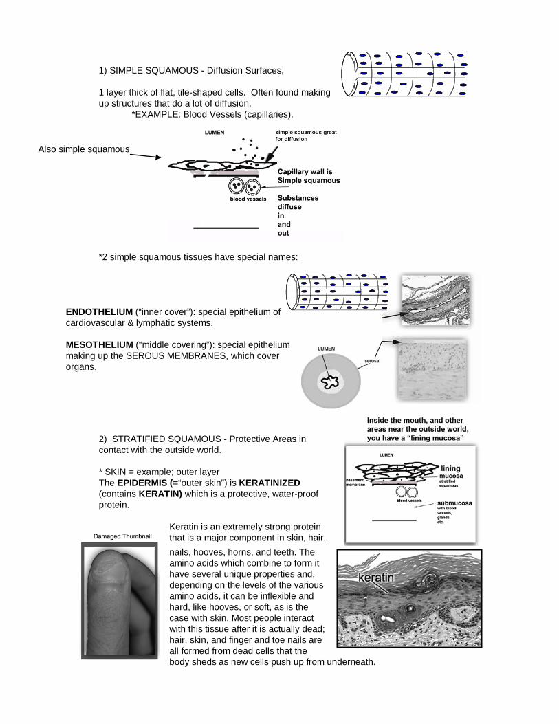
1) SIMPLE SQUAMOUS - Diffusion Surfaces, 1 layer thick of flat, tile-shaped cells. Often found making up structures that do a lot of diffusion. *EXAMPLE: Blood Vessels (capillaries).
*2 simple squamous tissues have special names:
ENDOTHELIUM (“inner cover”): special epithelium of cardiovascular & lymphatic systems. MESOTHELIUM (“middle covering”): special epithelium making up the SEROUS MEMBRANES, which cover organs.
2) STRATIFIED SQUAMOUS - Protective Areas in contact with the outside world. * SKIN = example; outer layer The EPIDERMIS (=“outer skin”) is KERATINIZED (contains KERATIN) which is a protective, water-proof protein.
Keratin is an extremely strong protein that is a major component in skin, hair, nails, hooves, horns, and teeth. The amino acids which combine to form it have several unique properties and, depending on the levels of the various amino acids, it can be inflexible and hard, like hooves, or soft, as is the case with skin. Most people interact with this tissue after it is actually dead; hair, skin, and finger and toe nails are all formed from dead cells that the body sheds as new cells push up from underneath.
Also simple squamous
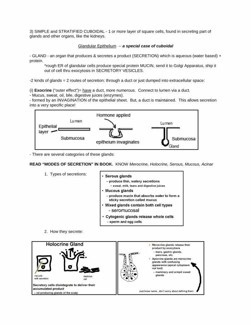
3) SIMPLE and STRATIFIED CUBOIDAL - 1 or more layer of square cells, found in secreting part of glands and other organs, like the kidneys.
Glandular Epithelium -- a special case of cuboidal
- GLAND - an organ that produces & secretes a product (SECRETION) which is aqueous (water based) + protein.
*rough ER of glandular cells produce special protein MUCIN, send it to Golgi Apparatus, ship it out of cell thru exocytosis in SECRETORY VESICLES.
-2 kinds of glands = 2 routes of secretion: through a duct or just dumped into extracellular space: (i) Exocrine (“outer effect”)= have a duct, more numerous. Connect to lumen via a duct. - Mucus, sweat, oil, bile, digestive juices (enzymes). - formed by an INVAGINATION of the epithelial sheet. But, a duct is maintained. This allows secretion into a very specific place!
- There are several categories of these glands: READ “MODES OF SECRETION" IN BOOK. KNOW Merocrine, Holocrine, Serous, Mucous, Acinar
1. Types of secretions:
2. How they secrete:
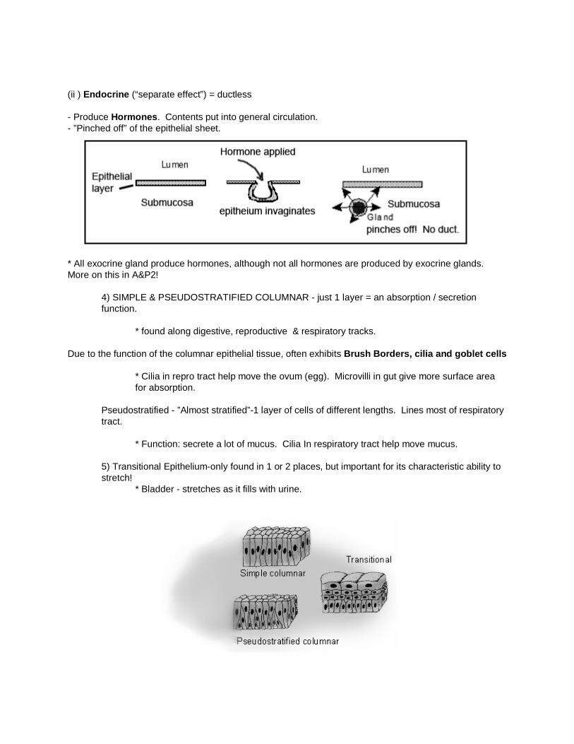
(ii ) Endocrine (“separate effect”) = ductless - Produce Hormones. Contents put into general circulation. - ”Pinched off” of the epithelial sheet.
* All exocrine gland produce hormones, although not all hormones are produced by exocrine glands. More on this in A&P2!
4) SIMPLE & PSEUDOSTRATIFIED COLUMNAR - just 1 layer = an absorption / secretion function.
* found along digestive, reproductive & respiratory tracks.
Due to the function of the columnar epithelial tissue, often exhibits Brush Borders, cilia and goblet cells
* Cilia in repro tract help move the ovum (egg). Microvilli in gut give more surface area for absorption.
Pseudostratified - ”Almost stratified”-1 layer of cells of different lengths. Lines most of respiratory tract.
* Function: secrete a lot of mucus. Cilia In respiratory tract help move mucus.
5) Transitional Epithelium-only found in 1 or 2 places, but important for its characteristic ability to stretch!
* Bladder - stretches as it fills with urine.