Embed Size (px)
Citation preview
PM205-B04 PII: B978-0-8194-8535-9.00013-0 ISBN: 978-0-8194-8535-9 PAGE: 89 (89–114)
Chapter 4X Rays and Gamma Rays:Crookes Tubes and Nuclear Light
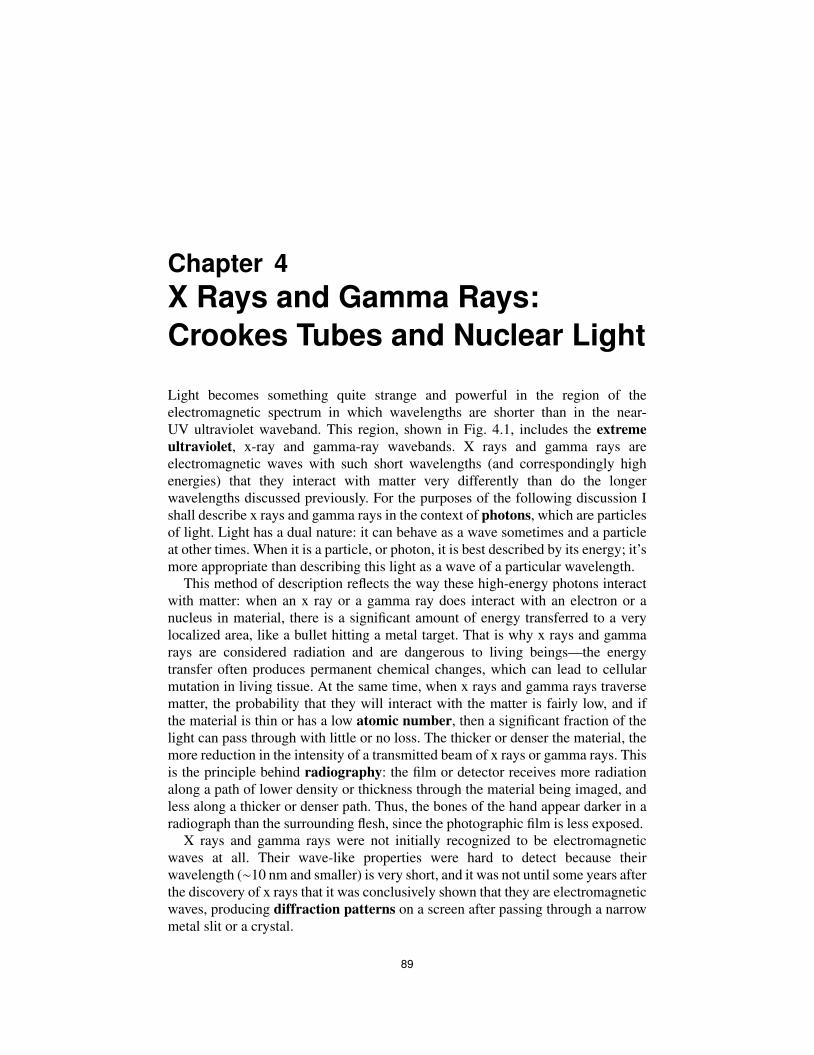
Light becomes something quite strange and powerful in the region of theelectromagnetic spectrum in which wavelengths are shorter than in the near-UV ultraviolet waveband. This region, shown in Fig. 4.1, includes the extremeultraviolet, x-ray and gamma-ray wavebands. X rays and gamma rays areelectromagnetic waves with such short wavelengths (and correspondingly highenergies) that they interact with matter very differently than do the longerwavelengths discussed previously. For the purposes of the following discussion Ishall describe x rays and gamma rays in the context of photons, which are particlesof light. Light has a dual nature: it can behave as a wave sometimes and a particleat other times. When it is a particle, or photon, it is best described by its energy; it’smore appropriate than describing this light as a wave of a particular wavelength.
This method of description reflects the way these high-energy photons interactwith matter: when an x ray or a gamma ray does interact with an electron or anucleus in material, there is a significant amount of energy transferred to a verylocalized area, like a bullet hitting a metal target. That is why x rays and gammarays are considered radiation and are dangerous to living beings—the energytransfer often produces permanent chemical changes, which can lead to cellularmutation in living tissue. At the same time, when x rays and gamma rays traversematter, the probability that they will interact with the matter is fairly low, and ifthe material is thin or has a low atomic number, then a significant fraction of thelight can pass through with little or no loss. The thicker or denser the material, themore reduction in the intensity of a transmitted beam of x rays or gamma rays. Thisis the principle behind radiography: the film or detector receives more radiationalong a path of lower density or thickness through the material being imaged, andless along a thicker or denser path. Thus, the bones of the hand appear darker in aradiograph than the surrounding flesh, since the photographic film is less exposed.
X rays and gamma rays were not initially recognized to be electromagneticwaves at all. Their wave-like properties were hard to detect because theirwavelength (∼10 nm and smaller) is very short, and it was not until some years afterthe discovery of x rays that it was conclusively shown that they are electromagneticwaves, producing diffraction patterns on a screen after passing through a narrowmetal slit or a crystal.
89
PM205-B04 PII: B978-0-8194-8535-9.00013-0 ISBN: 978-0-8194-8535-9 PAGE: 90 (89–114)
90 Chapter 4
Figure 4.1 The short-wavelength region of the electromagnetic spectrum.
X rays and gamma rays are best known for their ability to enable imagingthrough opaque material, since they are not absorbed by matter nearly as much as islight with longer wavelength. While microwave light can be used to image throughcertain materials such as dry dirt or sand quite well, it cannot penetrate effectivelythrough conductive materials such as water or metal. X rays and gamma rays,however, can penetrate all known materials. While visible light may only penetratea few millimeters deep into human tissue, x rays at modest energies will easilypass all the way through a person’s chest, and a person could easily look nearlytransparent in a gamma-ray image – bones and all. Electromagnetic waves at thesehigh energies are generally not encountered in daily life except when generated bymedical or scientific instrumentation, or (in the case of gamma rays) by radioactiveisotopes such as Cobalt-60. Luckily for us, the atmosphere blocks nearly all x-rayand gamma-ray light impinging on Earth from extraterrestrial sources.1
We can imagine what it would be like for a hypothetical creature that could seeonly in the x-ray or gamma-ray wavebands to be on Earth. Such a creature wouldhave a very hard time passively imaging anything in our atmosphere at sea level,since there would be virtually no available x-ray or gamma-ray illumination fromthe sun or other celestial sources, and none at all from ordinary (non-radioactive)objects. This situation is quite different from what a creature with thermal infraredvision (like the Predator in the movie of the same name) would encounter, forexample. People, animals, trees and machines all emit thermal infrared light andcan be observed without any active illumination being present. Observing peopleor trees or other objects using light in the x-ray and gamma-ray bands wouldrequire active illumination of a very specialized nature. Ordinary light sourcessuch as lightbulbs emit invisible light (infrared and ultraviolet), but they do notemit x rays or gamma rays because the filaments in lightbulbs are simply nothot enough. A lightbulb filament would have to be heated to millions of degreesto glow incandescently with x-ray light. Making incandescent x rays with evenmodest energies in a terrestrial laboratory is very hard to do.2 It takes extremeconditions such as those encountered in a nuclear explosion or nuclear fusionreactor to achieve these temperatures.
1X-ray and gamma-ray astronomy requires detectors mounted on high-altitude platforms such asrockets or satellites.2Incandescent means that the material producing the x rays is doing so by heating alone; this requiresenormously high temperatures (>1 million degrees C).
PM205-B04 PII: B978-0-8194-8535-9.00013-0 ISBN: 978-0-8194-8535-9 PAGE: 91 (89–114)
X Rays and Gamma Rays: Crookes Tubes and Nuclear Light 91
How, then, do we make x rays? For the most part, we make them thesame way they were made by their discoverer, Prof. Wilhelm Konrad Roentgen(1845–1923), a German physicist. Roentgen’s method is not incandescent—ratherit uses electrons as x-ray generating “bullets.” Roentgen used an evacuated tubewith two metal electrodes inside connected to a high voltage power supply. Theelectric field generated by the power supply accelerates electrons emitted from ahot wire filament inside the tube and smashes them into a metal plate. The electronspenetrate into atoms in the metal surface and are sharply deflected by the electricfields near the nuclei of the atoms. This deflection produces x rays. The energy ofthe resulting x rays is described in units that include the acceleration voltage ofthe x-ray tube. We thus speak of x rays as having energies of kilo-electron volts,or keV. One keV is the kinetic energy of an electron that has dropped througha potential difference of 1000 volts (1 kV). This same method of generating xrays is still used today. Typical medical x-ray tube voltages are 100 kV; we woulddescribe the resulting x rays as having energies of 100 keV, corresponding to awavelength of about 0.01 nm (10−11 m), or about a tenth of the radius of an atom.Electron-volt units are a convenient way to describe their energy, even when thesource has nothing to do with voltage, as in the case of thermal x rays. In fact,this descriptive convention applies to all sorts of high-energy particles producedby particle accelerators, radioactive decay, or astrophysical sources.
Gamma rays
Gamma rays areelectromagnetic waves with extremely short wavelengths, shorterthan x rays, and with correspondingly higher photon energies. They are not causedby the rearrangement of electrons within an atom; rather, they are generated bychanges in the nuclei of atoms, changes due to nuclear decay processes wherea nucleus changes internally and releases energy. Their energies correspond tobinding energies of nucleons in the nucleus. We can generate gamma rays byassembling concentrated masses of radioactive material or by using powerfulparticle accelerators to accelerate electrons to very high energies and then smashthem into a target material, causing nuclear processes to occur. The gamma-rayband encompasses all photons with energies above about 100 keV, correspondingto wavelengths of 0.01 nm (10−11m) and shorter. What is the difference between xrays and gamma rays? There is no particular boundary between the two (since thenaming convention is somewhat arbitrary), but most scientists consider x rays tobe produced by electron-atom interactions, whereas gamma rays are in the millionelectron-volt or higher energy range and are produced by nuclear processes suchas radioactive decay.
X-ray and gamma-ray imaging technology is quite different from imagingtechnology described in previous chapters. The wavelike properties of light atmedium wavelengths (visible, UV and IR) make it possible to image this lightonto detectors, much like our own eye, which like a standard film camera consistof a detector material in a focal-plane array (the retina) and an imaging optic (thecrystalline lens) that focuses a scene onto the retina. X rays and gamma rays do
PM205-B04 PII: B978-0-8194-8535-9.00013-0 ISBN: 978-0-8194-8535-9 PAGE: 92 (89–114)
92 Chapter 4
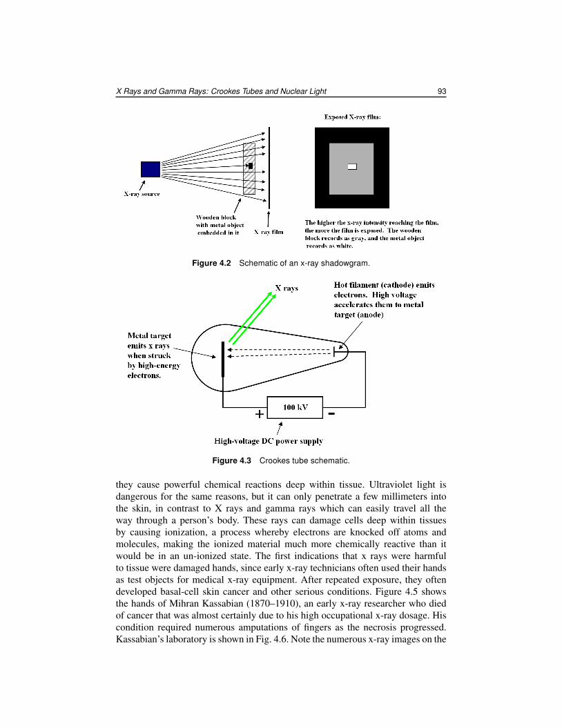
not lend themselves well to being focused by lenses or mirrors — they pass rightthrough the material of the optics without deflection. But lenses and focal-planearrays are not the only way to make an image from electromagnetic waves. As wesaw in the last chapter, microwave imaging systems create images through precisetiming of return pulses of microwave energy, or, in the case of passive imaging, byscanning a reflective parabolic antenna with a single detector at the focal point backand forth over a scene. Most x-ray and gamma-ray images are made an entirelydifferent way, by a method that does not require a lens. The images we traditionallyassociate with x rays are what are known as shadowgrams; that is, a recording ofan object’s shadow projected onto a detector material, which could be a piece offilm or a sheet of glass coated with phosphor. Thus, the object under investigationhas to be placed between the x-ray source and the detector, as shown in Fig. 4.2.It should be noted that there are techniques for focusing x rays—there will be anastronomical example later.
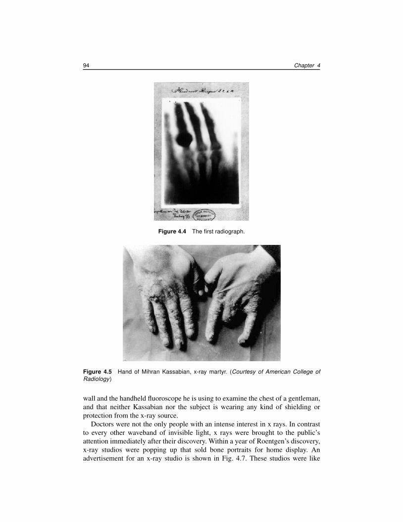
The discovery of x rays was one of the last great scientific achievements ofthe 19th century, and heralded a new age of physics. Wilhelm Roentgen wasawarded the first Nobel Prize in physics for a discovery he made in 1895: hefound that a Hittorf tube (a special evacuated tube with metal electrodes and ahigh-voltage current) produced penetrating rays that fogged photographic film andmade fluorescent materials glow even when the tube was covered with black paperto block the ultraviolet light that was also emitted by the tube.3 Figure 4.3 shows aschematic drawing of a Crookes tube, an x-ray tube similar to the Hittorf tube usedby Roentgen. The low-voltage power supply heats a filament called a cathode,causing electrons to “boil off”—a process known as thermionic emission. The 100kV supply accelerates the electrons towards the metal target, known as the anode.These high-voltage electrons strike the target, causing electrons in the metal atomsto emit x rays.
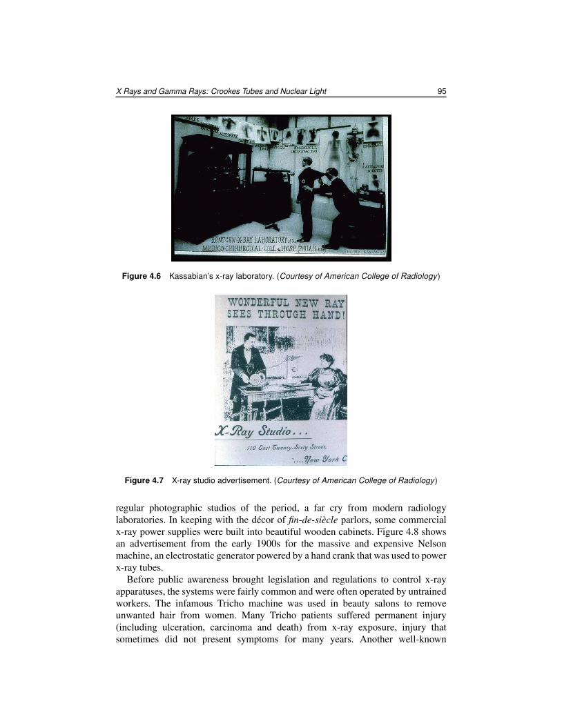
Roentgen’s system produced x rays with sufficient energy (about 40 keV) toimage the bones in his hand using a fluorescent screen. He soon discovered thatphotographic film is also sensitive to x-ray light. Figure 4.4 shows the first x-rayimage made with film; it is a picture of the hand of Roentgen’s wife Bertha withher wedding band clearly visible. X-ray images are also known as radiographs.
Doctors began using x rays as part of diagnosis and treatment of patientsalmost immediately after Roentgen announced his discoveries. The exploitationof x-ray imaging technology by the medical establishment raced far ahead ofany understanding of their effects, particularly on living tissue. The publicityblitz that followed the initial discovery engendered a great deal of quackery andmisinformation, some of it with tragic consequences. Some writers in the early1900s claimed that x rays could raise the dead, cure blindness, or treat skinconditions, and many people were injured permanently with large, uncontrolleddoses of x rays. X rays (and gamma rays) are dangerous in high doses because
3A translation of Roentgen’s original paper can be found in E.C. Watson, “The Discovery of X Rays,”American Journal of Physics 13, 284 (1945).
PM205-B04 PII: B978-0-8194-8535-9.00013-0 ISBN: 978-0-8194-8535-9 PAGE: 93 (89–114)
X Rays and Gamma Rays: Crookes Tubes and Nuclear Light 93
Figure 4.2 Schematic of an x-ray shadowgram.
Figure 4.3 Crookes tube schematic.
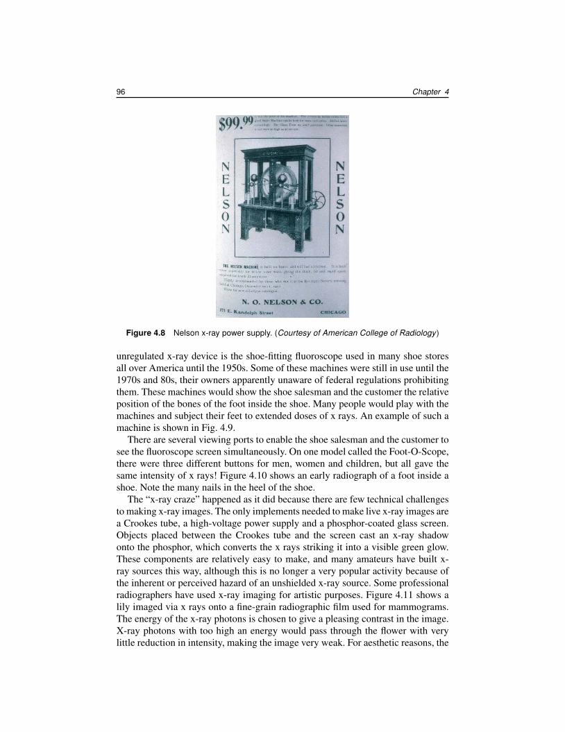
they cause powerful chemical reactions deep within tissue. Ultraviolet light isdangerous for the same reasons, but it can only penetrate a few millimeters intothe skin, in contrast to X rays and gamma rays which can easily travel all theway through a person’s body. These rays can damage cells deep within tissuesby causing ionization, a process whereby electrons are knocked off atoms andmolecules, making the ionized material much more chemically reactive than itwould be in an un-ionized state. The first indications that x rays were harmfulto tissue were damaged hands, since early x-ray technicians often used their handsas test objects for medical x-ray equipment. After repeated exposure, they oftendeveloped basal-cell skin cancer and other serious conditions. Figure 4.5 showsthe hands of Mihran Kassabian (1870–1910), an early x-ray researcher who diedof cancer that was almost certainly due to his high occupational x-ray dosage. Hiscondition required numerous amputations of fingers as the necrosis progressed.Kassabian’s laboratory is shown in Fig. 4.6. Note the numerous x-ray images on the
PM205-B04 PII: B978-0-8194-8535-9.00013-0 ISBN: 978-0-8194-8535-9 PAGE: 94 (89–114)
94 Chapter 4
Figure 4.4 The first radiograph.
Figure 4.5 Hand of Mihran Kassabian, x-ray martyr. (Courtesy of American College ofRadiology)
wall and the handheld fluoroscope he is using to examine the chest of a gentleman,and that neither Kassabian nor the subject is wearing any kind of shielding orprotection from the x-ray source.
Doctors were not the only people with an intense interest in x rays. In contrastto every other waveband of invisible light, x rays were brought to the public’sattention immediately after their discovery. Within a year of Roentgen’s discovery,x-ray studios were popping up that sold bone portraits for home display. Anadvertisement for an x-ray studio is shown in Fig. 4.7. These studios were like
PM205-B04 PII: B978-0-8194-8535-9.00013-0 ISBN: 978-0-8194-8535-9 PAGE: 95 (89–114)
X Rays and Gamma Rays: Crookes Tubes and Nuclear Light 95
Figure 4.6 Kassabian’s x-ray laboratory. (Courtesy of American College of Radiology)
Figure 4.7 X-ray studio advertisement. (Courtesy of American College of Radiology)
regular photographic studios of the period, a far cry from modern radiologylaboratories. In keeping with the décor of fin-de-siècle parlors, some commercialx-ray power supplies were built into beautiful wooden cabinets. Figure 4.8 showsan advertisement from the early 1900s for the massive and expensive Nelsonmachine, an electrostatic generator powered by a hand crank that was used to powerx-ray tubes.
Before public awareness brought legislation and regulations to control x-rayapparatuses, the systems were fairly common and were often operated by untrainedworkers. The infamous Tricho machine was used in beauty salons to removeunwanted hair from women. Many Tricho patients suffered permanent injury(including ulceration, carcinoma and death) from x-ray exposure, injury thatsometimes did not present symptoms for many years. Another well-known
PM205-B04 PII: B978-0-8194-8535-9.00013-0 ISBN: 978-0-8194-8535-9 PAGE: 96 (89–114)
96 Chapter 4
Figure 4.8 Nelson x-ray power supply. (Courtesy of American College of Radiology)
unregulated x-ray device is the shoe-fitting fluoroscope used in many shoe storesall over America until the 1950s. Some of these machines were still in use until the1970s and 80s, their owners apparently unaware of federal regulations prohibitingthem. These machines would show the shoe salesman and the customer the relativeposition of the bones of the foot inside the shoe. Many people would play with themachines and subject their feet to extended doses of x rays. An example of such amachine is shown in Fig. 4.9.
There are several viewing ports to enable the shoe salesman and the customer tosee the fluoroscope screen simultaneously. On one model called the Foot-O-Scope,there were three different buttons for men, women and children, but all gave thesame intensity of x rays! Figure 4.10 shows an early radiograph of a foot inside ashoe. Note the many nails in the heel of the shoe.
The “x-ray craze” happened as it did because there are few technical challengesto making x-ray images. The only implements needed to make live x-ray images area Crookes tube, a high-voltage power supply and a phosphor-coated glass screen.Objects placed between the Crookes tube and the screen cast an x-ray shadowonto the phosphor, which converts the x rays striking it into a visible green glow.These components are relatively easy to make, and many amateurs have built x-ray sources this way, although this is no longer a very popular activity because ofthe inherent or perceived hazard of an unshielded x-ray source. Some professionalradiographers have used x-ray imaging for artistic purposes. Figure 4.11 shows alily imaged via x rays onto a fine-grain radiographic film used for mammograms.The energy of the x-ray photons is chosen to give a pleasing contrast in the image.X-ray photons with too high an energy would pass through the flower with verylittle reduction in intensity, making the image very weak. For aesthetic reasons, the