Embed Size (px)
Citation preview
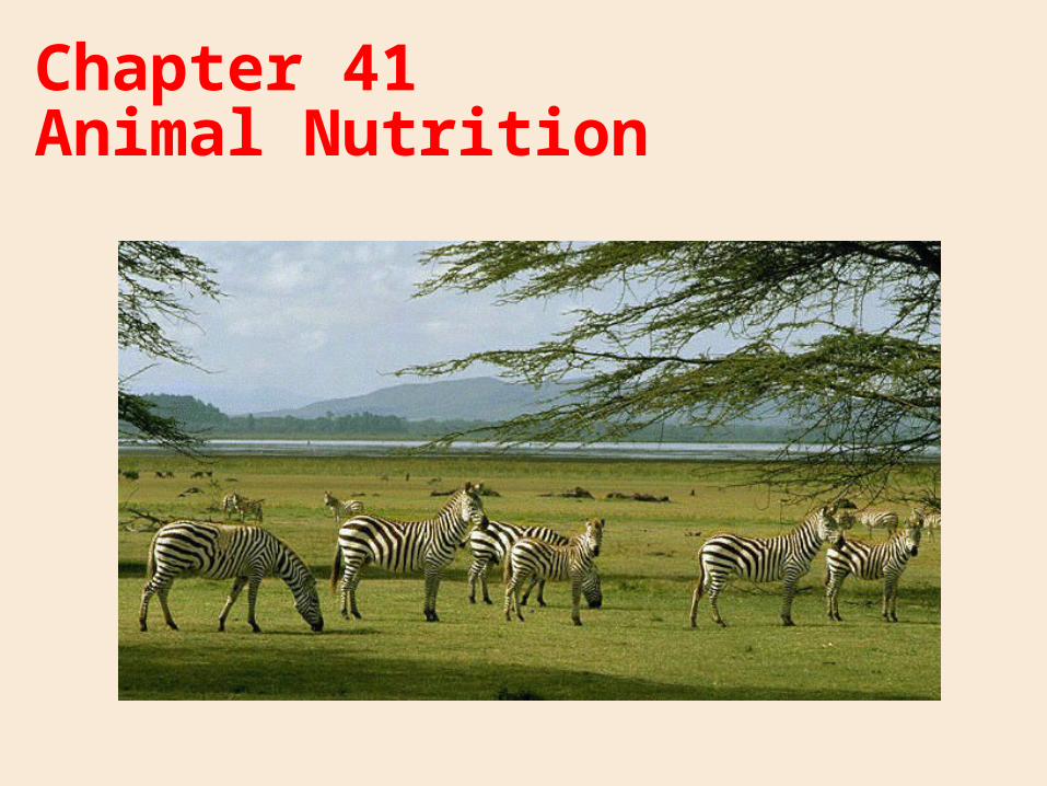
Chapter 41 Animal Nutrition
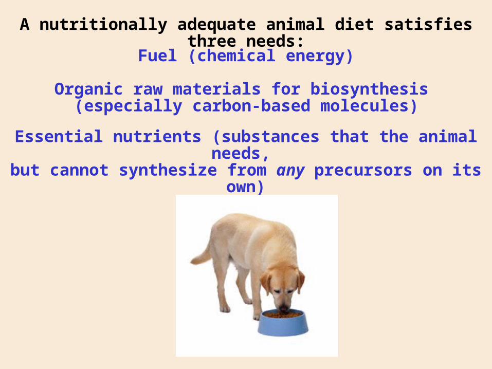
A nutritionally adequate animal diet satisfies three needs:
Fuel (chemical energy)
Organic raw materials for biosynthesis (especially carbon-based molecules)
Essential nutrients (substances that the animal needs, but cannot synthesize from any precursors on its own)
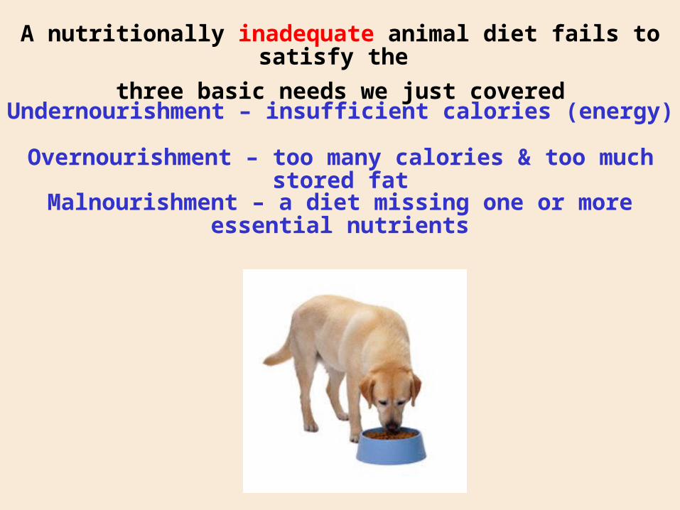
A nutritionally inadequate animal diet fails to satisfy the
three basic needs we just covered
Undernourishment – insufficient calories (energy)
Overnourishment – too many calories & too much stored fat
Malnourishment – a diet missing one or more essential nutrients
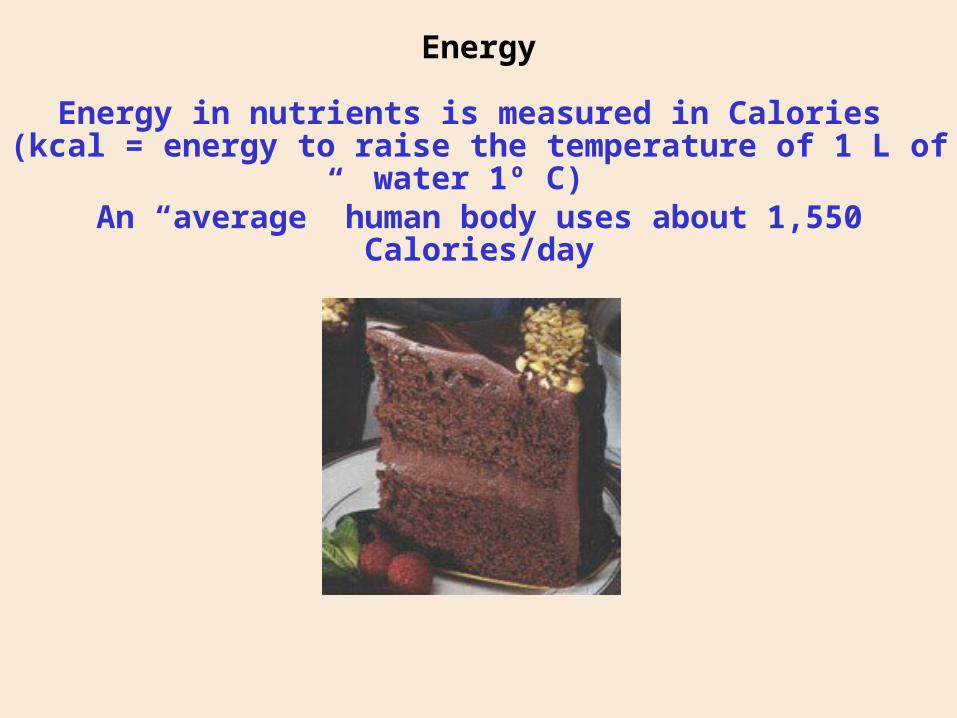
Energy
Energy in nutrients is measured in Calories (kcal = energy to raise the temperature of 1 L of water 1º C)
An “average” human body uses about 1,550 Calories/day
Principal categories of nutrients:
Lipids – found in lipid membranes, etc.;including essential fatty acids
9 Calories per gram (a principal energy source)
Principal categories of nutrients:
Proteins – building blocks and enzymes;animals require 20 amino acids, including essential amino acids
4 Calories per gram (usually a secondary energy source, since the breakdown of proteins produces urea,
a potentially toxic compound)
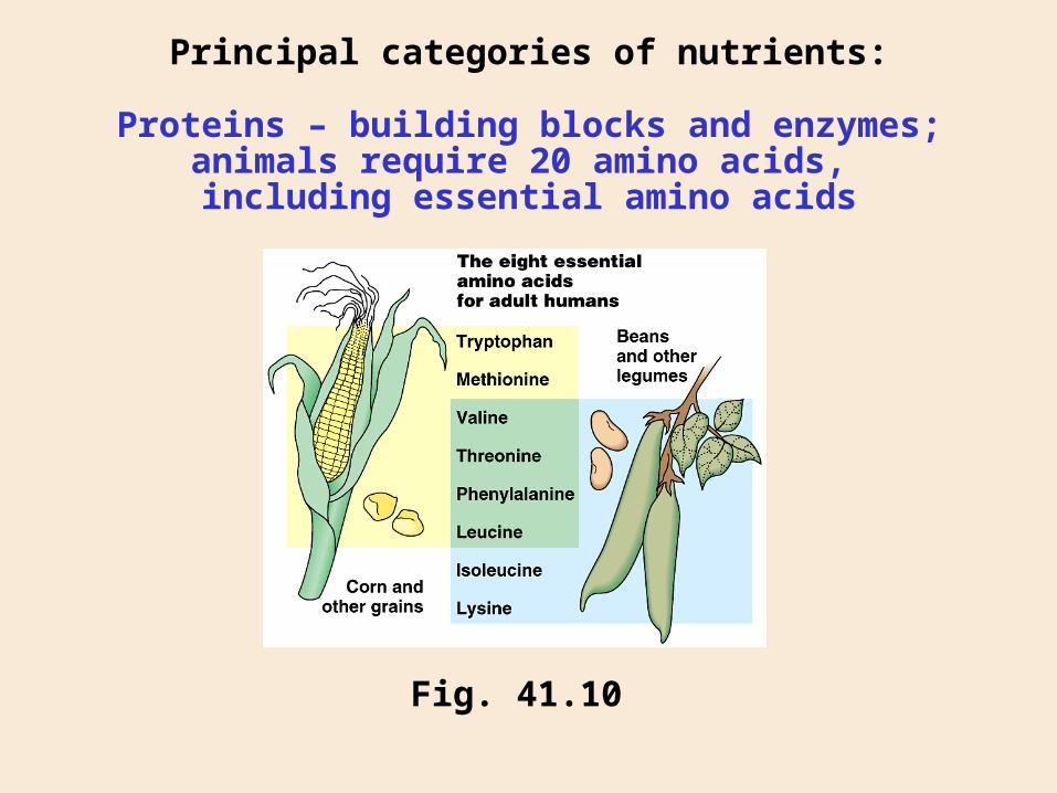
Principal categories of nutrients:
Fig. 41.10
Proteins – building blocks and enzymes;animals require 20 amino acids, including essential amino acids
Principal categories of nutrients:
Carbohydrates – C-based building blocks and energy
4 Calories per gram; can be a very quick energy source (e.g., glucose)
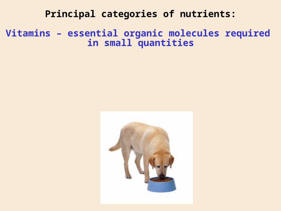
Principal categories of nutrients:
Vitamins – essential organic molecules required in small quantities
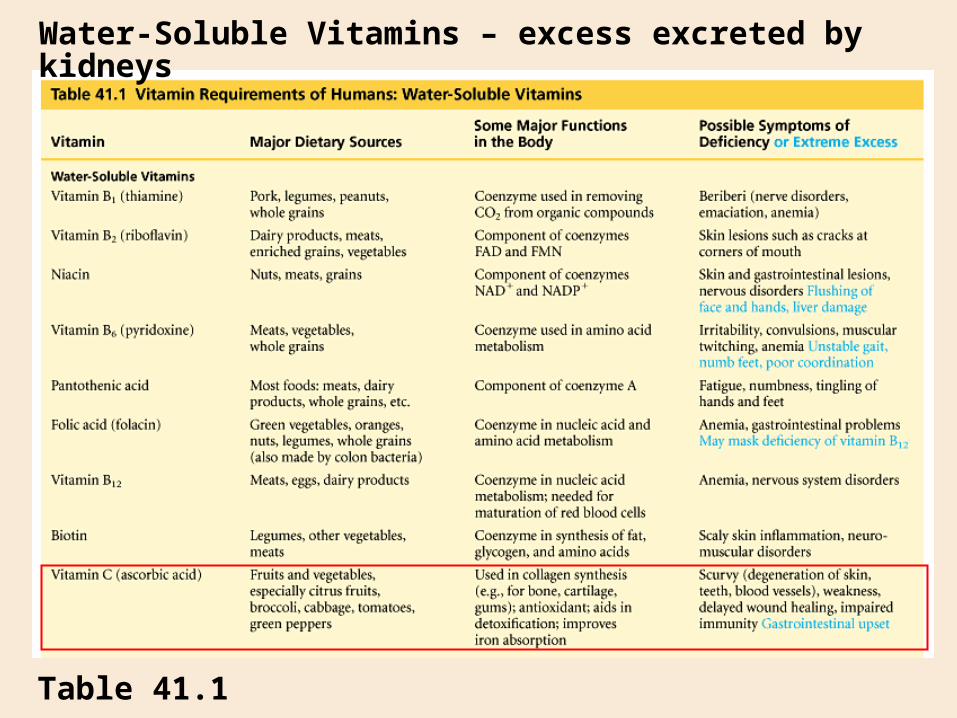
Water-Soluble Vitamins – excess excreted by kidneys
Table 41.1
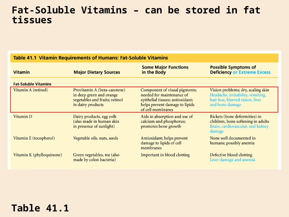
Fat-Soluble Vitamins – can be stored in fat tissues
Table 41.1
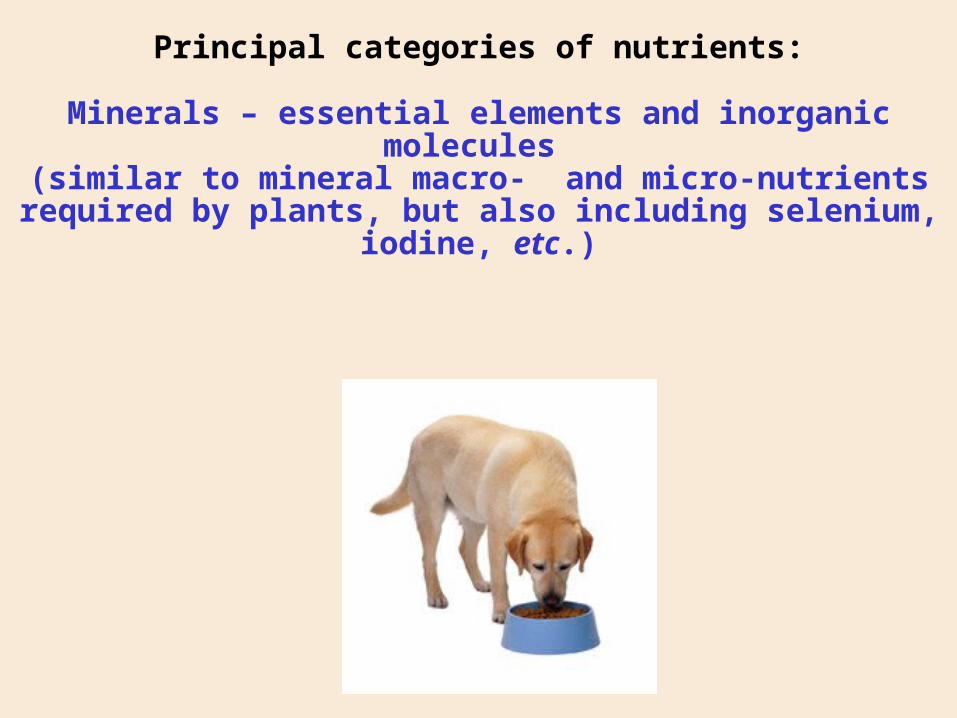
Principal categories of nutrients:
Minerals – essential elements and inorganic molecules (similar to mineral macro- and micro-nutrients required by
plants, but also including selenium, iodine, etc.)
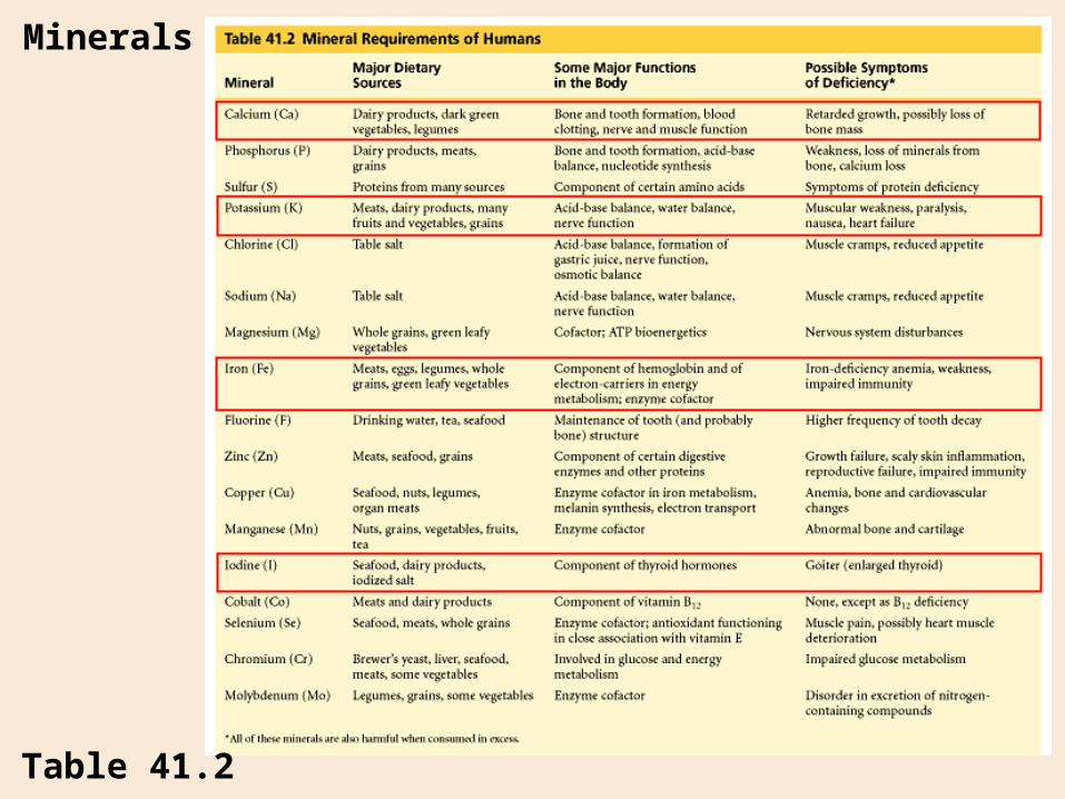
Minerals
Table 41.2
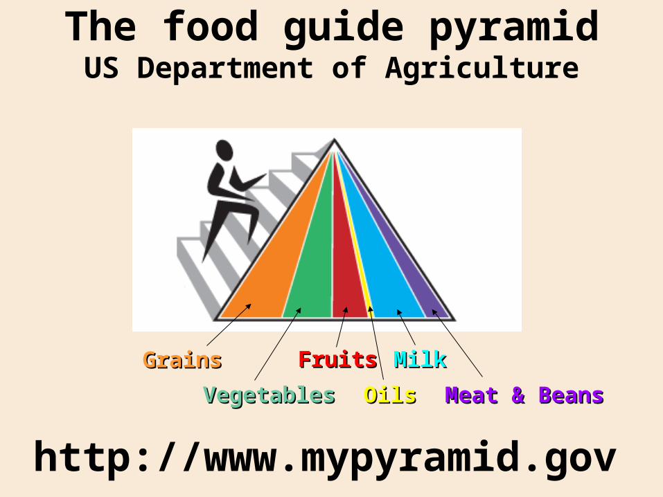
http://www.mypyramid.gov
The food guide pyramidUS Department of Agriculture
GrainsGrains
VegetablesVegetables
FruitsFruits
OilsOils
MilkMilk
Meat & BeansMeat & Beans
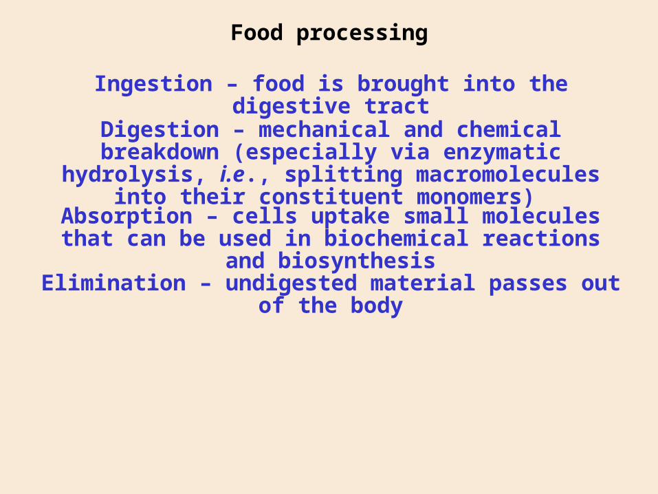
Food processing
Ingestion – food is brought into the digestive tract
Digestion – mechanical and chemical breakdown (especially via enzymatic hydrolysis, i.e., splitting macromolecules into their constituent monomers)
Absorption – cells uptake small molecules that can be used in biochemical reactions and biosynthesis
Elimination – undigested material passes out of the body
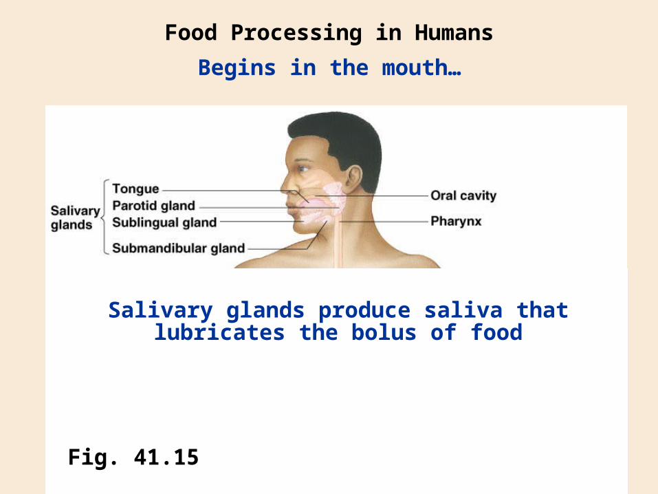
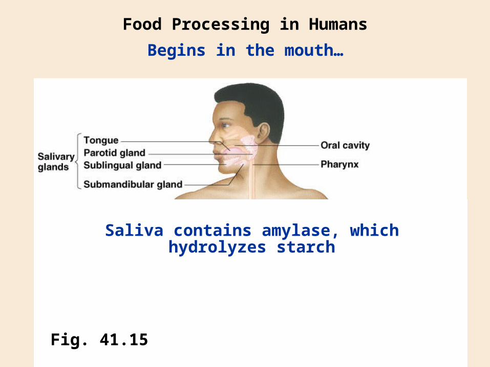
Food Processing in Humans
Begins in the mouth…
Fig. 41.15
Salivary glands produce saliva that lubricates the bolus of food
Food Processing in Humans
Begins in the mouth…
Fig. 41.15
Saliva contains amylase, which hydrolyzes starch
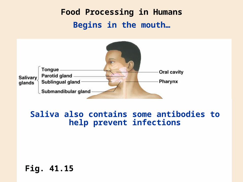
Food Processing in Humans
Begins in the mouth…
Fig. 41.15
Saliva also contains some antibodies to help prevent infections
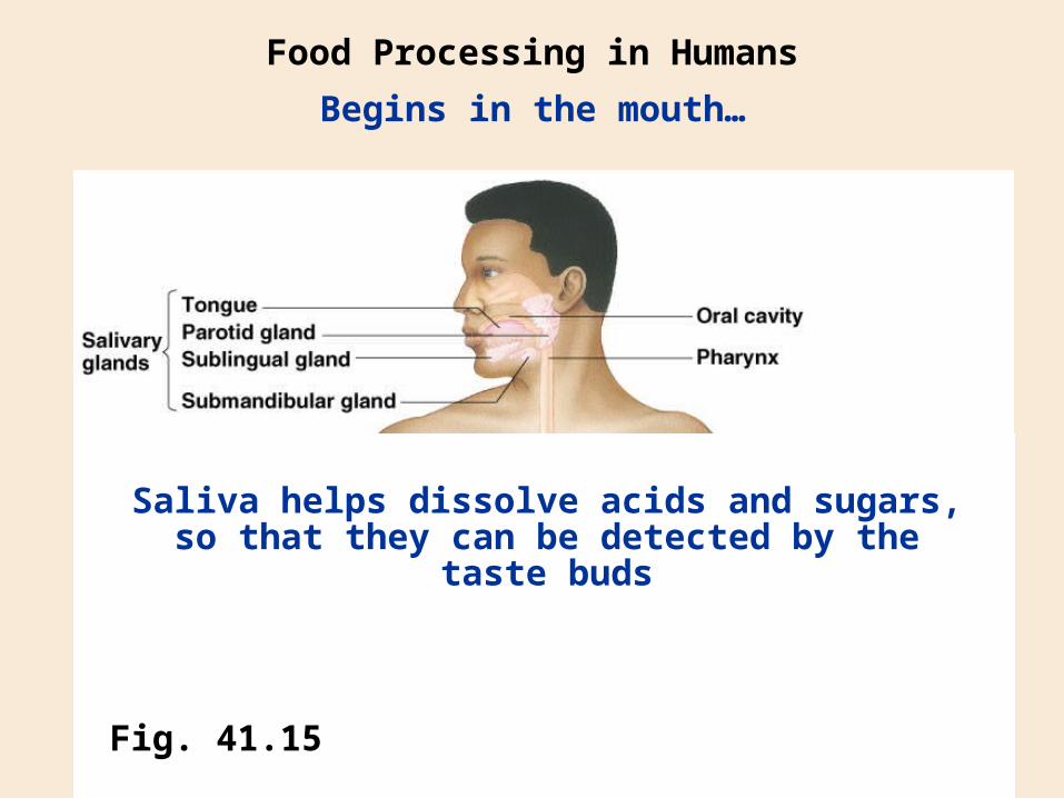
Food Processing in Humans
Begins in the mouth…
Fig. 41.15
Saliva helps dissolve acids and sugars, so that they can be detected by the taste buds
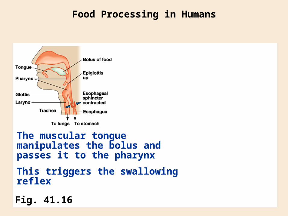
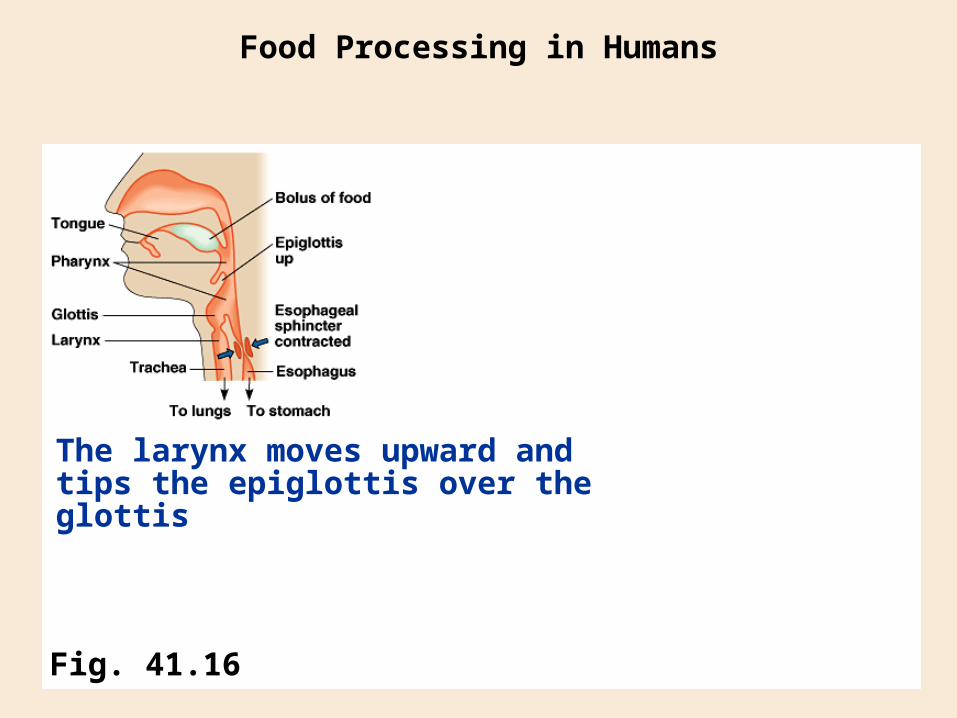
Food Processing in Humans
Fig. 41.16
The muscular tongue manipulates the bolus and passes it to the pharynx
This triggers the swallowing reflex
Food Processing in Humans
Fig. 41.16
The larynx moves upward and tips the epiglottis over the glottis
Food Processing in Humans
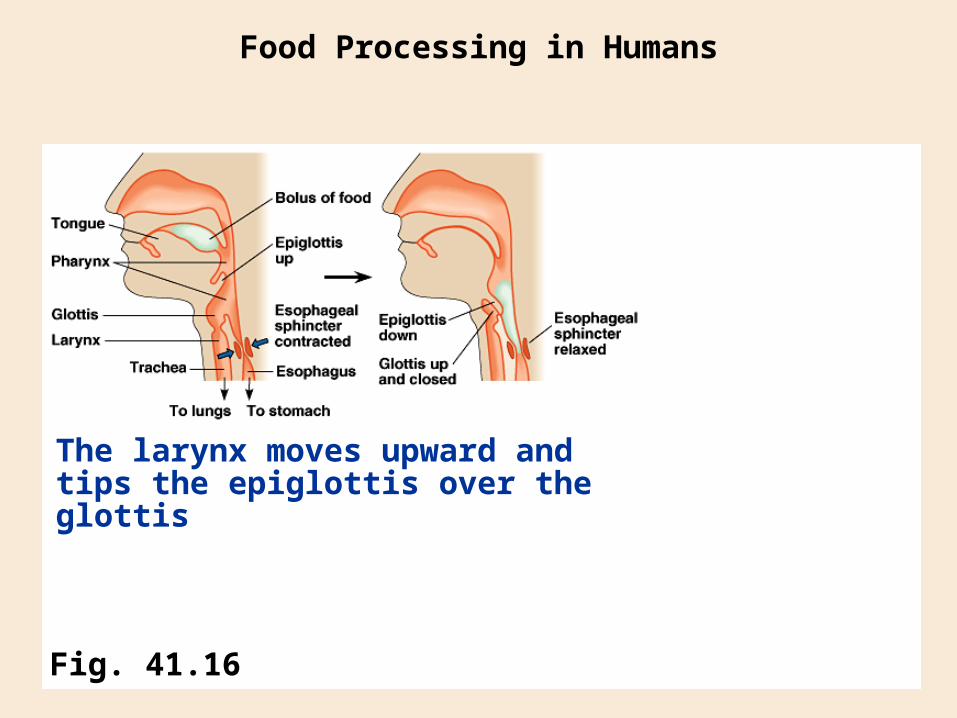
Fig. 41.16
The larynx moves upward and tips the epiglottis over the glottis

PharynxPharynxEpiglottisEpiglottis
EsophagusEsophagus
TracheaTrachea
Food Processing in Humans
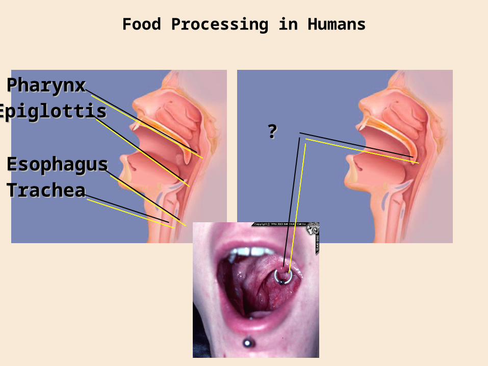
PharynxPharynxEpiglottisEpiglottis
EsophagusEsophagus
TracheaTrachea
??
Food Processing in Humans
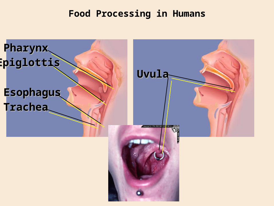
PharynxPharynxEpiglottisEpiglottis
EsophagusEsophagus
TracheaTrachea
UvulaUvula
Food Processing in Humans
Food Processing in Humans
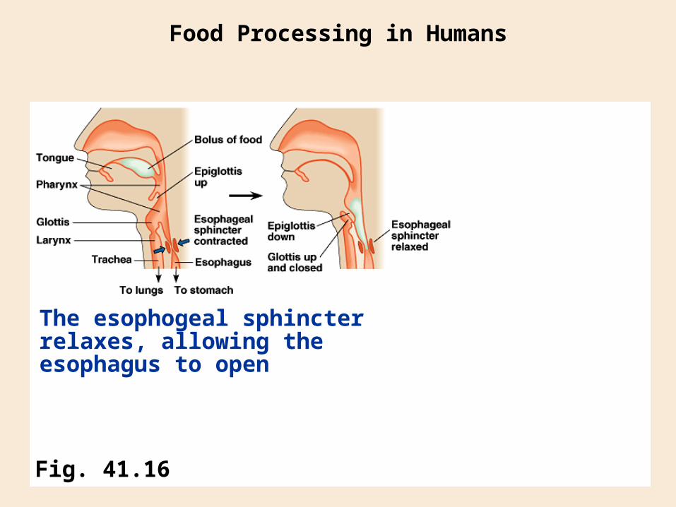
Fig. 41.16
The esophogeal sphincter relaxes, allowing the esophagus to open
Food Processing in Humans
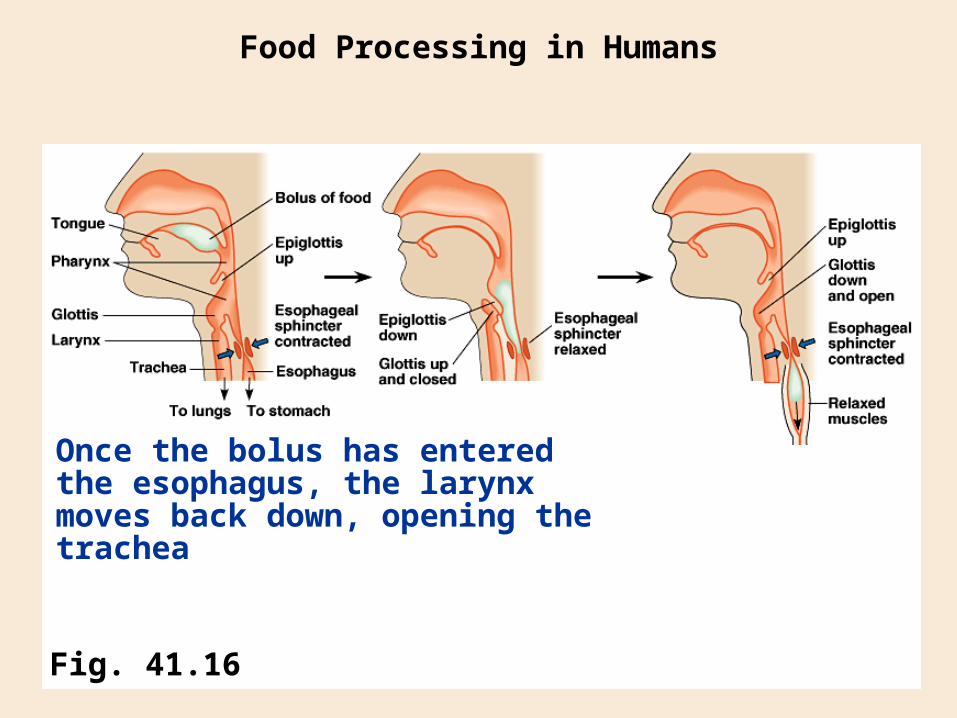
Fig. 41.16
Once the bolus has entered the esophagus, the larynx moves back down, opening the trachea
Food Processing in Humans
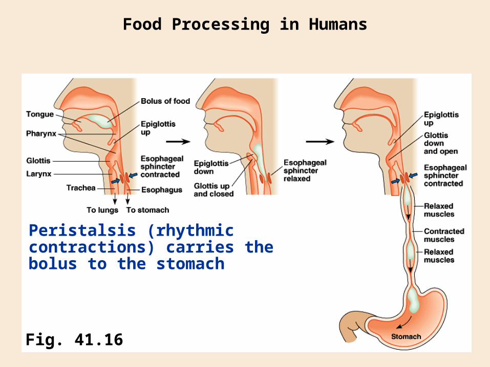
Fig. 41.16
Peristalsis (rhythmic contractions) carries the bolus to the stomach
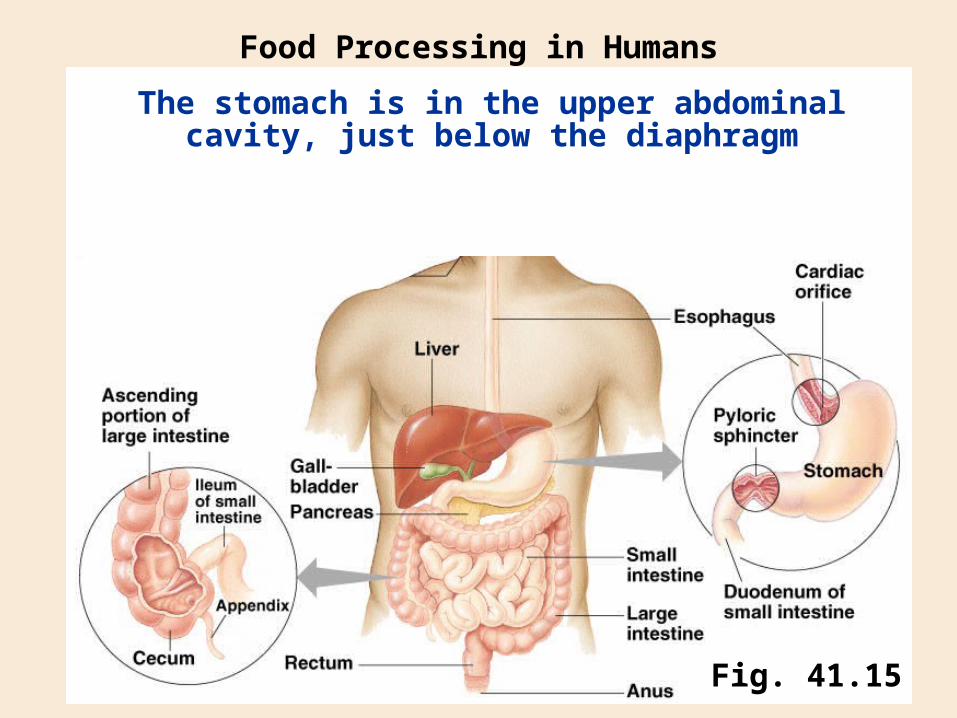
The stomach is in the upper abdominal cavity, just below the diaphragm
Food Processing in Humans
Fig. 41.15
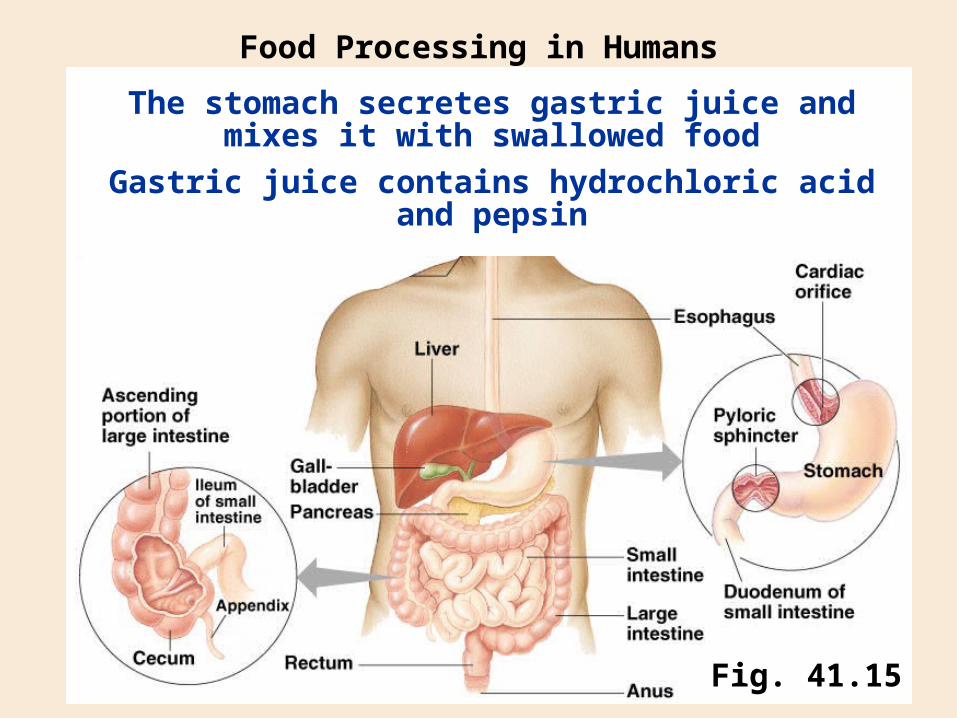
The stomach secretes gastric juice and mixes it with swallowed food
Food Processing in Humans
Fig. 41.15
Gastric juice contains hydrochloric acidand pepsin
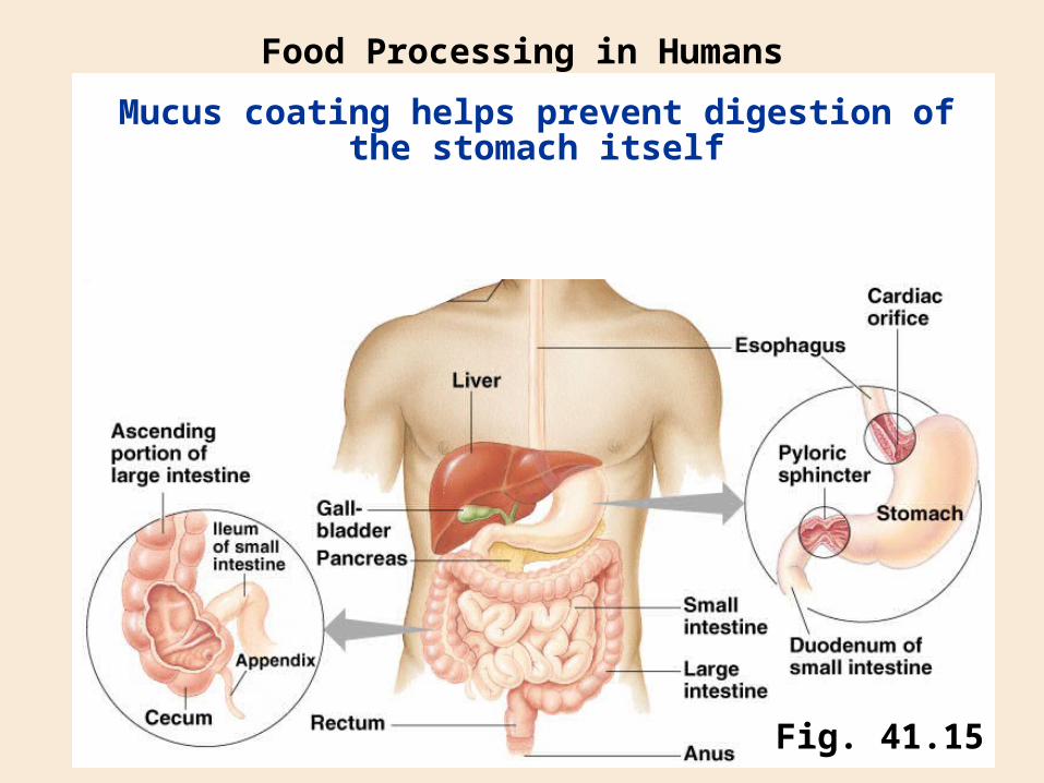
Mucus coating helps prevent digestion of the stomach itself
Food Processing in Humans
Fig. 41.15
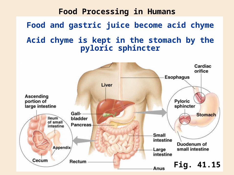
Food and gastric juice become acid chyme
Food Processing in Humans
Fig. 41.15
Acid chyme is kept in the stomach by the pyloric sphincter
Digestion continues in the small intestine
Food Processing in Humans
Fig. 41.15
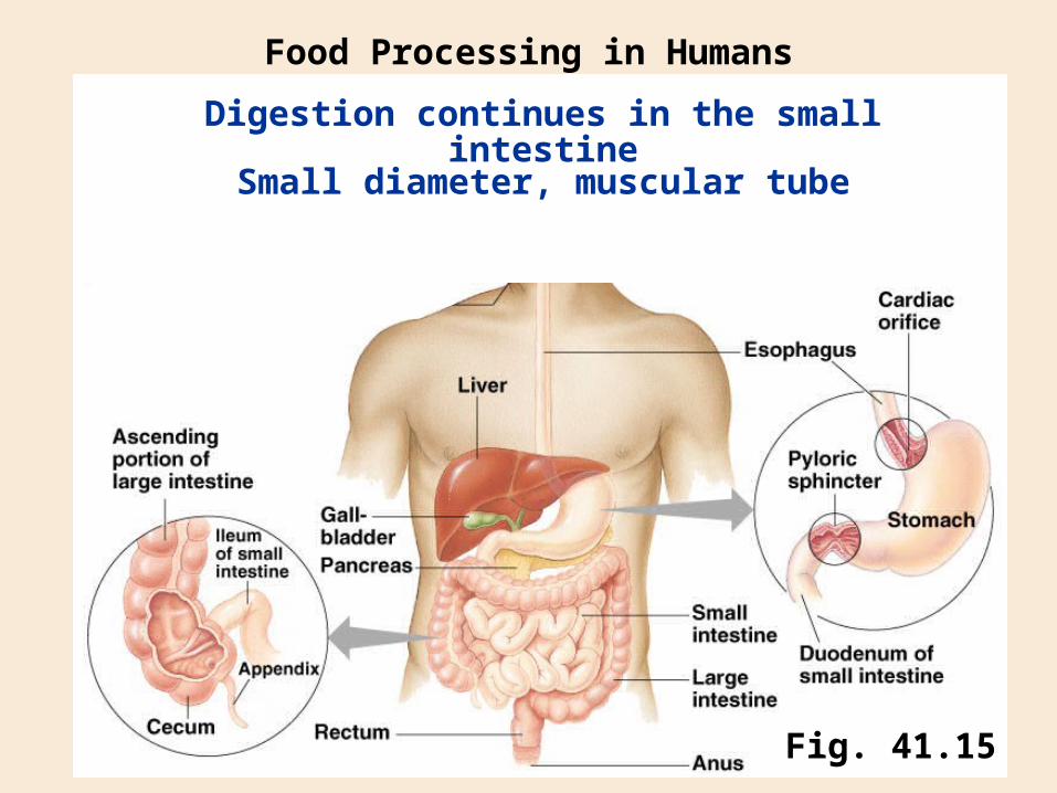
Small diameter, muscular tube
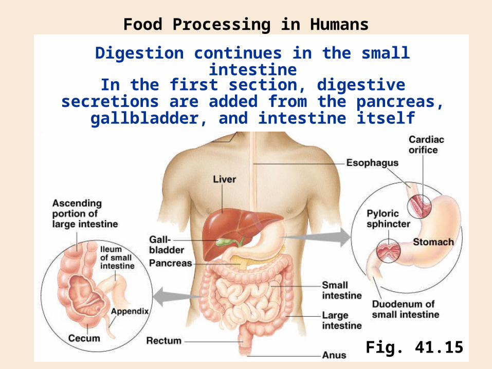
Digestion continues in the small intestine
Food Processing in Humans
Fig. 41.15
In the first section, digestive secretions are added from the pancreas, gallbladder, and intestine itself
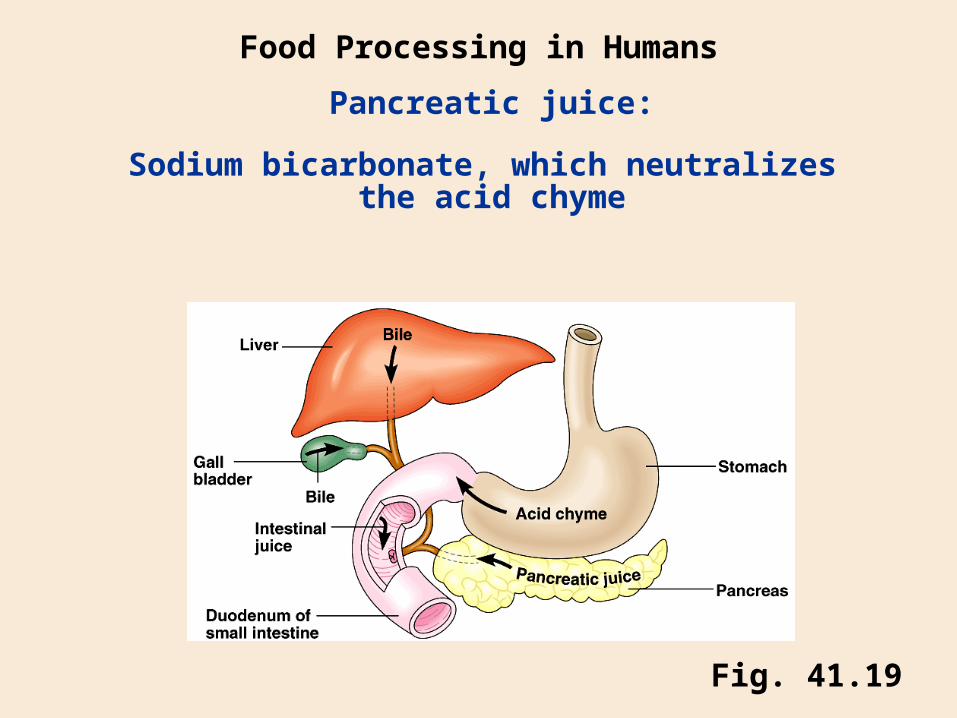
Fig. 41.19
Pancreatic juice:
Food Processing in Humans
Sodium bicarbonate, which neutralizes the acid chyme
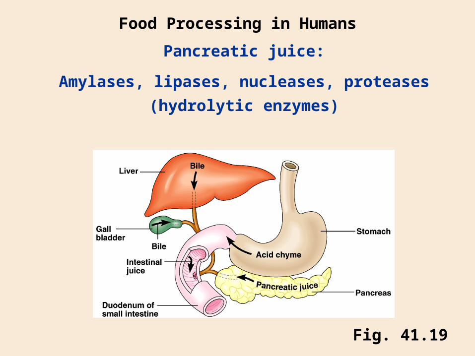
Fig. 41.19
Pancreatic juice:
Food Processing in Humans
Amylases, lipases, nucleases, proteases
(hydrolytic enzymes)
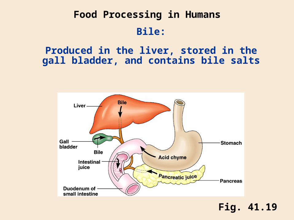
Fig. 41.19
Bile:
Food Processing in Humans
Produced in the liver, stored in the gall bladder, and contains bile salts
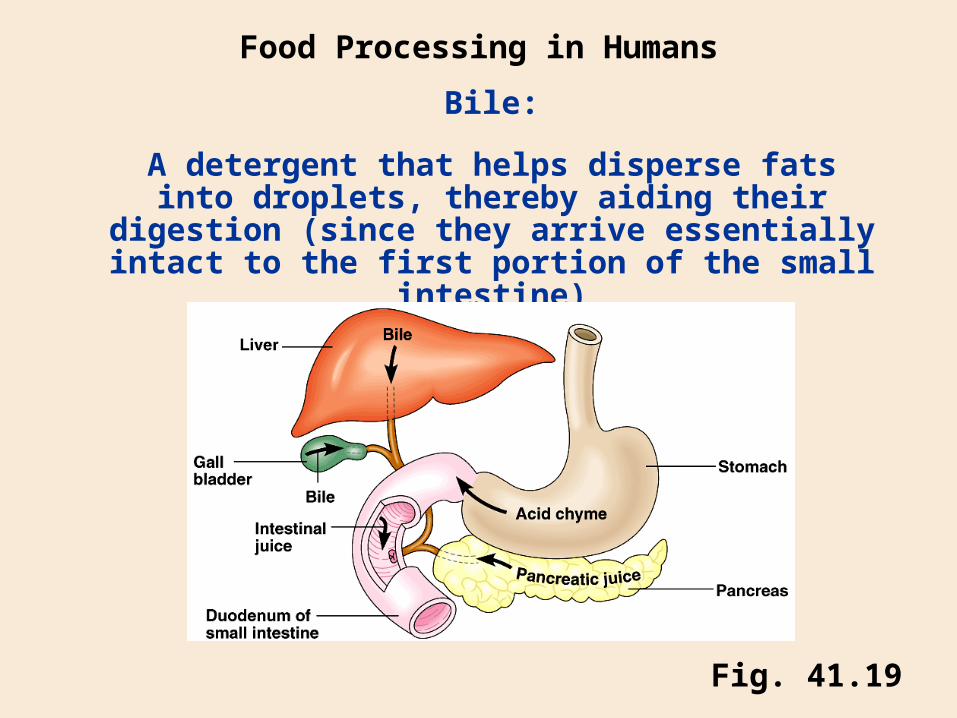
A detergent that helps disperse fats into droplets, thereby aiding their digestion (since they arrive essentially intact to the first portion of the small
intestine)
Fig. 41.19
Bile:
Food Processing in Humans
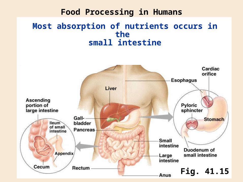
Most absorption of nutrients occurs in the small intestine
Food Processing in Humans
Fig. 41.15

Fig. 41.23
Most absorption of nutrients occurs in the small intestine
Food Processing in Humans
SEMSEM
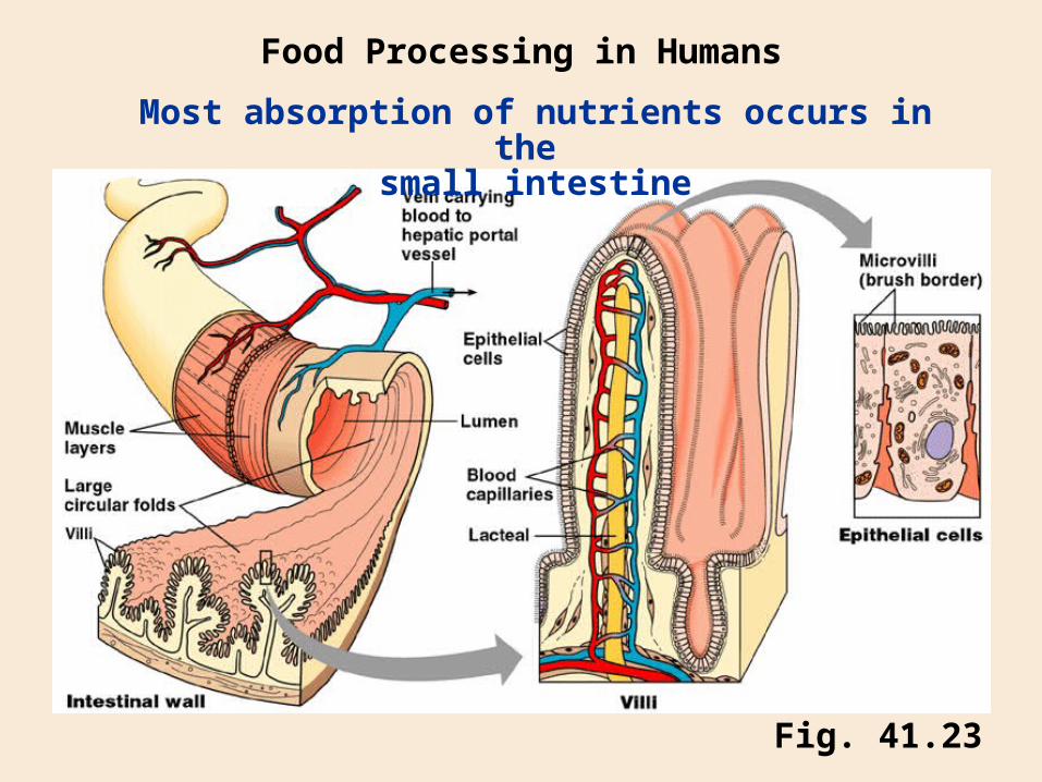
Fig. 41.23
Most absorption of nutrients occurs in the small intestine
Food Processing in Humans
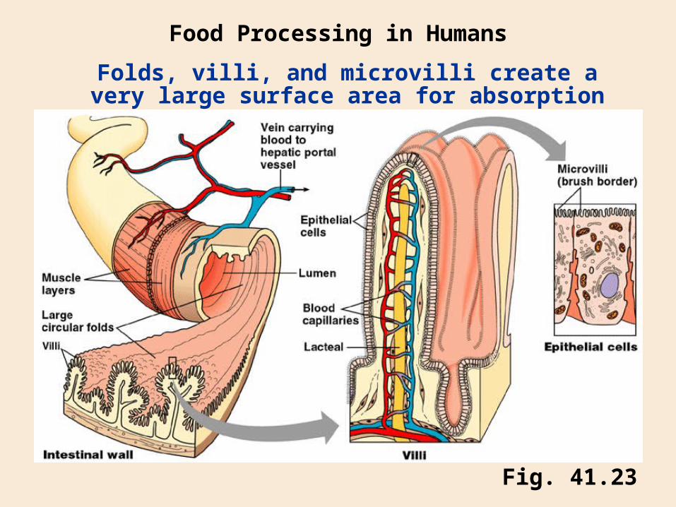
Fig. 41.23
Folds, villi, and microvilli create a very large surface area for absorption
Food Processing in Humans
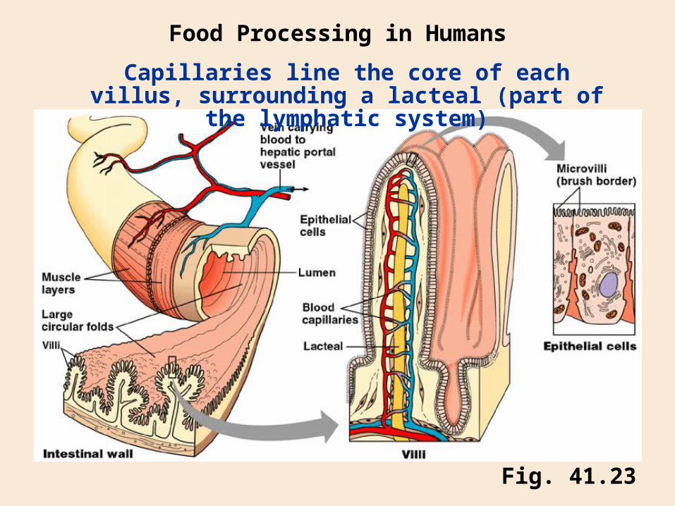
Fig. 41.23
Capillaries line the core of each villus, surrounding a lacteal (part of the lymphatic
system)
Food Processing in Humans
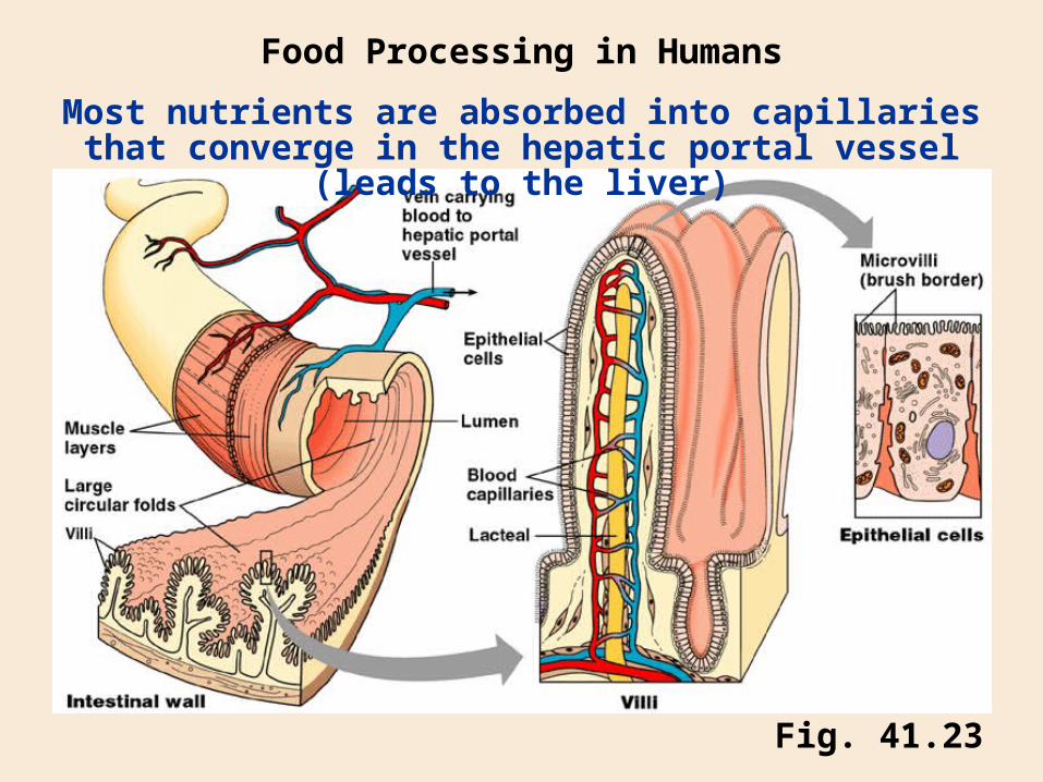
Fig. 41.23
Most nutrients are absorbed into capillaries that converge in the hepatic portal vessel (leads to the liver)
Food Processing in Humans
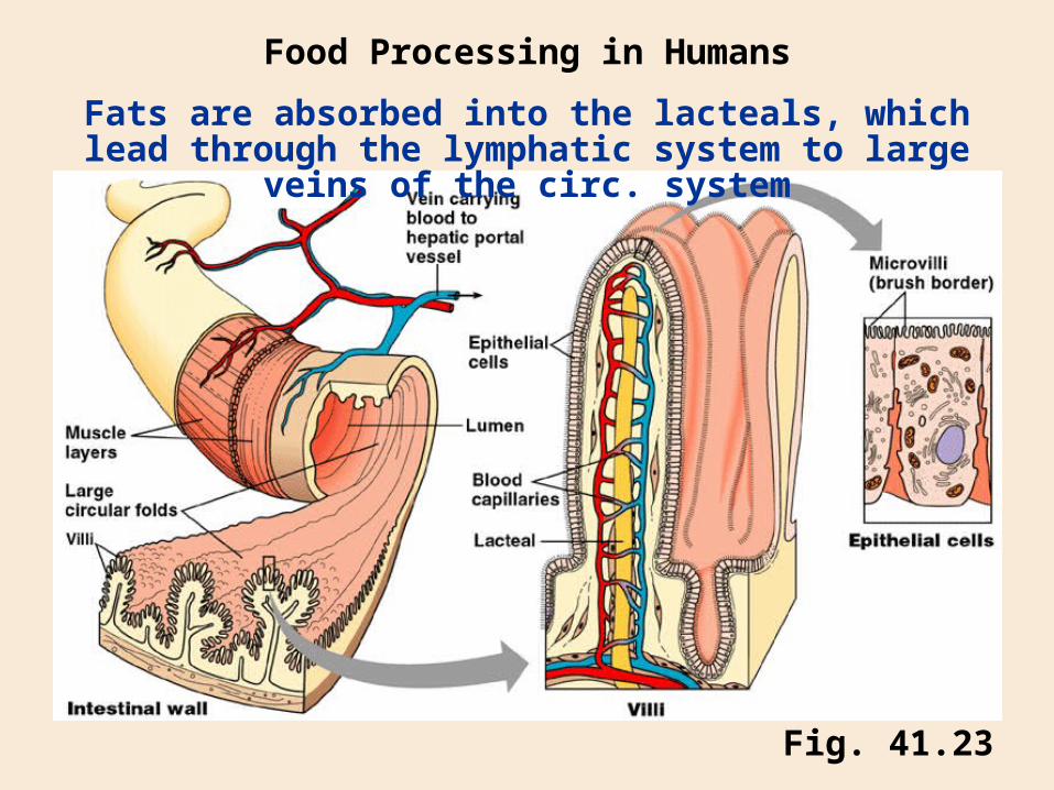
Fig. 41.23
Fats are absorbed into the lacteals, which lead through the lymphatic system to large veins of the circ. system
Food Processing in Humans
Chapter 1
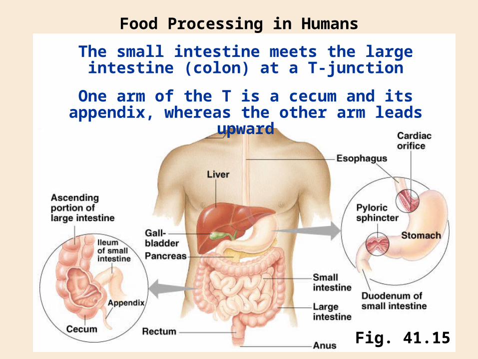
The small intestine meets the large intestine (colon) at a T-junction
Food Processing in Humans
Fig. 41.15
One arm of the T is a cecum and its appendix, whereas the other arm leads upward
Chapter 1
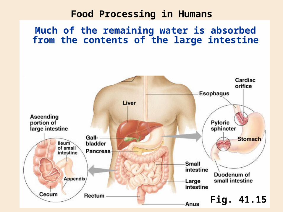
Much of the remaining water is absorbed from the contents of the large intestine
Food Processing in Humans
Fig. 41.15
Chapter 1
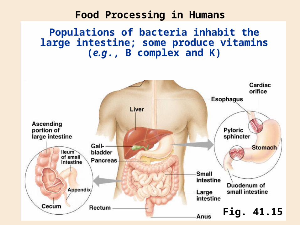
Populations of bacteria inhabit the large intestine; some produce vitamins (e.g., B complex and K)
Food Processing in Humans
Fig. 41.15
Chapter 1
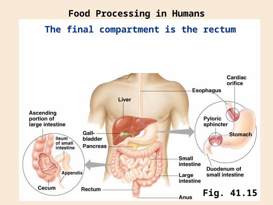
The final compartment is the rectum
Food Processing in Humans
Fig. 41.15
Chapter 1
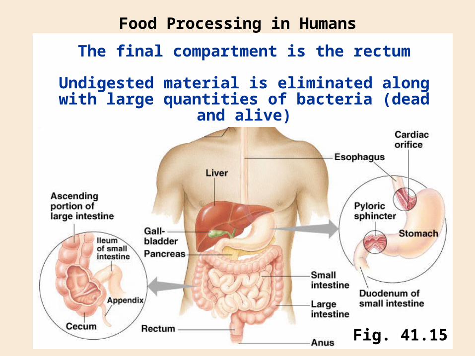
The final compartment is the rectum
Food Processing in Humans
Fig. 41.15
Undigested material is eliminated along with large quantities of bacteria (dead and alive)
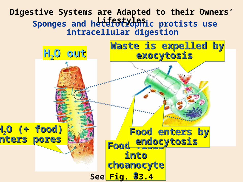
Sponges and heterotrophic protists use intracellular digestion
Digestive Systems are Adapted to their Owners’ Lifestyles
HH22O outO out
HH22O (+ food)O (+ food)enters pores enters pores HH22O (+ food)O (+ food)enters pores enters pores
Food flows into Food flows into choanocyteschoanocytes
Food flows into Food flows into choanocyteschoanocytes
Food enters byFood enters byendocytosis endocytosis
Food enters byFood enters byendocytosis endocytosis
Waste is expelled byWaste is expelled byexocytosis exocytosis
Waste is expelled byWaste is expelled byexocytosis exocytosis
See Fig. 33.4
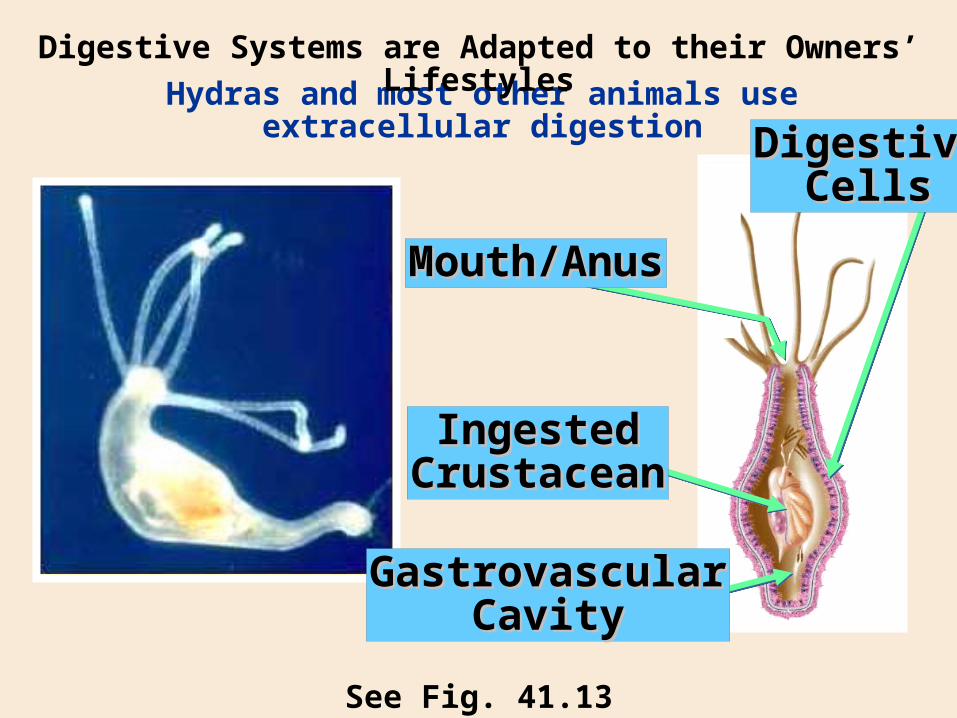
Hydras and most other animals use extracellular digestion
Digestive Systems are Adapted to their Owners’ Lifestyles
See Fig. 41.13
Mouth/AnusMouth/Anus
IngestedIngestedCrustaceanCrustacean
GastrovascularGastrovascularCavityCavity
DigestiveDigestiveCellsCells
Extracellular digestion in a tube (complete digestive tract or alimentary canal) is the most efficient and effective
Digestive Systems are Adapted to their Owners’ Lifestyles
The animal can eat frequently, even while digesting the previous meal
Specialized compartments and digestive organs can contribute to the process sequentially
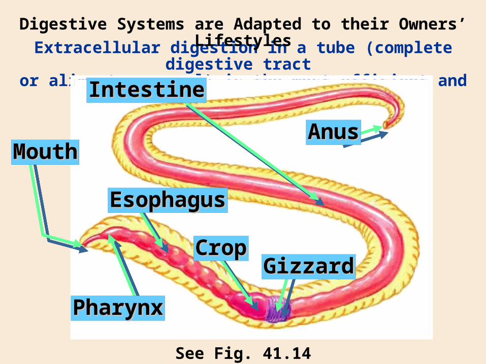
Extracellular digestion in a tube (complete digestive tract or alimentary canal) is the most efficient and effective
Digestive Systems are Adapted to their Owners’ Lifestyles
See Fig. 41.14
IntestineIntestine
AnusAnusMouthMouth
PharynxPharynx
EsophagusEsophagus
CropCropGizzardGizzard
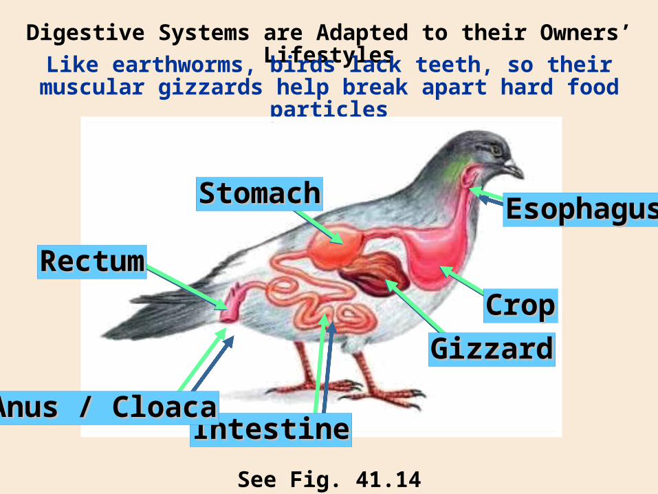
Digestive Systems are Adapted to their Owners’ Lifestyles
See Fig. 41.14
Like earthworms, birds lack teeth, so their muscular gizzards help break apart hard food particles
IntestineIntestineAnus / CloacaAnus / Cloaca
EsophagusEsophagus
CropCrop
GizzardGizzard
StomachStomach
RectumRectum
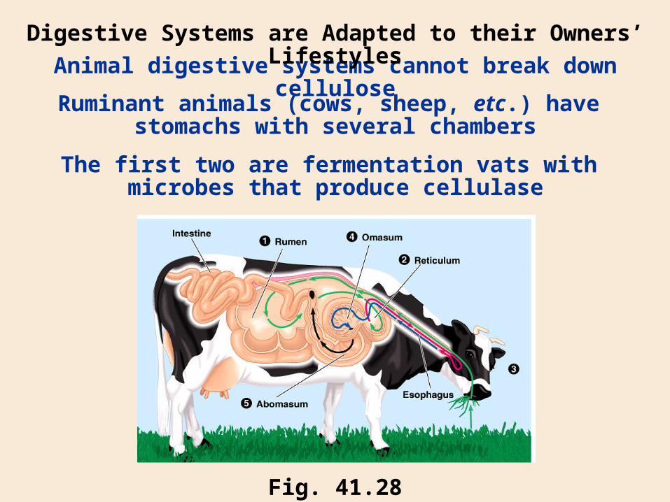
Animal digestive systems cannot break down cellulose
Digestive Systems are Adapted to their Owners’ Lifestyles
Fig. 41.28
Ruminant animals (cows, sheep, etc.) have stomachs with several chambers
The first two are fermentation vats with microbes that produce cellulase
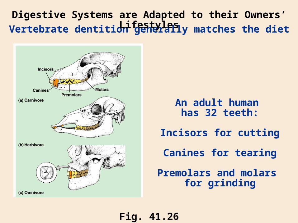
Vertebrate dentition generally matches the diet
Digestive Systems are Adapted to their Owners’ Lifestyles
Fig. 41.26
An adult human has 32 teeth:
Incisors for cutting
Canines for tearing
Premolars and molars for grinding
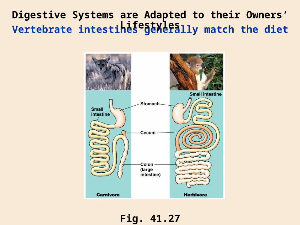
Vertebrate intestines generally match the diet
Digestive Systems are Adapted to their Owners’ Lifestyles
Fig. 41.27
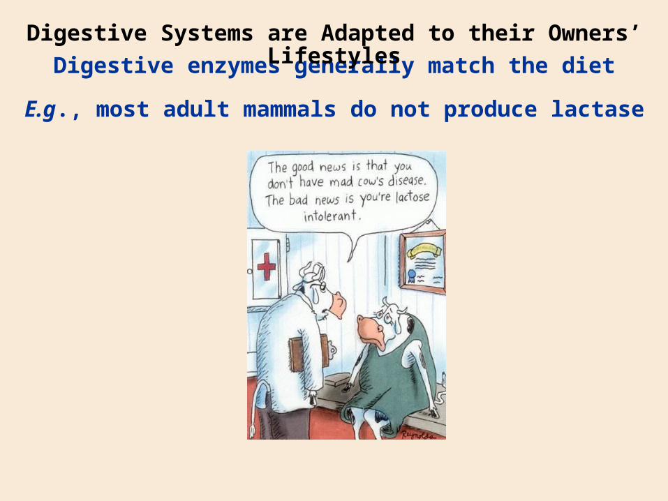
Digestive enzymes generally match the diet
Digestive Systems are Adapted to their Owners’ Lifestyles
E.g., most adult mammals do not produce lactase