Embed Size (px)
Citation preview

Chapter 6: A Tour of the Cell
1. Studying Cells2. Intracellular Structures3. The Cytoskeleton4. Extracellular Structures

1. Studying Cells

10 m
1 m
0.1 m
1 cm
1 mm
100 µm
10 µm
1 µm
100 nm
10 nm
1 nm
0.1 nm Atoms
Small molecules
Lipids
Proteins
Ribosomes
VirusesSmallest bacteria
Mitochondrion
NucleusMost bacteria
Most plant and animal cells
Frog egg
Chicken egg
Length of some nerve and muscle cells
Human height
Una
ided
eye
Ligh
t mic
rosc
ope
Elec
tron
mic
rosc
ope
Concepts of Microscopy
MAGNIFICATION• factor by which the image produced is larger than the actual object (e.g. “100X”)
RESOLUTION• minimum distance acrosswhich 2 points can be resolved or seen distinctly(limited by wavelength)
CONTRAST• degree to which objects differ
from background
Limits of Resolution
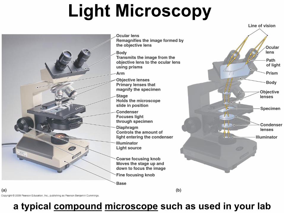
Light Microscopy
a typical compound microscope such as used in your lab
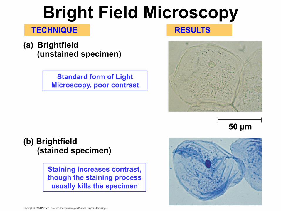
(a) Brightfield(unstained specimen)
(b) Brightfield(stained specimen)
TECHNIQUE RESULTS
50 µm
Bright Field Microscopy
Standard form of Light Microscopy, poor contrast
Staining increases contrast,though the staining processusually kills the specimen

(c) Phase-contrast
(d) Differential-interference-contrast (Nomarski)
TECHNIQUE RESULTS
Phase Contrast Microscopy
Enhances misalignment of light waves to create contrast
A variation of phase-contrast microscopy involving a more complex
combination of filters and prisms.
Reveals internal detailwithout staining, useful
for viewing live specimens
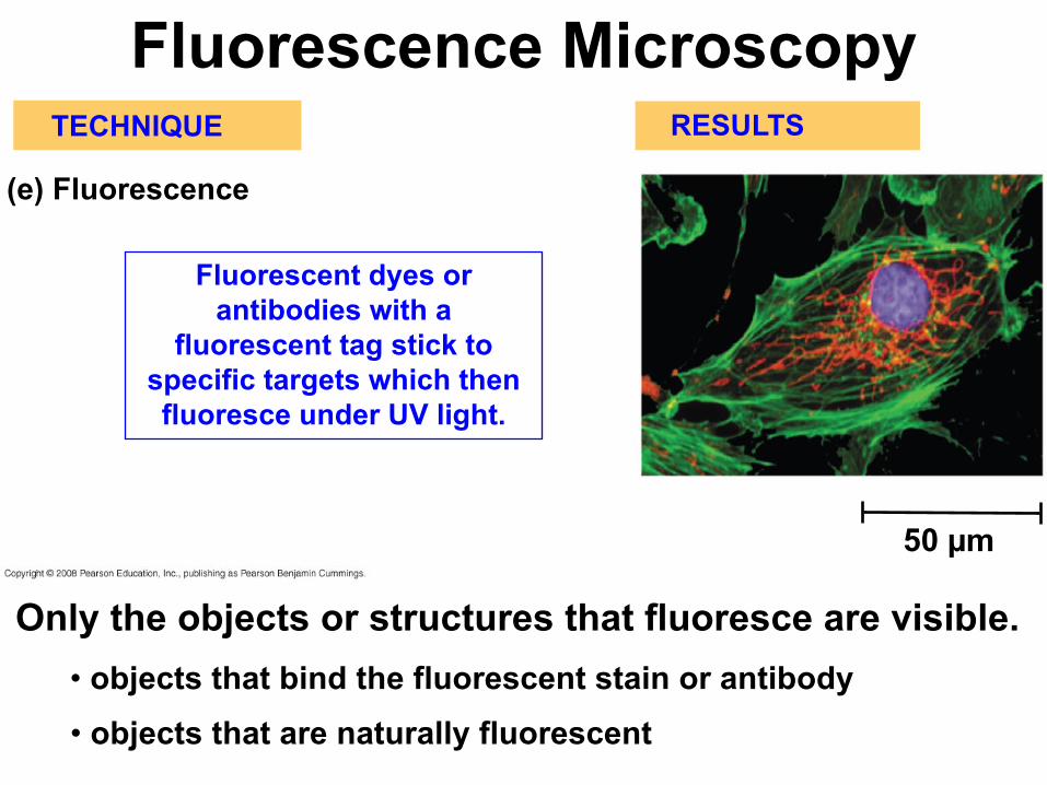
(e) Fluorescence
TECHNIQUE RESULTS
50 µm
Fluorescence Microscopy
Fluorescent dyes or antibodies with a
fluorescent tag stick to specific targets which then fluoresce under UV light.
Only the objects or structures that fluoresce are visible.• objects that bind the fluorescent stain or antibody• objects that are naturally fluorescent

Confocal Fluorescence Microscopy
Only light from a given depth or plane is transmitted, “out of focus” light is excluded

(a) Scanning electronmicroscopy (SEM)
TECHNIQUE RESULTS
(b) Transmission electronmicroscopy (TEM)
Cilia
Longitudinalsection ofcilium
Cross sectionof cilium
1 µm
1 µmElectron
Microscopy
specimen cut in thin sections, higher
resolution
view of whole specimen, reveals surface features
Electromagnetic lenses focus electron beam onto heavy metal-stained specimen.
• electron beams have very short wavelengths
• allows far greater resolution than with light microscopy

Homogenization
TECHNIQUE
HomogenateTissuecells
1,000 g(1,000 times theforce of gravity)
10 min Differential centrifugationSupernatant pouredinto next tube
20,000 g20 min
80,000 g60 minPellet rich in
nuclei andcellular debris
Pellet rich inmitochondria(and chloro-plasts if cellsare from a plant)
Pellet rich in“microsomes”(pieces of plasmamembranes andcells’ internalmembranes)
150,000 g3 hr
Pellet rich inribosomes
Fractionation by Centrifugation
In addition to microscopic examination, cells and their structures are also studied biochemically:
• in order to study a cellularcompartment biochemically,it must be separated from the rest of the cell
• this is accomplished through successive centrifugation steps at increasing speeds

Surface area increases whiletotal volume remains constant
5
11
6 150 750
125 1251
6 61.2
Total surface area[Sum of the surface areas(height ´ width) of all boxessides ´ number of boxes]
Total volume[height ´ width ´ length ´number of boxes]
Surface-to-volume(S-to-V) ratio[surface area ÷ volume]
Why are cells the size they are?

Why aren’t cells bigger?1) Cell size is limited by the rate of diffusion:• if cells get too large, it takes too much time for
nutrients, wastes, etc, to disperse in the cell
2) And also by the surface to volume ratio (S/V):surface area of sphere = 4pr2
volume of sphere = (4/3)pr3
*Surface Area increases by square of radius*Volume increases by cube of radius
***The larger the cell, the smaller the S/V ratio***

2. Intracellular Structures
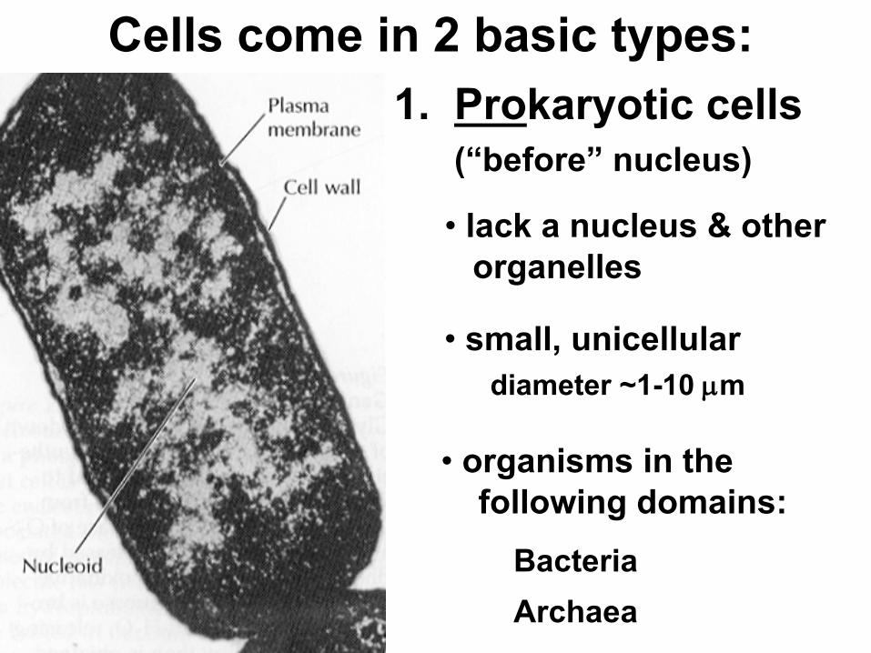
Cells come in 2 basic types:1. Prokaryotic cells
(“before” nucleus)
• lack a nucleus & otherorganelles
• small, unicellular
• organisms in thefollowing domains:
Bacteria
diameter ~1-10 µm
Archaea
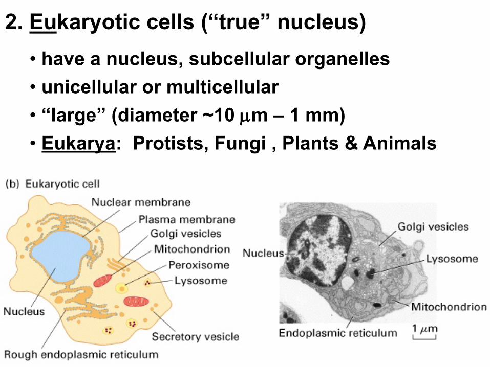
2. Eukaryotic cells (“true” nucleus)• have a nucleus, subcellular organelles• unicellular or multicellular
• Eukarya: Protists, Fungi , Plants & Animals• “large” (diameter ~10 µm – 1 mm)

Fimbriae
Nucleoid
Ribosomes
Plasma membrane
Cell wall
Capsule
Flagella
Bacterialchromosome
(a) A typical rod-shaped bacterium
(b) A thin section through the bacterium Bacillus coagulans (TEM)
0.5 µm
Prokaryotic Cells
Have intracellular organization despite no organelles.

ENDOPLASMIC RETICULUM (ER)
Smooth ERRough ERFlagellum
Centrosome
CYTOSKELETON:Microfilaments
Intermediatefilaments
Microtubules
Microvilli
Peroxisome
MitochondrionLysosome
Golgiapparatus
Ribosomes
Plasma membrane
Nuclearenvelope
Nucleolus
Chromatin
NUCLEUS
Animal Cell
*not in plant cells
*
*
*
*

NucleolusNucleus
Rough ER
Nuclear lamina (TEM)
Close-up of nuclear envelope
1 µm
1 µm
0.25 µm
Ribosome
Pore complex
Nuclear pore
Outer membraneInner membraneNuclear envelope:
Chromatin
Surface ofnuclear envelope
Pore complexes (TEM)
The NucleusWhere genetic material (DNA) is stored, gene expression begins.
Nucleolus
where ribosomalsubunits areassembledfrom rRNA& proteins
Chromatincomplexof DNA &histoneproteins

Cytosol
Endoplasmic reticulum (ER)
Free ribosomes
Bound ribosomes
Large subunit
Small subunit
Diagram of a ribosomeTEM showing ER and ribosomes
0.5 µm
RibosomesCarry out protein synthesis by the process of translation.
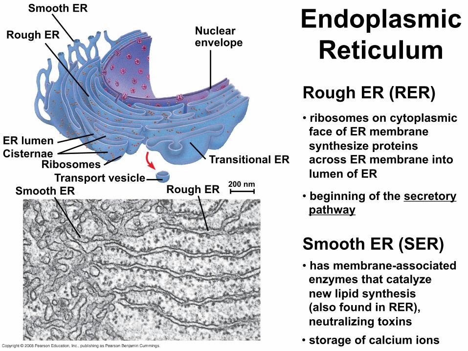
Smooth ER
Rough ER Nuclear envelope
Transitional ER
Rough ERSmooth ERTransport vesicle
RibosomesCisternaeER lumen
200 nm
Endoplasmic Reticulum
Rough ER (RER)• ribosomes on cytoplasmicface of ER membrane synthesize proteins across ER membrane into lumen of ER
Smooth ER (SER)• has membrane-associated enzymes that catalyze new lipid synthesis(also found in RER),neutralizing toxins
• beginning of the secretorypathway
• storage of calcium ions

cis face(“receiving” side of Golgi
apparatus)Cisternae
trans face(“shipping” side of Golgi
apparatus)TEM of Golgi apparatus
0.1 µm
The Golgi apparatus
• proteins destined to leave ER are transported to the Golgi where they are modified, sorted and sent to various destinations. • polysaccharides are produced in the Golgi apparatus as well

Nucleus 1 µm
Lysosome
Digestiveenzymes
Lysosome
Plasmamembrane
Food vacuole
(a) Phagocytosis
Digestion
(b) Autophagy
Peroxisome
Vesicle
Lysosome
Mitochondrion
Peroxisomefragment
Mitochondrionfragment
Vesicle containingtwo damaged organelles
1 µm
Digestion
Lysosomes
• acidic compartments full of enzymes for the breakdown or digestion of foreign or waste material
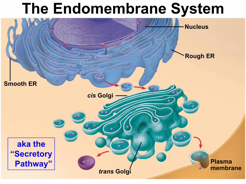
Smooth ER
Nucleus
Rough ER
Plasma membrane
cis Golgi
trans Golgi
The Endomembrane System
aka the “Secretory
Pathway”

Free ribosomesin the mitochondrial matrix
Intermembrane spaceOuter membrane
Inner membraneCristae
Matrix
0.1 µm
Mitochondria
ATP production via cellular respiration• convert energy from glucose, fatty acids, etc, to energy in ATP
Have ribosomes
that resemble those of
prokaryotes

NUCLEUSNuclear envelopeNucleolusChromatin
Rough endoplasmic reticulum
Smooth endoplasmic reticulum
Ribosomes
Central vacuole
MicrofilamentsIntermediate filamentsMicrotubules
CYTO-SKELETON
Chloroplast
PlasmodesmataWall of adjacent cell
Cell wall
Plasma membrane
Peroxisome
Mitochondrion
Golgiapparatus
Plant Cell*
*
*
*
*not in animal cells
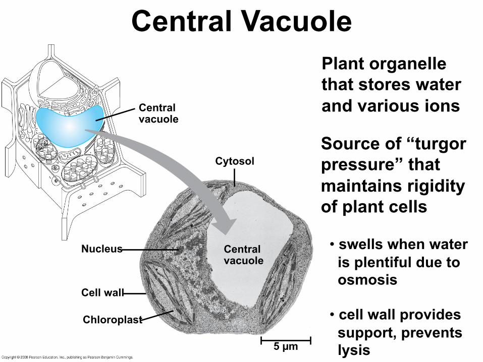
Central vacuole
Cytosol
Central vacuole
Nucleus
Cell wall
Chloroplast
5 µm
Central VacuolePlant organelle that stores water and various ions
Source of “turgorpressure” that maintains rigidity of plant cells
• swells when water is plentiful due to osmosis
• cell wall provides support, prevents lysis
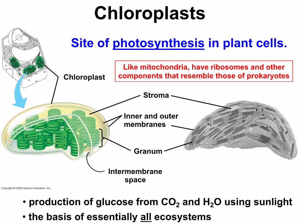
Chloroplast
Stroma
Inner and outermembranes
Granum
Intermembranespace
ChloroplastsSite of photosynthesis in plant cells.
• production of glucose from CO2 and H2O using sunlight • the basis of essentially all ecosystems
Like mitochondria, have ribosomes and other components that resemble those of prokaryotes

1 µm
ChloroplastPeroxisome
Mitochondrion
PeroxisomesContain enzymes that oxidize (i.e., remove H) various organic molecules thus forming H2O2from O2
• H2O2 is then converted to O2 and H2O
Involved in the detoxification oftoxic substances, breakdown of fatty acids

3. The Cytoskeleton

Microtubule
Microfilaments0.25 µm
The CytoskeletonA complex, highly dynamic intracellular network of protein filaments largely responsible for:
• cell shape, rigidity • cell movement, motility
• localization of organelles
• movement of vesicles (and organelles)
• dynamics of cell division

3 Basic Cytoskeletal FilamentsActin filaments/microfilaments (MF) intermediate filaments (IF) microtubules (MT)
IF
MT
MF

Actin subunit
10 µm
7 nm
Microfilaments

5 µm
Keratin proteins
Fibrous subunit (keratinscoiled together)
8–12 nm
Intermediate Filaments

10 µm
Column of tubulin dimers
Tubulin dimera b
25 nm
Microtubules

VesicleATP
Receptor for motor protein
Microtubuleof cytoskeleton
Motor protein (ATP powered)
(a)
Microtubule Vesicles
(b)
0.25 µm
VesicleTransport
• a variety of motor proteins are involved in binding and transporting vesicles along cytoskeletalfibers to their destination

Centrosome
Microtubule
Centrioles0.25 µm
Longitudinal section of one centriole
Microtubules Cross sectionof the other centriole
Centrosomes• centrosomes contain a pair of centrioles(animal cells only)
• these structures are involved in the formation of the mitotic spindle or “spindle fibers” that play such an important role in cell division

5 µm
Direction of swimming
(a) Motion of flagella
Direction of organism’s movement
Power stroke
Recovery stroke
(b) Motion of cilia15 µm
Flagella & CiliaFLAGELLAare involved in cell motility, are very long, and cells have relatively few (1 or several)
CILIAare involved in motility, moving material across the cell surface, and are present on the cell surface in high numbers

4. Extracellular Structures

Secondary cell wall
Primary cell wall
Middle lamella
Central vacuoleCytosol
Plasma membrane
Plant cell walls
Plasmodesmata
1 µm
Plant Cell Walls
Interior of cell
Interior of cell
0.5 µm Plasmodesmata Plasma membranes
Cell walls
Plant cell walls contain fibers of cellulose and other polysaccharides as well as proteins
• one or more layers of secondary cell wall may be produced in some plant cells

Tight junction
0.5 µm
1 µmDesmosome
Gap junctionExtracellularmatrix
0.1 µm
Plasma membranesof adjacent cells
Spacebetweencells
Gapjunctions
Desmosome
Intermediatefilaments
Tight junction
Tight junctions preventfluid from movingacross a layer of cells
IntercellularJunctions
TIGHT JUNCTIONSare impenetrable seals connecting adjacent cells that prevent fluid and other materials from passing between the cells
DESMOSOMESare strong connections between cells that create a very strong sheet of cells
GAP JUNCTIONS (animal) & PLASMODESMATA (plant)provide channels through which ions & other small molecules can pass from cell to cell

Polysaccharide molecule
Carbohydrates
Core protein
Proteoglycanmolecule
Proteoglycan complex
Collagen
Fibronectin
Plasma membrane
Proteoglycan complex
Integrins
CYTOPLASMMicro-filaments
EXTRACELLULAR FLUID
The Extracellular MatrixA meshwork of protein fibers and polysaccharides that retain fluid and produce gel-like matrix that holds cells together in tissues.

Key Terms for Chapter 6
• Golgi apparatus, lysosome, peroxisome, vesicle
• mitochondria, chloroplasts
• endomembrane system, central vacuole
• prokaryotic vs eukaryotic
• nucleus, nucleolus, endoplasmic reticulum, ribosome
• cell wall, capsule, flagella, nucleoid, cytoplasm
• magnification, resolution, contrast
• bright field, phase contrast, fluorescent, confocal, transmission & scanning electron microscopy

• cytoskeleton, cilia, flagella, centrosome, centriole
• microtubules, microfilaments, intermediate filaments
• motor proteins
• tight junctions, gap junctions, desmosomes,plasmodesmata
• extracellular matrix, proteoglycan
Relevant Chapter Questions
1-9


![Studying inter-core data reuse in multicoreshostel.ufabc.edu.br/~cak/inf103/studying_inter... · on-chip cache topologies, and interconnect structures [5]. Unfortunately, in this](https://img.pdfslide.net/doc/110x75/5fa45d8f707c5142fd49d083/studying-inter-core-data-reuse-in-cakinf103studyinginter-on-chip-cache-topologies.jpg)