Embed Size (px)
Citation preview
CHAPTER 6 CELLULAR
STRUCTURE
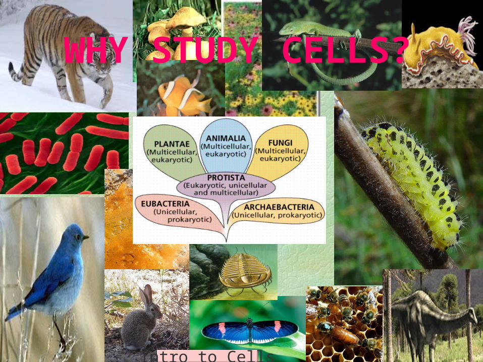
WHY STUDY CELLS?
Intro to Cells
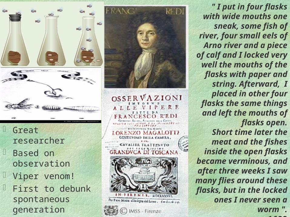
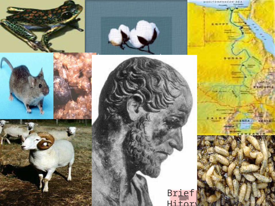
Great researcher Based on
observation Viper venom! First to debunk
spontaneous generation
" I put in four flasks with wide mouths one sneak, some fish of river, four
small eels of Arno river and a piece of calf and I locked very well the mouths of the
flasks with paper and string. Afterward, I placed
in other four flasks the same things and left the
mouths of flasks open.Short time later the meat and the fishes inside the
open flasks became verminous, and after three
weeks I saw many flies around these flasks, but in
the locked ones I never seen a worm ".
- 1688
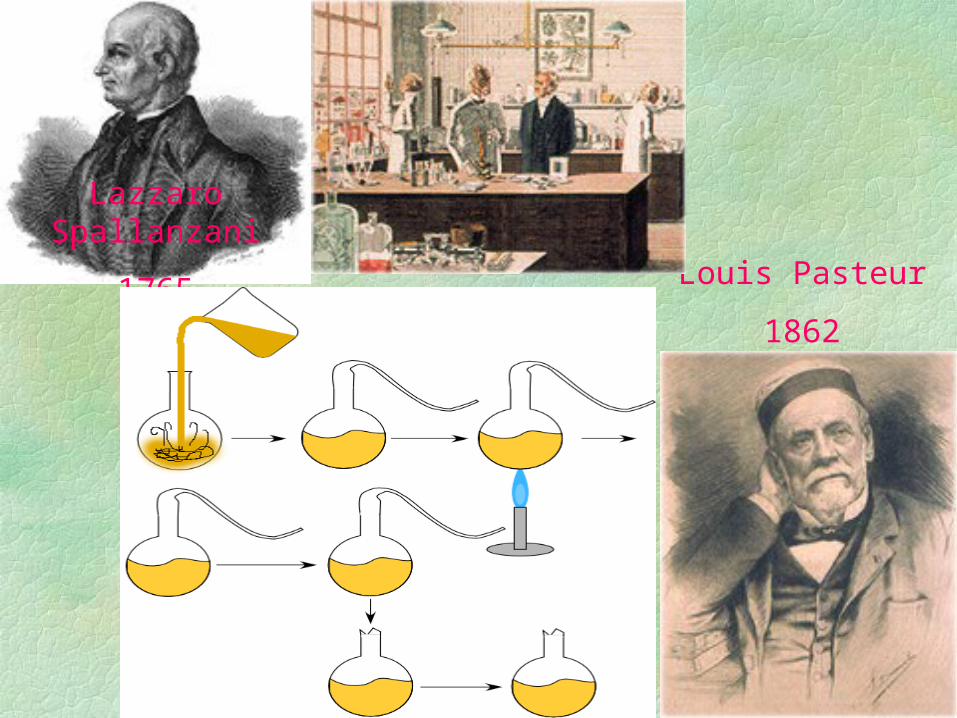
Lazzaro Spallanzani
1765Louis Pasteur
1862
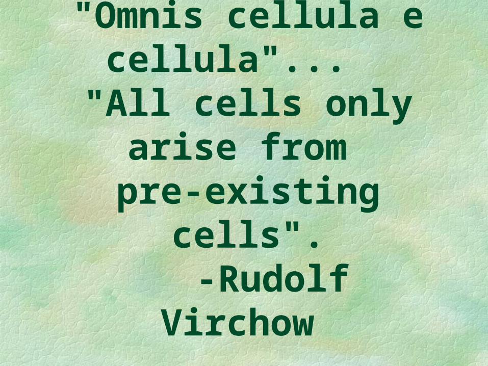
"Omnis cellula e cellula"...
"All cells only arise from pre-existing
cells".-Rudolf
Virchow
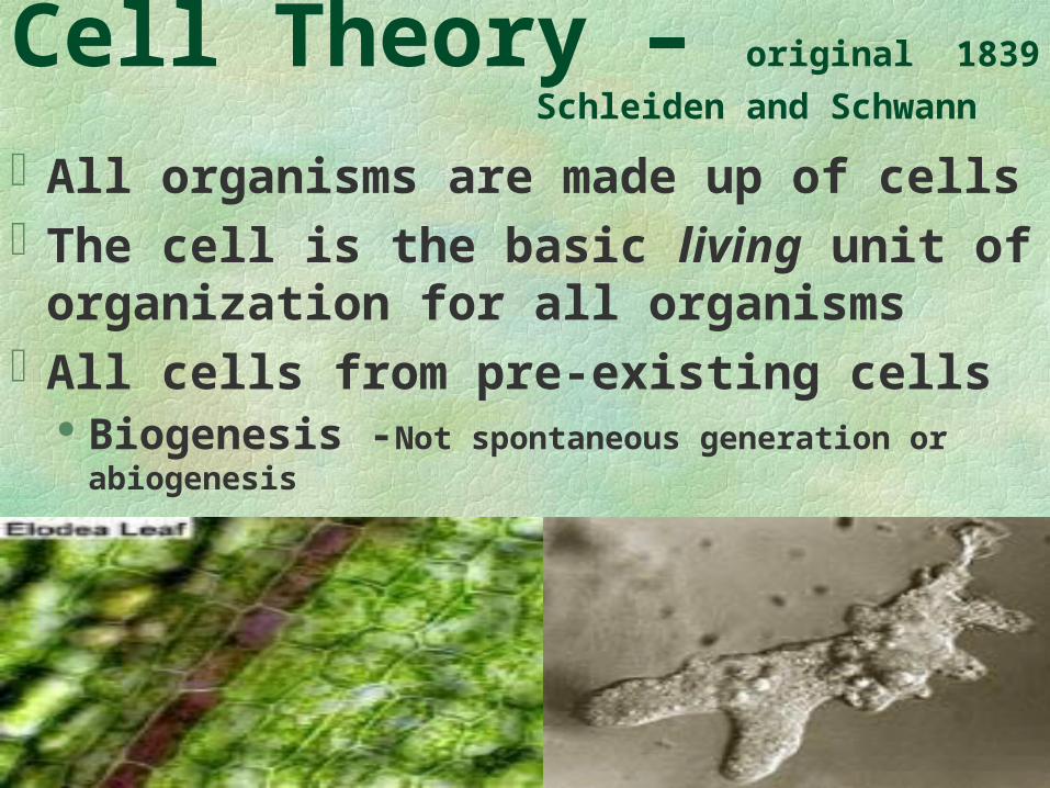
Cell Theory – original 1839
Schleiden and Schwann
All organisms are made up of cellsThe cell is the basic living unit of
organization for all organismsAll cells from pre-existing cells
Biogenesis -Not spontaneous generation or abiogenesis
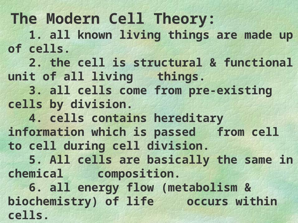
1. all known living things are made up of cells. 2. the cell is structural & functional unit of all living things. 3. all cells come from pre-existing cells by division. 4. cells contains hereditary information which is passed
from cell to cell during cell division. 5. All cells are basically the same in chemical composition. 6. all energy flow (metabolism & biochemistry) of life
occurs within cells.
The Modern Cell Theory:
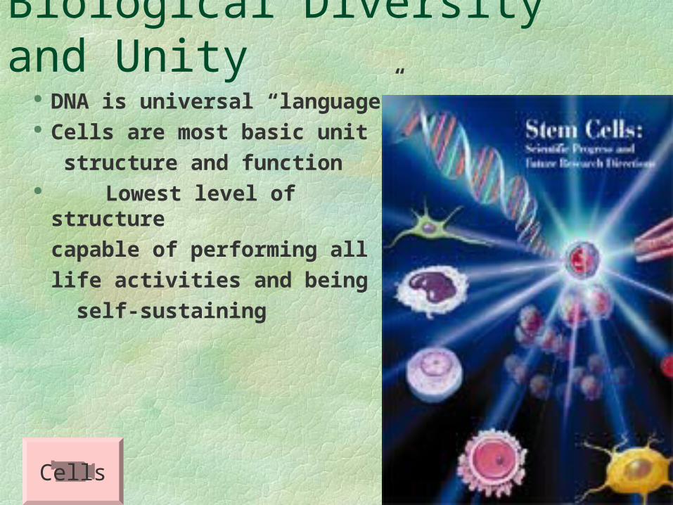
Biological Diversity and Unity DNA is universal “language” Cells are most basic unit of
structure and function Lowest level of structure
capable of performing all
life activities and being
self-sustaining
Cells
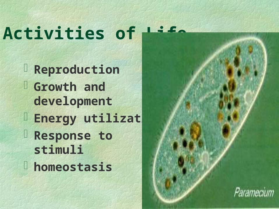
Activities of Life
ReproductionGrowth and
developmentEnergy utilizationResponse to stimulihomeostasis
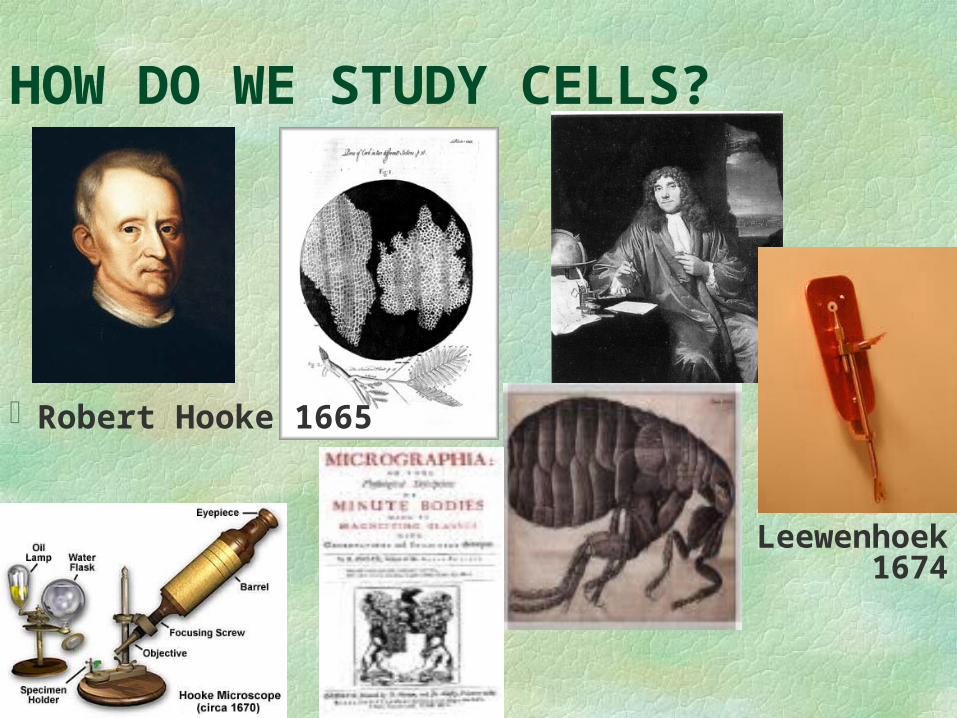
HOW DO WE STUDY CELLS?
Leewenhoek 1674
Robert Hooke 1665
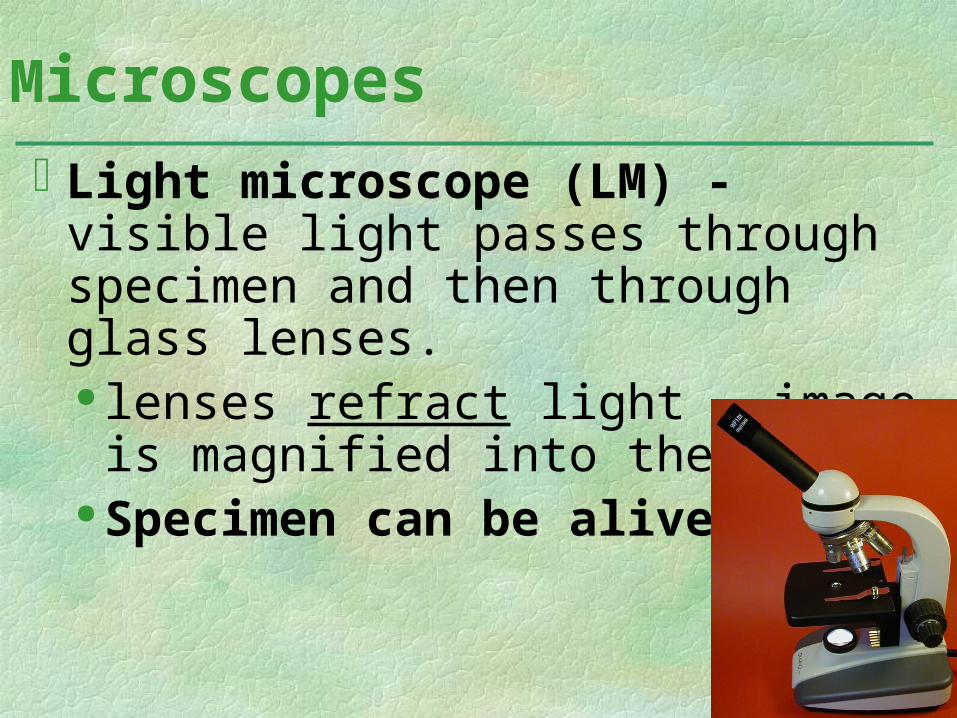
Light microscope (LM) - visible light passes through specimen and then through glass lenses. lenses refract light - image is
magnified into the eye Specimen can be alive!
Microscopes
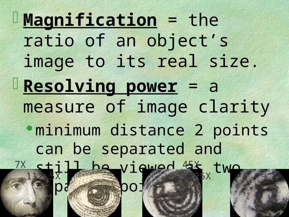
Magnification = the ratio of an object’s image to its real size.
Resolving power = a measure of image clarity minimum distance 2 points can be
separated and still be viewed as two separate points
7X 45X 112.5X 225X
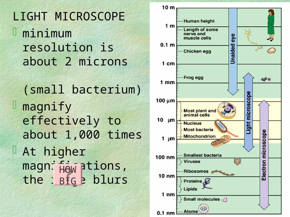
LIGHT MICROSCOPEminimum resolution is
about 2 microns (small bacterium)
magnify effectively to about 1,000 times
At higher magnifications, the image blurs
HOWBIG

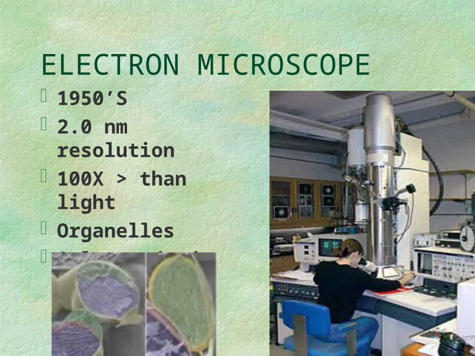
ELECTRON MICROSCOPE1950’S2.0 nm resolution100X > than lightOrganellesOnly on dead cells
electron microscope (EM)- beam of electrons through the
specimen or onto its surface - shorter wavelengths of light
greater resolution
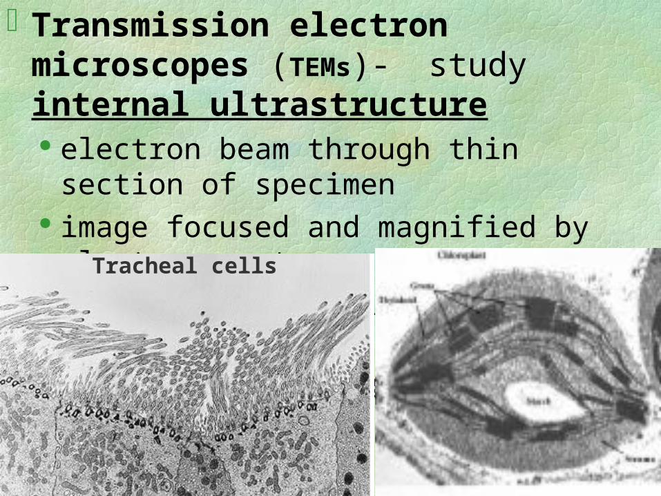
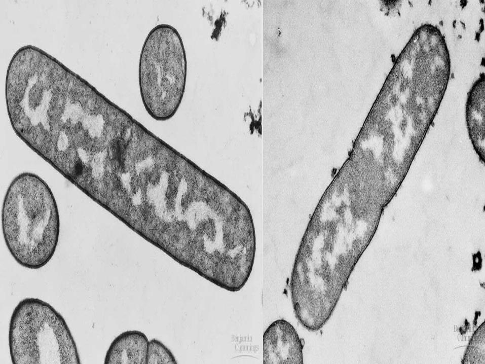
Transmission electron microscopes (TEMs)-study internal ultrastructure
electron beam through thin section of specimen image focused and magnified by electromagnets thin sections stained with atoms of heavy metals Dead; may leave debris/artifacts
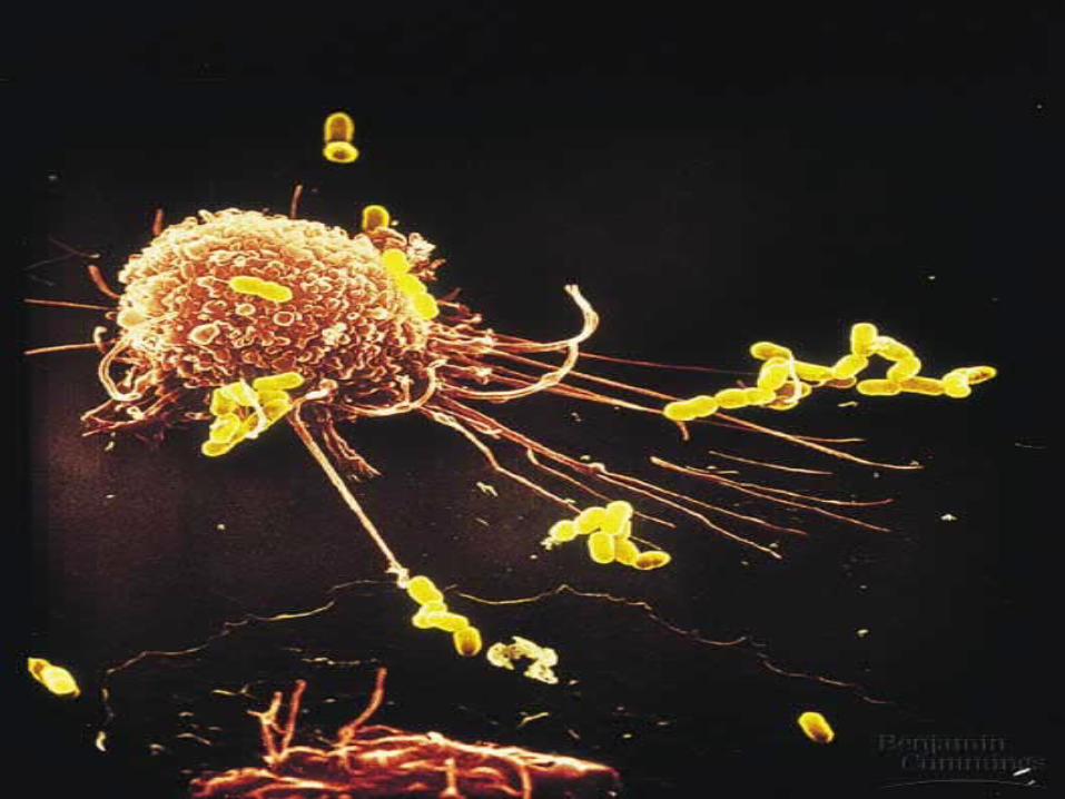
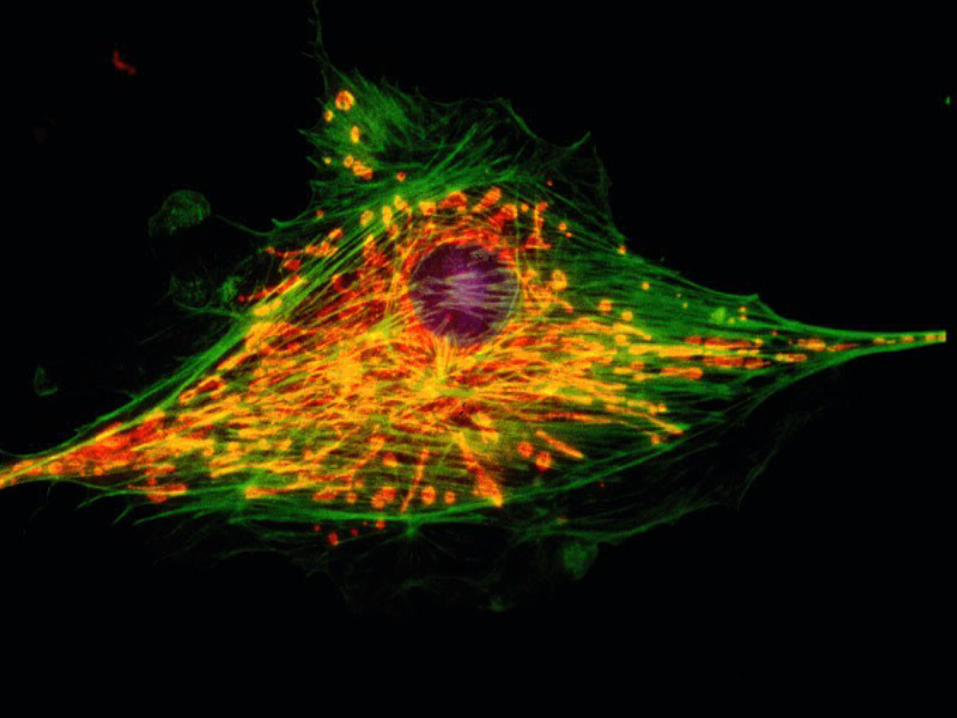
Tracheal cells
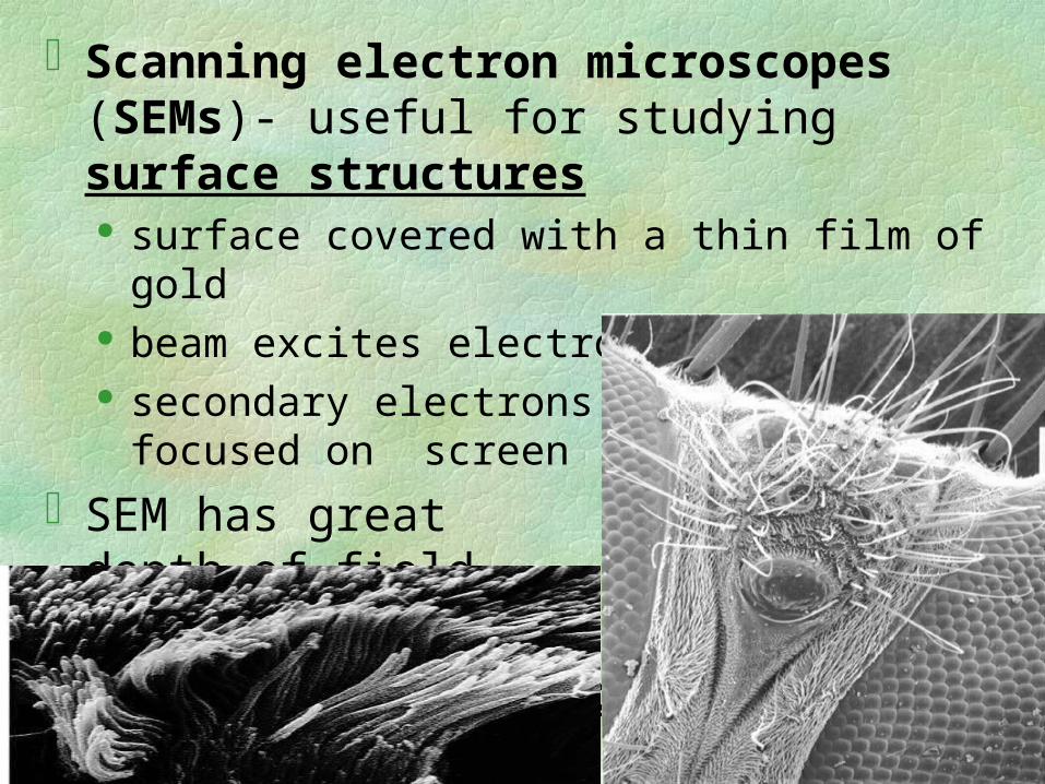
Scanning electron microscopes (SEMs)- useful for studying surface structures surface covered with a thin film of gold beam excites electrons on surface secondary electrons collected and focused on screen
SEM has great depth of field, image seems 3-D
Dead,debris/artifacts
LM’s -less resolution but livingcytology- study of cell structuresCytology + biochemistry =
modern cell biology
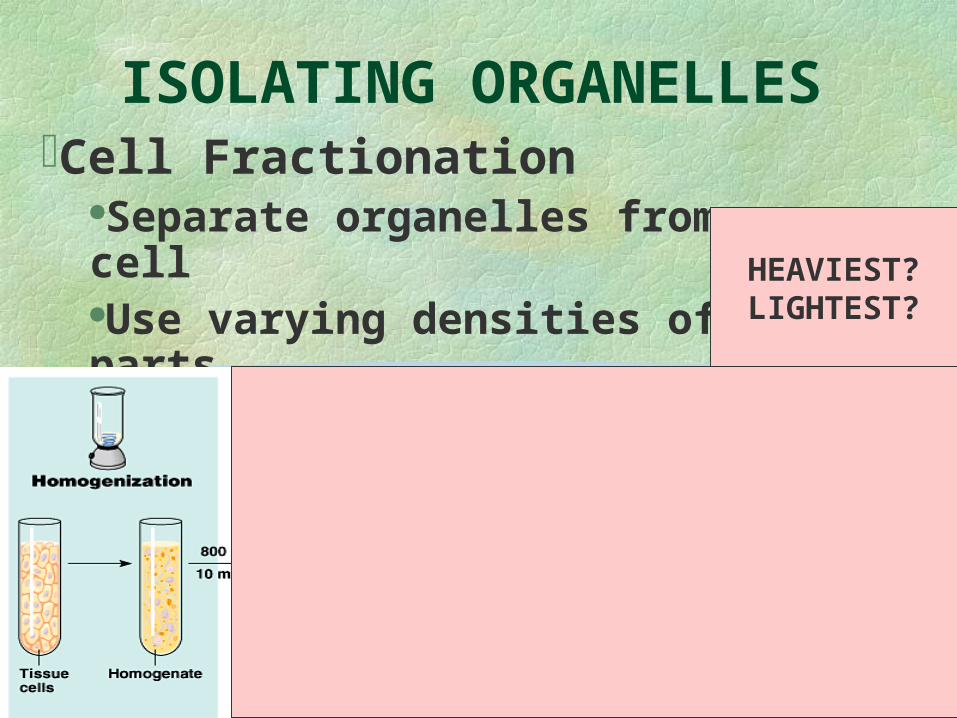
ISOLATING ORGANELLESCell Fractionation
Separate organelles from cellUse varying densities of parts
•Ultracetrifuge
HEAVIEST? LIGHTEST?
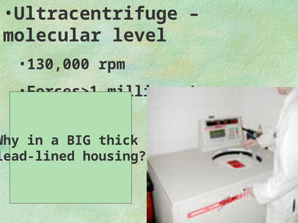
•Ultracentrifuge – molecular level
•130,000 rpm
•Forces>1 million g’s
Why in a BIG thick lead-lined housing?
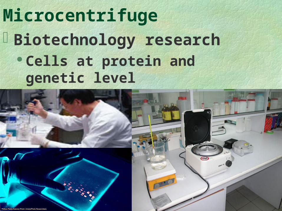
MicrocentrifugeBiotechnology research
Cells at protein and genetic level
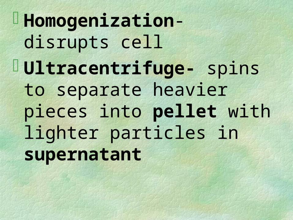
Homogenization- disrupts cellUltracentrifuge- spins to separate
heavier pieces into pellet with lighter particles in supernatant
CELLSALIVE
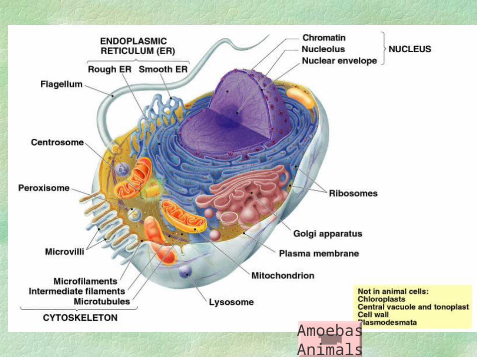
AmoebasAnimals
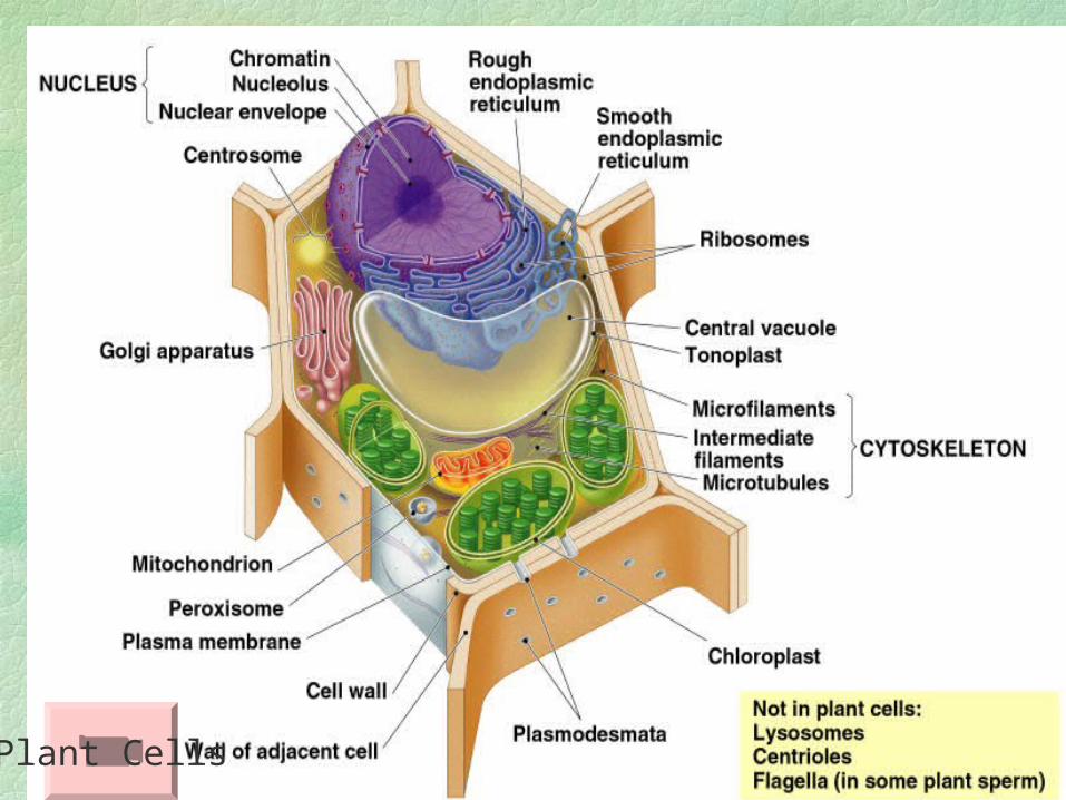
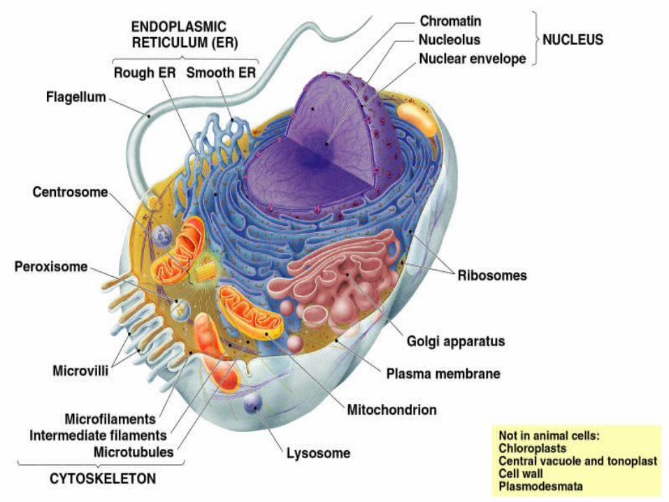
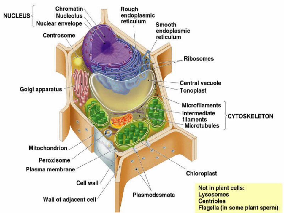
Plant Cells

Cell characteristics – All Cells
Plasma membraneCytosol
Semi-fluid substance w/ “solutes” Cytoplasm = cytosol + organelles(euk’s)
Contain chromosomes w/ genes in DNARibosomes
Protein synthesis; carry out gene instructions
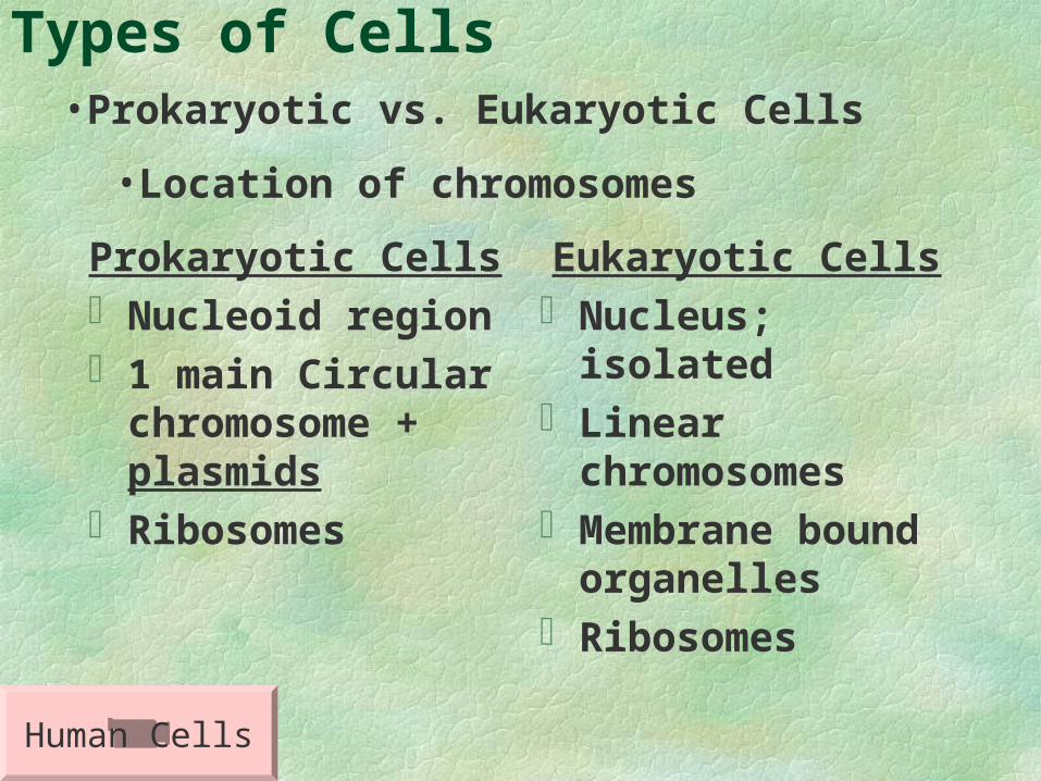
Types of Cells
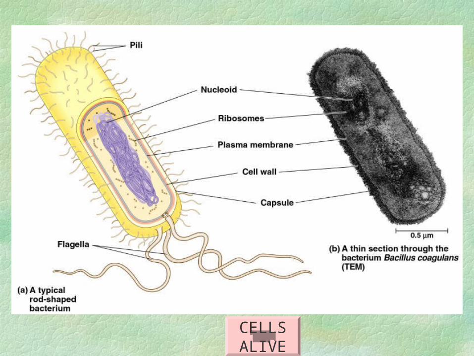
Prokaryotic CellsNucleoid region1 main Circular
chromosome + plasmids
Ribosomes
Eukaryotic CellsNucleus; isolatedLinear chromosomesMembrane bound
organellesRibosomes
•Prokaryotic vs. Eukaryotic Cells
•Location of chromosomes
Human Cells
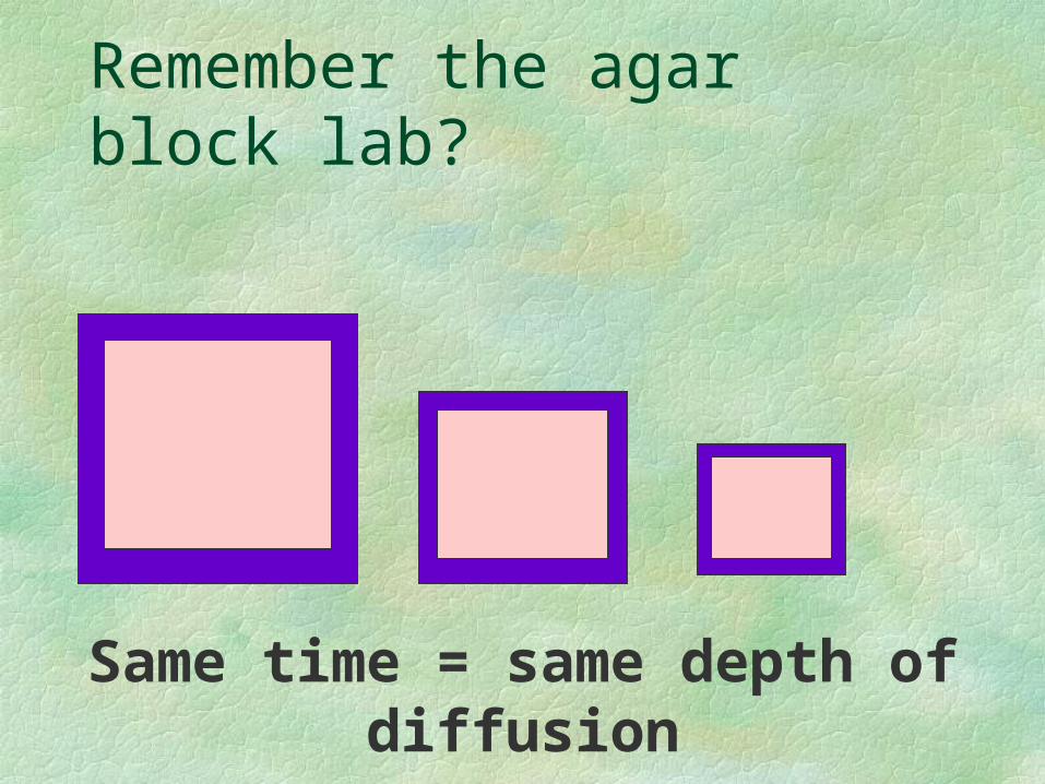
Remember the agar block lab?
Same time = same depth of diffusion
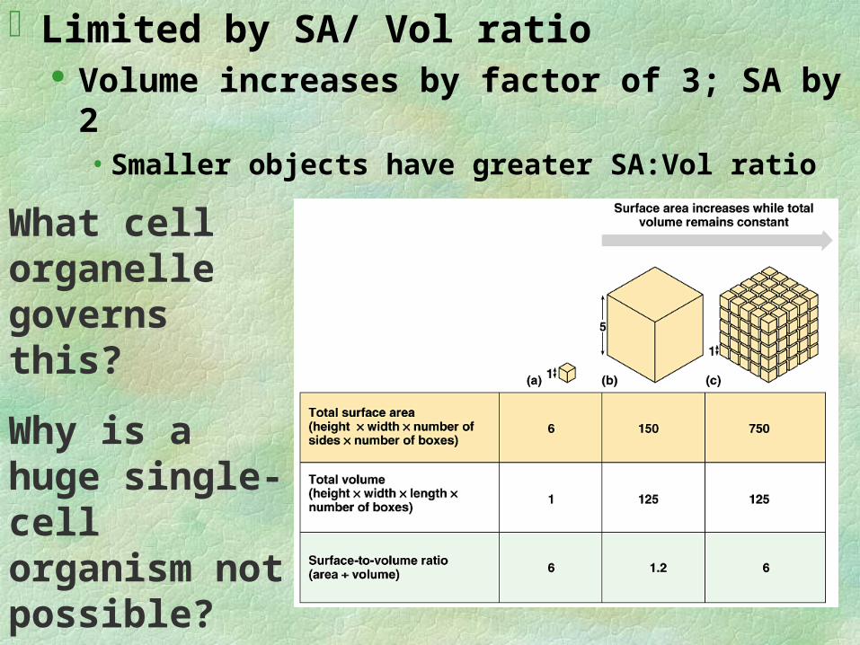
Limited by SA/ Vol ratio Volume increases by factor of 3; SA by 2
• Smaller objects have greater SA:Vol ratio
What cell organelle governs this?
Why is a huge single-cell organism not possible?
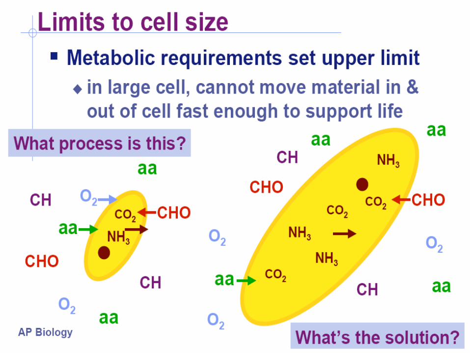
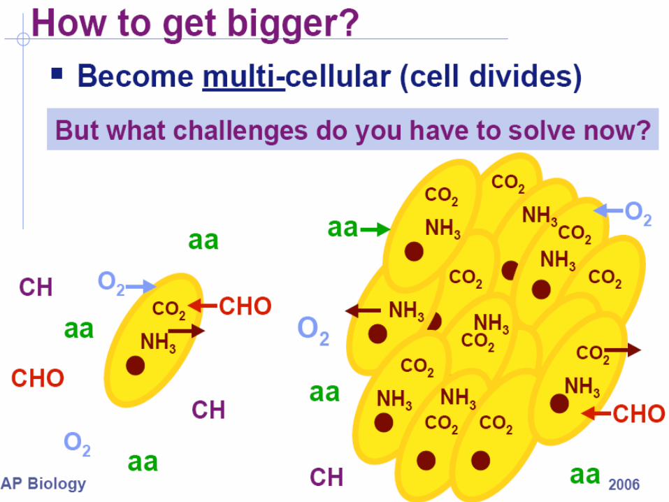
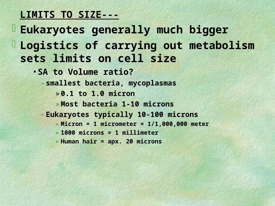
LIMITS TO SIZE---
Eukaryotes generally much bigger Logistics of carrying out metabolism sets
limits on cell size • SA to Volume ratio?
– smallest bacteria, mycoplasmas
» 0.1 to 1.0 micron
» Most bacteria 1-10 microns
– Eukaryotes typically 10-100 microns» Micron = 1 micrometer = 1/1,000,000 meter» 1000 microns = 1 millimeter» Human hair = apx. 20 microns
Size must be low to sustain life
• enough DNA to program metabolism
• enough ribosomes for protein synthesis
• enough enzymes for metabolism
• enough cellular components
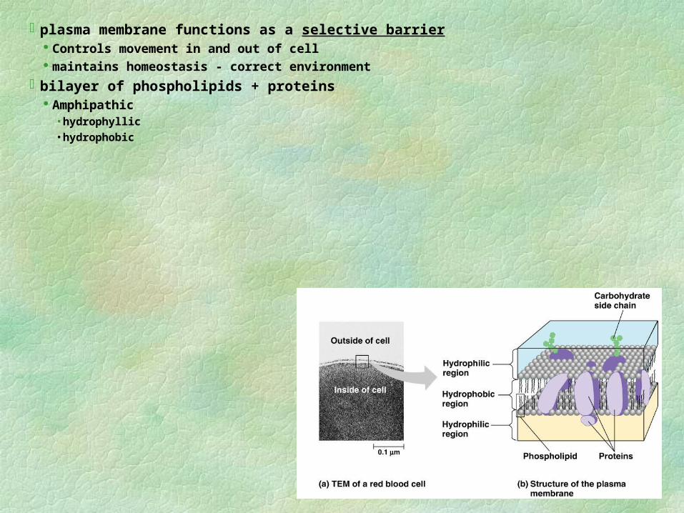
plasma membrane functions as a selective barrier Controls movement in and out of cell maintains homeostasis - correct environment
bilayer of phospholipids + proteins Amphipathic
• hydrophyllic• hydrophobic
TOUR OF THE CELL
BUCKLE UP!
The Nucleus and
Ribosomes

contains most of genes in euk. Celllargest organelledouble membrane
unique environment
membranes fuse to form pores/envelope large macromolecules & particles pass unique chemical signals viruses may break code
Nucleus contains a eukaryotic cell’s genetic library
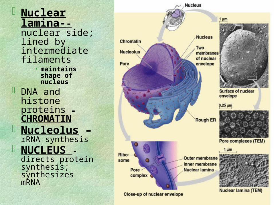
Nuclear lamina--nuclear side; lined by intermediate filaments
• maintains shape of nucleus
DNA and histone proteins = CHROMATIN
Nucleolus – rRNA synthesis
NUCLEUS - directs protein synthesis; synthesizes mRNA
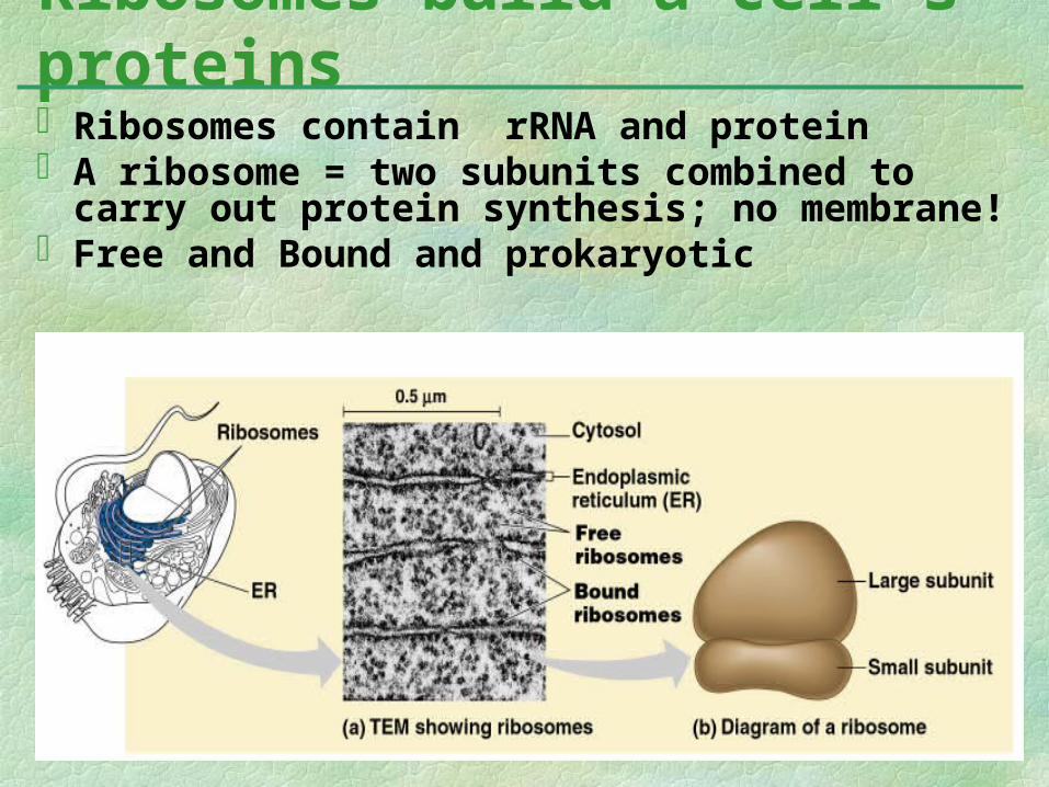
Ribosomes contain rRNA and proteinA ribosome = two subunits combined to carry out
protein synthesis; no membrane!Free and Bound and prokaryotic
Ribosomes build a cell’s proteins
The Endomembrane
System
Endo-Membrane
system
membranous tubules and internal, fluid-filled spaces = cisternae; storage area
Lumen is center of ERcontinuous with N. E.
ER manufactures membranes and performs many other biosynthetic functions
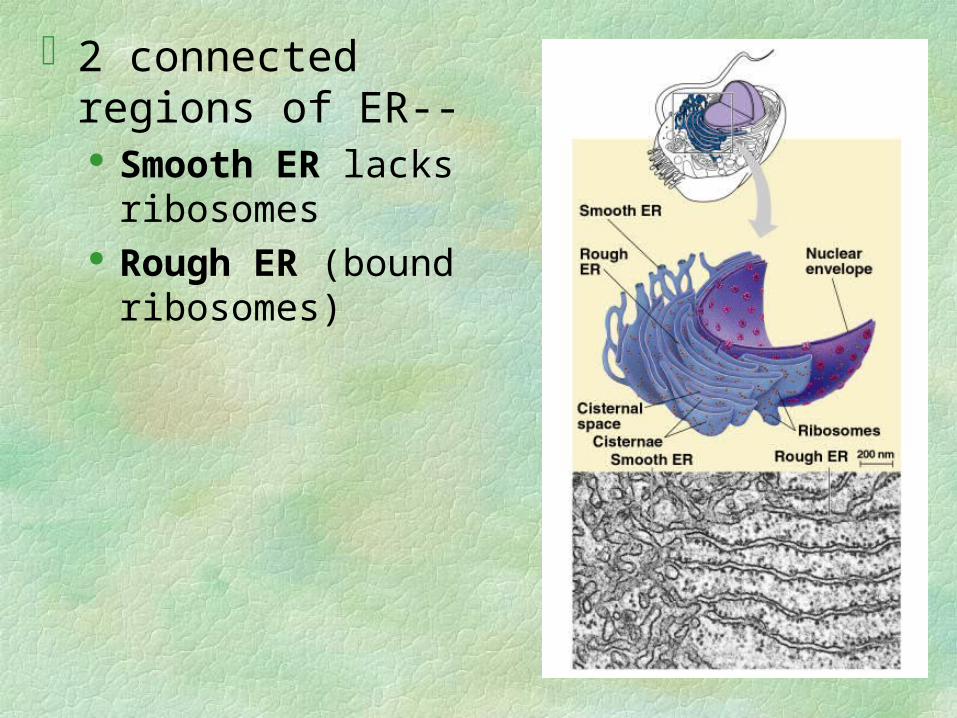
2 connected regions of ER-- Smooth ER lacks
ribosomes Rough ER (bound
ribosomes)
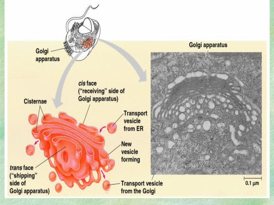
Receives transports vesicles from ERModifies contentsWarehousing, sorting, and shippingAbundant in secretory cells Produces lysosomes and cell wall
The Golgi Apparatus finishes, sorts, and ships cell products
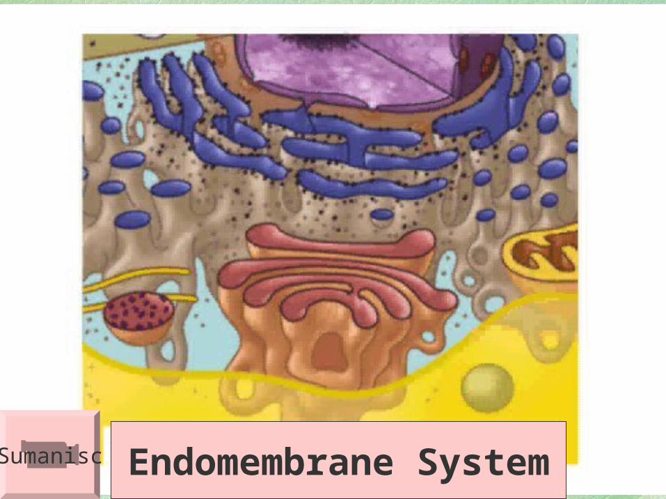
Endomembrane SystemSumanisc
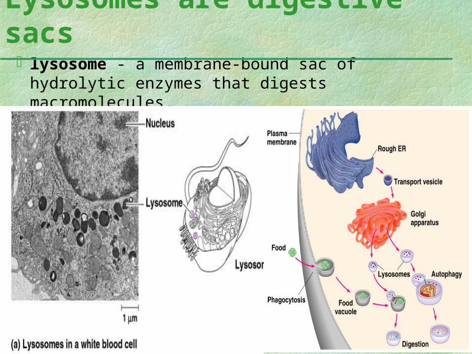
lysosome - a membrane-bound sac of hydrolytic enzymes that digests macromolecules.
Lysosomes are digestive sacs
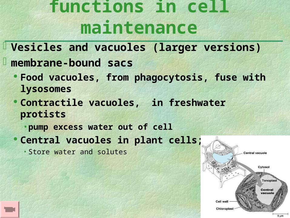
Vesicles and vacuoles (larger versions)membrane-bound sacs
Food vacuoles, from phagocytosis, fuse with lysosomes
Contractile vacuoles, in freshwater protists• pump excess water out of cell
Central vacuoles in plant cells; • Store water and solutes
Vacuoles have diverse functions in cell maintenance
Other Membranous Organelles
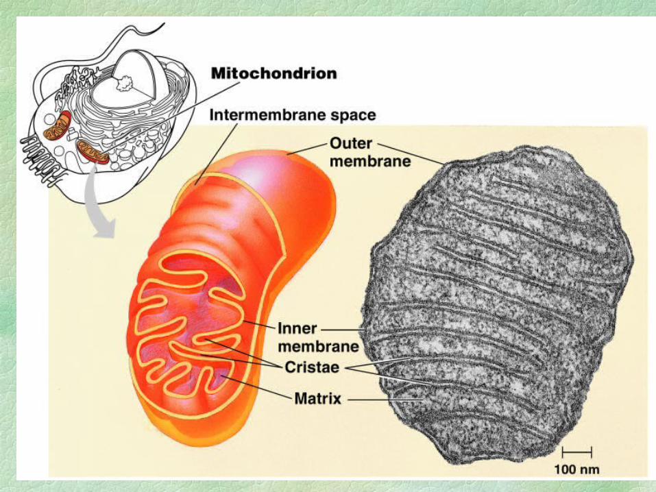
convert energy to usable forms for workMitochondria = sites of cell. respiration,
generate ATP from catabolism of sugars, fats, and other fuels in presence of oxygen
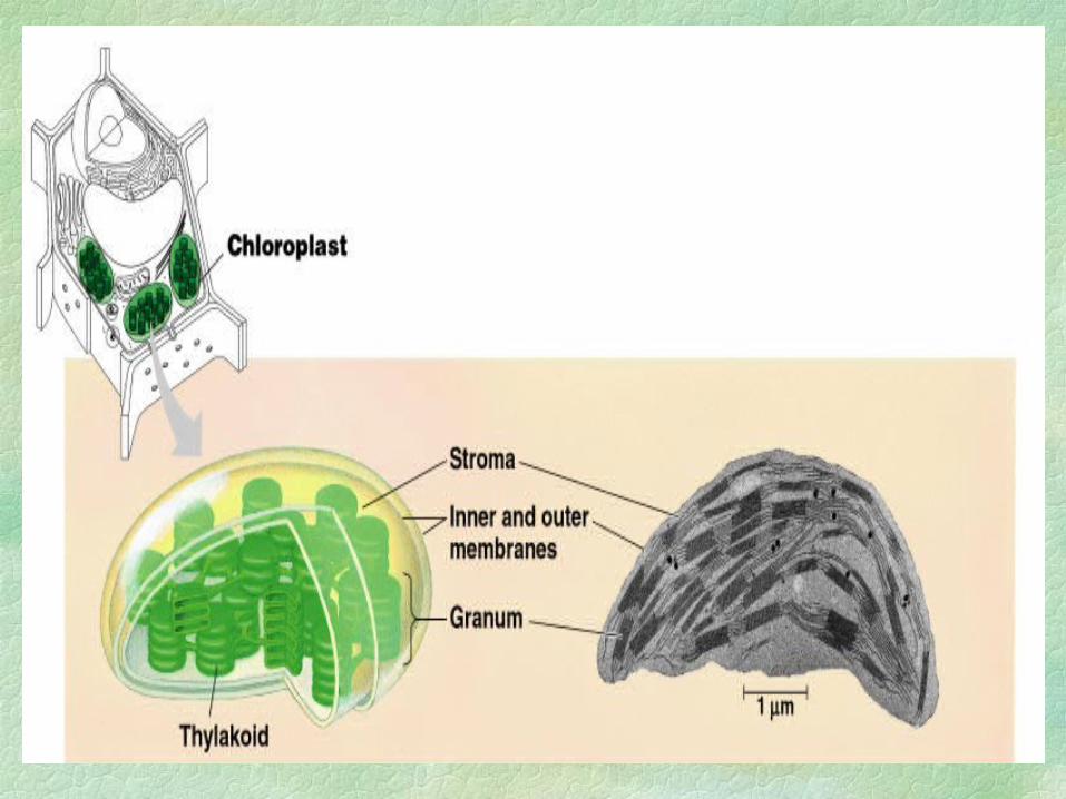
Chloroplasts - found in plants and eukaryotic algae; sites of photosynthesis convert solar energy to chemical energy and
synthesize new organic compounds from CO2 and H2O.
Mitochondria and chloroplasts are main energy transformers of cells
PLASTIDS - Amyloplasts/leucoplasts - store starch
in roots and tubers Chromoplasts store pigments Chloroplast
• produces sugar via photosynthesis
• color from chlorophyll pigment • in leaves and other green structures of plants and in eukaryotic algae
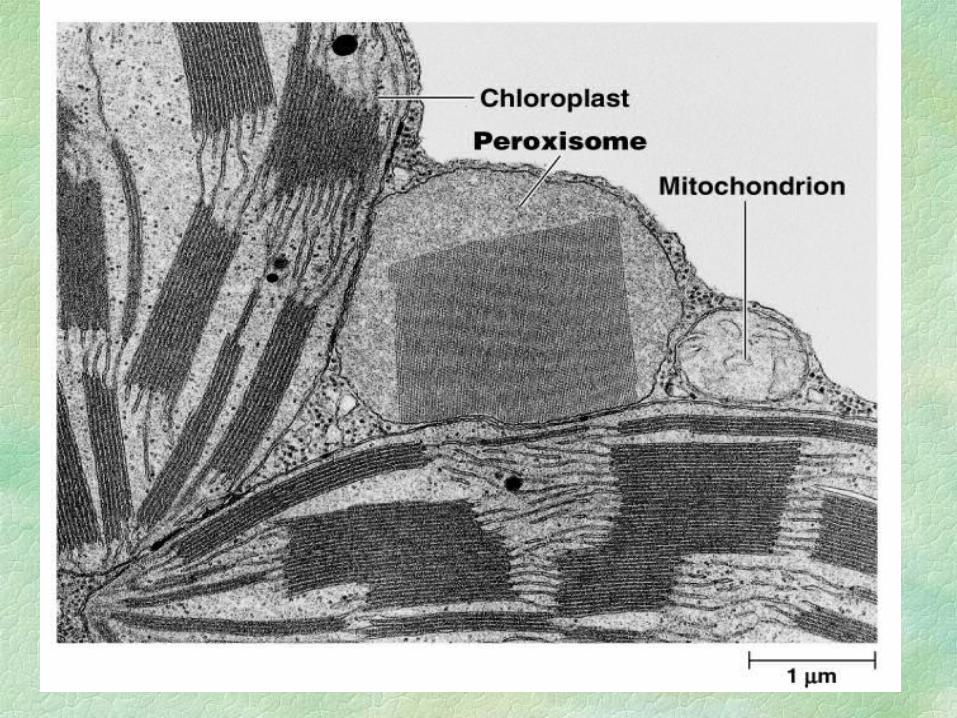
Peroxisomes - single membranecontain enzymes to break down H2O2
Some break fatty acids down for mitochondria for fuel Some detoxify alcohol and other harmful compounds
Glyoxysomes = Specialized peroxisomes,
in plants only, convert fatty acids to sugars in seeds
= easier energy and carbon source
The Cytoskeleton
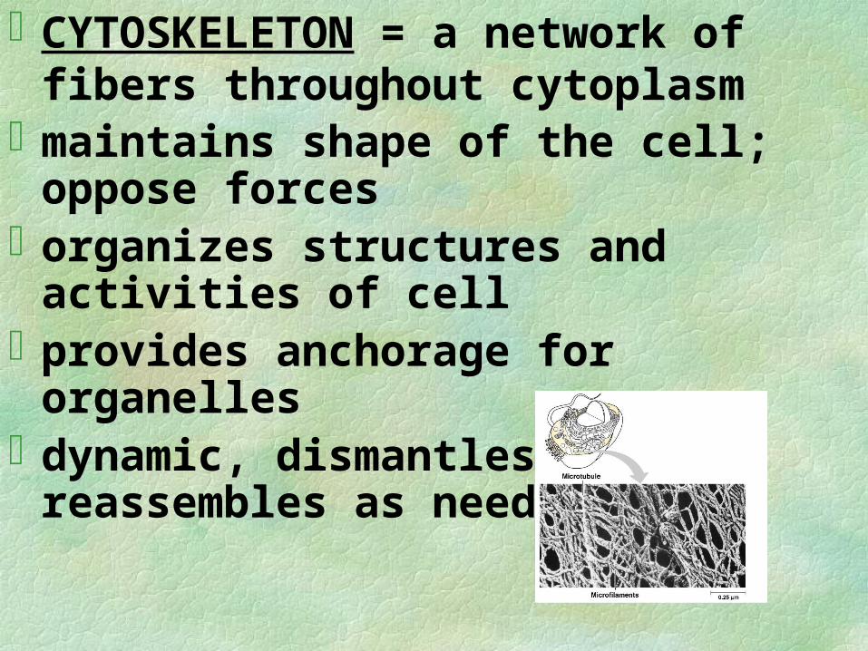
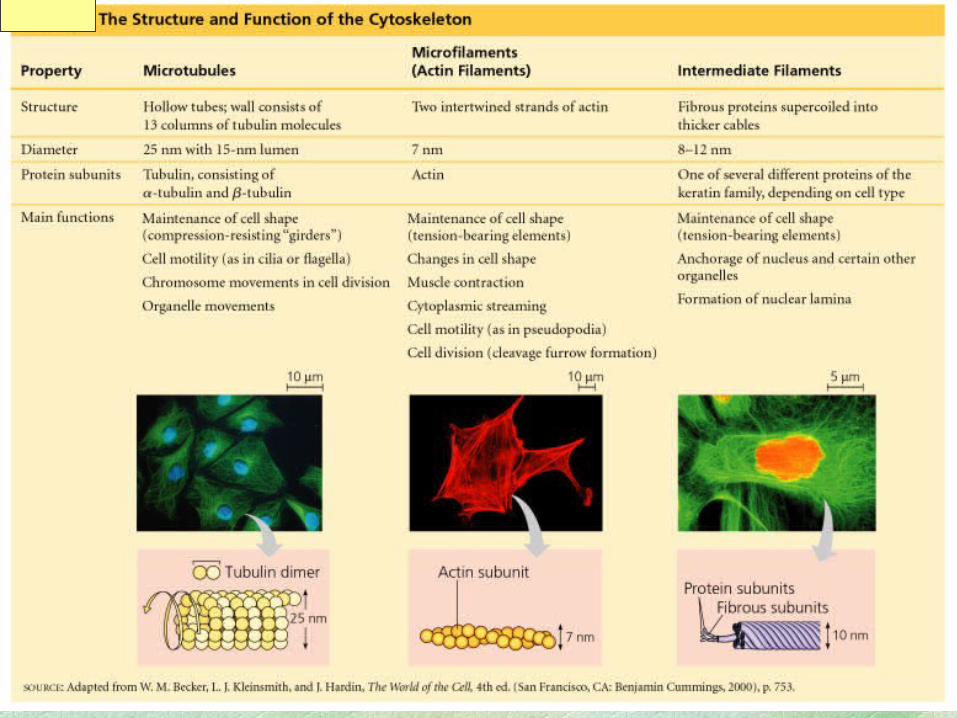
CYTOSKELETON = a network of fibers throughout cytoplasm
maintains shape of the cell; oppose forcesorganizes structures and activities of cellprovides anchorage for organellesdynamic, dismantles and reassembles as
needed
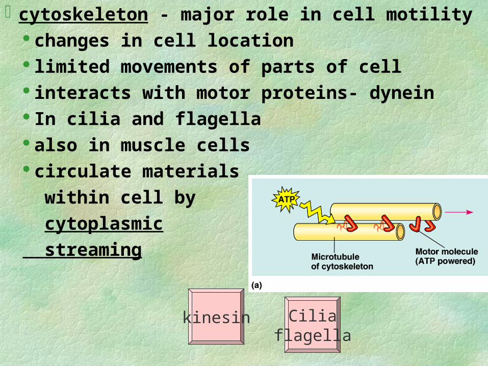
cytoskeleton - major role in cell motility changes in cell location limited movements of parts of cell interacts with motor proteins- dynein In cilia and flagella also in muscle cells circulate materials
within cell by
cytoplasmic
streaming
kinesin Ciliaflagella
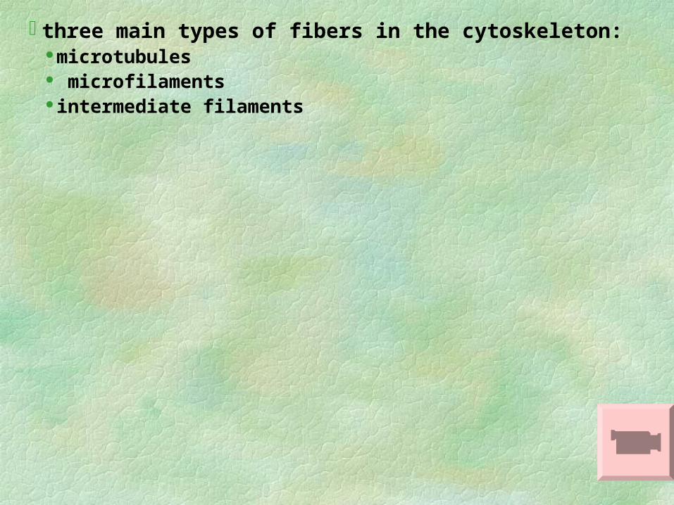
three main types of fibers in the cytoskeleton: microtubules microfilaments intermediate filaments
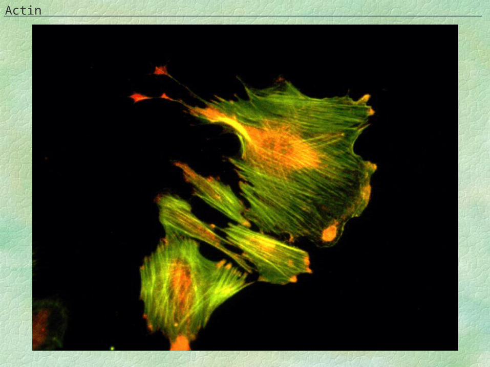
Actin
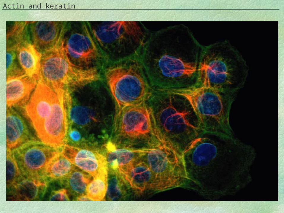
Actin and keratin
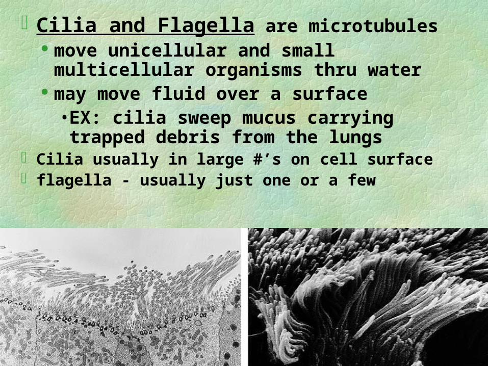
Cilia and Flagella are microtubules move unicellular and small multicellular
organisms thru water may move fluid over a surface
• EX: cilia sweep mucus carrying trapped debris from the lungs
Cilia usually in large #’s on cell surface flagella - usually just one or a few
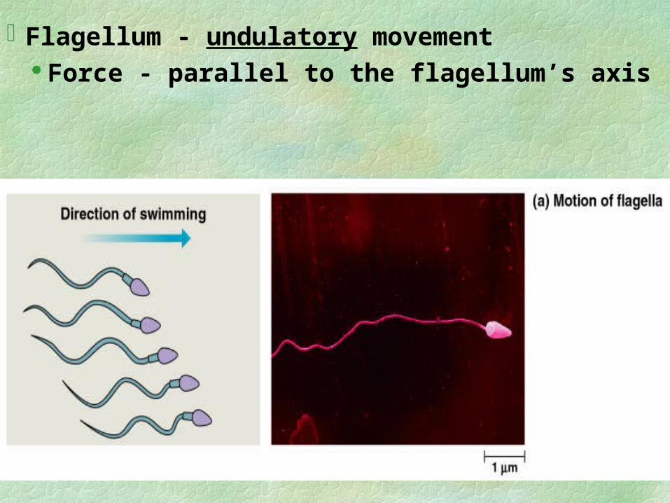
Flagellum - undulatory movement Force - parallel to the flagellum’s axis
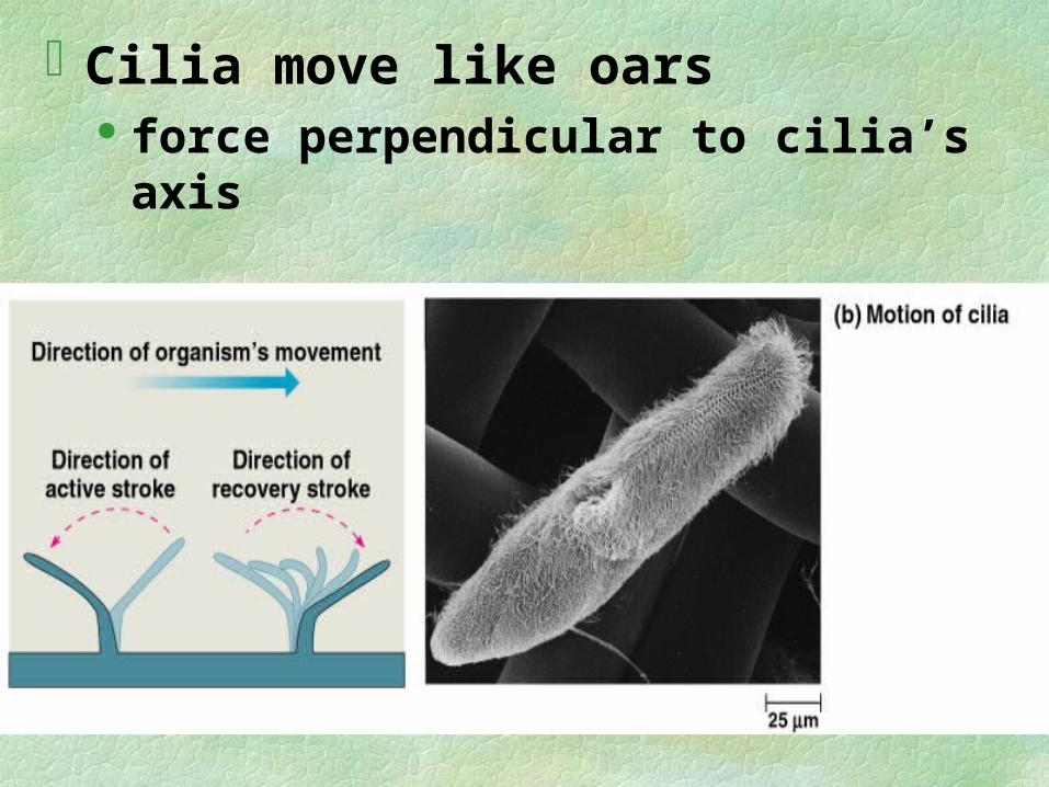
Cilia move like oars force perpendicular to cilia’s axis
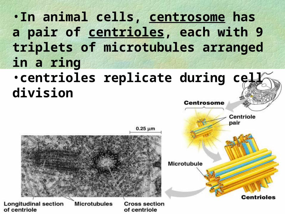
•In animal cells, centrosome has a pair of centrioles, each with 9 triplets of microtubules arranged in a ring•centrioles replicate during cell division
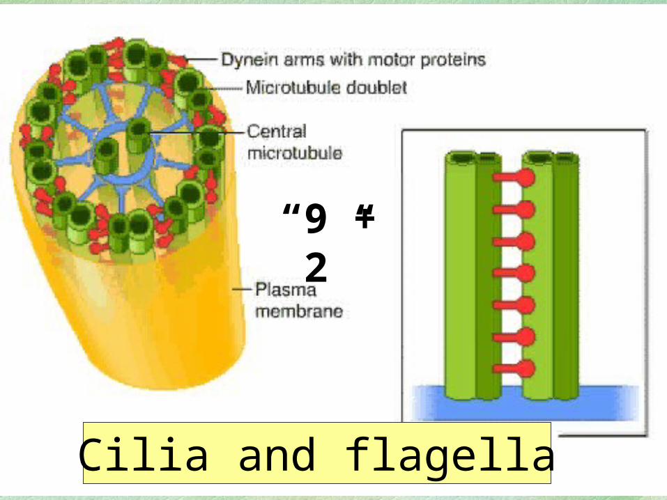
“9 + 2”
Cilia and flagella
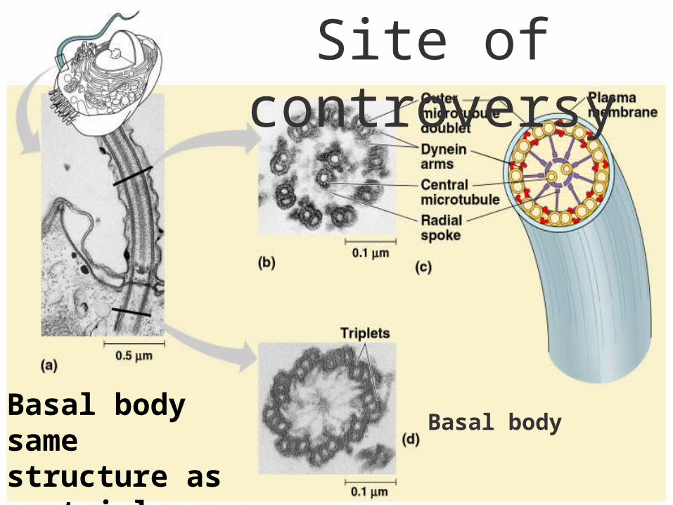
Basal bodyBasal body same structure as centriole
Site of controversy
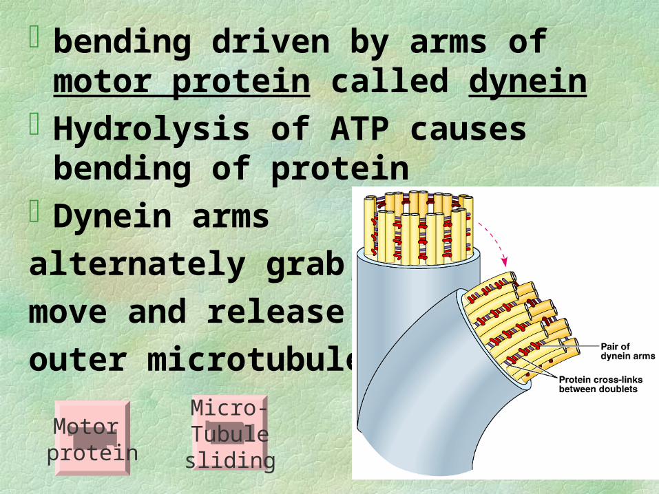
bending driven by arms of motor protein called dynein
Hydrolysis of ATP causes bending of protein
Dynein arms
alternately grab,
move and release
outer microtubulesMicro-Tubulesliding
Motor protein
Microfilaments= thinnest fibers; solid, globular protein actin microfilament of actin subunits
resist tension = pullinginteract with myosin for muscle
contractionA contracting belt- divides cytoplasm
animal cells during cell division
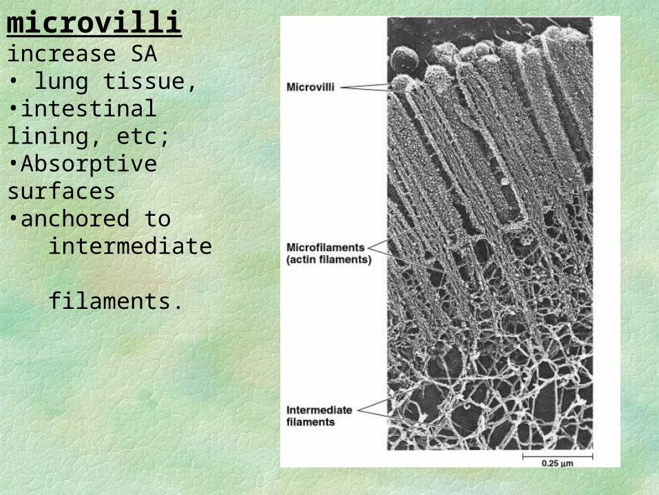
microvilliincrease SA • lung tissue,•intestinal lining, etc;•Absorptive surfaces•anchored to intermediate filaments.
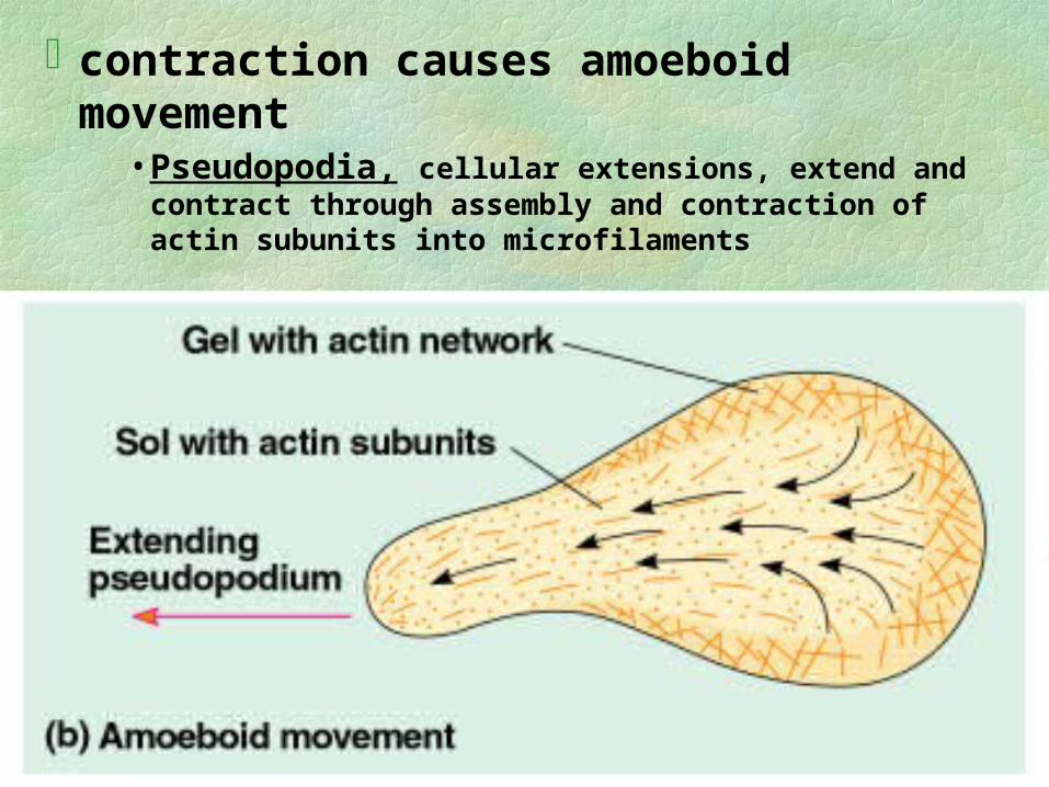
contraction causes amoeboid movement• Pseudopodia, cellular extensions, extend and contract
through assembly and contraction of actin subunits into microfilaments
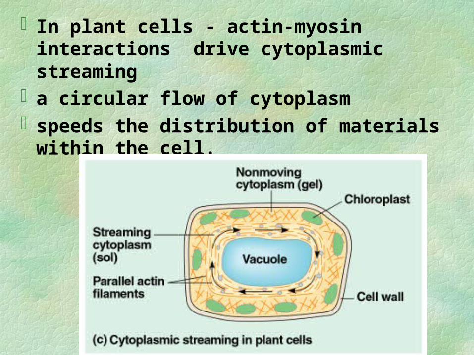
In plant cells - actin-myosin interactions drive cytoplasmic streaming
a circular flow of cytoplasmspeeds the distribution of materials within the
cell.
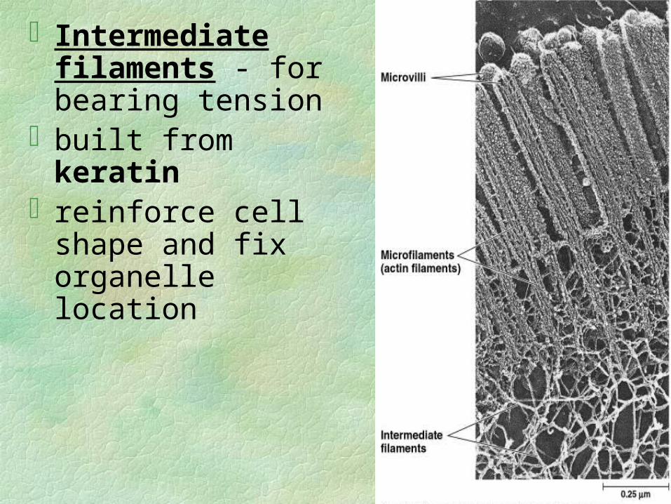
Intermediate filaments - for bearing tension
built from keratinreinforce cell shape and
fix organelle location
Cell Surfaces and Junctions
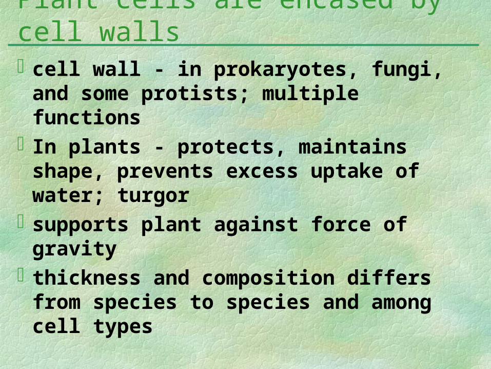
cell wall - in prokaryotes, fungi, and some protists; multiple functions
In plants - protects, maintains shape, prevents excess uptake of water; turgor
supports plant against force of gravitythickness and composition differs from
species to species and among cell types
Plant cells are encased by cell walls
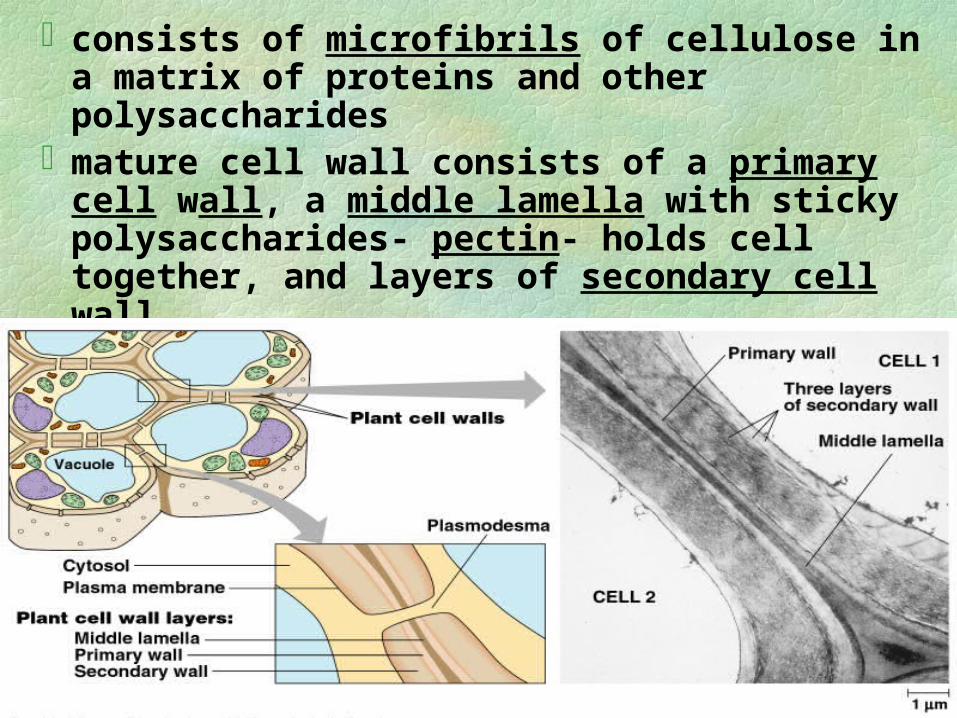
consists of microfibrils of cellulose in a matrix of proteins and other polysaccharides
mature cell wall consists of a primary cell wall, a middle lamella with sticky polysaccharides- pectin- holds cell together, and layers of secondary cell wall
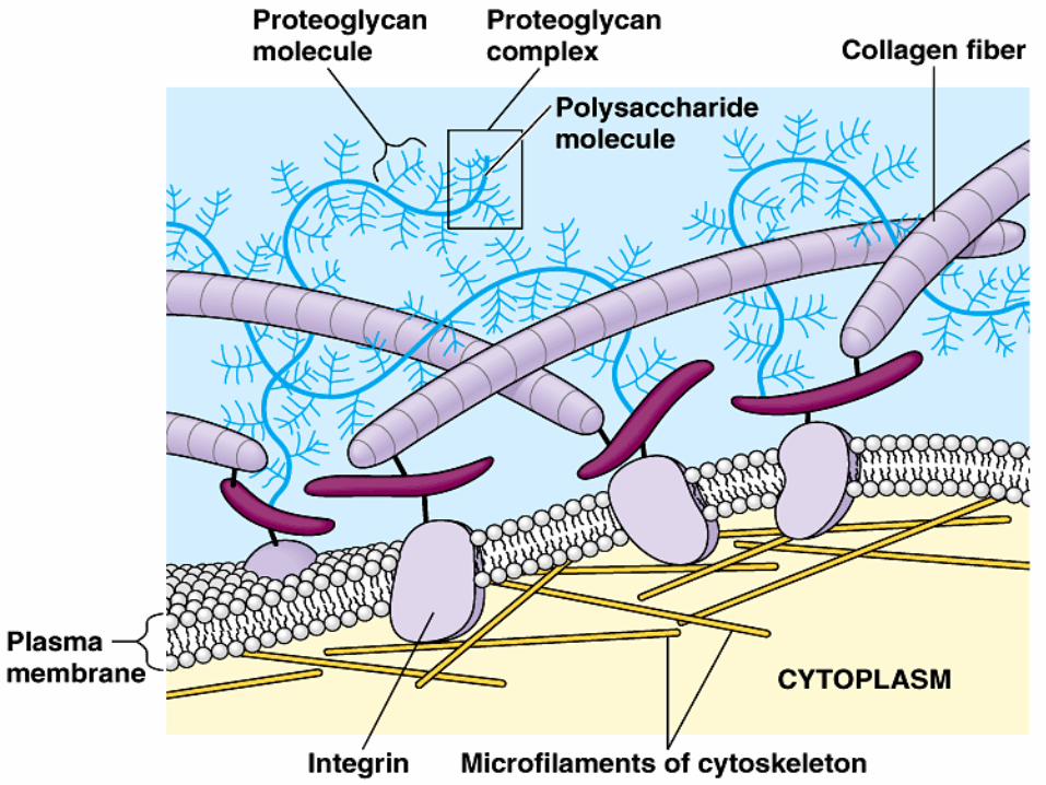
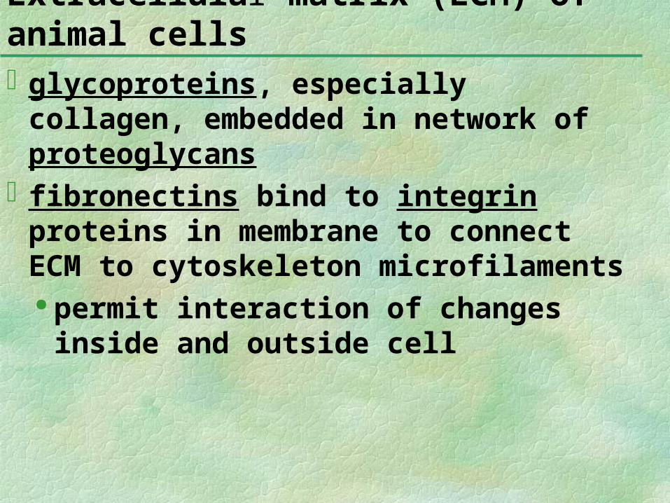
glycoproteins, especially collagen, embedded in network of proteoglycans
fibronectins bind to integrin proteins in membrane to connect ECM to cytoskeleton microfilaments permit interaction of changes inside
and outside cell
Extracellular matrix (ECM) of animal cells
The ECM can regulate cell behavior Embryonic cells migrate along specific
pathways by matching the orientation of their microfilaments to the “grain” of fibers in the extracellular matrix.
ECM can influence activity of genes in nucleus via a combination of chemical and mechanical signaling pathways• This may coordinate all the cells within a tissue.
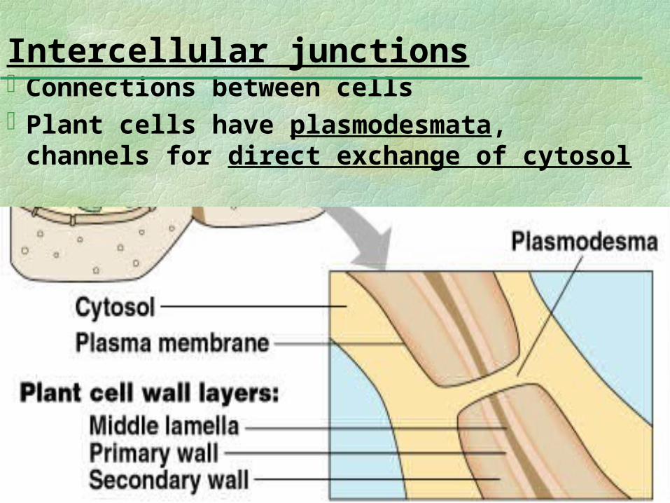
Connections between cellsPlant cells have plasmodesmata,
channels for direct exchange of cytosol
Intercellular junctions
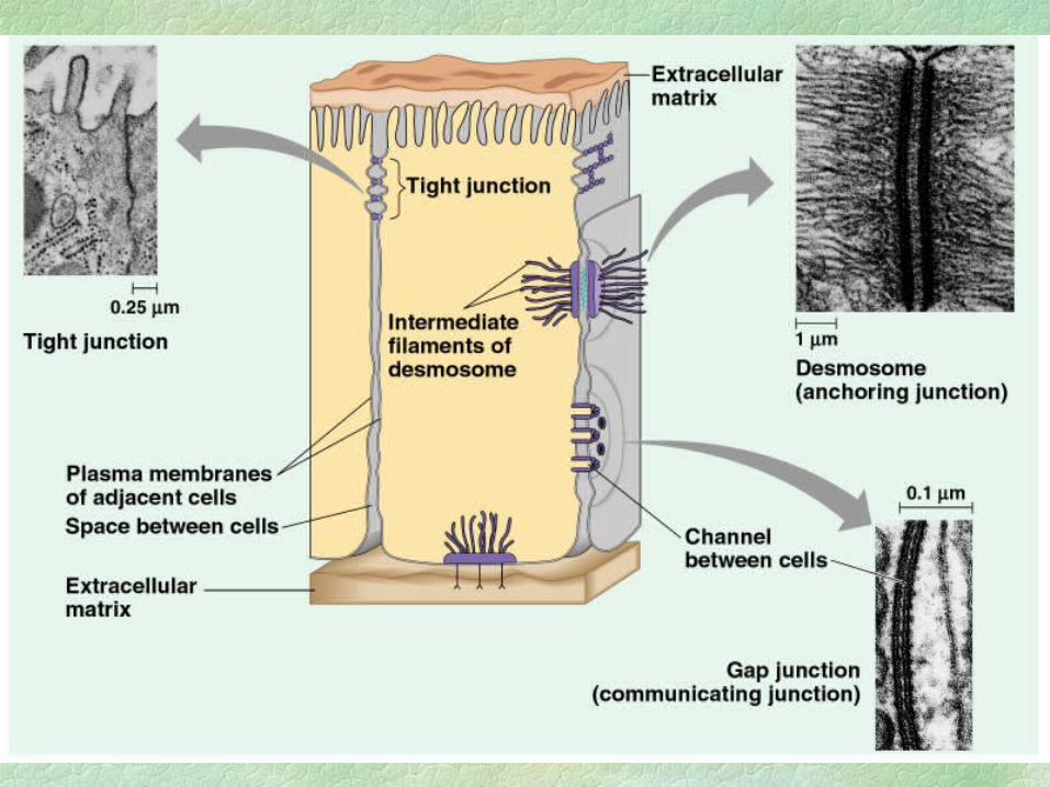
Animal have 3 main types of intercellular links:
tight junctions, membranes are fused, form continuous belts around cells-prevents leakage of extracellular fluid
Desmosomes fasten cells together into strong sheets - keratin intermediate filaments
Gap junctions provide cytoplasmic channels between adjacent cells