Embed Size (px)
Citation preview
Chapter 10Methods commonly used in
Molecular Genetics

outline
1.Nucleic acid extraction
2.Gel electrophoresis
3.Nucleic acid hybridization
4.Polymerase chain reaction
5.Sanger DNA sequencing
6.DNA recombination technology
7.Gene transfer
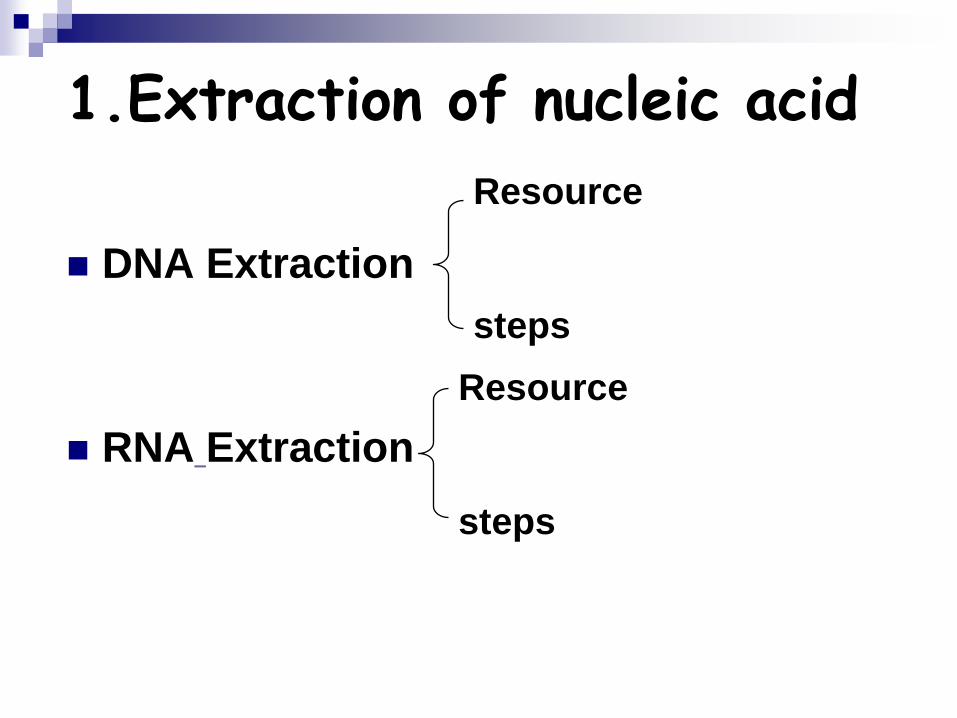
1.Extraction of nucleic acid
DNA Extraction
RNA Extraction
Resource
steps
Resource
steps
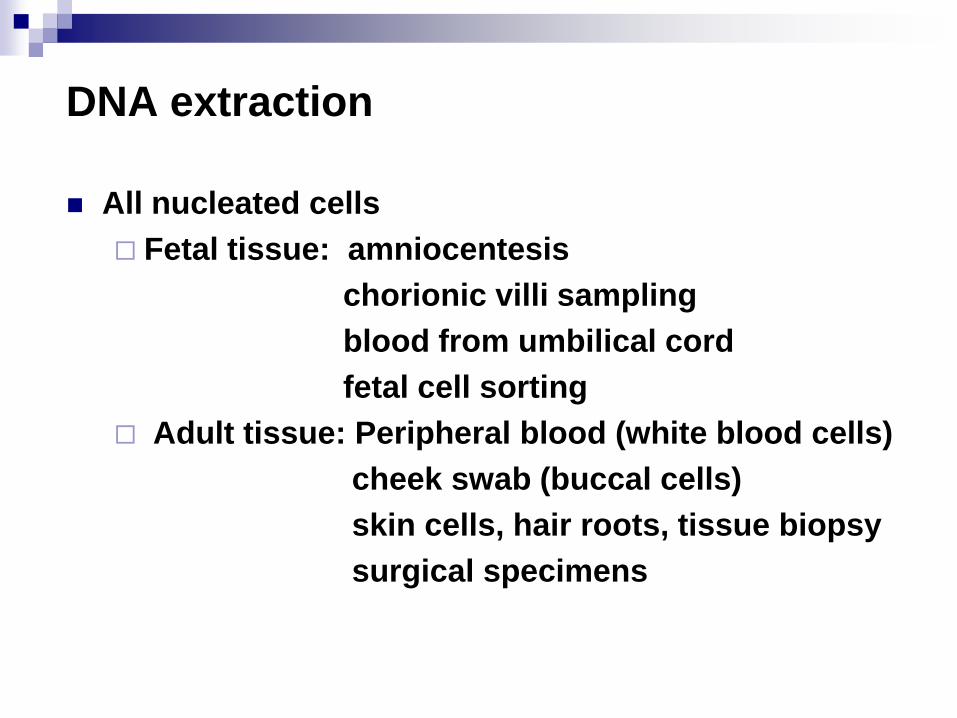
DNA extraction
All nucleated cells
Fetal tissue: amniocentesis
chorionic villi sampling
blood from umbilical cord
fetal cell sorting
Adult tissue: Peripheral blood (white blood cells)
cheek swab (buccal cells)
skin cells, hair roots, tissue biopsy
surgical specimens
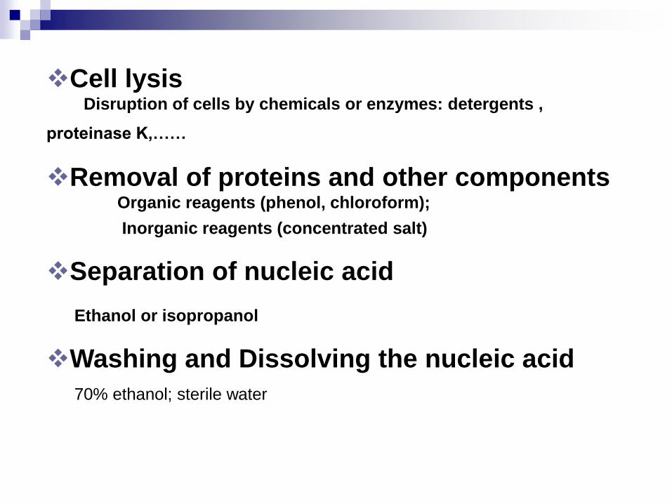
Cell lysisDisruption of cells by chemicals or enzymes: detergents ,
proteinase K,……
Removal of proteins and other componentsOrganic reagents (phenol, chloroform);
Inorganic reagents (concentrated salt)
Separation of nucleic acid
Ethanol or isopropanol
Washing and Dissolving the nucleic acid
70% ethanol; sterile water
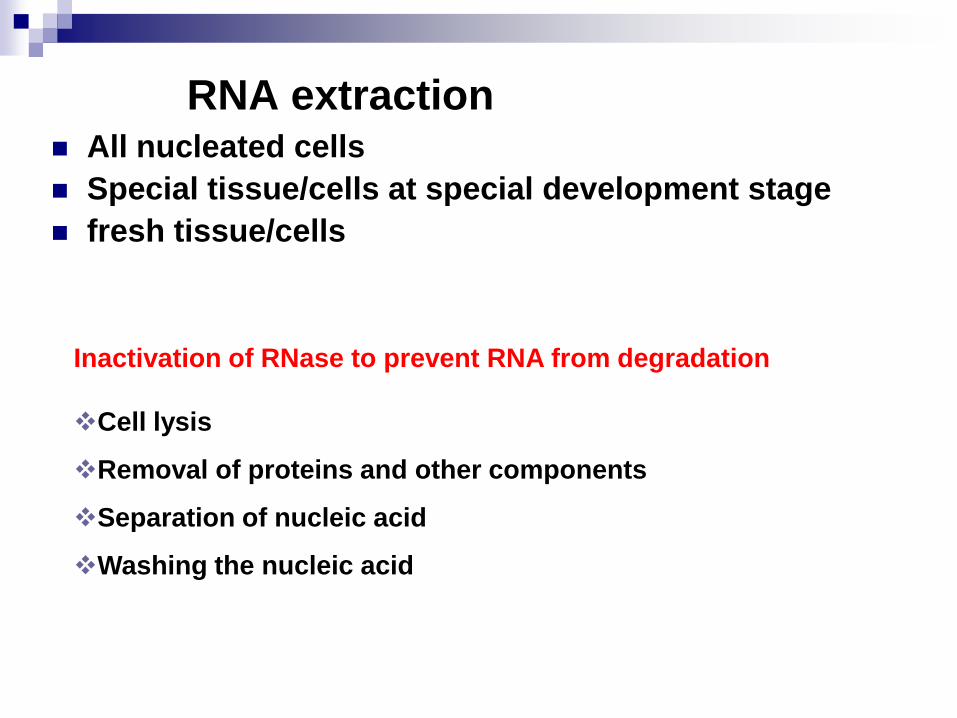
RNA extraction All nucleated cells
Special tissue/cells at special development stage
fresh tissue/cells
Cell lysis
Removal of proteins and other components
Separation of nucleic acid
Washing the nucleic acid
Inactivation of RNase to prevent RNA from degradation
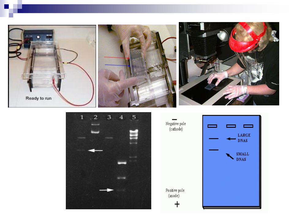
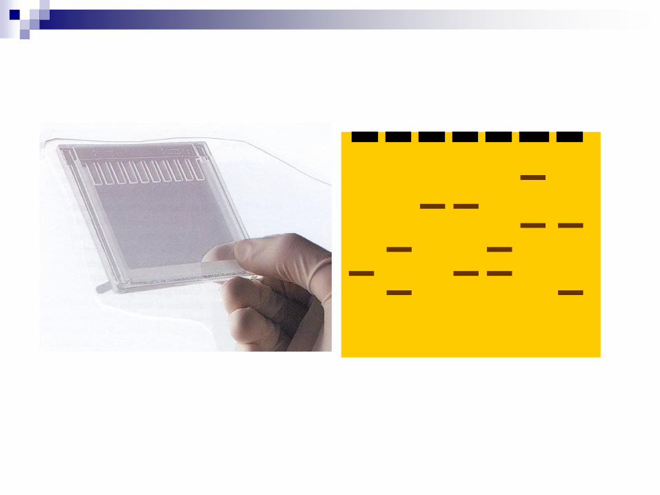
2.Gel electrophoresis
Gel electrophoresis is a procedure for
separating a mixture of molecules through a
stationary material (gel) in an electrical field.
Electromotive force moves charged
molecules through a porous gel.
Separates molecules from each other on the
basis of size, electric charge , other physical
properties.
Separation depends on how the sample and
gel are prepared.
Gel
Agarose gel:a reversible matrix
Acrylamide gel:a permanent matrix
Translucent, porous, colloid, solid materials
Very stable
Pore size can also be controlled.
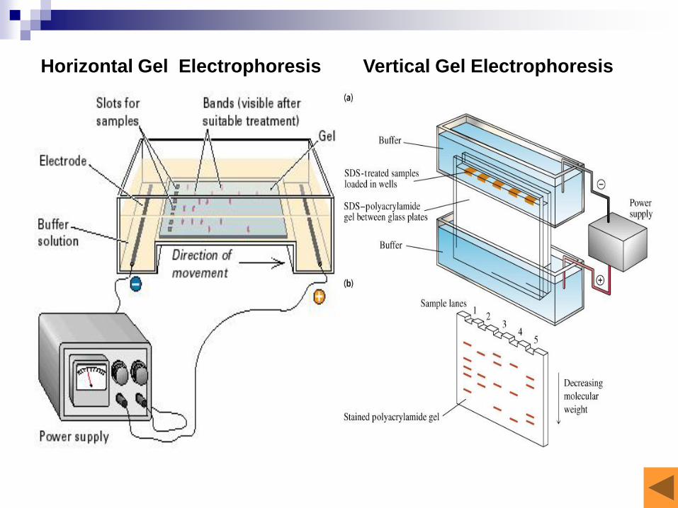
Vertical Gel ElectrophoresisHorizontal Gel Electrophoresis
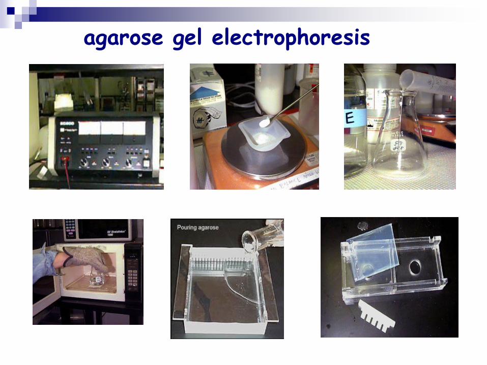
agarose gel electrophoresis
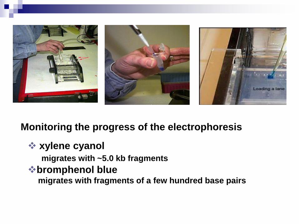
xylene cyanol
migrates with ~5.0 kb fragments
bromphenol bluemigrates with fragments of a few hundred base pairs
Monitoring the progress of the electrophoresis
Polyacrylamide Gel Electrophoresis(PAGE)
Polyacrylamide has a smaller pore size than agarose, so it can
Resolve short ssDNA of the same length that differ in sequence.
Resolve short fragments of dsDNA that differ in length by only a few oligonucleotides.
Resolve fragments of denatured ssDNA that differ in length by only a single nucleotide.
Resolve proteins.
3. Molecular Hybridization
Definition
Membrane hybridization
Southern blotting: DNA
Northern blotting: RNA
Dot/slot blotting
Western Blotting
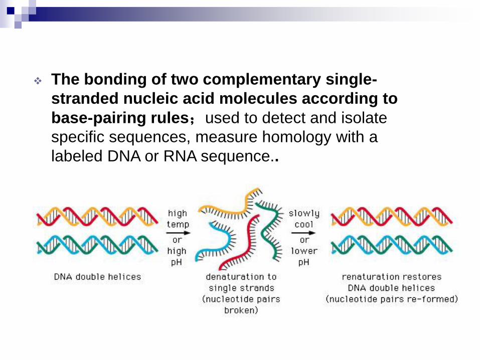
The bonding of two complementary single-
stranded nucleic acid molecules according to
base-pairing rules;used to detect and isolate
specific sequences, measure homology with a
labeled DNA or RNA sequence..
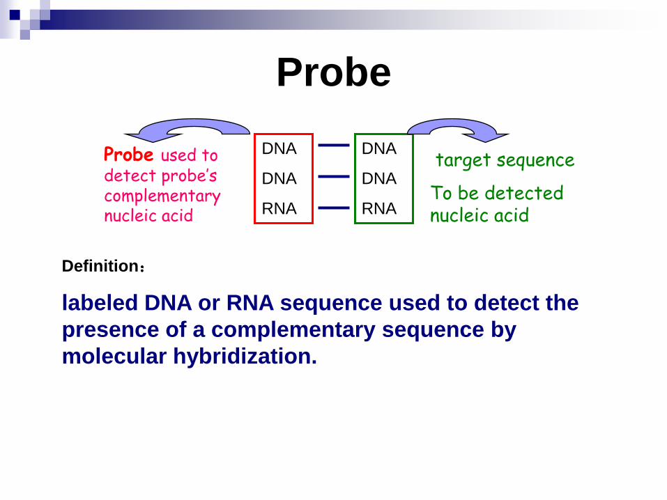
Probe used to detect probe’s complementary nucleic acid
target sequence
To be detected nucleic acid
DNA
DNA
RNA
DNA
DNA
RNA
Probe
Definition:
labeled DNA or RNA sequence used to detect the
presence of a complementary sequence by
molecular hybridization.
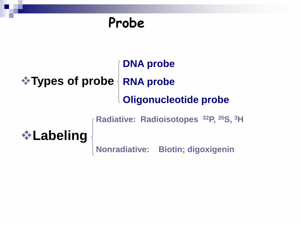
Probe
Types of probe
Labeling
Radiative: Radioisotopes 32P, 35S, 3H
Nonradiative: Biotin; digoxigenin
DNA probe
RNA probe
Oligonucleotide probe
Type: DNA
Origin: Cell-based DNA cloning or PCR
Characteristics: double-stranded
Labeling: DNA polymerase-based DNA strand synthesis
DNA Probes
RNA probes
Type : RNA
Origin: Transcription from insert DNA cloned in
suitable vectors
Characteristics: single-stranded,
Labeling: RNA polymerase-based RNA synthesis
Oligonucleotides probe
Type: oligonucleotide
Origin: chemical synthesis
Characteristics: single-stranded, 15-50nt long
Labeling: end-labeling by polynucleotide kinase
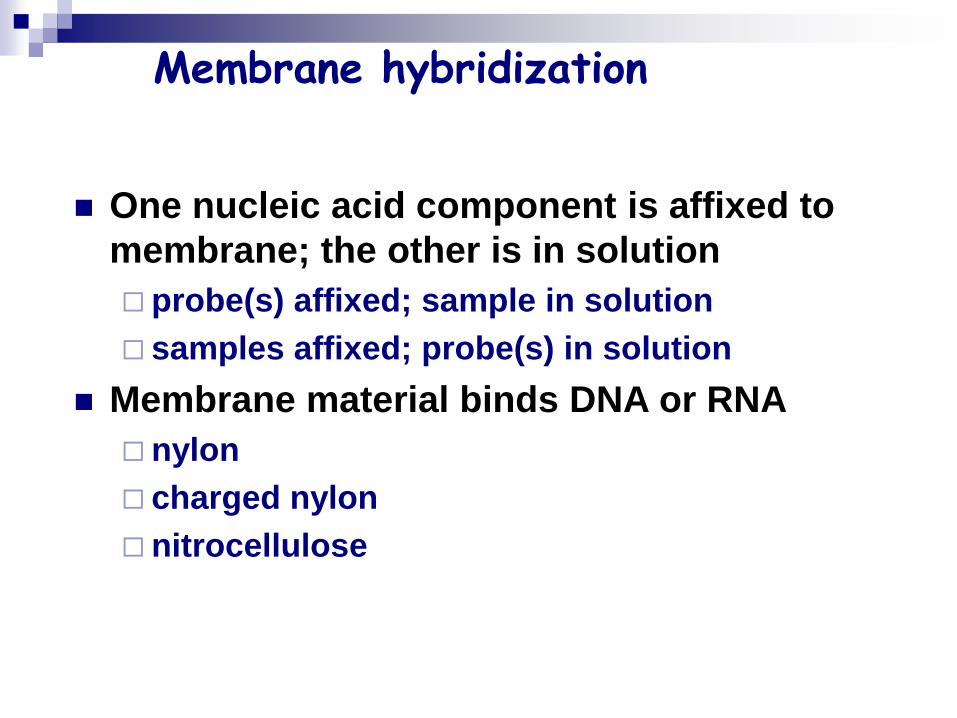
Membrane hybridization
One nucleic acid component is affixed to
membrane; the other is in solution
probe(s) affixed; sample in solution
samples affixed; probe(s) in solution
Membrane material binds DNA or RNA
nylon
charged nylon
nitrocellulose
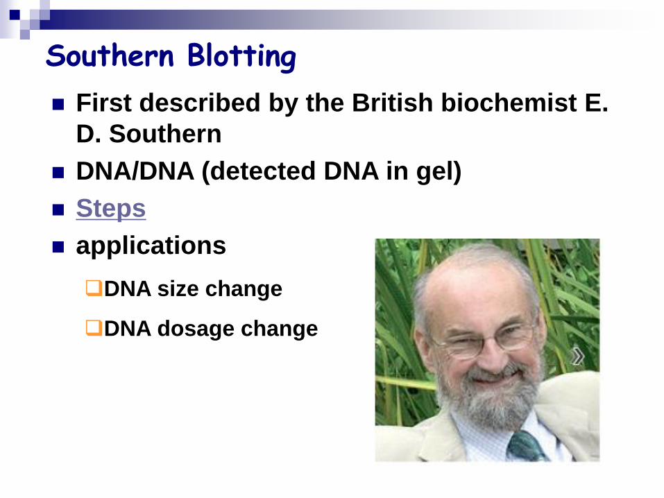
Southern Blotting
First described by the British biochemist E.
D. Southern
DNA/DNA (detected DNA in gel)
Steps
applications
DNA size change
DNA dosage change
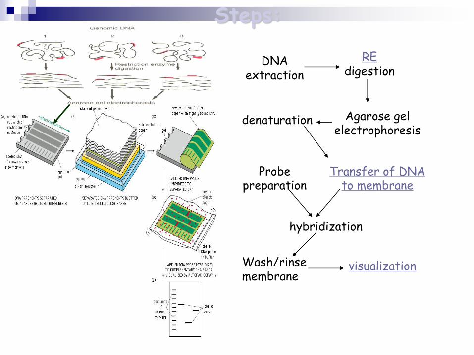
Steps:
DNA extraction
REdigestion
Agarose gel electrophoresis
Transfer of DNA to membrane
Probe preparation
hybridization
Wash/rinse membrane
visualization
denaturation
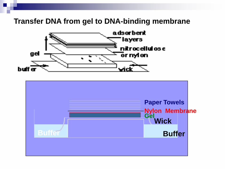
Paper Towels
Nylon Membrane
Buffer Buffer
GelWick
Transfer DNA from gel to DNA-binding membrane
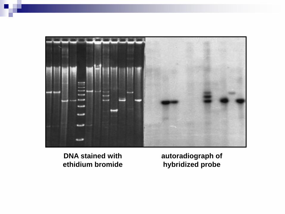
DNA stained with
ethidium bromide
autoradiograph of
hybridized probe
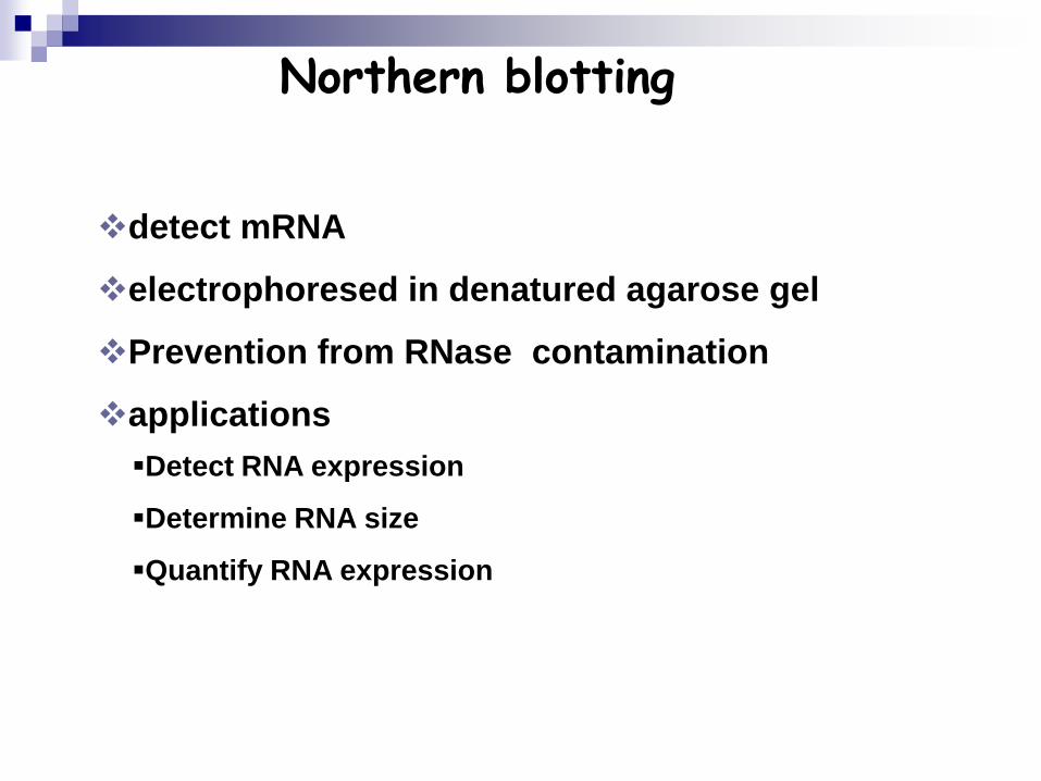
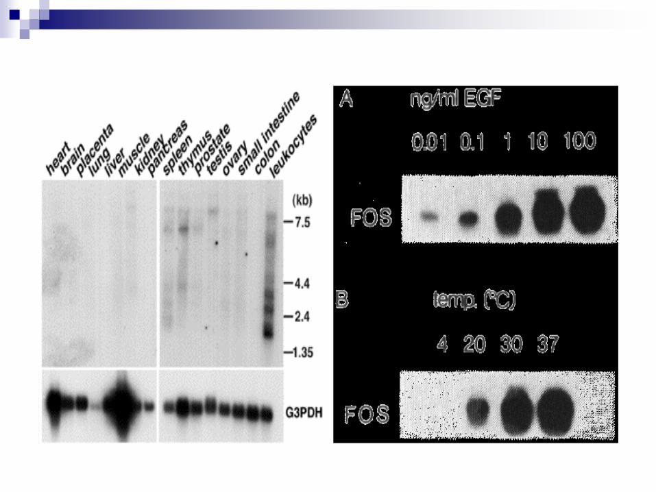
Northern blotting
detect mRNA
electrophoresed in denatured agarose gel
Prevention from RNase contamination
applications
Detect RNA expression
Determine RNA size
Quantify RNA expression
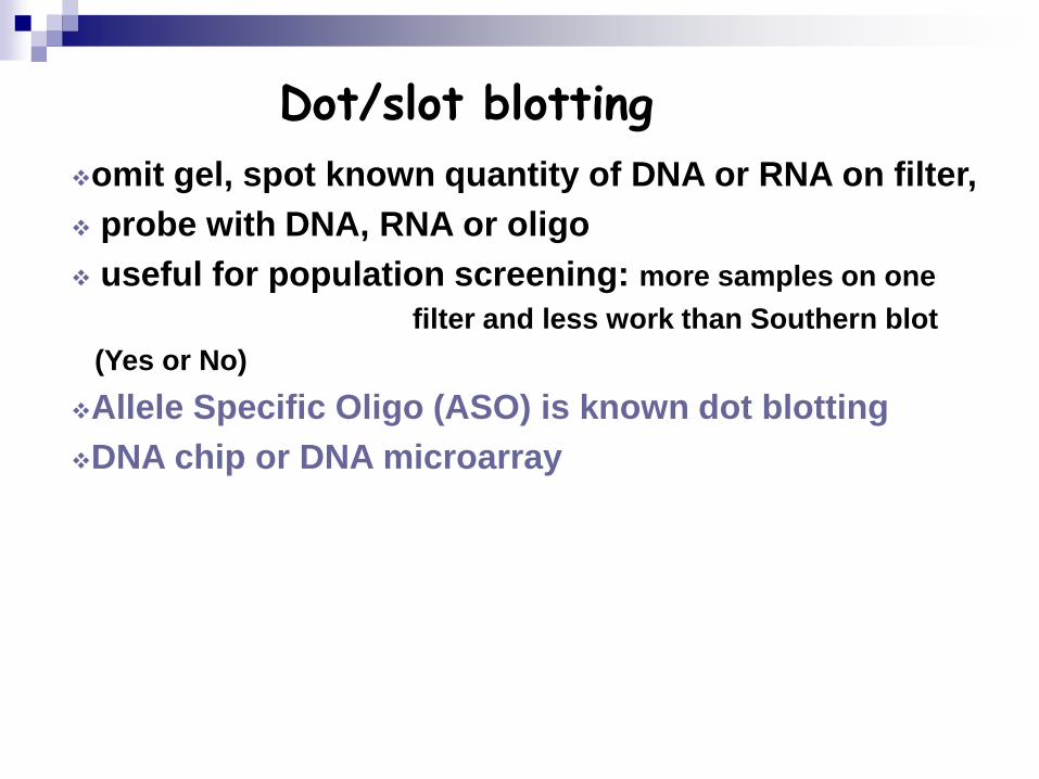
Dot/slot blotting
omit gel, spot known quantity of DNA or RNA on filter,
probe with DNA, RNA or oligo
useful for population screening: more samples on one
filter and less work than Southern blot
(Yes or No)
Allele Specific Oligo (ASO) is known dot blotting
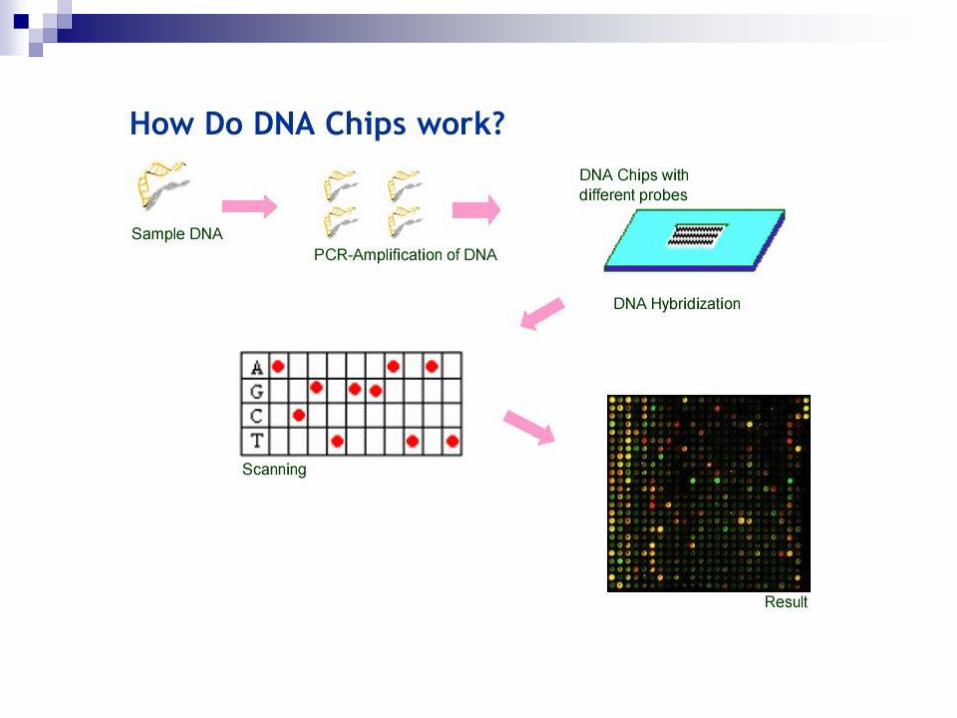
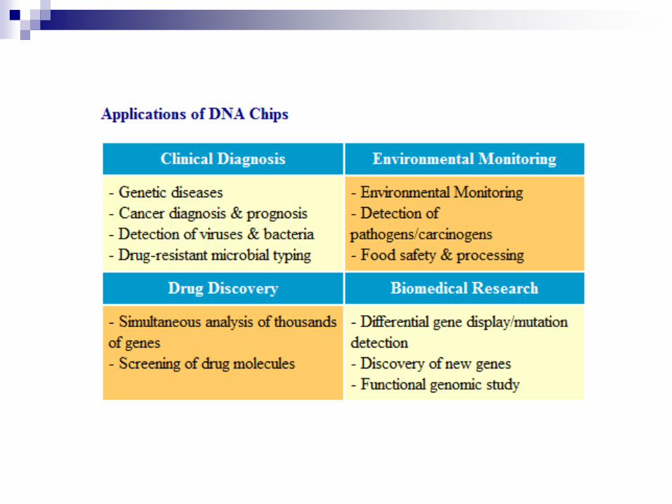
DNA chip or DNA microarray
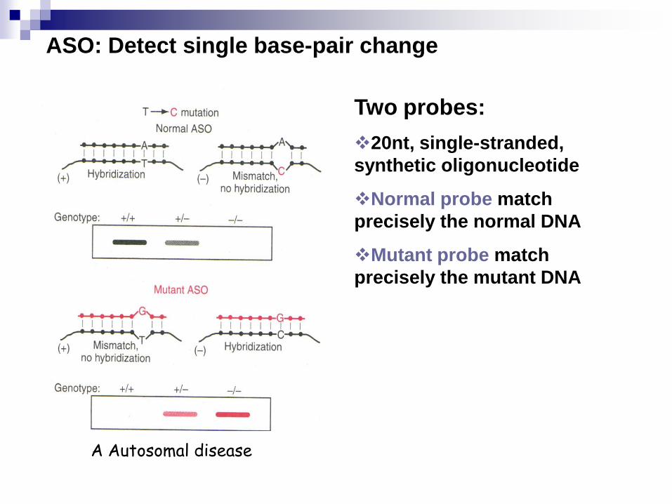
Two probes:
20nt, single-stranded,
synthetic oligonucleotide
Normal probe match
precisely the normal DNA
Mutant probe match
precisely the mutant DNA
ASO: Detect single base-pair change
A Autosomal disease
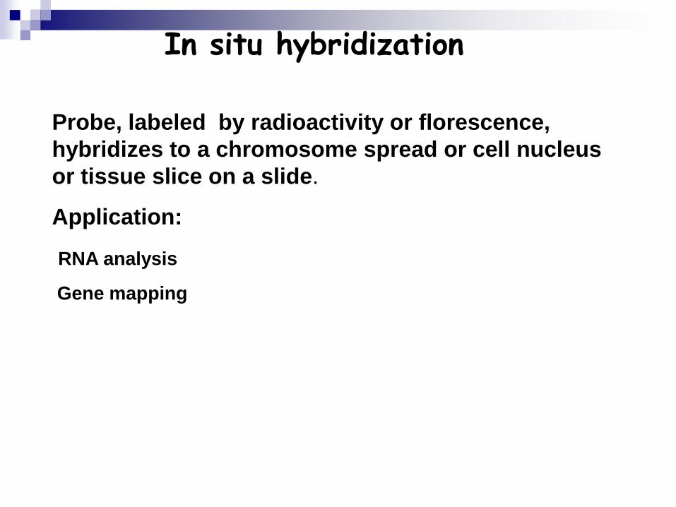
In situ hybridization
Probe, labeled by radioactivity or florescence,
hybridizes to a chromosome spread or cell nucleus
or tissue slice on a slide.
Application:
RNA analysis
Gene mapping
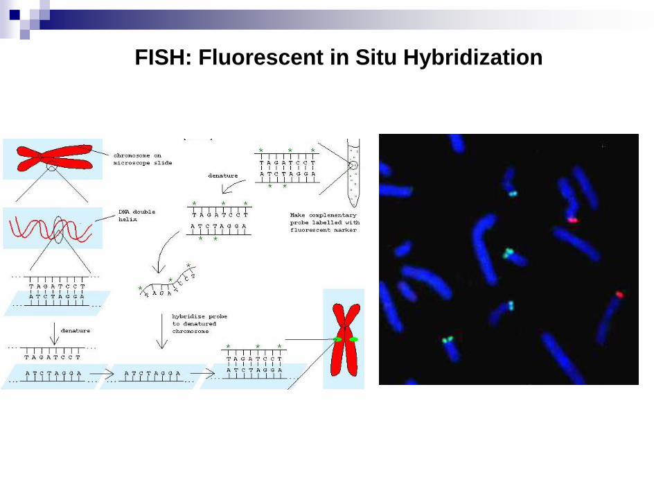
FISH: Fluorescent in Situ Hybridization
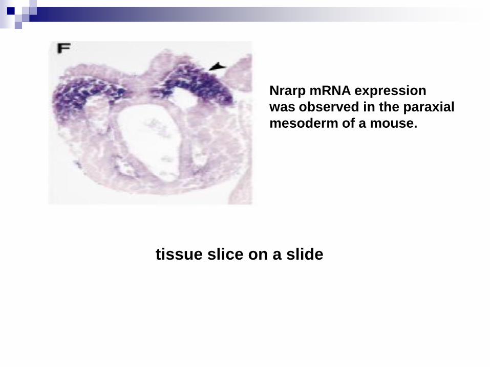
Nrarp mRNA expression
was observed in the paraxial
mesoderm of a mouse.
tissue slice on a slide
Western Blotting
Proteins from cells are separated based on their size using an
electric current. An antibody to the protein of interest is used
to determine whether that protein is present in the cells.
SDS-PAGE
electroblotting :The transfer of separated proteins from
polyacrylamide gel onto a membrane .
Protein and Antibody
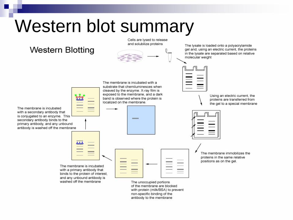
Western blot summary
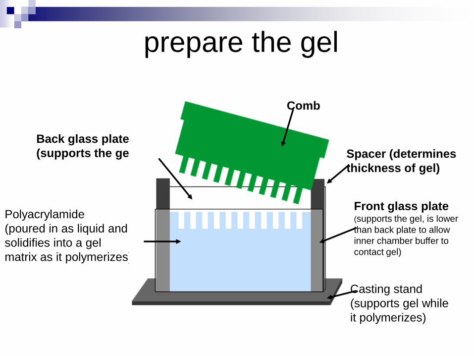
prepare the gel
Back glass plate
(supports the gel) Spacer (determines
thickness of gel)
Front glass plate(supports the gel, is lower
than back plate to allow
inner chamber buffer to
contact gel)
Casting stand
(supports gel while
it polymerizes)
Polyacrylamide
(poured in as liquid and
solidifies into a gel
matrix as it polymerizes)
Comb
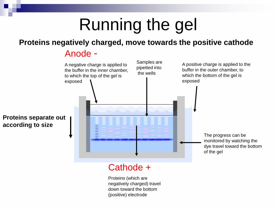
Running the gel
Anode -A negative charge is applied to
the buffer in the inner chamber,
to which the top of the gel is
exposed
A positive charge is applied to the
buffer in the outer chamber, to
which the bottom of the gel is
exposed
Cathode +Proteins (which are
negatively charged) travel
down toward the bottom
(positive) electrode
The progress can be
monitored by watching the
dye travel toward the bottom
of the gel
Proteins separate out
according to size
Proteins negatively charged, move towards the positive cathode
Samples are
pipetted into
the wells
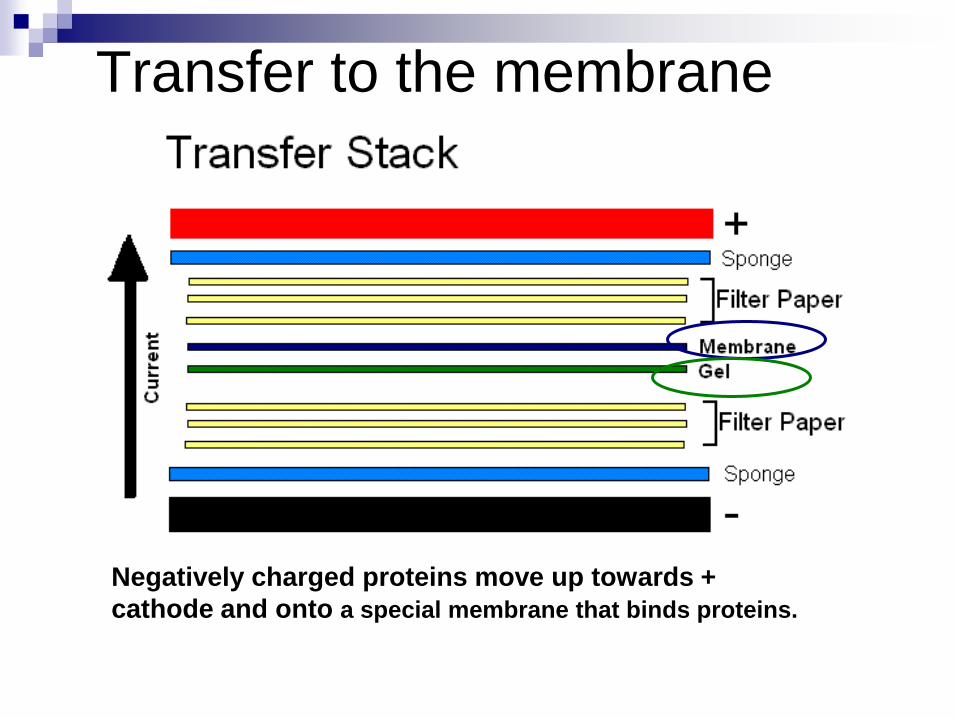
Transfer to the membrane
Negatively charged proteins move up towards +
cathode and onto a special membrane that binds proteins.
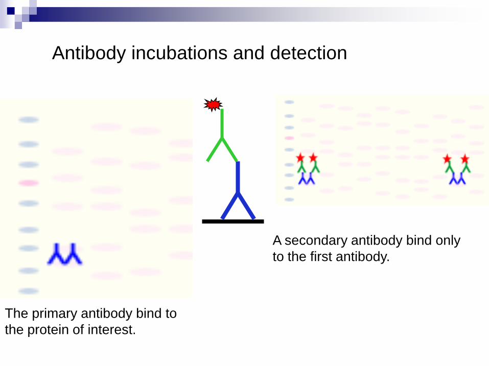
Antibody incubations and detection
The primary antibody bind to
the protein of interest.
A secondary antibody bind only
to the first antibody.
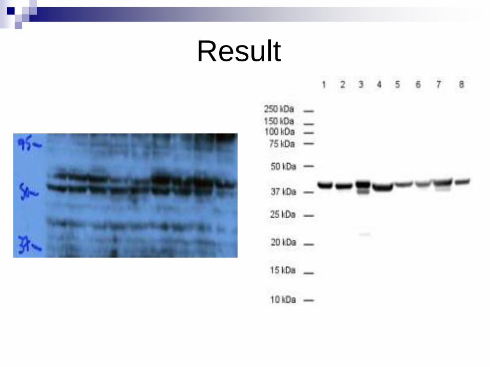
Result
4.Polymerase Chain Reaction
(PCR)
Introduction
Cycles
Essential reagent
Process
application
Described in 1985 by Kary Mullis
Nobel Prize in 1993
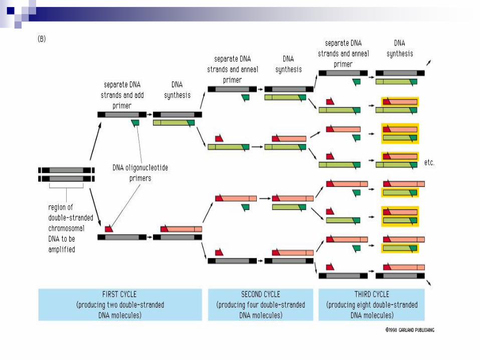
A simple rapid, sensitive and versatile in vitro
method for selectively amplifying defined
sequences/regions of DNA/RNA from an initial
complex source of nucleic acid by means of two
flanking oligonucleotide primers used in repeated
cycles -generates sufficient product for
subsequent analysis and/or manipulation.
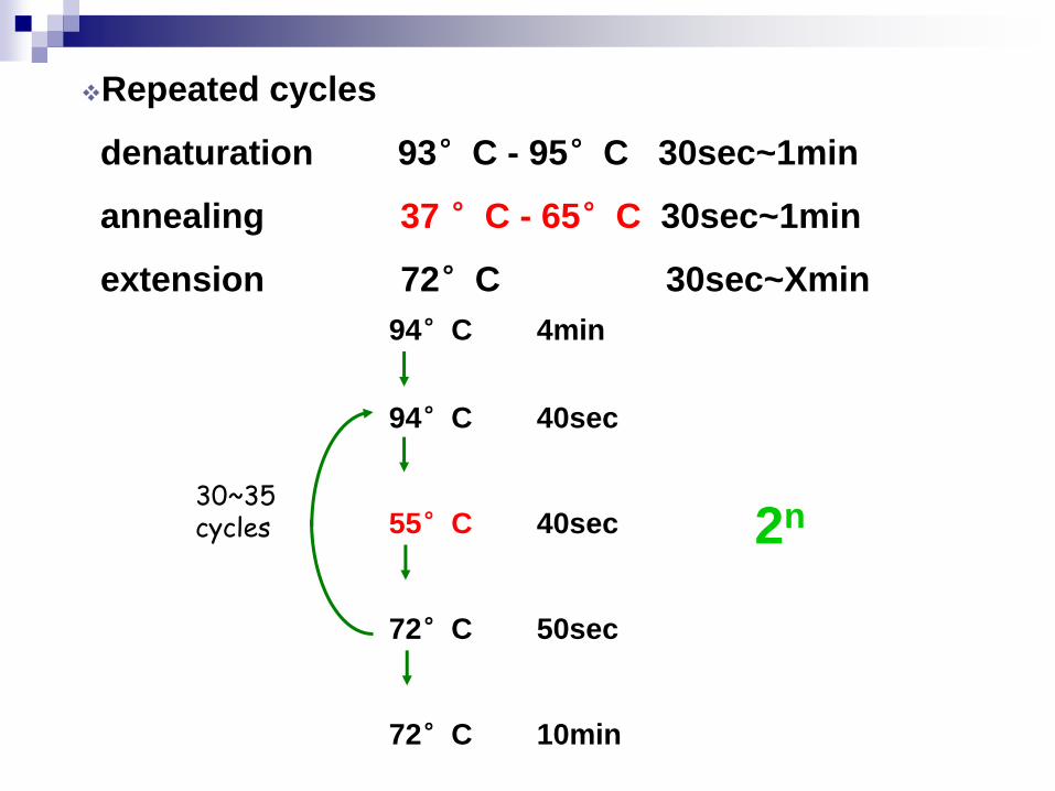
Repeated cycles
denaturation 93°C - 95°C 30sec~1min
annealing 37 °C - 65°C 30sec~1min
extension 72°C 30sec~Xmin
94°C 4min
94°C 40sec
55°C 40sec
72°C 50sec
72°C 10min
30~35 cycles 2n
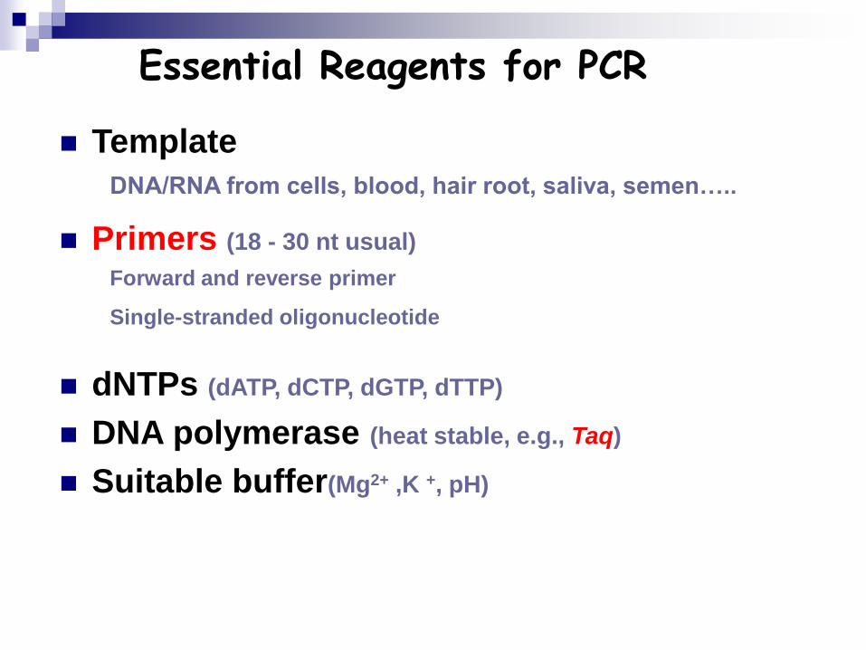
Essential Reagents for PCR
Template
Primers (18 - 30 nt usual)
dNTPs (dATP, dCTP, dGTP, dTTP)
DNA polymerase (heat stable, e.g., Taq)
Suitable buffer(Mg2+ ,K +, pH)
DNA/RNA from cells, blood, hair root, saliva, semen…..
Forward and reverse primer
Single-stranded oligonucleotide
•
Application of PCR
Cloning of genes or gene fragments
Genetic diagnosis - Mutation detection
Paternity testing( STR,VNTR)
Mutagenesis to investigate protein function
Quantitate differences in gene expression
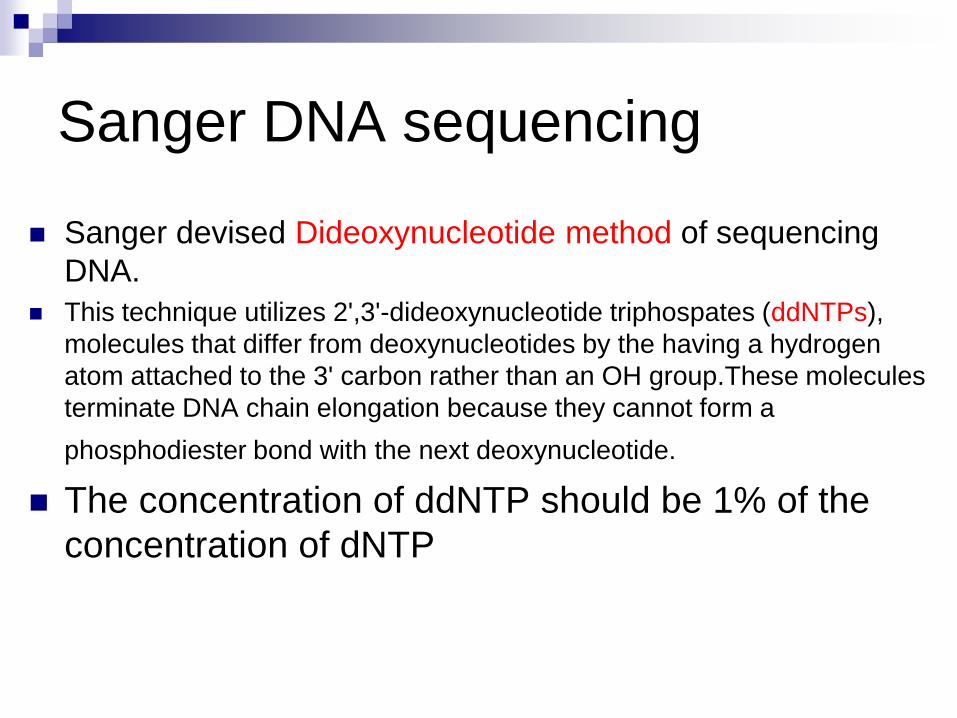
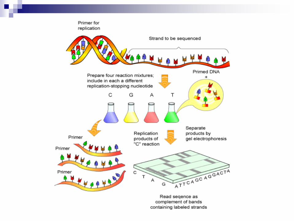
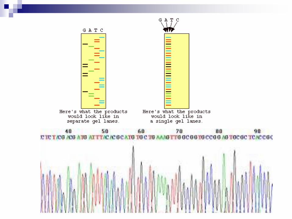
Sanger DNA sequencing
Sanger devised Dideoxynucleotide method of sequencing
DNA.
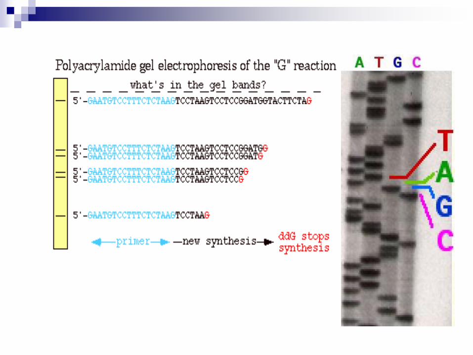
This technique utilizes 2',3'-dideoxynucleotide triphospates (ddNTPs),
molecules that differ from deoxynucleotides by the having a hydrogen
atom attached to the 3' carbon rather than an OH group.These molecules
terminate DNA chain elongation because they cannot form a
phosphodiester bond with the next deoxynucleotide.
The concentration of ddNTP should be 1% of the
concentration of dNTP
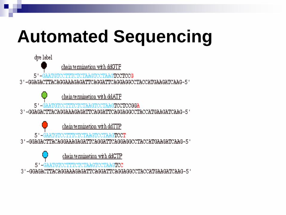
Automated Sequencing
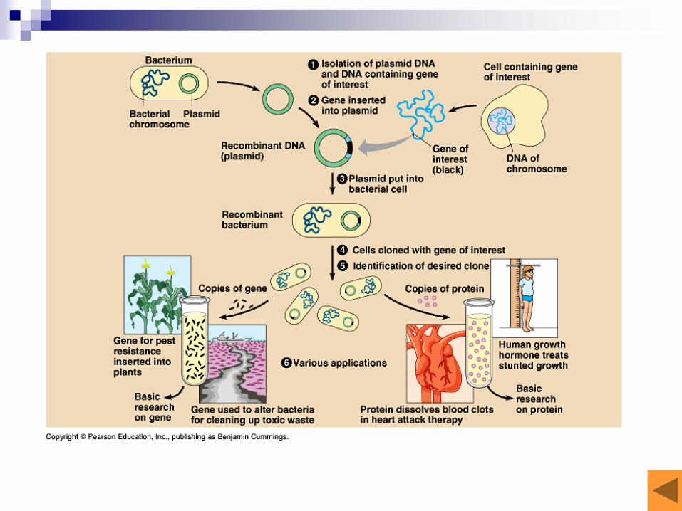
6. Recombinant DNA technology
Definition
Recombinant DNA Tool Kit
Process
Applications
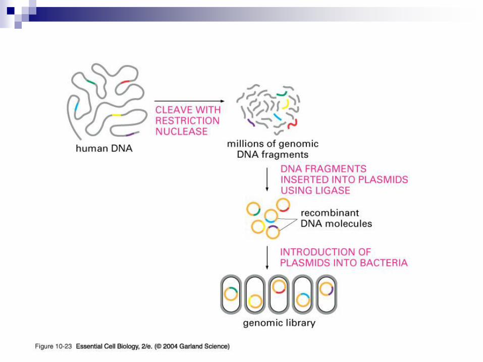
genomic library
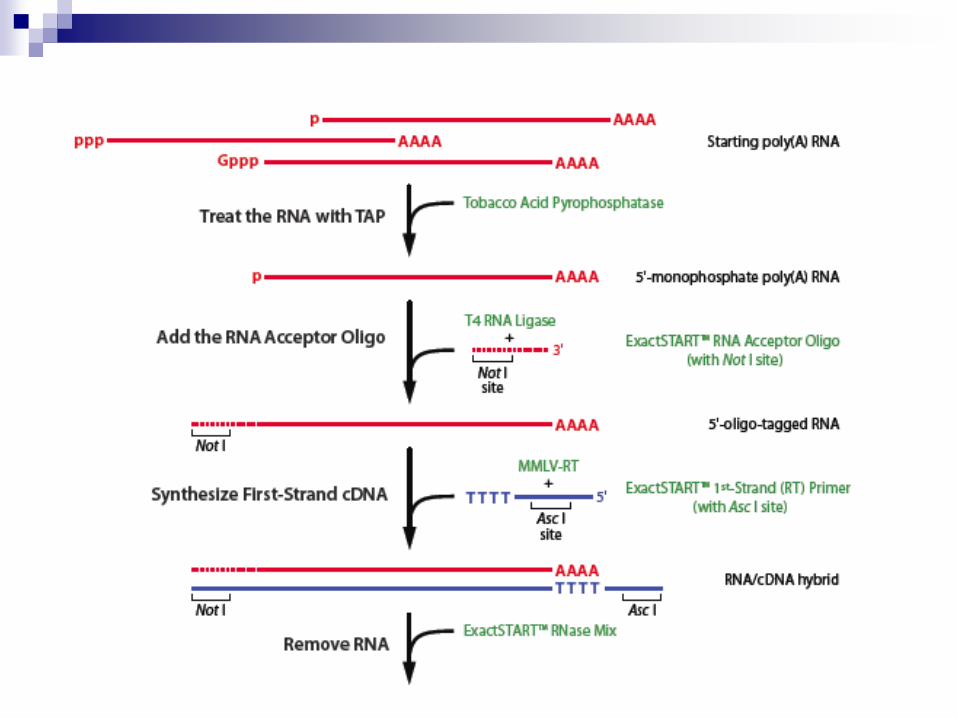
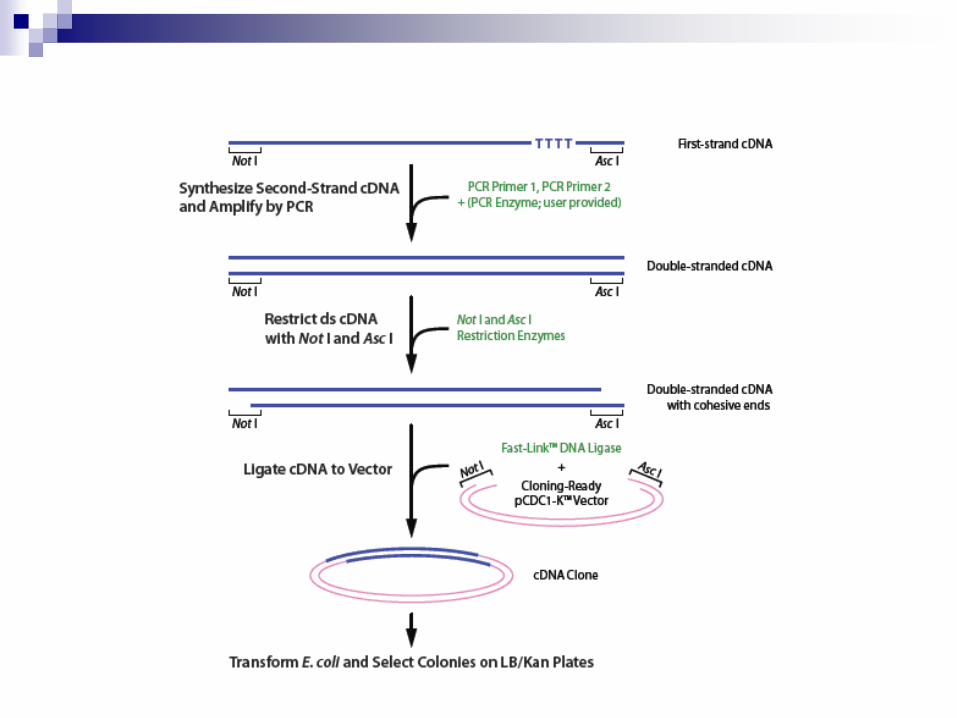
cDNA library
Cell-based DNA amplification
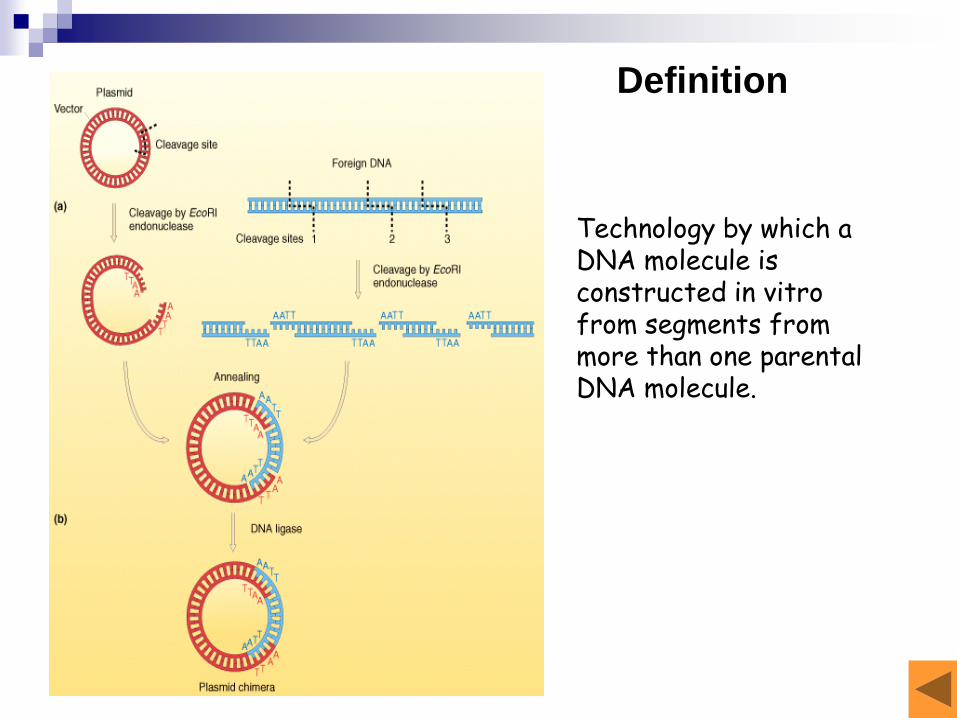
Technology by which a DNA molecule is constructed in vitro from segments from more than one parental DNA molecule.
Definition
Recombinant DNA Tool Kit
Restriction Enzymes(RE)
Vectors (pieces of DNA that allow
maintenance in a cell)
Transformation of cells
Other enzymes that can be used in vitro -
Ligase, Polymerase, etc.
Restriction EnzymesorRestriction Endonuclease (RE)
Derived from bacteria, not human cells.
Natural role - protect the bacteria containing them
against intruding DNA from other organisms (e.g.
viruses).
Recognize a specific sequence of DNA and cut
the DNA molecule within the recognition site or at
some nearby site.
type I, II, III
Allow the production of recombinant molecules
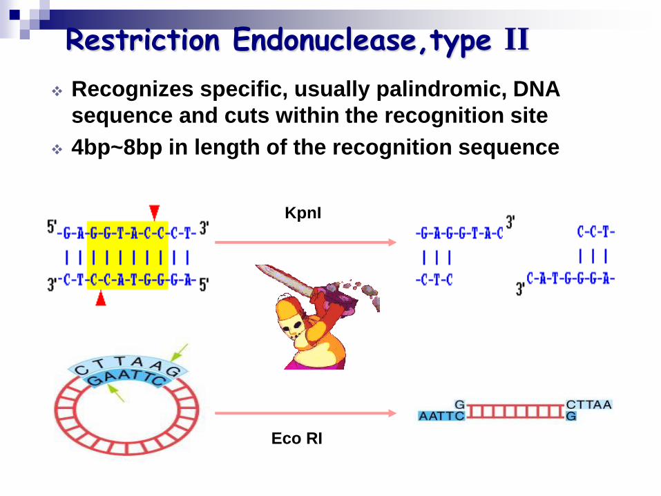
Restriction Endonuclease,type II
Recognizes specific, usually palindromic, DNA
sequence and cuts within the recognition site
4bp~8bp in length of the recognition sequence
KpnI
Eco RI
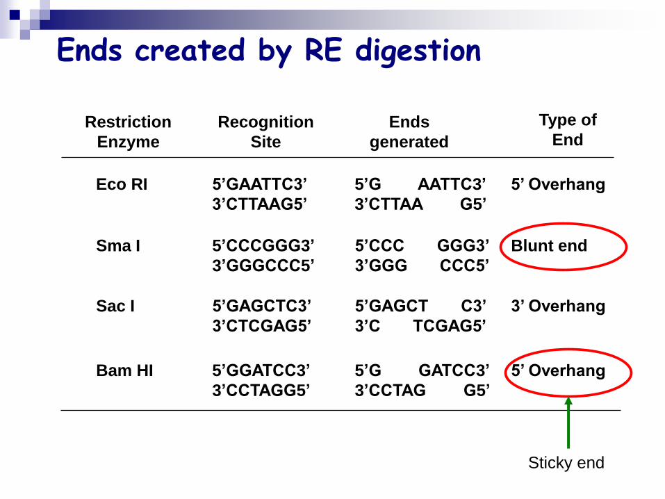
Ends created by RE digestion
Restriction
Enzyme
Recognition
Site
Ends
generated
Type of
End
Eco RI 5’GAATTC3’
3’CTTAAG5’
5’G AATTC3’
3’CTTAA G5’
5’ Overhang
Sma I 5’CCCGGG3’
3’GGGCCC5’
5’CCC GGG3’
3’GGG CCC5’
Blunt end
Sac I 5’GAGCTC3’
3’CTCGAG5’
5’GAGCT C3’
3’C TCGAG5’
3’ Overhang
Bam HI 5’GGATCC3’
3’CCTAGG5’
5’G GATCC3’
3’CCTAG G5’
5’ Overhang
Sticky end
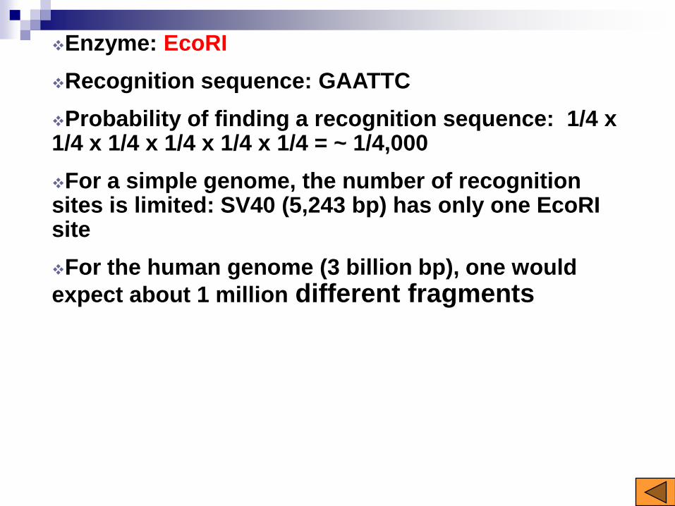
Enzyme: EcoRI
Recognition sequence: GAATTC
Probability of finding a recognition sequence: 1/4 x 1/4 x 1/4 x 1/4 x 1/4 x 1/4 = ~ 1/4,000
For a simple genome, the number of recognition sites is limited: SV40 (5,243 bp) has only one EcoRI site
For the human genome (3 billion bp), one would
expect about 1 million different fragments
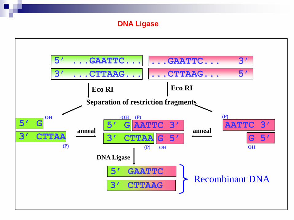
5’ ...GAATTC...
3’ ...CTTAAG...
...GAATTC... 3’
...CTTAAG... 5’
Eco RI Eco RI
Separation of restriction fragments
(P)
-OH
5’ G
3’ CTTAAOH
(P)
AATTC 3’
G 5’(P)
-OH
5’ G
3’ CTTAAOH
(P)
AATTC 3’
G 5’anneal anneal
5’ GAATTC
3’ CTTAAG
DNA Ligase
Recombinant DNA
DNA Ligase
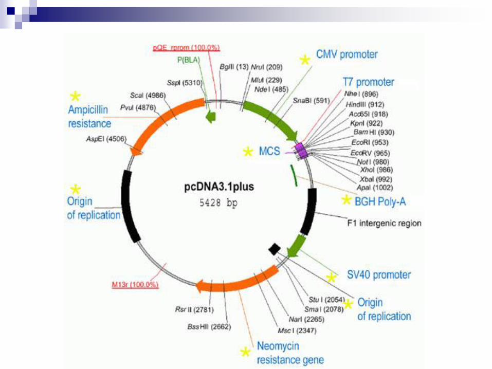
Cloning Vectors
Definition
The DNA molecule into which DNA fragment has been
cloned,capable of replicating in a special host and
thereby propagating the cloned DNA fragment.
Essential features:
a. Small size
b. Origin of replication or promoter
c. Dominant selectable marker
d. Single restriction sites
Vector Type
Plasmid
Bacteriophage lambda
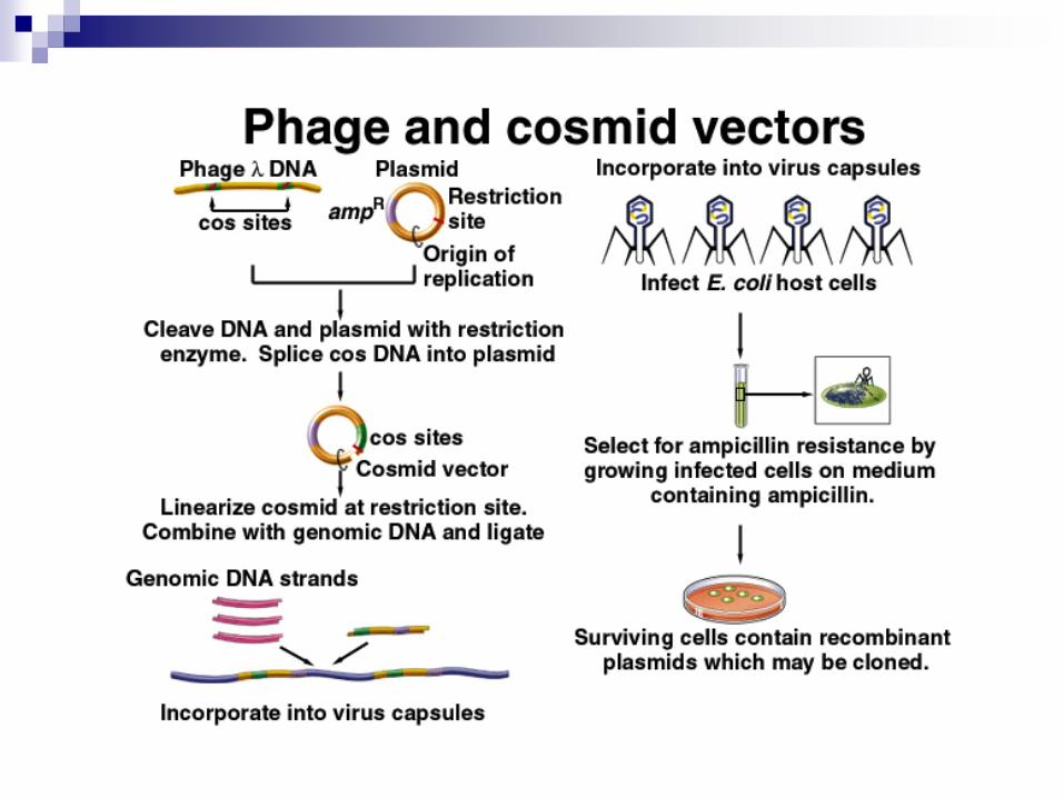
Cosmid
BAC(bacterial artificial chromosome)
YAC (yest artificial chromosome)
Cloning vector: DNA propagation
Expression vector: protein
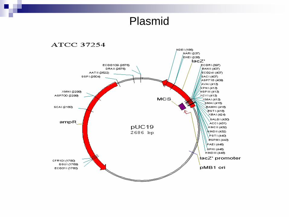
Plasmid
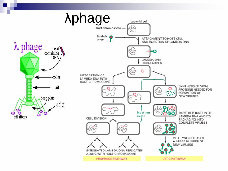
λphage
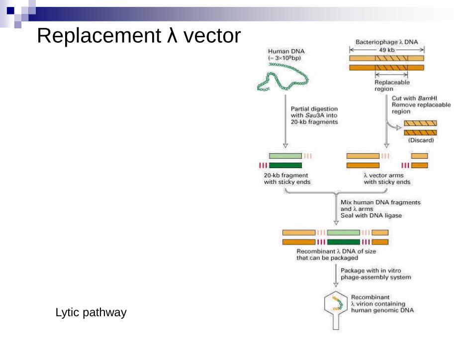
Replacement λ vector
Lytic pathway
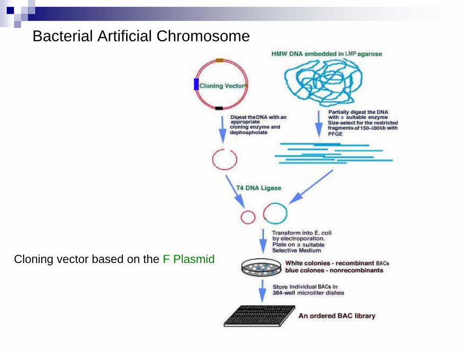
Bacterial Artificial Chromosome
Cloning vector based on the F Plasmid
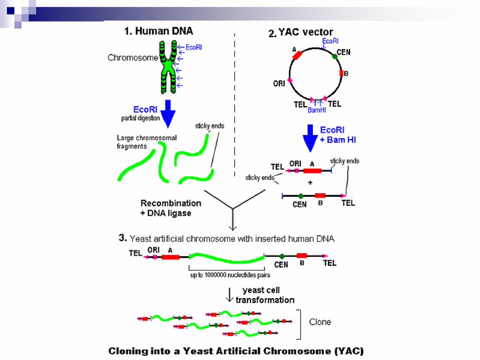
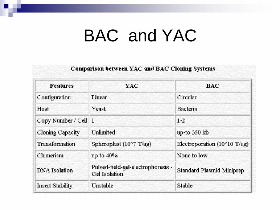
BAC and YAC
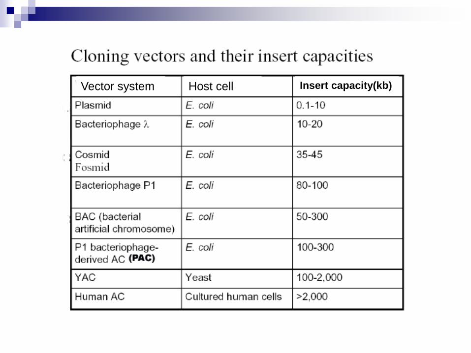
Vector system Host cell Insert capacity(kb)
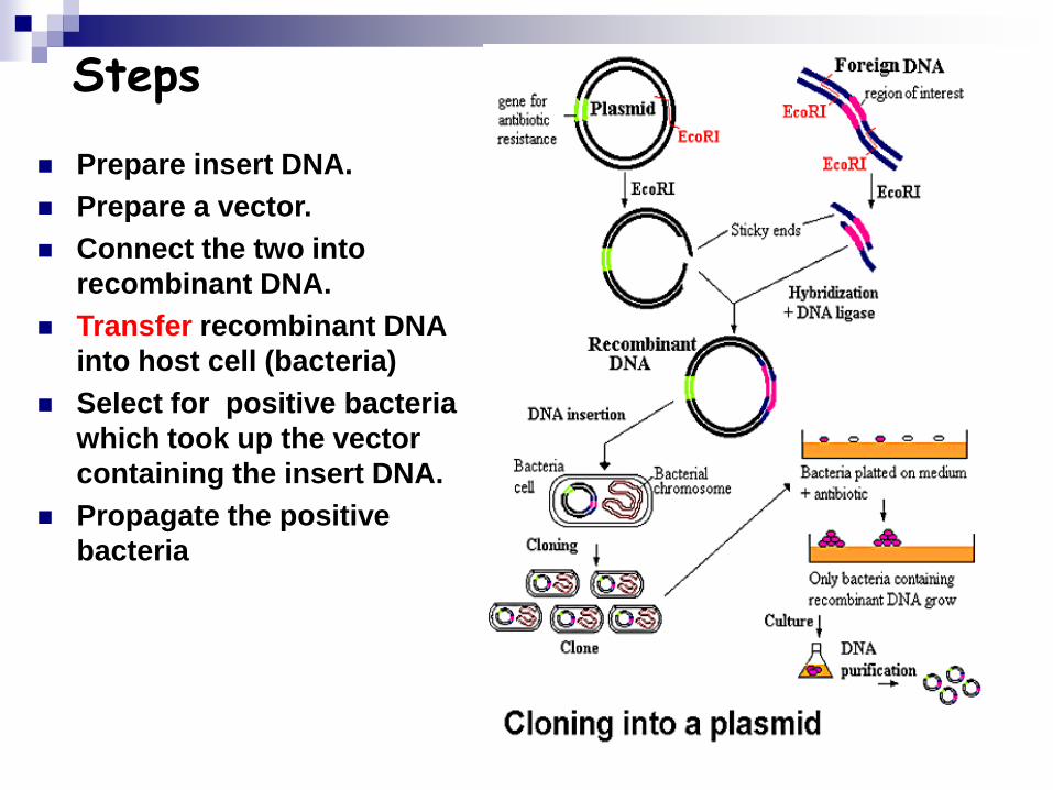
Steps
Prepare insert DNA.
Prepare a vector.
Connect the two into
recombinant DNA.
Transfer recombinant DNA
into host cell (bacteria)
Select for positive bacteria
which took up the vector
containing the insert DNA.
Propagate the positive
bacteria
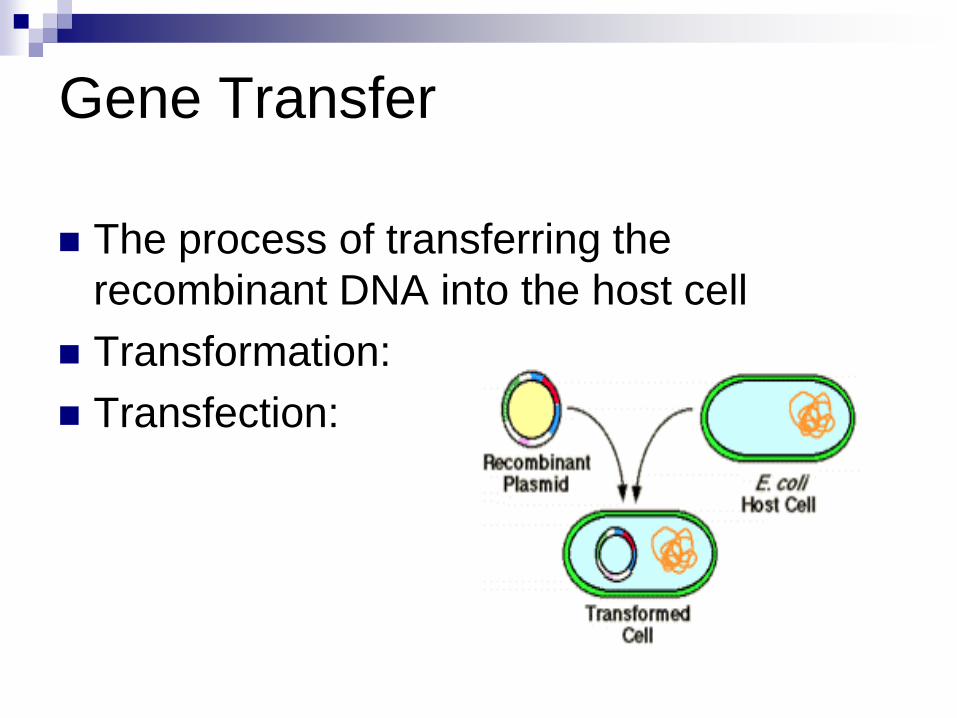
Gene Transfer
The process of transferring the
recombinant DNA into the host cell
Transformation:
Transfection:
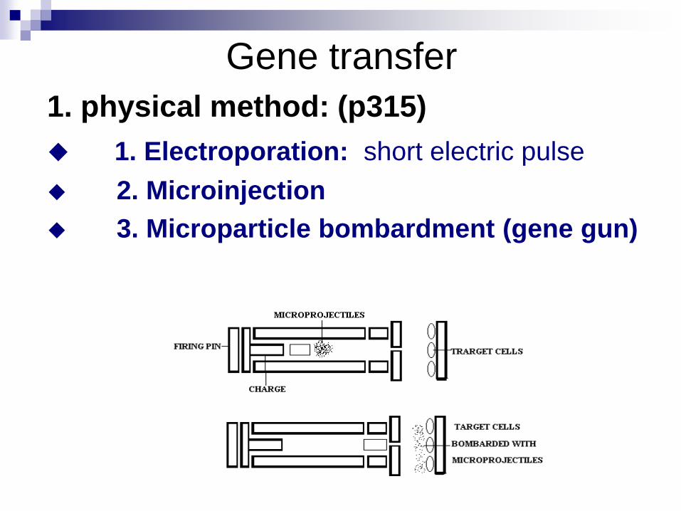
Gene transfer
1. physical method: (p315)
1. Electroporation: short electric pulse
2. Microinjection
3. Microparticle bombardment (gene gun)
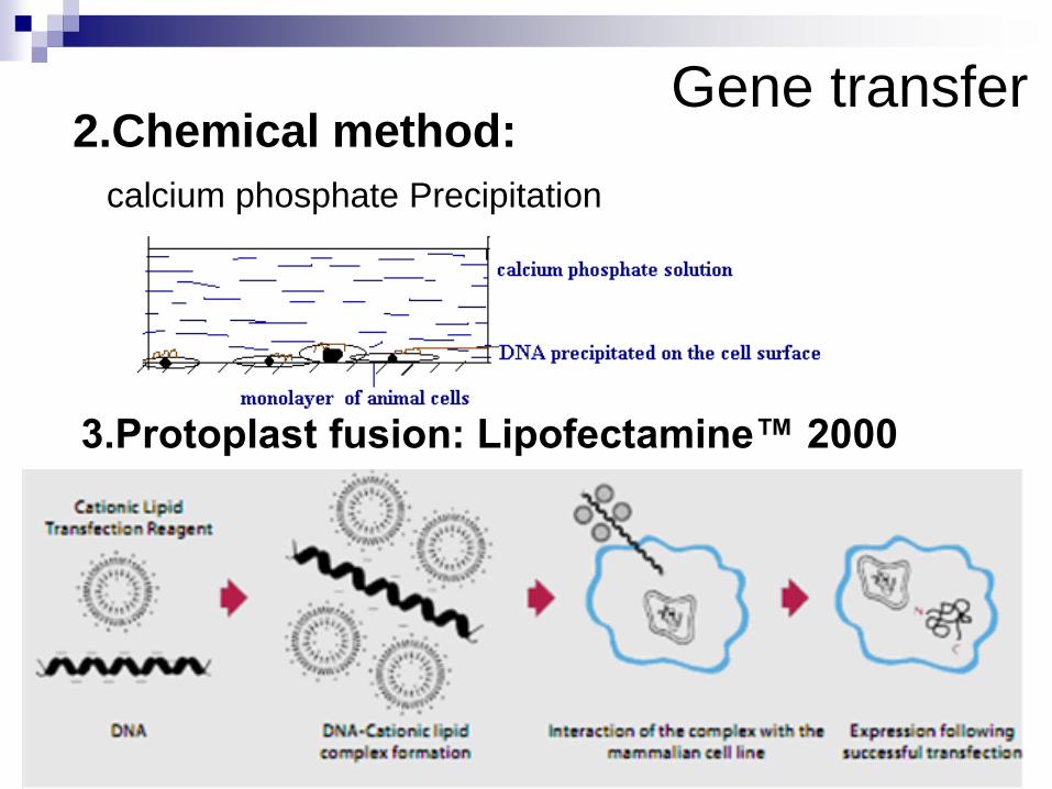
Gene transfer2.Chemical method:
calcium phosphate Precipitation
3.Protoplast fusion: Lipofectamine™ 2000
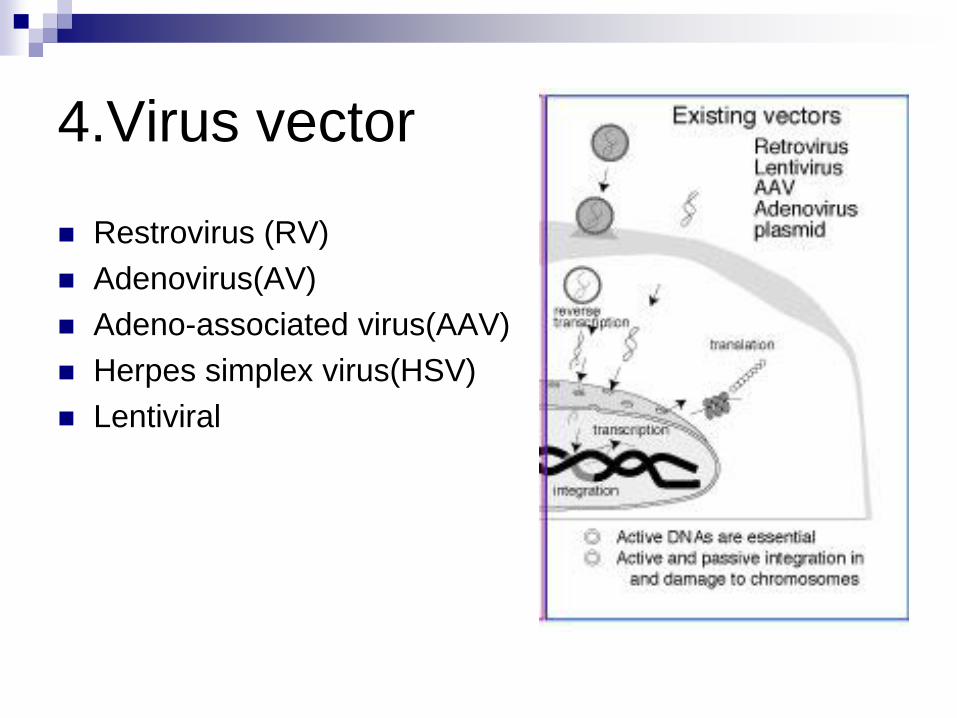
4.Virus vector
Restrovirus (RV)
Adenovirus(AV)
Adeno-associated virus(AAV)
Herpes simplex virus(HSV)
Lentiviral
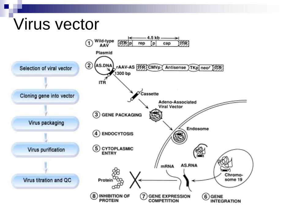
Virus vector
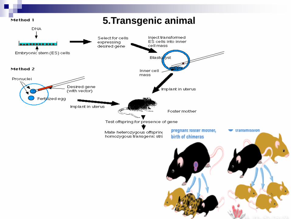
5.Transgenic animal
DNA Library Libraries are constructed by inserting a variety of
sequences into a cloning vector, and simultaneously
maintaining all of these clones; each clone
representing an individual insert.
Genomic DNA Library: randomly cut the genome into
appropriate size for vector, ligate with vector, replicate in host.
Each clone (a vector with an insert) represents a unique region
of the genome, but each region of the genome is represented
equally in overlapping clones.
cDNA Library: isolate mRNA from a cell or tissue type, convert
into DNA, and ligate with vector. Each clone represents a DNA
copy of an mRNA, and each mRNA is represented in the library
based on its level of transcription.