Embed Size (px)
Citation preview
Chapter 6Chapter 6
VisionVision
IntroductionIntroduction
Sensory receptorsSensory receptors – a specialized neuron – a specialized neuron that detects a particular category of that detects a particular category of physical eventsphysical events
Sensory transductionSensory transduction – the process by – the process by which sensory stimuli are transduced into which sensory stimuli are transduced into slow, graded slow, graded receptor potentialsreceptor potentials
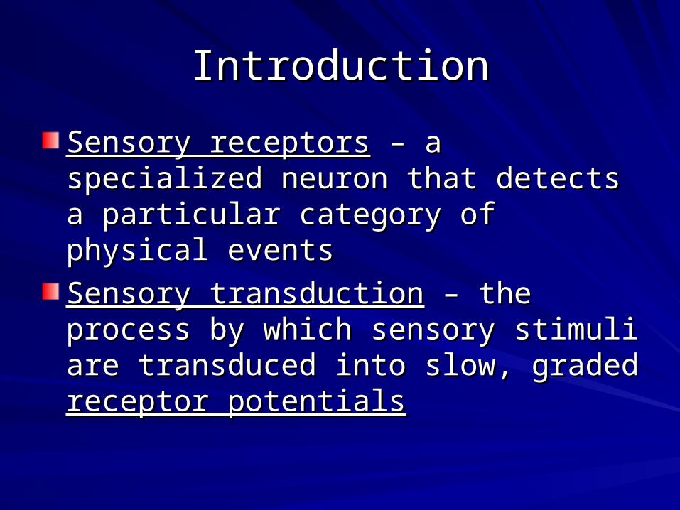
The StimulusThe Stimulus
The perceived color of light is determined by 3 The perceived color of light is determined by 3 dimensions:dimensions:– HueHue – the dominant wavelength – the dominant wavelength– SaturationSaturation - purity - purity– BrightnessBrightness - intensity - intensity
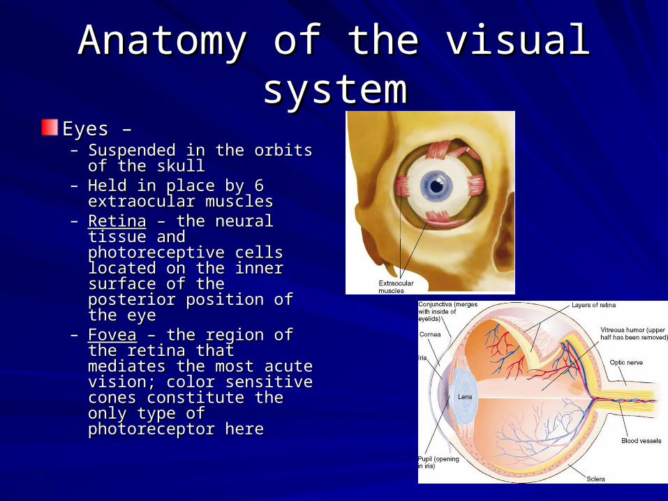
Anatomy of the visual systemAnatomy of the visual system
Eyes –Eyes –– Suspended in the orbits of Suspended in the orbits of
the skullthe skull– Held in place by 6 Held in place by 6
extraocular musclesextraocular muscles– RetinaRetina – the neural tissue – the neural tissue
and photoreceptive cells and photoreceptive cells located on the inner located on the inner surface of the posterior surface of the posterior position of the eyeposition of the eye
– FoveaFovea – the region of the – the region of the retina that mediates the retina that mediates the most acute vision; color most acute vision; color sensitive cones constitute sensitive cones constitute the only type of the only type of photoreceptor herephotoreceptor here
Anatomy of the visual systemAnatomy of the visual system
EyesEyes– PhotoreceptorPhotoreceptor – one of the receptor cells of the retina; – one of the receptor cells of the retina;
transduces photic energy into electrical potentialstransduces photic energy into electrical potentialsRodRod – sensitive to light of low intensity – sensitive to light of low intensityConesCones – maximally sensitive to one of 3 different wavelengths of – maximally sensitive to one of 3 different wavelengths of light and hence encodes color visionlight and hence encodes color vision
– Optic diskOptic disk – the location of the exit point from the retina of the – the location of the exit point from the retina of the fibers of the ganglion cells that form the optic nerve; responsible fibers of the ganglion cells that form the optic nerve; responsible for the blind spotfor the blind spot
– 3 types of movements:3 types of movements:Vergence movementsVergence movements – the cooperative movements that keep both – the cooperative movements that keep both eyes fixed on the same targeteyes fixed on the same targetSaccadic movementsSaccadic movements – rapid, jerky movements of the eyes used in – rapid, jerky movements of the eyes used in scanning a visual scenescanning a visual scenePursuit movementPursuit movement – the movement made to maintain an image of a – the movement made to maintain an image of a moving object on the foveamoving object on the fovea
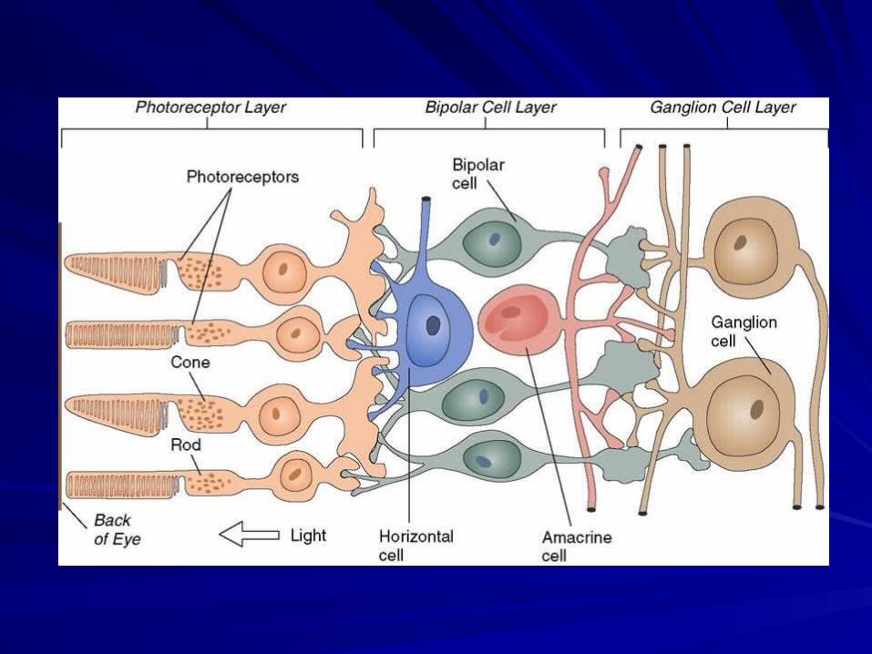
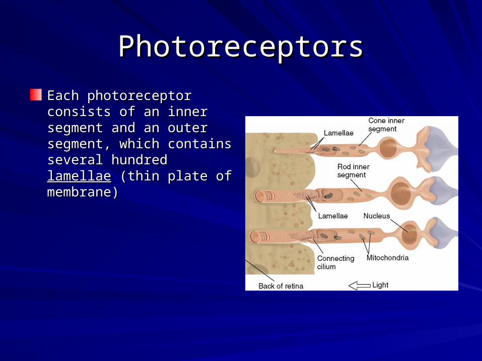
PhotoreceptorsPhotoreceptors
Each photoreceptor consists of Each photoreceptor consists of an inner segment and an outer an inner segment and an outer segment, which contains segment, which contains several hundred several hundred lamellaelamellae (thin (thin plate of membrane)plate of membrane)
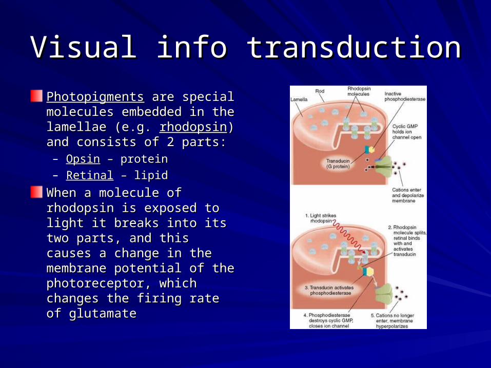
Visual info transductionVisual info transduction
PhotopigmentsPhotopigments are special are special molecules embedded in the molecules embedded in the lamellae (e.g. lamellae (e.g. rhodopsinrhodopsin) and ) and consists of 2 parts:consists of 2 parts:– OpsinOpsin – protein – protein
– RetinalRetinal – lipid – lipid
When a molecule of rhodopsin When a molecule of rhodopsin is exposed to light it breaks is exposed to light it breaks into its two parts, and this into its two parts, and this causes a change in the causes a change in the membrane potential of the membrane potential of the photoreceptor, which changes photoreceptor, which changes the firing rate of glutamatethe firing rate of glutamate
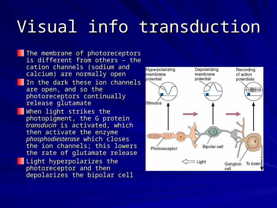
Visual info transductionVisual info transduction
The membrane of photoreceptors is The membrane of photoreceptors is different from others – the cation different from others – the cation channels (sodium and calcium) are channels (sodium and calcium) are normally opennormally openIn the dark these ion channels are In the dark these ion channels are open, and so the photoreceptors open, and so the photoreceptors continually release glutamatecontinually release glutamateWhen light strikes the photopigment, When light strikes the photopigment, the G protein the G protein transducintransducin is activated, is activated, which then activate the enzyme which then activate the enzyme phosphodiesterasephosphodiesterase which closes the which closes the ion channels; this lowers the rate of ion channels; this lowers the rate of glutamate releaseglutamate releaseLight hyperpolarizes the Light hyperpolarizes the photoreceptor and then depolarizes photoreceptor and then depolarizes the bipolar cellthe bipolar cell
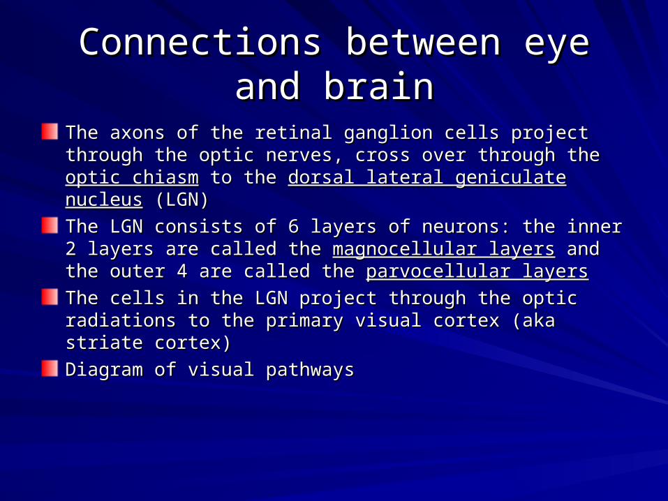
Connections between eye and Connections between eye and brainbrain
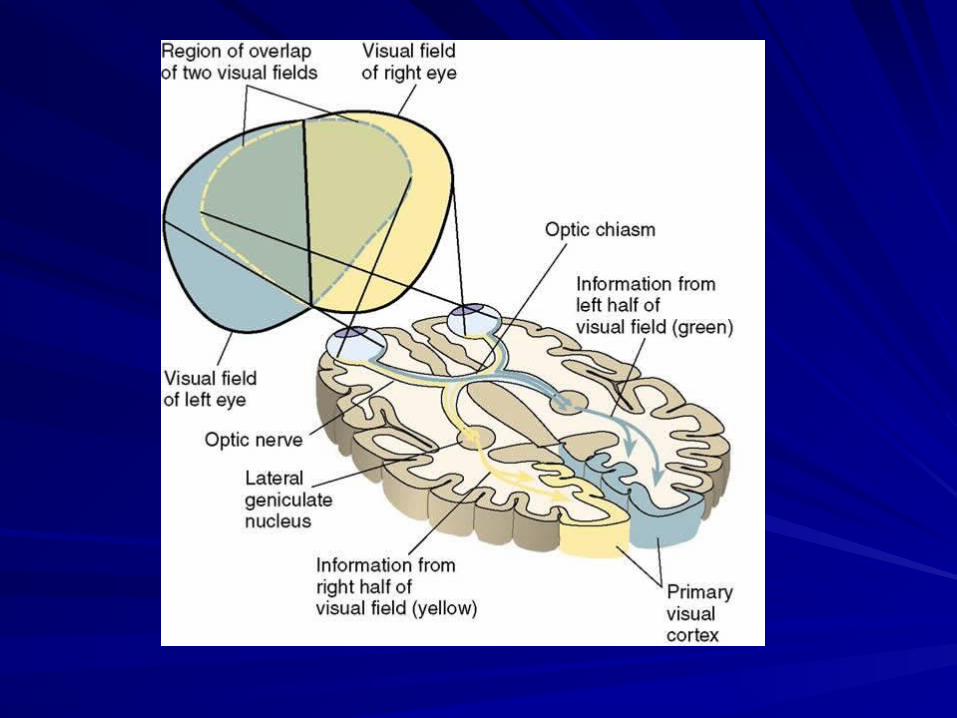
The axons of the retinal ganglion cells project through the optic The axons of the retinal ganglion cells project through the optic nerves, cross over through the nerves, cross over through the optic chiasmoptic chiasm to the to the dorsal lateral dorsal lateral geniculate nucleusgeniculate nucleus (LGN) (LGN)
The LGN consists of 6 layers of neurons: the inner 2 layers are The LGN consists of 6 layers of neurons: the inner 2 layers are called the called the magnocellular layersmagnocellular layers and the outer 4 are called the and the outer 4 are called the parvocellular layersparvocellular layers
The cells in the LGN project through the optic radiations to the The cells in the LGN project through the optic radiations to the primary visual cortex (aka striate cortex)primary visual cortex (aka striate cortex)
Diagram of visual pathwaysDiagram of visual pathways
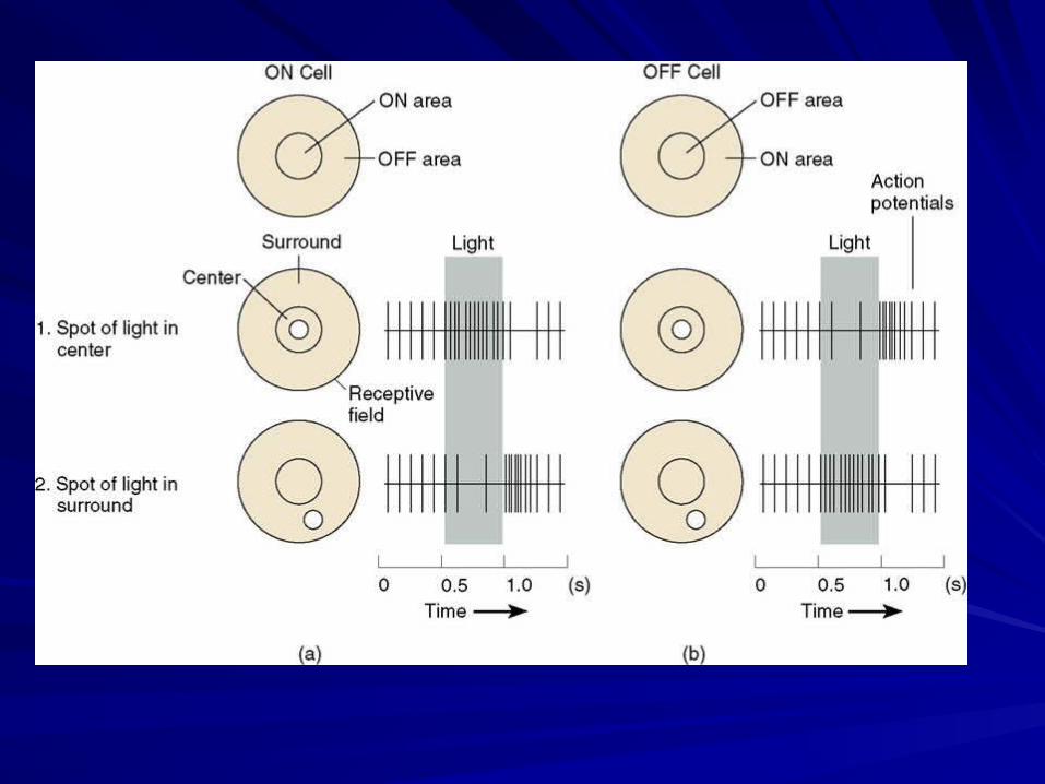
Coding of light and darkCoding of light and dark
The The receptive fieldreceptive field of a neuron in the visual system is the part of the of a neuron in the visual system is the part of the visual field that an individual neuron “sees”, i.e. the part in which visual field that an individual neuron “sees”, i.e. the part in which light must fall for the neuron to be stimulatedlight must fall for the neuron to be stimulated
2 major types of retinal ganglion cells, ON center and OFF center 2 major types of retinal ganglion cells, ON center and OFF center cellscells– ON center cells are excited by light falling in the center of the field ON center cells are excited by light falling in the center of the field
(center), and inhibited by light falling in the surrounding field (surround)(center), and inhibited by light falling in the surrounding field (surround)
– OFF center cells are excited by light in the surround, and inhibited by OFF center cells are excited by light in the surround, and inhibited by light in the centerlight in the center
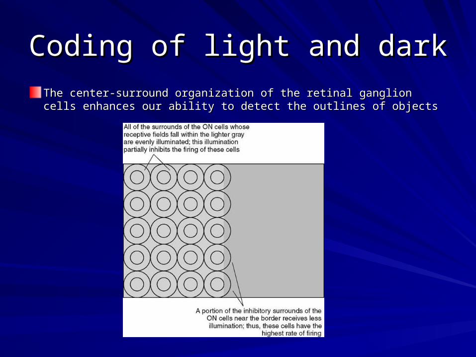
Coding of light and darkCoding of light and dark
The center-surround organization of the retinal ganglion cells The center-surround organization of the retinal ganglion cells enhances our ability to detect the outlines of objectsenhances our ability to detect the outlines of objects
Coding of colorCoding of color
The retinas of humans, Old World monkeys and apes contain 3 The retinas of humans, Old World monkeys and apes contain 3 different types of cones which provide us with an elaborate form of different types of cones which provide us with an elaborate form of color visioncolor vision
All visible colors (for humans at least) can be mixed from the 3 main All visible colors (for humans at least) can be mixed from the 3 main colors: red (long), green (medium), and blue (short); due to the colors: red (long), green (medium), and blue (short); due to the wavelengths absorbed by the 3 different cones (wavelengths absorbed by the 3 different cones (trichromatic theory)trichromatic theory)
Genetic defects can cause one or more of the 3 types of cones to Genetic defects can cause one or more of the 3 types of cones to not function properly, resulting in either not function properly, resulting in either protanopiaprotanopia, where red and , where red and green are confused because the red cones respond to green; green are confused because the red cones respond to green; deuteranopiadeuteranopia, where where red and green are confused also, but , where where red and green are confused also, but because the green cones respond to red; or because the green cones respond to red; or tritanopiatritanopia, where they , where they lack blue coneslack blue cones
Anatomy of the striate cortexAnatomy of the striate cortex
Consists of 6 principle layers, arranged in bands parallel to the Consists of 6 principle layers, arranged in bands parallel to the surfacesurface
The striate cortex of one hemisphere contains info from the The striate cortex of one hemisphere contains info from the contralateral visual fieldcontralateral visual field
Approx. 25% of the striate cortex is devoted to anlaysis of info from Approx. 25% of the striate cortex is devoted to anlaysis of info from the foveathe fovea
Neurons in the visual cortex selectively respond to specific features Neurons in the visual cortex selectively respond to specific features of the visual world, not just to lightof the visual world, not just to light
Orientation and MovementOrientation and Movement
Simple cellSimple cell – an orientation-sensitive neuron whose receptive field is – an orientation-sensitive neuron whose receptive field is organized in an opponent fashionorganized in an opponent fashion
Complex cellComplex cell – a neuron that responds to the presence of a line – a neuron that responds to the presence of a line segment with a particular orientation located within it s receptor segment with a particular orientation located within it s receptor field, especially when the line moves perpendicularly to its field, especially when the line moves perpendicularly to its orientationorientation
Hypercomplex cellHypercomplex cell – a neuron that responds to the presence of a – a neuron that responds to the presence of a line segment with a particular orientation that ends at a particluar line segment with a particular orientation that ends at a particluar point within the cell’s receptive fieldpoint within the cell’s receptive field
Spatial FrequencySpatial Frequency
Neurons in the primary visual cortex respond best to Neurons in the primary visual cortex respond best to sine-wave sine-wave gratingsgratings, which are a series of straight parallel bands varying , which are a series of straight parallel bands varying continuously in brightness according to a sine-wave function, along continuously in brightness according to a sine-wave function, along a line perpendicular to their lengthsa line perpendicular to their lengths
A sine-wave grating is designated by its A sine-wave grating is designated by its spatial frequencyspatial frequency, or the , or the relative width of the bands, measured in cycles per degree of visual relative width of the bands, measured in cycles per degree of visual angleangle
The most important visual information is that contained in low spatial The most important visual information is that contained in low spatial frequenciesfrequencies
Retinal disparityRetinal disparity
Binocular vision (i.e. from 2 eyes) provides a vivid perception of Binocular vision (i.e. from 2 eyes) provides a vivid perception of depth through the process of stereoscopic vision, or depth through the process of stereoscopic vision, or stereopsisstereopsis
Most neurons in the primary visual cortex are binocular, they Most neurons in the primary visual cortex are binocular, they respond to info of either eyerespond to info of either eye
The cells respond most vigorously when each eye sees a stimulus The cells respond most vigorously when each eye sees a stimulus in a slightly different location, called in a slightly different location, called retinal disparityretinal disparity
ColorColor
In the striate cortex, info from color-sensitive ganglion cells is In the striate cortex, info from color-sensitive ganglion cells is transmitted through to special cells grouped into transmitted through to special cells grouped into cytochrome cytochrome oxidase (CO) blobsoxidase (CO) blobs
Modular organization of the striate Modular organization of the striate cortexcortex
The striate cortex is divided into ~2500 modules, each containing The striate cortex is divided into ~2500 modules, each containing ~150,000 neurons~150,000 neurons
The neurons in each module are devoted to the analysis of various The neurons in each module are devoted to the analysis of various features contained in one very small portion of the visual field, and features contained in one very small portion of the visual field, and combine together to form a complete wholecombine together to form a complete whole
Info from the layers of the LGN project to the different layers of the Info from the layers of the LGN project to the different layers of the modulesmodules
The modules consist of 2 segments, each surrounding a CO blobThe modules consist of 2 segments, each surrounding a CO blob– The neurons in the CO blobs are sensitive to color and low spatial The neurons in the CO blobs are sensitive to color and low spatial
frequenciesfrequencies
– The neurons outside of the blobs are sensitive to orientation, movement, The neurons outside of the blobs are sensitive to orientation, movement, spatial freq, texture and binocular disparityspatial freq, texture and binocular disparity
Role of the visual association Role of the visual association cortexcortex
Two streams of visual analysisTwo streams of visual analysis– Visual info receive from the striate cortex is analyzed in the visual assc Visual info receive from the striate cortex is analyzed in the visual assc
cortexcortex
– Neurons in the striate cortex project to the Neurons in the striate cortex project to the extrastriate cortexextrastriate cortex, which , which surrounds the visual assc cortexsurrounds the visual assc cortex
– The primate extrastriate cortex consists of several specialized regions The primate extrastriate cortex consists of several specialized regions that respond to particular features of a visual stimulusthat respond to particular features of a visual stimulus
– Contains 2 streams of analysis:Contains 2 streams of analysis:Dorsal streamDorsal stream – a system of interconnected regions of visual cortex involved – a system of interconnected regions of visual cortex involved in the perception of spatial location, beginning with the striate cortex and in the perception of spatial location, beginning with the striate cortex and ending with the posterior parietal cortexending with the posterior parietal cortex
Ventral streamVentral stream – a system of interconnected regions of visual cortex – a system of interconnected regions of visual cortex involved in the perception of form, beginning in the striate cortex and ending involved in the perception of form, beginning in the striate cortex and ending in the inferior temporal cortexin the inferior temporal cortex
Perception of colorPerception of color
Color constancyColor constancy – the relatively constant appearance of the colors – the relatively constant appearance of the colors of objects under varying lighting conditions; the visual system of objects under varying lighting conditions; the visual system compensates for the source of light when process visual information compensates for the source of light when process visual information about colorsabout colors
AchromatopsiaAchromatopsia – inability to discriminate among different hues; – inability to discriminate among different hues; caused by damage to the visual assc cortexcaused by damage to the visual assc cortex
Analysis of formAnalysis of formIn primates the recognition of visual patterns and identification of particular objects In primates the recognition of visual patterns and identification of particular objects take place in the take place in the inferior temporal cortexinferior temporal cortex, located at the end of the ventral stream on , located at the end of the ventral stream on the ventral part of the temporal lobethe ventral part of the temporal lobe
– Analyses of form and color are put together here and perceptions of 3D objects and Analyses of form and color are put together here and perceptions of 3D objects and backgrounds are achievedbackgrounds are achieved
– Consists of 2 major regions: TE and TEOConsists of 2 major regions: TE and TEO
Damage to the human visual assc cortex can result in a Damage to the human visual assc cortex can result in a visual agnosiavisual agnosia, which results , which results in an inability to perceive or identify a visual stimulusin an inability to perceive or identify a visual stimulus
– Apperceptive visual agnosiaApperceptive visual agnosia – failure to perceive objects, even though visual acuity is – failure to perceive objects, even though visual acuity is normal (e.g. cannot name an object by looking at it, but can if allowed to touch it)normal (e.g. cannot name an object by looking at it, but can if allowed to touch it)
– ProsopagnosiaProsopagnosia – failure to recognize particular people by the sight of their faces (i.e. can – failure to recognize particular people by the sight of their faces (i.e. can recognize by voice, hair color, etc.)recognize by voice, hair color, etc.)
Fusiform face areaFusiform face area – region of the extrastriate cortex located at the base of the brain; – region of the extrastriate cortex located at the base of the brain; involved in perception of faces and other complex objects that require expertise to involved in perception of faces and other complex objects that require expertise to recognizerecognizeAssociative visual agnosiaAssociative visual agnosia – inability to identify objects that are perceived visually, – inability to identify objects that are perceived visually, even though the form of the perceived object can be drawn or matched with similar even though the form of the perceived object can be drawn or matched with similar objects; appears to involve difficulty in transferring visual info to verbal mechanismsobjects; appears to involve difficulty in transferring visual info to verbal mechanisms
Perception of movementPerception of movement
Area V5 of the extrastriate cortex contains neurons that respond to Area V5 of the extrastriate cortex contains neurons that respond to movement, and show directional sensitivitymovement, and show directional sensitivity
An area adjacent to V5 receives info from V5 about movement and An area adjacent to V5 receives info from V5 about movement and respond to more complex movement features, such as radial, respond to more complex movement features, such as radial, circular or spiral motioncircular or spiral motion– Optic flowOptic flow – the complex motion of points in the visual field caused by – the complex motion of points in the visual field caused by
relative movement b/t the observer and env’t; provides info about the relative movement b/t the observer and env’t; provides info about the relative distance of objects from the observer and of the relative relative distance of objects from the observer and of the relative direction of movementdirection of movement
AkinetopsiaAkinetopsia – inability to perceive movement, caused by damage to – inability to perceive movement, caused by damage to area V5 of the visual assc cortexarea V5 of the visual assc cortex
Perception of spatial locationPerception of spatial location
3 subareas of the extrastriate cortex send info through area V5 to 3 subareas of the extrastriate cortex send info through area V5 to the parietal cortex, which is involved in spatial perceptionthe parietal cortex, which is involved in spatial perception
Damage to this area disrupts performance on tasks that require Damage to this area disrupts performance on tasks that require perceiving and remembering the location of objectsperceiving and remembering the location of objects
Balint’s syndromeBalint’s syndrome – caused by bilateral damage to the parieto- – caused by bilateral damage to the parieto-occipital region; includes occipital region; includes optic ataxiaoptic ataxia (difficulty in reaching for (difficulty in reaching for objects under visual guidance), objects under visual guidance), ocular apraxiaocular apraxia (difficulty in visual (difficulty in visual scanning), and scanning), and simultanagnosiasimultanagnosia (difficulty in perceiving more than (difficulty in perceiving more than one object at a time)one object at a time)