Embed Size (px)
Citation preview
139
Chapter 7Mechanotransduction, Metastasis and Genomic Instability
Allison K. Simi, Alexandra S. Piotrowski and Celeste M. Nelson
© Springer International Publishing Switzerland 2015C. Maxwell, C. Roskelley (eds.), Genomic Instability and Cancer Metastasis, Cancer Metastasis - Biology and Treatment 20, DOI 10.1007/978-3-319-12136-9_7
C. M. Nelson () · A. K. Simi · A. S. PiotrowskiDepartment of Chemical & Biological Engineering, Princeton University, 303 Hoyt Laboratory, William Street, Princeton, NJ 08544, USAe-mail: [email protected]
C. M. NelsonDepartment of Molecular Biology, Princeton University, 303 Hoyt Laboratory, William Street, Princeton, NJ 08544, USA
Abstract Cells translate mechanical forces in the environment into biochemical signals in a process called mechanotransduction. In this way, mechanical forces direct cell behavior, including motility, proliferation, and differentiation, and become important in physiological processes such as development and wound healing. Abnormalities in mechanotransduction can lead to aberrant cell behavior and disease, including cancer. Changes in extracellular mechanical forces or defects in mechanosensors can result in misregulation of signaling pathways inside the cell, and ultimately lead to malignancy. Here, we discuss the ways in which physical attributes of the tumor microenvironment can promote metastasis and genomic instability, two hallmark features of cancer.
Keywords Mechanical stress · EMT · Stiffness
Abbreviations
2D Two-dimensional3D Three-dimensionalαSMA α-smooth muscle actinbFGF Basic fibroblast growth factorECM Extracellular matrixEGF Epidermal growth factorEMT Epithelial-mesenchymal transitionERK Extracellular-signal-regulated kinaseFAK Focal adhesion kinaseFGF Fibroblast growth factorGIN Genomic instability
Authors Allison Simi and Alexandra Piotrowski have contributed equally to this work.
A. K. Simi et al.140
IFP Interstitial fluid pressureILK Integrin-linked kinaseMET Mechanoelectrical transductionMLC Myosin light chainMMP Matrix metalloproteinasePDGF Platelet-derived growth factorPI3K Phosphoinositide 3-kinasePTEN Phosphatase and tensin homologROCK Rho-associated kinaseROS Reactive oxygen speciesRTK Receptor tyrosine kinaseTAZ transcriptional co-activator with PDZ-binding motifTGF-β Transforming growth factor βVEGF Vascular endothelial growth factorYAP Yes-associated protein
Introduction
Over a decade ago, Hanahan and Weinberg defined several features of cancer that they considered essential for the acquisition of a malignant phenotype, including replicative immortality, evasion of growth suppressors, evasion of apoptosis, stim-ulation of angiogenesis, stimulation of proliferation, and invasion and metastasis [1]. Since then, a flood of cancer research has led to modification and expansion of the proposed hallmarks; metastasis and genomic instability are two that per-sist [2]. Cancer is widely regarded as a disease of the cell, and cell behavior is directed by both biochemical and physical cues, which can work independently or synergistically [3]. Accordingly, the tumor microenvironment has been shown to affect tumor progression [4, 5]. This chapter focuses on the physical factors and mechanical forces that tumor cells encounter in the tumor microenvironment, which can in turn alter their behavior. Cells convert the physical signals they receive into biological responses via a process known as mechanotransduction [6].
Mechanotransduction involves both the external environment and internal sig-naling [7]. The transmission of external forces to intracellular signaling is centered on proteins that are activated by force, such as integrins [8, 9] and T-cell receptors [10]. Many cellular phenotypes, including morphology, motility, and proliferation, are governed by external mechanical forces [11–13]. Thus, mechanotransduction is central to a variety of physiologically normal processes, including embryonic development, differentiation, wound healing, and angiogenesis [14, 15]. Defects in mechanotransduction are known to be involved in several diseases, including cancer [16]. Understanding how defects in mechanotransduction affect tumor pro-gression will add to our fundamental knowledge of cancer biology and may suggest new approaches for treatment.
7 Mechanotransduction, Metastasis and Genomic Instability 141
How Mechanotransduction Regulates Normal Cell Behavior
Extracellular Factors Affecting Mechanotransduction in Normal Cells
Most cells are anchorage dependent: they need to adhere to a substratum to prevent apoptosis and promote cell cycle progression [17]. Thus, the mechanical microen-vironment is important for cell survival. Cells sense their environment via confor-mational changes in mechanically responsive proteins, known as mechanosensors. Physical forces induce these conformational changes, which result in downstream signaling inside the cell [18, 14]. Forces can originate from a variety of features, including the rigidity of the extracellular matrix (ECM), static or dynamic fluid flow, and tissue growth [6]. These forces are further classified into specific types of loads that cells can detect. For example, forces incurred by blood flow include hydrodynamic pressure, shear stress, and cyclic strain, and all of these help regulate endothelial cell behaviors [19] such as cell reorientation [20].
Cells can also respond to mechanical loads by secreting biochemical factors, some of which result in subsequent ECM remodeling. Growth factors comprise one class of proteins that are important in this respect. Transforming growth factor β (TGF-β) is sequestered in the ECM, and is released when internal contractility of myofibroblasts is balanced externally by a stiff matrix, causing conforma-tional changes in protein complexes embedded in the ECM. Free TGF-β starts a feed-forward loop, causing increased deposition of ECM proteins and additional (increased) expression of TGF-β [21]. Various other growth factors increase activity as a result of mechanical load, as evidenced by endothelial secretion of basic fibro-blast growth factor (bFGF) in response to shear stress and hydrostatic pressure [22, 23]. Mechanical forces also regulate the expression of matrix remodeling proteins such matrix metalloproteinases (MMPs). This is seen in human monocytes/mac-rophages, which have been shown to increase expression of MMPs under cyclic strain, and thus contribute to ECM degradation [24].
Intracellular Factors Affecting Mechanotransduction in Normal Cells
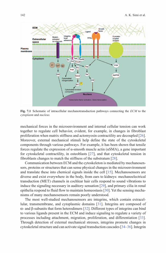
There are several intracellular components involved in receiving mechanical signals and eliciting a response (Fig. 7.1). A feature that is particularly important to mechanical sensing is contractility; all cells have a network of cytoskeletal proteins (actin, microtubules, intermediate filaments) that aid in cell structure and mobil-ity [17]. Cytoskeletal contractility creates a balance between intra- and extracel-lular forces acting on the cell, and thus is important for cells to be able to respond to forces in the surrounding microenvironment [25]. This balance exists so that
A. K. Simi et al.142
mechanical forces in the microenvironment and internal cellular tension can work together to regulate cell behavior, evident, for example, in changes in fibroblast proliferation when matrix stiffness and actomyosin contractility are decoupled [26]. Moreover, external mechanical stimuli help define the state of the cytoskeletal components through various pathways. For example, it has been shown that tensile forces regulate the expression of α-smooth muscle actin (αSMA), a gene important for cytoskeletal contractility, in osteoblasts [27], and that cytoskeletal tension in fibroblasts changes to match the stiffness of the substratum [28].
Communication between ECM and the cytoskeleton is mediated by mechanosen-sors, proteins or structures that can sense physical changes in the microenvironment and translate these into chemical signals inside the cell [15]. Mechanosensors are diverse and exist everywhere in the body, from ears to kidneys: mechanoelectrical transduction (MET) channels in cochlear hair cells respond to sound vibrations to induce the signaling necessary in auditory sensation [29], and primary cilia in renal epithelia respond to fluid flow to maintain homeostasis [30]. Yet the sensing mecha-nisms of many mechanosensors remain poorly understood.
The most well-studied mechanosensors are integrins, which contain extracel-lular, transmembrane, and cytoplasmic domains [31]. Integrins are composed of α- and β-subunits that form heterodimers [32]. Different types of integrins can bind to various ligands present in the ECM and induce signaling to regulate a variety of processes including attachment, migration, proliferation, and differentiation [33]. Through detection of external mechanical stresses, integrins promote changes in cytoskeletal structure and can activate signal transduction cascades [34–36]. Integrin
Fig. 7.1 Schematic of intracellular mechanotransduction pathways connecting the ECM to the cytoplasm and nucleus.
7 Mechanotransduction, Metastasis and Genomic Instability 143
activity is also essential for the formation of focal adhesions, which act as centers of mechanotransduction [37]. Focal adhesions are protein complexes localized at the plasma membrane that link the ECM to the actin cytoskeleton. In addition to integrins, focal adhesions include hundreds of proteins, the most well-characterized of which are talin, paxillin, vinculin, focal adhesion kinase (FAK) and Src family kinases, which act as signaling molecules [38]. The formation of focal adhesions is regulated by both external forces and cytoskeletal contractility [39].
Other intracellular components involved in mechanotransduction include G proteins, receptor tyrosine kinases (RTKs), extracellular-signal-regulated kinases (ERKs), and stretch-activated ion channels [6].
G proteins are localized at focal adhesion sites and can undergo conformational changes induced by mechanical stress to promote cell growth. G proteins are acti-vated in cardiac fibroblasts in response to stretch, as well as in endothelial cells and osteocytes in response to shear stress [40–42].
RTKs are transmembrane proteins that dimerize to become activated, and are involved in integrin-mediated mechanotransduction downstream of G proteins. Dimerization is triggered by binding of the receptor to extracellular ligands such as epidermal growth factor (EGF) and platelet-derived growth factor (PDGF), leading to further signaling [43]. RTKs can also activate ERKs, which are important for gene expression and protein synthesis [44].
ERKs are kinases that play an important role in intracellular signaling, such as the activation of cytoplasmic and nuclear regulatory proteins. These kinases can be activated in response to mechanical stimuli. Shear stress and stretch have been shown to activate ERKs in aortic endothelial cells and pulmonary epithelial cells, respectively [45, 46].
Stretch-activated ion channels allow ions such as Ca2 + to move in and out of cells, which regulates several cellular processes. Cell stretching has been shown to increase intracellular levels of Ca2 + in several cell types [47, 48]. Intracellular Ca2 + levels are also important for the activation of other proteins in the mechanotrans-duction signaling cascade, such as ERKs [49].
Mechanotransduction and Metastasis
The invasion of primary tumors into their surrounding tissue and subsequent meta-static spread to other organs are among the largest obstacles to cancer treatment, and metastasis is the main cause of cancer-related deaths [50]. Metastasis relies on the ability of tumor cells to migrate from the primary tumor and form new lesions at distant locations [51]. Invasion and metastasis require physical interactions be-tween malignant cells and the microenvironment, a process that inherently involves mechanosensing and mechanotransduction [16]. Both extracellular factors in the physical tumor microenvironment and intracellular factors within cancer cells con-tribute to mechanotransduction during invasion and metastasis. Identifying how mechanotransduction becomes abnormally regulated in cancer cells is necessary to understand the mechanisms that underlie invasion and metastasis.
A. K. Simi et al.144
Extracellular Factors Affecting Mechanotransduction in Tumors
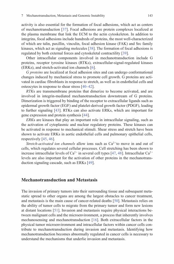
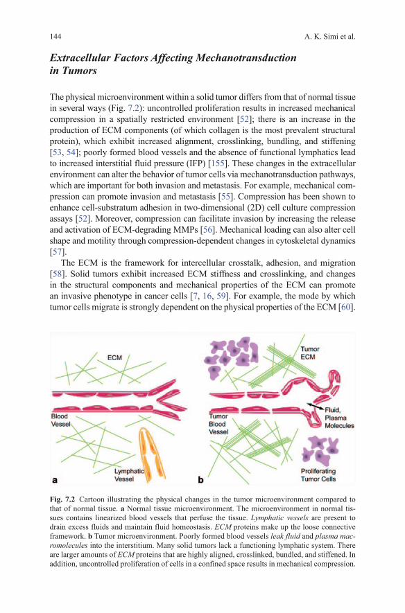
The physical microenvironment within a solid tumor differs from that of normal tissue in several ways (Fig. 7.2): uncontrolled proliferation results in increased mechanical compression in a spatially restricted environment [52]; there is an increase in the production of ECM components (of which collagen is the most prevalent structural protein), which exhibit increased alignment, crosslinking, bundling, and stiffening [53, 54]; poorly formed blood vessels and the absence of functional lymphatics lead to increased interstitial fluid pressure (IFP) [155]. These changes in the extracellular environment can alter the behavior of tumor cells via mechanotransduction pathways, which are important for both invasion and metastasis. For example, mechanical com-pression can promote invasion and metastasis [55]. Compression has been shown to enhance cell-substratum adhesion in two-dimensional (2D) cell culture compression assays [52]. Moreover, compression can facilitate invasion by increasing the release and activation of ECM-degrading MMPs [56]. Mechanical loading can also alter cell shape and motility through compression-dependent changes in cytoskeletal dynamics [57].
The ECM is the framework for intercellular crosstalk, adhesion, and migration [58]. Solid tumors exhibit increased ECM stiffness and crosslinking, and changes in the structural components and mechanical properties of the ECM can promote an invasive phenotype in cancer cells [7, 16, 59]. For example, the mode by which tumor cells migrate is strongly dependent on the physical properties of the ECM [60].
Fig. 7.2 Cartoon illustrating the physical changes in the tumor microenvironment compared to that of normal tissue. a Normal tissue microenvironment. The microenvironment in normal tis-sues contains linearized blood vessels that perfuse the tissue. Lymphatic vessels are present to drain excess fluids and maintain fluid homeostasis. ECM proteins make up the loose connective framework. b Tumor microenvironment. Poorly formed blood vessels leak fluid and plasma mac-romolecules into the interstitium. Many solid tumors lack a functioning lymphatic system. There are larger amounts of ECM proteins that are highly aligned, crosslinked, bundled, and stiffened. In addition, uncontrolled proliferation of cells in a confined space results in mechanical compression.
7 Mechanotransduction, Metastasis and Genomic Instability 145
Changes in ECM composition and architecture also affect the distribution and activa-tion of soluble factors (e.g., growth factors, cytokines, MMPs) that are themselves involved in cell behavioral changes and mechanotransduction [61]. ECM stiffness can promote the malignant behavior of tumor cells by increasing the expression and activity of adhesion receptors, thereby also activating mechanotransduction path-ways [12]. For example, force has been shown to influence the development of focal adhesions since maturation of these complexes requires mechanical tension [62].
Increased ECM stiffness also directs cell behavior by increasing external resis-tance forces experienced by the cell [63]. Links to the ECM via integrins and focal adhesions can relay these stresses to the cytoskeleton, alter the balance of intracel-lular forces, and stimulate signal transduction cascades that influence cell behavior [7]. Moreover, increased ECM stiffness can disrupt epithelial polarity and induce mi-gration and metastasis [64]. Cells have also been shown to migrate preferentially to regions of increased ECM stiffness via mechanotaxis/durotaxis [65, 66]. Finally, the crosslinking of ECM by lysyl oxidase, which can also stiffen the matrix and induce fibrosis, can promote tumorigenesis via enhanced integrin signaling [58].
ECM remodeling by tumor and stromal cells is important for both invasion and metastasis. For example, migrating tumor cells exhibit pericellular proteolytic deg-radation to make room for further migration [67]. Proteases such as MMPs are re-cruited to integrin assemblies and other adhesion receptors at the leading edge of a migrating cell to model and degrade the ECM [68]. Cancer cells have also been shown to realign their surrounding ECM perpendicular to the tumor boundary, alter-ing its architecture for improved adhesion and migration, creating diverse routes for dissemination [69]. Migration is mediated by several types of proteolytic structures enriched with F-actin, β1-integrins, and MMPs, which are key players in mechano-transduction [70]. Single cell migration can also occur without proteolytic degrada-tion under the mode of amoeboid migration [71]. The microscale architecture of the ECM, including the alignment of fibers and the location and size of pores, dictates the mechanisms of invasion and metastasis applied by cancer cells [72].
IFP and interstitial fluid flow have also been shown to affect the migratory and invasive behaviors of tumor cells [73, 156, 157]. In a three-dimensional (3D) cul-ture model in which single tumor cells were suspended in ECM, fluid flow was shown to increase the percentage of migratory cells as well as their speed [73]. In a similar study, interstitial fluid flow was shown to result in the upstream migration of cancer cells as a result of asymmetry in matrix adhesion stresses needed to bal-ance drag from fluid flow [74]. The stresses induced by flow created a gradient of integrin activation across the cells. Components of focal adhesions, including FAK, paxillin, and vinculin, localized at the upstream side of the migrating cells.
Intracellular Factors Affecting Mechanotransduction in Tumors
It is well known that changes in mechanotransduction promote invasion and metastasis [75]. The intracellular factors affecting mechanotransduction pathways in tumor cells may be altered in response to changes in the tumor microenvironment,
A. K. Simi et al.146
or to genetic mutations and changes in gene expression within the tumor cells. Intracellular mechanotransduction can, in turn, lead to changes in gene expression to promote invasion and metastasis.
Cytoskeletal reorganization is important for changes in cell shape and motility, and therefore migration and metastasis [16]. Cytoskeletal tension is primarily regulated by ERKs and the Rho family of small GTPases. One effector of Rho is Rho-associated kinase (ROCK), which regulates actin cytoskeletal contractility via myosin light chain (MLC) phosphorylation [64]. Rho activity has been shown to be elevated in some tumors, though decreases in its activity have also been reported [76, 77]. Cytoskeletal tension is also affected by the mechanical properties of the ECM, such as stiffness and crosslinking [7]. Increased matrix stiffness promotes the clustering of integrins and the formation of focal adhesions, in addition to increas-ing activation of FAK and ERK, and enhancing ROCK-mediated cytoskeletal con-tractility [64]. ROCK is also involved in the disruption of adherens junctions and moving the tail end of the cell behind the leading edge to assist in cell locomotion [78–80]. Moreover, cell migration involves the extension of membrane protrusions resulting from the cycling of actin polymerization and depolymerization, which are regulated by Rho GTPases via the cofilin pathway [81, 82].
ECM crosslinking has also been shown to result in the aggregation and clustering of integrins as well as enhanced signaling via phosphoinositide 3-kinase (PI3K) to induce invasion [58, 64]. Other components of focal adhesions have also been implicated in tumor progression, including Src, the activity of which has been shown to influence proliferation, invasion and metastasis [83, 84]. Src activation is required for ECM degradation during migration [85]. In 3D culture studies of breast tumor cells, Src activity increases the strength of cellular forces on the ECM as well as the duration and length of cell membrane protrusions [86].
Whereas some cells in the tumor become stiffer, metastatic cells are more deformable and exhibit reduced cytoskeletal stiffness [87]. Lower levels of integrin expression along with decreased adhesion to the ECM have been associated with oncogenic transformation [88, 60]. This increased deformability is correlated with enhanced metastatic potential. For example, enhanced deformability enables meta-static cells to move through tight spaces, such as between endothelial cells, during intravasation and extravasation [89].
In addition to regulating the cytoskeleton and associated proteins, mechanotrans-duction can lead to gene expression changes that promote invasion and metastasis. Cancer cells undergo a variety of genetic mutations and gene expression changes during tumor progression, which can affect their interactions with the microenvi-ronment and subsequent mechanotransduction. Mechanotransduction itself is one source of changes in gene expression in cancer cells. A major way that mechano-transduction can affect gene expression is via the epithelial-mesenchymal transition (EMT). EMT, in which epithelial genes are downregulated and mesenchymal genes are upregulated, is thought to be an important mechanism in both invasion and metastasis [90, 91]. ECM stiffness has been shown to promote EMT, through which cancer cells acquire a migratory phenotype via a variety of pathways, some of which include key players in mechanotransduction, such as RTKs [92]. In one pathway,
7 Mechanotransduction, Metastasis and Genomic Instability 147
EMT results from stiffness-mediated localization and signaling of Rac GTPases downstream of MMPs [93]. Mechanical stress and matrix rigidity can also induce EMT downstream of TGF-β [94, 95]. Furthermore, the activation of Rho GTPases is thought to contribute to EMT via the loss of adherens junctions between cells and the gain of mesenchymal characteristics [96].
Induction of EMT in tumor cells, which affects cytoskeletal organization and cell-cell and cell-matrix adhesions, can also alter how the cells sense exogenous forces, and therefore their responses to those forces [97, 98]. The downregulation of epithelial keratins results in reduced cytoskeletal stiffness and greater cell deform-ability, directly influencing the metastatic potential of tumor cells [99]. In addition to being more deformable than non-metastatic cells, metastatic cells also lose their anchorage dependence [100, 101]. Anoikis, or apoptosis induced by the loss of ad-hesion to the ECM, is suppressed in metastatic cells, allowing them to migrate and traverse through the bloodstream to distant organs [102, 103]. Anoikis is believed to be mediated by integrin signaling [104]. The activation of integrins and their associated proteins, including FAK and integrin-linked kinase (ILK), can suppress anoikis, indicating that mechanotransduction and apoptotic pathways are linked [105]. EMT can also suppress anoikis [106]. In particular, the downregulation of E-cadherin can protect cells against anoikis [107]. It is clear that several extracel-lular and intracellular components of mechanotransduction are altered in tumors, which promotes progression to invasive disease. Mechanotransduction, it seems, is another mechanism that can be hijacked to support malignant transformation.
Mechanotransduction and Genomic Instability
The term genomic instability (GIN) broadly describes the inability of a cell to pass on a copy of its DNA with fidelity. GIN can manifest itself in several ways, each the result of replicative stress caused by errors in DNA replication or the DNA damage response [108]. Microsatellite instability is the expansion or contraction of oligo-nucleotide repeats and results from mutations in mismatch repair genes [109, 110]; nucleotide excision-repair-related instability results from an impaired ability of the cell to remove and replace damaged nucleotides [111]; and chromosomal instability is a change in the structure or number of chromosomes, which typically occurs as a result of errors in DNA replication or mitosis [112, 113].
GIN is a defining feature of cancers, and is believed to be the driving force behind tumor progression. Various errors in DNA replication or repair processes lead to an abnormal genotype that continues to change with each generation of cells. As a result of GIN, tumors that originate from the same tissue and cell type can have wildly varying genetic profiles [114]. This intertumor heterogeneity, as well as subclonal heterogeneity within a single tumor, has been largely attributed to the Darwinian characteristics of cancer; that is, the evolution and adaptation of a cancer clone in response to external selective pressures [115]. Ultimately, this results in the acquisition of survival-enhancing features that allow a cancer to develop.
A. K. Simi et al.148
The local microenvironment is one source of pressure that results in GIN [116] and increased survival. Mouse embryonic stem cells exposed to radiation develop a high frequency of mutation in vivo but not in culture, suggesting that the microenvi-ronment of the cells contributed to their development [117]. More specifically, both physical features of the tumor microenvironment as well as onslaughts by external agents have been shown to increase the frequency of mutation, thus increasing the chances that one of these mutations will affect maintenance of genomic integrity. Hypoxia is one hallmark characteristic of the tumor microenvironment known to play a role in promoting GIN. Hypoxia induces an elevated frequency of mutation in tumorigenic mammalian cell lines [118]. Similarly, exposure to heat and serum-starvation increases mutations in mouse mammary carcinoma cells [119]. Little is known about how GIN may arise from mechanical aspects of the microenviron-ment; the following describes a body of work that supports this idea.
Mechanical Forces Affect Mitosis and Cell Cycle Progression
One risk factor for the development of GIN is an increase in cellular proliferation, and hence the chance for DNA copy errors to arise. Recently, the mechanical prop-erties of the microenvironment have been considered a major factor in its influence on cell behavior, specifically the regulation of cell cycle progression and mitosis and subsequent maintenance of the genome. Several studies have shown that modu-lating mechanical forces acting on cells can affect proliferation: mechanical stretch can reduce proliferation of podocytes [120], enhance differentiation and reduce proliferation of preadipocytes [121], and in endothelial cells, directed mechanical forces (specifically, shear and stretch) promote homeostasis but non-uniform forces can result in sustained pro-inflammatory and proliferative signaling [122]. These effects can be mediated by cell-cell contact, such as through VE-cadherin in endo-thelial cells [123].
The adhesion of a cell to its surroundings can alone induce changes in prolif-eration. Micropatterning techniques have been used to isolate the effects of cell spreading and cell-cell junctions from the effects of substratum adhesion on cell behavior. Such studies have revealed that E-cadherin is sufficient to induce epithelial cell proliferation via Rac1 signaling, and both proteins are required for cell-cell contact-dependent proliferation [124]. Similar findings hold for endothelial or smooth muscle cells via PI3K signaling [125]. Cytoskeletal structure and associated signaling have also proven to be important in cell-cell adhesion-mediated prolif-eration, based on studies regarding the role of VE-cadherin in vascular endothelial cells [126]. Additionally, simply varying the nature of the substratum also affects proliferative behavior. The basement membrane interacts differently with normal or cancerous epithelial cell lines, affecting growth and differentiation [127].
There is also evidence that mechanotransduction can influence various aspects of mitosis, and thus the segregation of the genome into daughter cells. Physical features of the microenvironment are one avenue of mechanical influence on

7 Mechanotransduction, Metastasis and Genomic Instability 149
mitosis. For example, in HeLa cells (human cervical cancer cells), retraction fibers, which bind mitotic cells to the substratum, exert forces on the cell that dictate the orientation of the spindle during mitosis. This is mediated by regulation of the sub-cortical actin network [128]. Another study in HeLa cells similarly showed that the spatial distribution of ECM proteins helps determine the axis of division by regulating actin dynamics [129].
It would follow from these studies that mechanosensors and other intracellu-lar mechanotransduction machinery are involved in the regulation of mitosis, and indeed this has been shown. Integrin-mediated adhesion is required for the cells to reorient the mitotic spindle parallel to the substratum [130]. Here again, cytoskeletal components are key communicators. G proteins and the motor protein dynein, both important in transmitting mechanical force, are also known to direct orientation of the spindle in development [131]. One can imagine that abnormal mechanical sig-naling, common to many diseases including cancer, could disrupt mitosis in a cell and thus generate genomically unstable progeny.
Mechanotransduction Regulates Biochemical Cues That Promote GIN
One way that mechanical stimuli ultimately promote changes in cell behavior is through intracellular signaling pathways that conclude with control of gene tran-scription. Genes regulated by mechanotransduction can affect a myriad of both normal and pathological processes in the body [14]. In the context of cancer, recent studies have suggested that important molecular targets of mechanotransduction include mitotic checkpoint genes and other cell-cycle regulators, which have long been associated with maintaining genomic stability [112, 132].
To discover mechanically-regulated genes associated with GIN, several studies have used polyacrylamide gels of varying stiffness to mimic the mechanical prop-erties of the ECM, and thus determine the effects of substratum stiffness on cell behavior in culture [133]. Recent findings from these experiments show that the transcription factors YAP (Yes-associated protein) and TAZ (transcriptional coacti-vator with PDZ-binding motif), which have implications in growth, proliferation, and differentiation, become activated in response to cytoskeletal tension and cell spreading induced by a stiff substratum [134]. In human mammary epithelial cells, expression of tumor suppressor phosphatase and tensin homolog (PTEN) is reduced in the presence of microRNA miR-18a, which is modulated by ECM stiffness [135]. PTEN is antagonistic to PI3K, a protein involved in many pathways important for cell growth and survival that promotes cancer when misregulated [136]. Polyacryl-amide gels were also used to show that matrix rigidity induces integrin clustering in mammary epithelial cells, which induces the formation of focal adhesions and generates cytoskeletal tension. This in turn activates ERK and enhances EGF-dependent pathways that activate ERK, which is known for its involvement in cell cycle regulation [137, 64].
A. K. Simi et al.150
Other cell cycle-regulators are activated by adhesion to or disruption of the sub-stratum. The protein p38 is best known for its role as a tumor suppressor, but also regulates mitotic entry and the spindle assembly checkpoint [138], and negatively regulates cell proliferation through a reactive oxygen species (ROS)-mediated re-sponse to stress [139]. When mammary epithelial cells lose adhesion to the substra-tum, p38 is activated and can induce apoptosis [140]. NM23-H1 is another protein associated with growth arrest, and this function was shown to be correlated with basement membrane assembly in human breast cancer cells [141].
Aside from the effects of mechanically-regulated gene transcription, cytokines and other signaling factors that contribute to cancer progression are often triggered by mechanical forces, and can induce GIN. For example, lung cancer cells show an increased production of ROS in response to shear stress [142]. ROS are well known to promote genetic mutations and cancer progression [143]. Furthermore, a xenograft of human skin overexpressing bFGF (in a cocktail with stem cell factor and endothelin-3) causes replication stress [144], the major source of GIN [108]. As previously described, bFGF is regulated by shear stress and hydrostatic pressure [23, 22].
Restructuring of the Stroma Results in GIN
In addition to signaling mediated by mechanosensors, cells can communicate with the microenvironment through various soluble factors that serve to restructure the surrounding stroma. In cancer, misregulation of these proteins has been linked to GIN. MMPs make up one class of proteins that remodel the ECM. Overexpression of MMPs can induce cell cycle progression, activate genotoxic pathways, and inhibit cytokinesis [145]. Furthermore, cells overexpressing MMPs often exhibit patterns of genomic irregularities [146]. The stroma is also heavily remodeled during the formation of new vasculature. Both cyclic and constant static stretch of endothelial cells increase the expression of vascular endothelial growth factor (VEGF) receptor and promote VEGF-induced proliferation, vasculogenesis, and angiogenesis [147]. VEGF has been shown to regulate the axis of division in endothelial cells, poten-tiating GIN [148]. Thus, through restructuring of the stroma, in addition to control of the cell cycle and associated proteins and cytokines by external forces, GIN is mediated by mechanotransduction in cancer cells.
Synopsis and Outlook
Aberrant mechanotransduction is a major contributor to tumor progression, metas-tasis, and GIN. Both mechanosensing and subsequent intracellular signaling alter properties of the cell that can lead to malignant transformation in cancer. Mechano-transduction is therefore important to study in order to understand the progression of this disease. Developing improved 2D and 3D cell culture models to mimic
7 Mechanotransduction, Metastasis and Genomic Instability 151
the tumor microenvironment will enable us to determine the effects of abnormal mechanotransduction in cancer progression. Beyond experimental models, compu-tational models can characterize the effects of mechanical stretch on cell behavior [121]. Others begin to account for intratumor heterogeneity when predicting therapeutic response [149]. However, current computational models cannot cope with mutational frequency of cancer cells, and thus there is a disconnect between investigations of the causes and consequences of this feature.
Although many of the proteins involved in mechanotransduction are known (e.g. integrins, cytoskeleton, myosins, kinases), the precise mechanisms by which a cell perceives the mechanical information of its environment remain unclear [150]. In addition, mechanical forces in the microenvironment are known to affect the cell cycle, and abnormal expression of cell-cycle regulators can result in GIN [132]; however, a clear mechanotransduction pathway linking these two events has not been elucidated. Similarly, current knowledge on the mechanosensing capabilities of stem cells is limited; verifying which forces, molecular pathways, and mecha-nosensing proteins are most important in directing construction of the stem cell niche and stem cell differentiation could lead to clinical applications (for example, targeting cancer stem cells) [151, 152].
Components of mechanotransduction pathways are starting to be considered as potential therapeutic targets. For example, it has been shown that the disruption of Rho or ERK signaling results in a reduction of cytoskeletal tension that leads to a decrease in tumor cell proliferation and the repression of malignant progression [16, 64]. Targeting Src activity could reduce proliferation, invasion, and metastasis [153]. Restoring anoikis response might curb metastasis [154], and the inhibition of collagen crosslinking and integrin signaling might reduce invasion. In addition, the mechanical properties of isolated metastatic cancer cells could be diagnostic indicators for prognosis. As we broaden our current understanding of mechano-transduction as it relates to both normal cell functions and disease, we will be able to integrate this knowledge into a synergistic treatment strategy for cancer.
References
1. Hanahan D, Weinberg RA (2000) The hallmarks of cancer. Cell 100(1):57–702. Hanahan D, Weinberg RA (2011) Hallmarks of cancer: the next generation. Cell 144(5):646–6743. Ingber DE (2008) Tensegrity-based mechanosensing from macro to micro. Prog Biophys Mol
Biol 97(2–3):163–1794. Fang H, Declerck YA (2013) Targeting the tumor microenvironment: from understanding path-
ways to effective clinical trials. Cancer Res 73(16):4965–49775. Fidler IJ, Poste G (2008) The “seed and soil” hypothesis revisited. Lancet Oncol 9(8):8086. Wang JH, Thampatty BP (2006) An introductory review of cell mechanobiology. Biomech
Model Mechanobiol 5(1):1–167. Huang S, Ingber DE (2005) Cell tension, matrix mechanics, and cancer development. Cancer
Cell 8(3):175–1768. Friedland JC, Lee MH, Boettiger D (2009) Mechanically activated integrin switch controls
alpha5beta1 function. Science 323(5914):642–644
A. K. Simi et al.152
9. Paszek MJ, Boettiger D, Weaver VM, Hammer DA (2009) Integrin clustering is driven by mechanical resistance from the glycocalyx and the substrate. PLoS Comput Biol 5(12):e1000604
10. Ma Z, Finkel TH (2010) T cell receptor triggering by force. Trends Immunol 31(1):1–611. Pelham RJ Jr, Wang Y (1997) Cell locomotion and focal adhesions are regulated by substrate
flexibility. Proc Natl Acad Sci U S A 94(25):13661–1366512. Yeung T, Georges PC, Flanagan LA, Marg B, Ortiz M, Funaki M, Zahir N, Ming W, Weaver
V, Janmey PA (2005) Effects of substrate stiffness on cell morphology, cytoskeletal structure, and adhesion. Cell Motil Cytoskeleton 60(1):24–34
13. Assoian RK, Klein EA (2008) Growth control by intracellular tension and extracellular stiff-ness. Trends Cell Biol 18(7):347–352
14. Orr AW, Helmke BP, Blackman BR, Schwartz MA (2006) Mechanisms of mechanotransduc-tion. Dev Cell 10(1):11–20
15. Vogel V, Sheetz M (2006) Local force and geometry sensing regulate cell functions. Nat Rev Mol Cell Biol 7(4):265–275
16. Jaalouk DE, Lammerding J (2009) Mechanotransduction gone awry. Nat Rev Mol Cell Biol 10(1):63–73
17. Discher DE, Janmey P, Wang YL (2005) Tissue cells feel and respond to the stiffness of their substrate. Science 310(5751):1139–1143
18. Ingber DE (2006) Cellular mechanotransduction: putting all the pieces together again. FASEB J 20(7):811–827
19. Resnick N, Yahav H, Shay-Salit A, Shushy M, Schubert S, Zilberman LC, Wofovitz E (2003) Fluid shear stress and the vascular endothelium: for better and for worse. Prog Biophys Mol Biol 81(3):177–199
20. Wang JH, Goldschmidt-Clermont P, Wille J, Yin FC (2001) Specificity of endothelial cell reorientation in response to cyclic mechanical stretching. J Biomech 34(12):1563–1572
21. Wells RG, Discher DE (2008) Matrix elasticity, cytoskeletal tension, and TGF-beta: the insoluble and soluble meet. Sci Signal 1(10):pe13
22. Gloe T, Sohn HY, Meininger GA, Pohl U (2002) Shear stress-induced release of basic fibroblast growth factor from endothelial cells is mediated by matrix interaction via integrin alpha(v)beta3. J Biol Chem 277(26):23453–23458
23. Acevedo AD, Bowser SS, Gerritsen ME, Bizios R (1993) Morphological and proliferative responses of endothelial cells to hydrostatic pressure: role of fibroblast growth factor. J Cell Physiol 157(3):603–614
24. Yang JH, Sakamoto H, Xu EC, Lee RT (2000) Biomechanical regulation of human mono-cyte/macrophage molecular function. Am J Pathol 156(5):1797–1804
25. Ingber DE (1997) Tensegrity: the architectural basis of cellular mechanotransduction. Annu Rev Physiol 59: 575–599
26. Mih JD, Marinkovic A, Liu F, Sharif AS, Tschumperlin DJ (2012) Matrix stiffness reverses the effect of actomyosin tension on cell proliferation. J Cell Sci 125(Pt 24):5974–5983
27. Wang J, Su M, Fan J, Seth A, McCulloch CA (2002) Transcriptional regulation of a con-tractile gene by mechanical forces applied through integrins in osteoblasts. J Biol Chem 277(25):22889–22895
28. Solon J, Levental I, Sengupta K, Georges PC, Janmey PA (2007) Fibroblast adaptation and stiffness matching to soft elastic substrates. Biophys J 93(12):4453–4461
29. Fettiplace R, Hackney CM (2006) The sensory and motor roles of auditory hair cells. Nat Rev Neurosci 7(1):19–29
30. Praetorius HA, Spring KR (2005) A physiological view of the primary cilium. Annu Rev Physiol 67:515–529
31. Juliano RL, Haskill S (1993) Signal transduction from the extracellular matrix. J Cell Biol 120(3):577–585
32. Guo W, Giancotti FG (2004) Integrin signalling during tumour progression. Nat Rev Mol Cell Biol 5(10):816–826
33. Ross TD, Coon BG, Yun S, Baeyens N, Tanaka K, Ouyang M, Schwartz MA (2013) Integrins in mechanotransduction. Curr Opin Cell Biol 25(5):613–618
7 Mechanotransduction, Metastasis and Genomic Instability 153
34. Schmidt CE, Horwitz AF, Lauffenburger DA, Sheetz MP (1993) Integrin-cytoskeletal interactions in migrating fibroblasts are dynamic, asymmetric, and regulated. J Cell Biol 123(4):977–991
35. Urbich C, Dernbach E, Reissner A, Vasa M, Zeiher AM, Dimmeler S (2002) Shear stress-induced endothelial cell migration involves integrin signaling via the fibronectin receptor subunits alpha(5) and beta(1). Arterioscler Thromb Vasc Biol 22(1):69–75
36. Wang N, Butler JP, Ingber DE (1993) Mechanotransduction across the cell surface and through the cytoskeleton. Science 260(5111):1124–1127
37. Burridge K, Chrzanowska-Wodnicka M (1996) Focal adhesions, contractility, and signaling. Annu Rev Cell Dev Biol 12:463–518
38. Seong J, Wang N, Wang Y (2013) Mechanotransduction at focal adhesions: from physiology to cancer development. J Cell Mol Med 17(5):597–604
39. Goldmann WH (2012) Mechanotransduction and focal adhesions. Cell Biol Int 36(7):649–652
40. Govey PM, Jacobs JM, Tilton SC, Loiselle AE, Zhang Y, Freeman WM, Waters KM, Kar-in NJ, Donahue HJ (2014) Integrative transcriptomic and proteomic analysis of osteocytic cells exposed to fluid flow reveals novel mechano-sensitive signaling pathways. J Biomech 47(8):1838–1845
41. Gudi SR, Clark CB, Frangos JA (1996) Fluid flow rapidly activates g proteins in human en-dothelial cells. Involvement of G proteins in mechanochemical signal transduction. Circ Res 79(4):834–839
42. Gudi SR, Lee AA, Clark CB, Frangos JA (1998) Equibiaxial strain and strain rate stimulate early activation of G proteins in cardiac fibroblasts. Am J Physiol 274(5 Pt 1):C1424–1428
43. Ullrich A, Schlessinger J (1990) Signal transduction by receptors with tyrosine kinase activity. Cell 61(2):203–212
44. Cobb MH, Robbins DJ, Boulton TG (1991) Erks, extracellular signal-regulated map-2 ki-nases. Curr Opin Cell Biol 3(6):1025–1032
45. Chess PR, Toia L, Finkelstein JN (2000) Mechanical strain-induced proliferation and signal-ing in pulmonary epithelial h441 cells. Am J Physiol Lung Cell Mol Physiol 279(1):L43–L51
46. Jo H, Sipos K, Go YM, Law R, Rong J, McDonald JM (1997) Differential effect of shear stress on extracellular signal-regulated kinase and n-terminal jun kinase in endothelial cells. Gi2- and gbeta/gamma-dependent signaling pathways. J Biol Chem 272(2):1395–1401
47. Pommerenke H, Schreiber E, Durr F, Nebe B, Hahnel C, Moller W, Rychly J (1996) Stimu-lation of integrin receptors using a magnetic drag force device induces an intracellular free calcium response. Eur J Cell Biol 70(2):157–164
48. Shen J, Luscinskas FW, Connolly A, Dewey CF, Jr, Gimbrone MA Jr (1992) Fluid shear stress modulates cytosolic free calcium in vascular endothelial cells. Am J Physiol 262(2 Pt 1):C384–390
49. Iwasaki H, Eguchi S, Ueno H, Marumo F, Hirata Y (2000) Mechanical stretch stimulates growth of vascular smooth muscle cells via epidermal growth factor receptor. Am J Physiol Heart Circ Physiol 278(2):H521–529
50. Geiger TR, Peeper DS (2009) Metastasis mechanisms. Biochim Biophys Acta 1796(2): 293–308
51. Mareel M, Leroy A (2003) Clinical, cellular, and molecular aspects of cancer invasion. Physiol Rev 83(2):337–376
52. Tse JM, Cheng G, Tyrrell JA, Wilcox-Adelman SA, Boucher Y, Jain RK, Munn LL (2012) Mechanical compression drives cancer cells toward invasive phenotype. Proc Natl Acad Sci U S A 109(3):911–916
53. Provenzano PP, Inman DR, Eliceiri KW, Knittel JG, Yan L, Rueden CT, White JG, Keely PJ (2008) Collagen density promotes mammary tumor initiation and progression. BMC Med 6:11
54. Ronnov-Jessen L, Petersen OW, Bissell MJ (1996) Cellular changes involved in conversion of normal to malignant breast: importance of the stromal reaction. Physiol Rev 76(1):69–125
55. Paszek MJ, Weaver VM (2004) The tension mounts: mechanics meets morphogenesis and malignancy. J Mammary Gland Biol Neoplasia 9(4):325–342
A. K. Simi et al.154
56. Reno F, Grazianetti P, Stella M, Magliacani G, Pezzuto C, Cannas M (2002) Release and acti-vation of matrix metalloproteinase-9 during in vitro mechanical compression in hypertrophic scars. Arch Dermatol 138(4):475–478
57. Joshi HC, Chu D, Buxbaum RE, Heidemann SR (1985) Tension and compression in the cytoskeleton of pc 12 neurites. J Cell Biol 101(3):697–705
58. Levental KR, Yu H, Kass L, Lakins JN, Egeblad M, Erler JT, Fong SF, Csiszar K, Giaccia A, Weninger W, Yamauchi M, Gasser DL, Weaver VM (2009) Matrix crosslinking forces tumor progression by enhancing integrin signaling. Cell 139(5):891–906
59. Suresh S (2007) Biomechanics and biophysics of cancer cells. Acta Biomater 3(4):413–43860. Petrie RJ, Yamada KM (2012) At the leading edge of three-dimensional cell migration. J Cell
Sci 125(Pt 24):5917–592661. McCawley LJ, Matrisian LM (2001) Matrix metalloproteinases: they’re not just for matrix
anymore! Curr Opin Cell Biol 13(5):534–54062. Balaban NQ, Schwarz US, Riveline D, Goichberg P, Tzur G, Sabanay I, Mahalu D, Safran
S, Bershadsky A, Addadi L, Geiger B (2001) Force and focal adhesion assembly: a close relationship studied using elastic micropatterned substrates. Nat Cell Biol 3(5):466–472
63. Krouskop TA, Wheeler TM, Kallel F, Garra BS, Hall T (1998) Elastic moduli of breast and prostate tissues under compression. Ultrason Imaging 20(4):260–274
64. Paszek MJ, Zahir N, Johnson KR, Lakins JN, Rozenberg GI, Gefen A, Reinhart-King CA, Margulies SS, Dembo M, Boettiger D, Hammer DA, Weaver VM (2005) Tensional homeo-stasis and the malignant phenotype. Cancer Cell 8(3):241–254
65. Gray DS, Tien J, Chen CS (2003) Repositioning of cells by mechanotaxis on surfaces with micropatterned young's modulus. J Biomed Mater Res A 66(3):605–614
66. Lo CM, Wang HB, Dembo M, Wang YL (2000) Cell movement is guided by the rigidity of the substrate. Biophys J 79(1):144–152
67. Wolf K, Wu YI, Liu Y, Geiger J, Tam E, Overall C, Stack MS, Friedl P (2007) Multi-step pericellular proteolysis controls the transition from individual to collective cancer cell inva-sion. Nat Cell Biol 9(8):893–904
68. Friedl P, Wolf K (2003) Tumour-cell invasion and migration: diversity and escape mecha-nisms. Nat Rev Cancer 3(5):362–374
69. Provenzano PP, Eliceiri KW, Campbell JM, Inman DR, White JG, Keely PJ (2006) Collagen reorganization at the tumor-stromal interface facilitates local invasion. BMC Med 4(1):38
70. Wolf K, Friedl P (2009) Mapping proteolytic cancer cell-extracellular matrix interfaces. Clin Exp Metastasis 26(4):289–298
71. Wolf K, Mazo I, Leung H, Engelke K, von Andrian UH, Deryugina EI, Strongin AY, Brocker EB, Friedl P (2003) Compensation mechanism in tumor cell migration: mesenchymal-amoe-boid transition after blocking of pericellular proteolysis. J Cell Biol 160(2):267–277
72. Friedl P, Wolf K (2010) Plasticity of cell migration: a multiscale tuning model. J Cell Biol 188(1):11–19
73. Haessler U, Teo JC, Foretay D, Renaud P, Swartz MA (2012) Migration dynamics of breast cancer cells in a tunable 3D interstitial flow chamber. Integr Biol (Camb) 4(4):401–409
74. Polacheck WJ, German AE, Mammoto A, Ingber DE, Kamm RD (2014) Mechanotransduc-tion of fluid stresses governs 3D cell migration. Proc Natl Acad Sci U S A 111(7):2447–2452
75. Hebner C, Weaver VM, Debnath J (2008) Modeling morphogenesis and oncogenesis in three-dimensional breast epithelial cultures. Annu Rev Pathol 3:313–339
76. Horiuchi A, Imai T, Wang C, Ohira S, Feng Y, Nikaido T, Konishi I (2003) Up-regulation of small GTPases, RhoA and RhoC, is associated with tumor progression in ovarian carcinoma. Lab Invest 83(6):861–870
77. Sahai E, Marshall CJ (2003) Differing modes of tumour cell invasion have distinct require-ments for Rho/rock signalling and extracellular proteolysis. Nat Cell Biol 5 (8):711–719
78. Ridley AJ (2001) Rho GTPases and cell migration. J Cell Sci 114(Pt 15):2713–272279. Sahai E, Marshall CJ (2002) Rho-GTPases and cancer. Nat Rev Cancer 2(2):133–14280. Lozano E, Betson M, Braga VM (2003) Tumor progression: small GTPases and loss of cell-
cell adhesion. Bioessays 25 (5):452–463
7 Mechanotransduction, Metastasis and Genomic Instability 155
81. Pollard TD, Borisy GG (2003) Cellular motility driven by assembly and disassembly of actin filaments. Cell 112(4):453–465
82. Wang W, Eddy R, Condeelis J (2007) The cofilin pathway in breast cancer invasion and metastasis. Nat Rev Cancer 7(6):429–440
83. Parsons SJ, Parsons JT (2004) Src family kinases, key regulators of signal transduction. Oncogene 23(48):7906–7909
84. Thomas SM, Brugge JS (1997) Cellular functions regulated by Src family kinases. Annu Rev Cell Dev Biol 13:513–609
85. Kelley LC, Ammer AG, Hayes KE, Martin KH, Machida K, Jia L, Mayer BJ, Weed SA (2010) Oncogenic Src requires a wild-type counterpart to regulate invadopodia maturation. J Cell Sci 123(Pt 22):3923–3932
86. Polackwich RJ, Koch D, Arevalo R, Miermont AM, Jee KJ, Lazar J, Urbach J, Mueller SC, McAllister RG (2013) A novel 3d fibril force assay implicates Src in tumor cell force generation in collagen networks. PLoS ONE 8(3):e58138
87. Guck J, Schinkinger S, Lincoln B, Wottawah F, Ebert S, Romeyke M, Lenz D, Erickson HM, Ananthakrishnan R, Mitchell D, Kas J, Ulvick S, Bilby C (2005) Optical deformability as an inherent cell marker for testing malignant transformation and metastatic competence. Biophys J 88(5):3689–3698
88. Plantefaber LC, Hynes RO (1989) Changes in integrin receptors on oncogenically trans-formed cells. Cell 56(2):281–290
89. Ochalek T, Nordt FJ, Tullberg K, Burger MM (1988) Correlation between cell deform-ability and metastatic potential in b16-f1 melanoma cell variants. Cancer Res 48(18):5124–5128
90. Kalluri R, Weinberg RA (2009) The basics of epithelial-mesenchymal transition. J Clin Invest 119(6):1420–1428
91. Thiery JP (2002) Epithelial-mesenchymal transitions in tumour progression. Nat Rev Cancer 2(6):442–454
92. Huber MA, Kraut N, Beug H (2005) Molecular requirements for epithelial-mesenchymal transition during tumor progression. Curr Opin Cell Biol 17(5):548–558
93. Lee K, Chen QK, Lui C, Cichon MA, Radisky DC, Nelson CM (2012) Matrix compliance regulates Rac1b localization, NADPH oxidase assembly, and epithelial-mesenchymal tran-sition. Mol Biol Cell 23(20):4097–4108
94. Gomez EW, Chen QK, Gjorevski N, Nelson CM (2010) Tissue geometry patterns epi-thelial-mesenchymal transition via intercellular mechanotransduction. J Cell Biochem 110(1):44–51
95. Leight JL, Wozniak MA, Chen S, Lynch ML, Chen CS (2012) Matrix rigidity regulates a switch between tgf-beta1-induced apoptosis and epithelial-mesenchymal transition. Mol Biol Cell 23(5):781–791
96. Bhowmick NA, Ghiassi M, Bakin A, Aakre M, Lundquist CA, Engel ME, Arteaga CL, Moses HL (2001) Transforming growth factor-beta1 mediates epithelial to mesenchymal transdifferentiation through a RhoA-dependent mechanism. Mol Biol Cell 12(1):27–36
97. Huang H, Kamm RD, Lee RT (2004) Cell mechanics and mechanotransduction: pathways, probes, and physiology. Am J Physiol Cell Physiol 287(1):C1–11
98. Janmey PA, Weitz DA (2004) Dealing with mechanics: mechanisms of force transduction in cells. Trends Biochem Sci 29(7):364–370
99. Seltmann K, Fritsch AW, Kas JA, Magin TM (2013) Keratins significantly contribute to cell stiffness and impact invasive behavior. Proc Natl Acad Sci U S A 110(46):18507–18512
100. Huang S, Ingber DE (1999) The structural and mechanical complexity of cell-growth control. Nat Cell Biol 1(5):E131–E138
101. Wang HB, Dembo M, Wang YL (2000) Substrate flexibility regulates growth and apoptosis of normal but not transformed cells. Am J Physiol Cell Physiol 279(5):C1345–C1350
102. Geiger TR, Peeper DS (2005) The neurotrophic receptor trkb in anoikis resistance and metastasis: a perspective. Cancer Res 65(16):7033–7036
A. K. Simi et al.156
103. Zhu Z, Sanchez-Sweatman O, Huang X, Wiltrout R, Khokha R, Zhao Q, Gorelik E (2001) Anoikis and metastatic potential of cloudman s91 melanoma cells. Cancer Res 61(4):1707–1716
104. Frisch SM, Ruoslahti E (1997) Integrins and anoikis. Curr Opin Cell Biol 9(5):701–706105. Attwell S, Roskelley C, Dedhar S (2000) The integrin-linked kinase (ilk) suppresses
anoikis. Oncogene 19(33):3811–3815106. Frisch SM, Schaller M, Cieply B (2013) Mechanisms that link the oncogenic epithelial-
mesenchymal transition to suppression of anoikis. J Cell Sci 126(Pt 1):21–29107. Derksen PW, Liu X, Saridin F, van der Gulden H, Zevenhoven J, Evers B, van Beijnum JR,
Griffioen AW, Vink J, Krimpenfort P, Peterse JL, Cardiff RD, Berns A, Jonkers J (2006) Somatic inactivation of e-cadherin and p53 in mice leads to metastatic lobular mam-mary carcinoma through induction of anoikis resistance and angiogenesis. Cancer Cell 10(5):437–449
108. Aguilera A, Garcia-Muse T (2013) Causes of genome instability. Annu Rev Genet 47:1–32109. Fishel R, Lescoe MK, Rao MR, Copeland NG, Jenkins NA, Garber J, Kane M, Kolodner R
(1993) The human mutator gene homolog msh2 and its association with hereditary nonpol-yposis colon cancer. Cell 75(5):1027–1038
110. Thibodeau SN, Bren G, Schaid D (1993) Microsatellite instability in cancer of the proximal colon. Science 260(5109):816–819
111. Al-Tassan N, Chmiel NH, Maynard J, Fleming N, Livingston AL, Williams GT, Hodges AK, Davies DR, David SS, Sampson JR, Cheadle JP (2002) Inherited variants of myh as-sociated with somatic G:C → T:A mutations in colorectal tumors. Nat Genet 30(2):227–232
112. Coschi CH, Dick FA (2012) Chromosome instability and deregulated proliferation: an un-avoidable duo. Cell Mol Life Sci 69(12):2009–2024
113. Negrini S, Gorgoulis VG, Halazonetis TD (2010) Genomic instability—an evolving hall-mark of cancer. Nat Rev Mol Cell Biol 11(3):220–228
114. Burrell RA, McGranahan N, Bartek J, Swanton C (2013) The causes and consequences of genetic heterogeneity in cancer evolution. Nature 501(7467):338–345
115. Greaves M, Maley CC (2012) Clonal evolution in cancer. Nature 481(7381):306–313116. Bindra RS, Glazer PM (2005) Genetic instability and the tumor microenvironment: towards
the concept of microenvironment-induced mutagenesis. Mutat Res 569(1–2):75–85117. Paquette B, Little JB (1994) In vivo enhancement of genomic instability in minisatel-
lite sequences of mouse c3h/10t1/2 cells transformed in vitro by x-rays. Cancer Res 54(12):3173–3178
118. Reynolds TY, Rockwell S, Glazer PM (1996) Genetic instability induced by the tumor microenvironment. Cancer Res 56(24):5754–5757
119. Li CY, Little JB, Hu K, Zhang W, Zhang L, Dewhirst MW, Huang Q (2001) Persistent genetic instability in cancer cells induced by non-DNA-damaging stress exposures. Cancer Res 61(2):428–432
120. Petermann AT, Hiromura K, Blonski M, Pippin J, Monkawa T, Durvasula R, Couser WG, Shankland SJ (2002) Mechanical stress reduces podocyte proliferation in vitro. Kidney Int 61(1):40–50
121. Shoham N, Gefen A (2012) The influence of mechanical stretching on mitosis, growth, and adipose conversion in adipocyte cultures. Biomech Model Mechanobiol 11(7):1029–1045
122. Chien S (2007) Mechanotransduction and endothelial cell homeostasis: the wisdom of the cell. Am J Physiol Heart Circ Physiol 292(3):H1209–1224
123. Liu WF, Nelson CM, Tan JL, Chen CS (2007) Cadherins, RhoA, and Rac1 are differentially required for stretch-mediated proliferation in endothelial versus smooth muscle cells. Circ Res 101(5):e44–52
124. Liu WF, Nelson CM, Pirone DM, Chen CS (2006) E-cadherin engagement stimulates pro-liferation via Rac1. J Cell Biol 173(3):431–441
125. Nelson CM, Chen CS (2002) Cell-cell signaling by direct contact increases cell prolifera-tion via a PI3K-dependent signal. FEBS Lett 514(2–3):238–242
126. Nelson CM, Chen CS (2003) Ve-cadherin simultaneously stimulates and inhibits cell pro-liferation by altering cytoskeletal structure and tension. J Cell Sci 116(Pt 17):3571–3581
7 Mechanotransduction, Metastasis and Genomic Instability 157
127. Petersen OW, Ronnov-Jessen L, Howlett AR, Bissell MJ (1992) Interaction with basement membrane serves to rapidly distinguish growth and differentiation pattern of normal and malignant human breast epithelial cells. Proc Natl Acad Sci U S A 89(19):9064–9068
128. Fink J, Carpi N, Betz T, Betard A, Chebah M, Azioune A, Bornens M, Sykes C, Fetler L, Cuvelier D, Piel M (2011) External forces control mitotic spindle positioning. Nat Cell Biol 13(7):771–778
129. Thery M, Racine V, Pepin A, Piel M, Chen Y, Sibarita JB, Bornens M (2005) The extracel-lular matrix guides the orientation of the cell division axis. Nat Cell Biol 7(10):947–953
130. Toyoshima F, Nishida E (2007) Integrin-mediated adhesion orients the spindle parallel to the substratum in an eb1- and myosin x-dependent manner. EMBO J 26(6):1487–1498
131. Ahringer J (2003) Control of cell polarity and mitotic spindle positioning in animal cells. Curr Opin Cell Biol 15(1):73–81
132. Cahill DP, Lengauer C, Yu J, Riggins GJ, Willson JK, Markowitz SD, Kinzler KW, Vogelstein B (1998) Mutations of mitotic checkpoint genes in human cancers. Nature 392(6673):300–303
133. Pelham RJ, Jr., Wang YL (1998) Cell locomotion and focal adhesions are regulated by the mechanical properties of the substrate. Biol Bull 194(3):348–349. (discussion 349–350)
134. Dupont S, Morsut L, Aragona M, Enzo E, Giulitti S, Cordenonsi M, Zanconato F, Le Digabel J, Forcato M, Bicciato S, Elvassore N, Piccolo S (2011) Role of yap/taz in mecha-notransduction. Nature 474(7350):179–183
135. Mouw JK, Yui Y, Damiano L, Bainer RO, Lakins JN, Acerbi I, Ou G, Wijekoon AC, Levental KR, Gilbert PM, Hwang ES, Chen YY, Weaver VM (2014) Tissue mechanics modulate microrna-dependent pten expression to regulate malignant progression. Nat Med 20(4):360–367
136. Krasilnikov MA (2000) Phosphatidylinositol-3 kinase dependent pathways: the role in control of cell growth, survival, and malignant transformation. Biochemistry (Mosc) 65(1):59–67
137. McCubrey JA, Steelman LS, Chappell WH, Abrams SL, Wong EW, Chang F, Lehmann B, Terrian DM, Milella M, Tafuri A, Stivala F, Libra M, Basecke J, Evangelisti C, Martelli AM, Franklin RA (2007) Roles of the raf/mek/erk pathway in cell growth, malignant trans-formation and drug resistance. Biochim Biophys Acta 1773(8):1263–1284
138. Takenaka K, Moriguchi T, Nishida E (1998) Activation of the protein kinase p38 in the spindle assembly checkpoint and mitotic arrest. Science 280(5363):599–602
139. Avivar-Valderas A, Wen HC, Aguirre-Ghiso JA (2014) Stress signaling and the shaping of the mammary tissue in development and cancer. Oncogene. doi:10.1038/onc.2013.554
140. Wen HC, Avivar-Valderas A, Sosa MS, Girnius N, Farias EF, Davis RJ, Aguirre-Ghiso JA (2011) P38alpha signaling induces anoikis and lumen formation during mammary morpho-genesis. Sci Signal 4(174):ra34
141. Howlett AR, Petersen OW, Steeg PS, Bissell MJ (1994) A novel function for the nm23-h1 gene: overexpression in human breast carcinoma cells leads to the formation of basement membrane and growth arrest. J Natl Cancer Inst 86(24):1838–1844
142. Lo KY, Zhu Y, Tsai HF, Sun YS (2013) Effects of shear stresses and antioxidant concentra-tions on the production of reactive oxygen species in lung cancer cells. Biomicrofluidics 7(6):64108
143. Waris G, Ahsan H (2006) Reactive oxygen species: role in the development of cancer and various chronic conditions. J Carcinog 5:14
144. Gorgoulis VG, Vassiliou LV, Karakaidos P, Zacharatos P, Kotsinas A, Liloglou T, Venere M, Ditullio RA Jr, Kastrinakis NG, Levy B, Kletsas D, Yoneta A, Herlyn M, Kittas C, Halazonetis TD (2005) Activation of the DNA damage checkpoint and genomic instability in human precancerous lesions. Nature 434(7035):907–913
145. Radisky DC, Bissell MJ (2006) Matrix metalloproteinase-induced genomic instability. Curr Opin Genet Dev 16(1):45–50
146. Radisky DC, Levy DD, Littlepage LE, Liu H, Nelson CM, Fata JE, Leake D, Godden EL, Albertson DG, Nieto MA, Werb Z, Bissell MJ (2005) Rac1b and reactive oxygen species mediate mmp-3-induced emt and genomic instability. Nature 436(7047):123–127
A. K. Simi et al.158
147. Zheng W, Christensen LP, Tomanek RJ (2008) Differential effects of cyclic and static stretch on coronary microvascular endothelial cell receptors and vasculogenic/angiogenic responses. Am J Physiol Heart Circ Physiol 295(2):H794–800
148. Zeng G, Taylor SM, McColm JR, Kappas NC, Kearney JB, Williams LH, Hartnett ME, Bautch VL (2007) Orientation of endothelial cell division is regulated by VEGF signaling during blood vessel formation. Blood 109(4):1345–1352
149. Fedele C, Tothill RW, McArthur GA (2014) Navigating the challenge of tumor heterogene-ity in cancer therapy. Cancer Discov 4(2):146–148
150. Janmey PA, Miller RT (2011) Mechanisms of mechanical signaling in development and disease. J Cell Sci 124(Pt 1):9–18
151. Liu YS, Lee OK (2014) In search of the pivot point of mechanotransduction: mechanosens-ing of stem cells. Cell Transplant 23(1):1–11
152. Hao J, Zhang Y, Ye R, Zheng Y, Zhao Z, Li J (2013) Mechanotransduction in cancer stem cells. Cell Biol Int 37(9):888–891
153. Gnoni A, Marech I, Silvestris N, Vacca A, Lorusso V (2011) Dasatinib: an anti-tumour agent via Src inhibition. Curr Drug Targets 12(4):563–578
154. Tan K, Goldstein D, Crowe P, Yang JL (2013) Uncovering a key to the process of metas-tasis in human cancers: a review of critical regulators of anoikis. J Cancer Res Clin Oncol 139(11):1795–1805
155. Chauhan et al. (2011) Delivery of molecular and nanoscale medicine to tumors: transport barriers and strategies. Annu Rev Chem Biomol Eng
156. Polacheck et al. (2011) Interstitial flow influences direction of tumor cell migration through competing mechanisms. PNAS
157. Tien et al. (2012) Modulation of invasive phenotype by interstitial pressure-driven convec-tion in aggregates of human breast cancer cells. Plos One


![Tumour tissue sampling for lung cancer …genomic spectrum of the metastasis, and the primary tumour tissue can be a surrogate for genomic profiling of all tumours [14]. A majority](https://img.pdfslide.net/doc/110x75/5f9b7ef46a58796d2940cf72/tumour-tissue-sampling-for-lung-cancer-genomic-spectrum-of-the-metastasis-and-the.jpg)