Embed Size (px)
Citation preview

Chapter 9 Muscle Tissue
Lab exam next Thurs. 2/12
CPR practice, essay entry complete this morning, remainder due next Tuesday
Knee dissection Tuesday2/5

Human Anatomy Spring 2012 2
Objectives
Discuss organization and functions of muscle as an organ including all tissue types
Describe the structural modifications of muscle cells and their functional significance
Describe the neuromuscular junction and events Discuss the sliding filament theory Describe a motor unit and neural control of muscle Explain the link between anatomy & physiology exemplified in
the length/tension curve Compare and contrast the three types of skeletal muscle cells
and relate these to muscular performance Compare and contrast the three types of muscle tissue Discuss developmental changes of muscle Apply knowledge of levers to human skeletal muscles(if time)

Human Anatomy Spring 2012 3
Muscle Function
1. Movement – including moving substances within body
• Contract against resistance Skeletal move against bone Cardiac move against fluid – blood Smooth move against other contents
2. Muscles also maintain posture & stabilize joints
3. Regulate organ volume
4. Generate heat
Human Anatomy Spring 2012 4
Functional Characteristics of Muscle Tissue
Contractility – the ability to shorten forcibly is the unique feature of muscle
Excitability, or irritability – the ability to receive and respond to stimuli, have action potentials like neurons
Extensibility – the ability to be stretched or extended
Elasticity – the ability to recoil and resume the original resting length
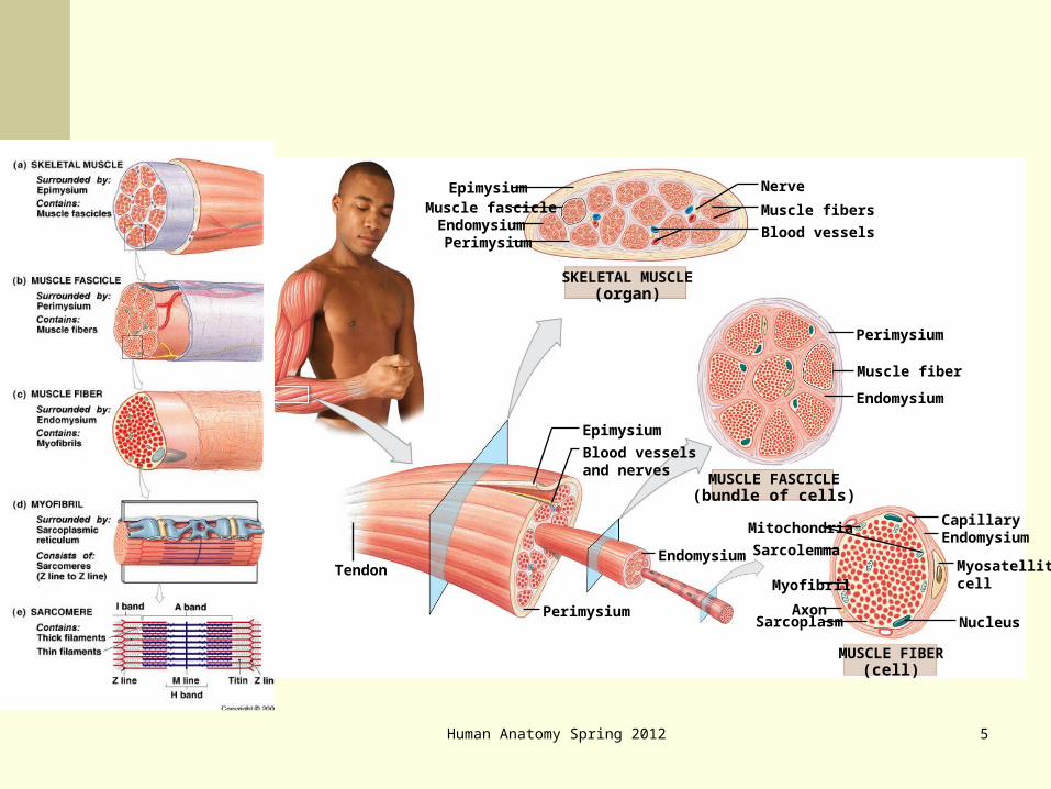
EpimysiumMuscle fascicle
EndomysiumPerimysium
Nerve
Muscle fibers
Blood vessels
SKELETAL MUSCLE(organ)
MUSCLE FASCICLE(bundle of cells)
Perimysium
Muscle fiber
Endomysium
Epimysium
Blood vesselsand nerves
Endomysium
Perimysium
Tendon
MUSCLE FIBER(cell)
Mitochondria
Sarcolemma
Myofibril
AxonSarcoplasm
CapillaryEndomysium
Myosatellitecell
Nucleus
Human Anatomy Spring 2012 5
Human Anatomy 6
Human Anatomy Spring 2012 7
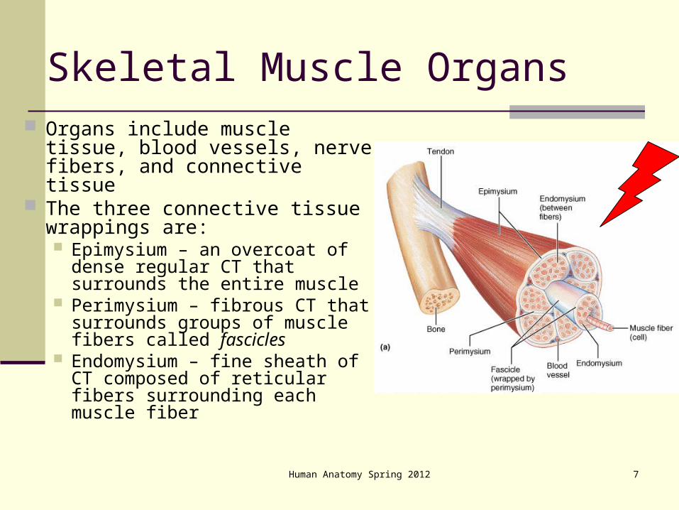
Skeletal Muscle Organs Organs include muscle tissue,
blood vessels, nerve fibers, and connective tissue
The three connective tissue wrappings are: Epimysium – an overcoat of
dense regular CT that surrounds the entire muscle
Perimysium – fibrous CT that surrounds groups of muscle fibers called fascicles
Endomysium – fine sheath of CT composed of reticular fibers surrounding each muscle fiber
Human Anatomy Spring 2012 8
Skeletal Muscle: Nerve and Blood Supply
Each muscle is served by at least one nerve, artery, and vein Each skeletal muscle fiber is supplied with a
nerve ending that controls contraction – a neuromuscular junction
Contracting fibers require continuous delivery of oxygen and nutrients via arteries
Wastes must be removed via veins

Human Anatomy Spring 2012 9
Skeletal Muscle: Attachments
Muscles span joints and are attached in at least two places
When muscles contract the movable bone, the muscle’s insertion moves toward the immovable bone – the muscle’s origin (i.e., origin is stationary— flawed concept, but customary)
Muscles attach: Directly – epimysium of the muscle is fused to the
periosteum of a bone Indirectly (more common) – CT wrappings extend
beyond the muscle as ropelike tendon or sheetlike aponeurosis

Human Anatomy Spring 2012 10
Microscopic Anatomy of a Skeletal Muscle Fiber
Each fiber is a long, cylindrical cell with multiple nuclei just beneath the sarcolemma
Fibers are 10 to 100 m in diameter, and up to hundreds of centimeters long
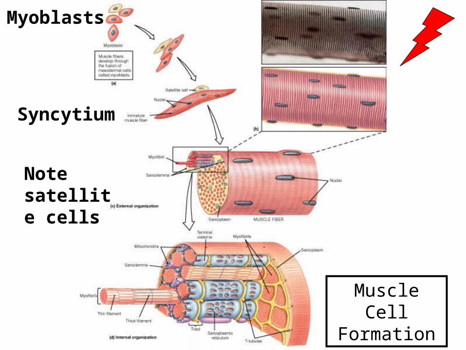
Each cell is a syncytium produced by fusion of myoblasts (embryonic cells)
Sarcoplasm has a unique oxygen-binding protein called myoglobin
Fibers contain the usual organelles plus myofibrils, sarcoplasmic reticulum, and T tubules
Human Anatomy Spring 2012 11
Muscle Cell Formation
Myoblasts
Syncytium
Note satellite cells
Human Anatomy Spring 2012 12
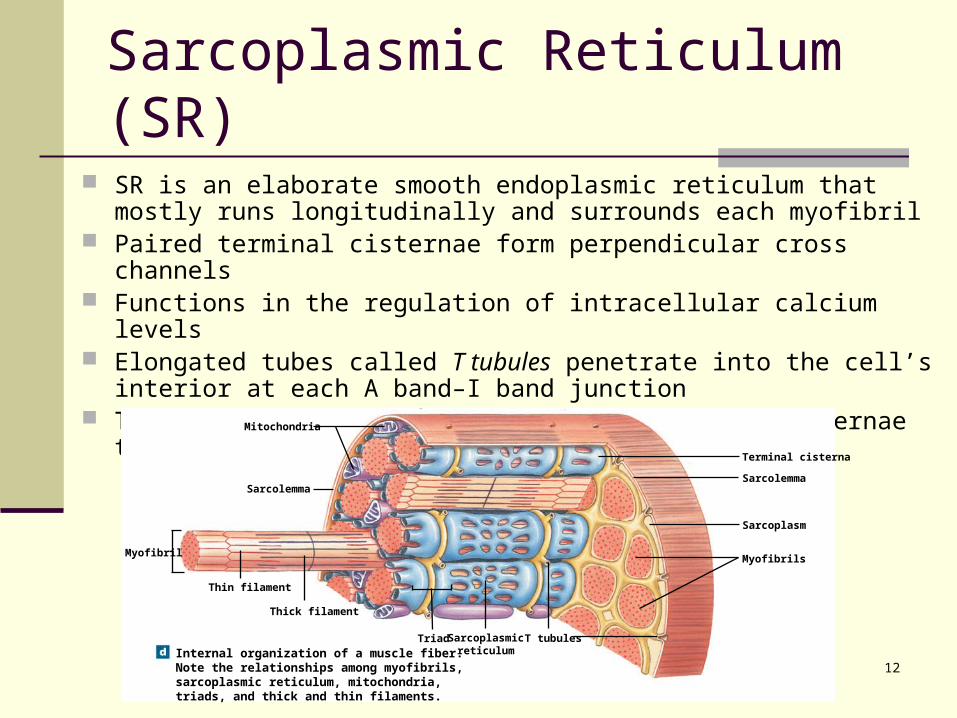
Sarcoplasmic Reticulum (SR)
SR is an elaborate smooth endoplasmic reticulum that mostly runs longitudinally and surrounds each myofibril
Paired terminal cisternae form perpendicular cross channels Functions in the regulation of intracellular calcium levels Elongated tubes called T tubules penetrate into the cell’s interior at
each A band–I band junction T tubules associate with the paired terminal cisternae to form triads
Internal organization of a muscle fiber.Note the relationships among myofibrils,sarcoplasmic reticulum, mitochondria,triads, and thick and thin filaments.
Mitochondria
Sarcolemma
Myofibril
Thin filament
Thick filament
Triad T tubulesSarcoplasmicreticulum
Terminal cisterna
Sarcolemma
Sarcoplasm
Myofibrils
Human Anatomy Spring 2012 13
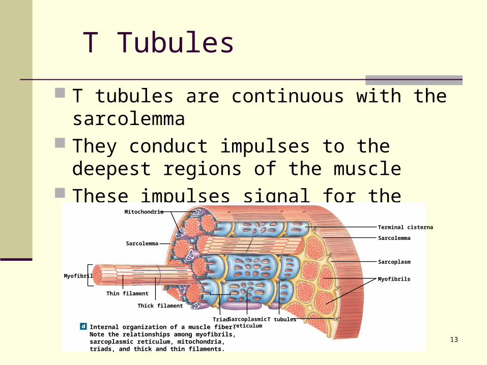
T Tubules
T tubules are continuous with the sarcolemma They conduct impulses to the deepest regions
of the muscle These impulses signal for the release of Ca2+
from adjacent terminal cisternae
Internal organization of a muscle fiber.Note the relationships among myofibrils,sarcoplasmic reticulum, mitochondria,triads, and thick and thin filaments.
Mitochondria
Sarcolemma
Myofibril
Thin filament
Thick filament
Triad T tubulesSarcoplasmicreticulum
Terminal cisterna
Sarcolemma
Sarcoplasm
Myofibrils
Human Anatomy Spring 2012 14
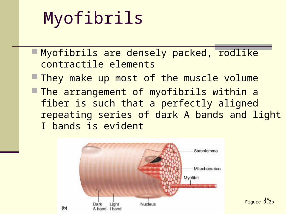
Myofibrils
Myofibrils are densely packed, rodlike contractile elements
They make up most of the muscle volume The arrangement of myofibrils within a fiber is
such that a perfectly aligned repeating series of dark A bands and light I bands is evident
Figure 9.2b
Human Anatomy Spring 2012 15
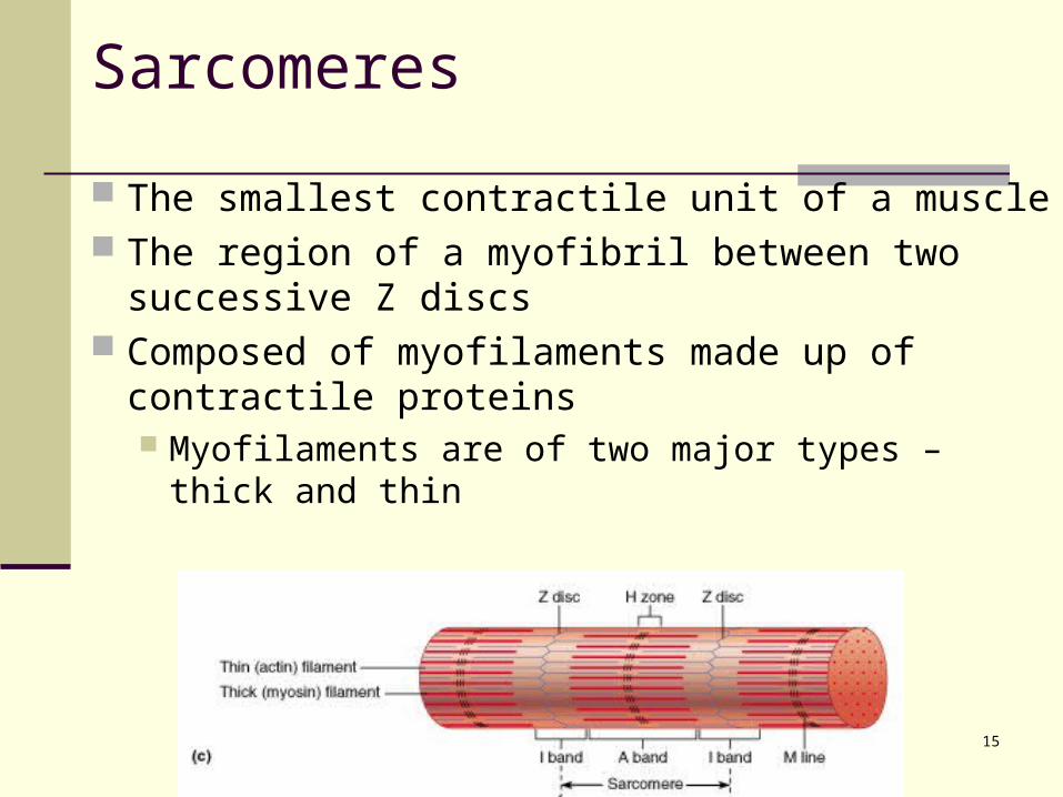
Sarcomeres
The smallest contractile unit of a muscle The region of a myofibril between two successive
Z discs Composed of myofilaments made up of contractile
proteins Myofilaments are of two major types – thick and
thin
Human Anatomy Spring 2012 16
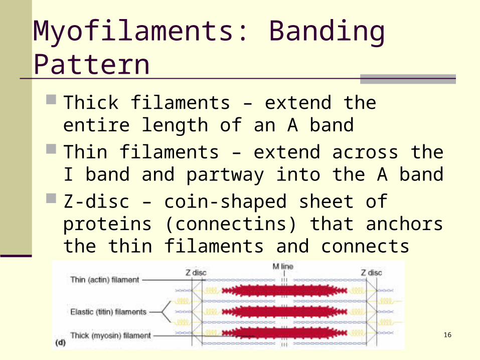
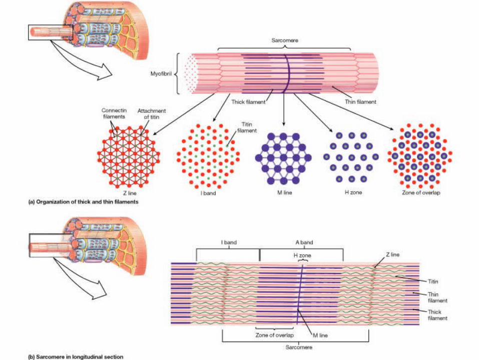
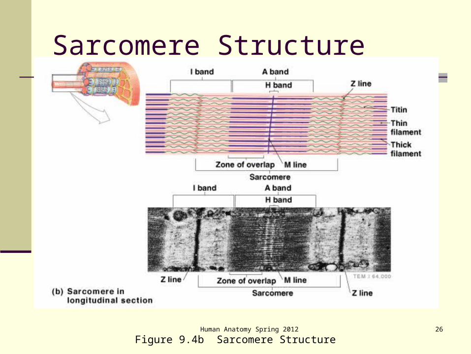
Myofilaments: Banding Pattern
Thick filaments – extend the entire length of an A band
Thin filaments – extend across the I band and partway into the A band
Z-disc – coin-shaped sheet of proteins (connectins) that anchors the thin filaments and connects myofibrils to one another
Human Anatomy Spring 2012 17
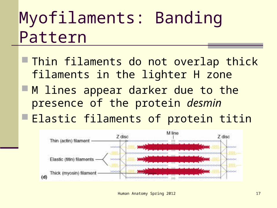
Myofilaments: Banding Pattern
Thin filaments do not overlap thick filaments in the lighter H zone
M lines appear darker due to the presence of the protein desmin
Elastic filaments of protein titin
Human Anatomy Spring 2012 18
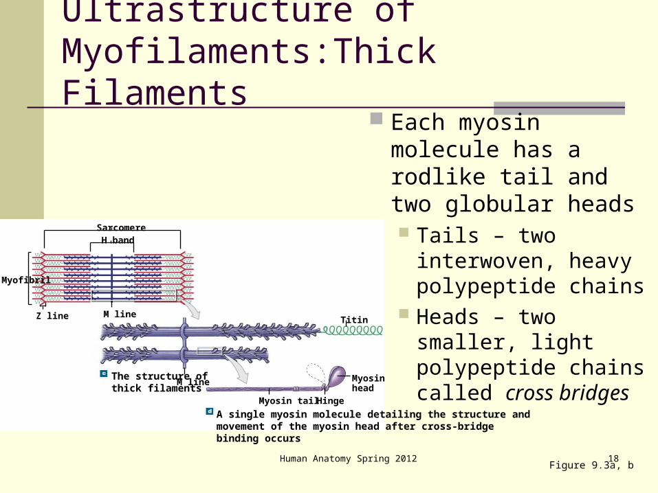
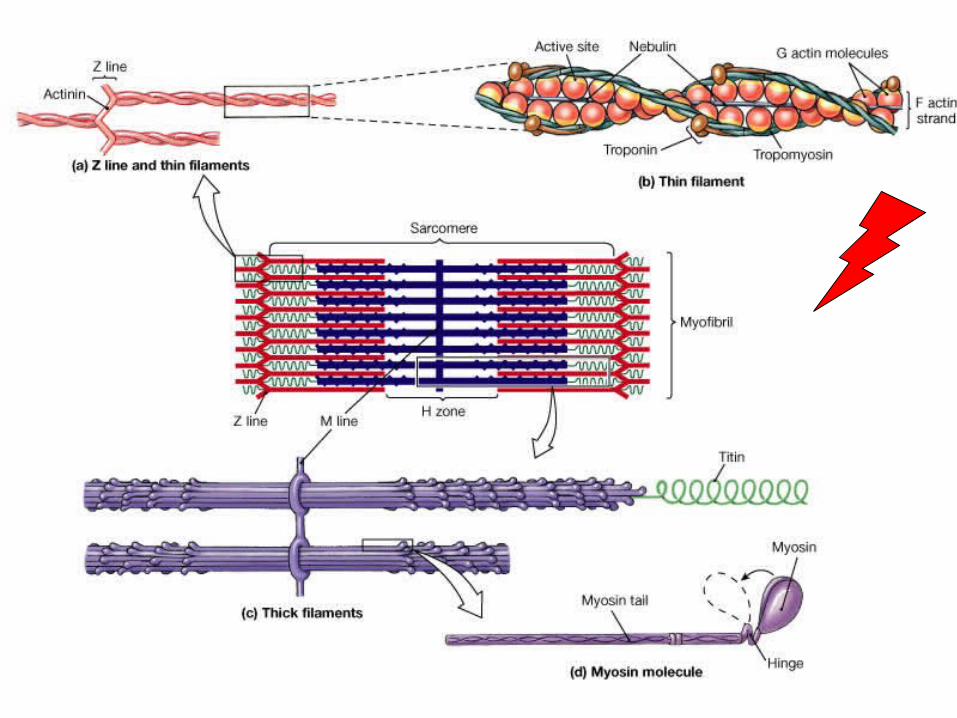
Ultrastructure of Myofilaments:Thick Filaments
Each myosin molecule has a rodlike tail and two globular heads Tails – two
interwoven, heavy polypeptide chains
Heads – two smaller, light polypeptide chains called cross bridges
Figure 9.3a, b
Myofibril
Z line M line
H bandSarcomere
A single myosin molecule detailing the structure andmovement of the myosin head after cross-bridgebinding occurs
The structure ofthick filaments
Titin
M line
Myosin tail
Myosin head
Hinge
19
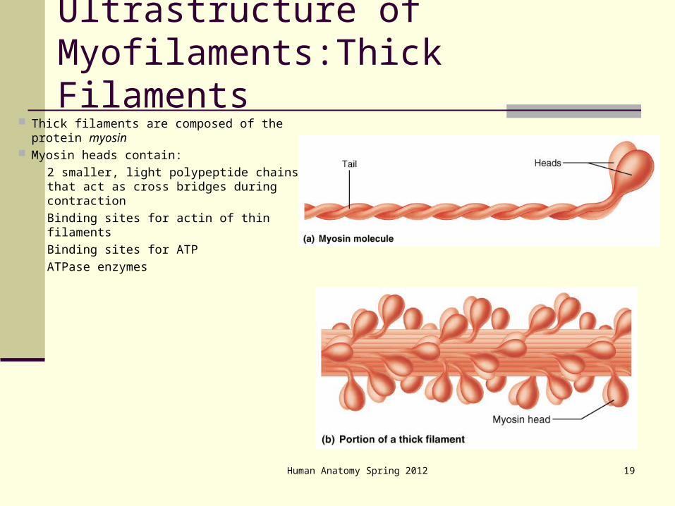
Ultrastructure of Myofilaments:Thick Filaments
Thick filaments are composed of the protein myosin
Myosin heads contain: 2 smaller, light polypeptide chains that
act as cross bridges during contraction Binding sites for actin of thin filaments Binding sites for ATP ATPase enzymes
Human Anatomy Spring 2012

The attachmentof thin filamentsto the Z line
The detailed structure of a thin filament showingthe organization of G actin, troponin, andtropomyosin
Myofibril
Z line M line
H band
Sarcomere
Actinin Z line Titin
Troponin Nebulin TropomyosinActive
siteG actin
molecules
F actinstrand
Human Anatomy Spring 2012 20
Ultrastructure of Myofilaments: Thin Filaments
Thin filaments are chiefly composed of protein actin Each actin molecule is a helical polymer of globular
subunits called G actin The subunits contain the active sites to which myosin
heads attach during contraction Tropomyosin (filamentous protein) and troponin are
regulatory subunits bound to actin
Human Anatomy Spring 2012 21
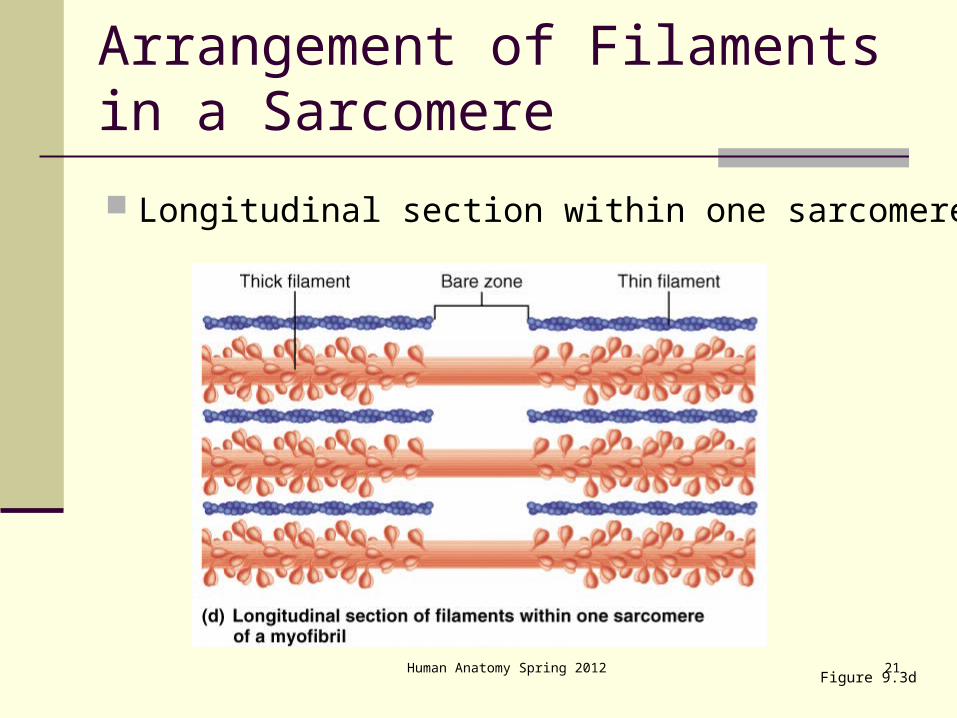
Arrangement of Filaments in a Sarcomere
Longitudinal section within one sarcomere
Figure 9.3d
22
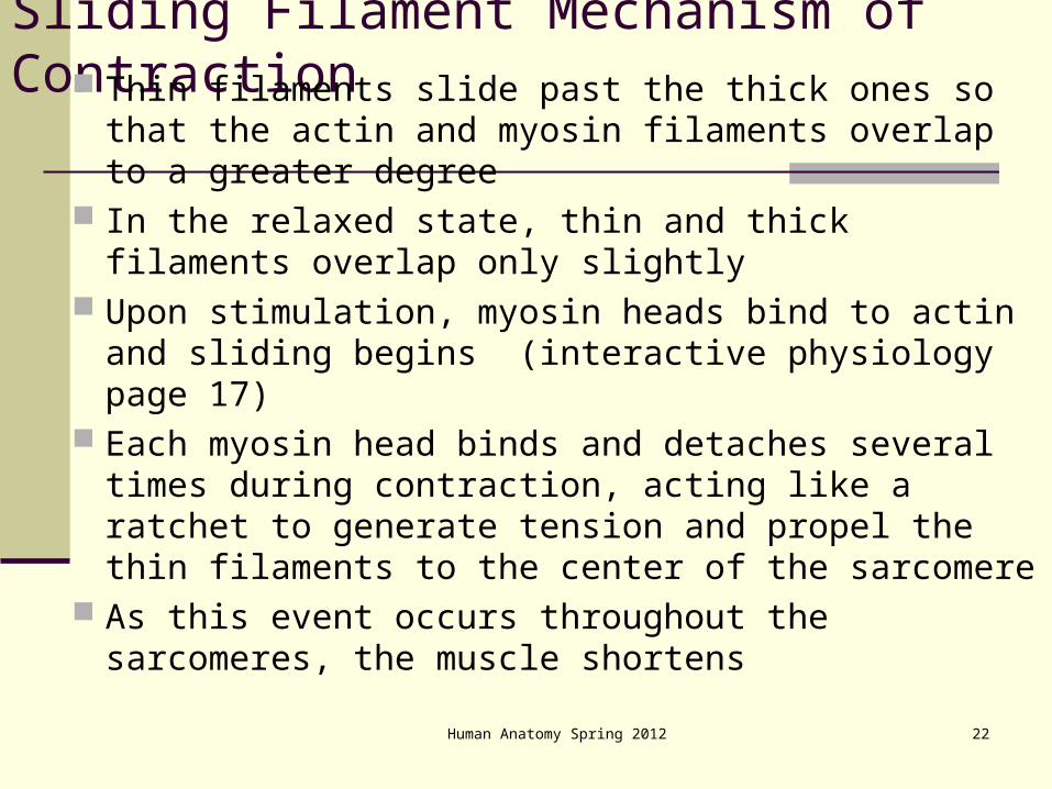
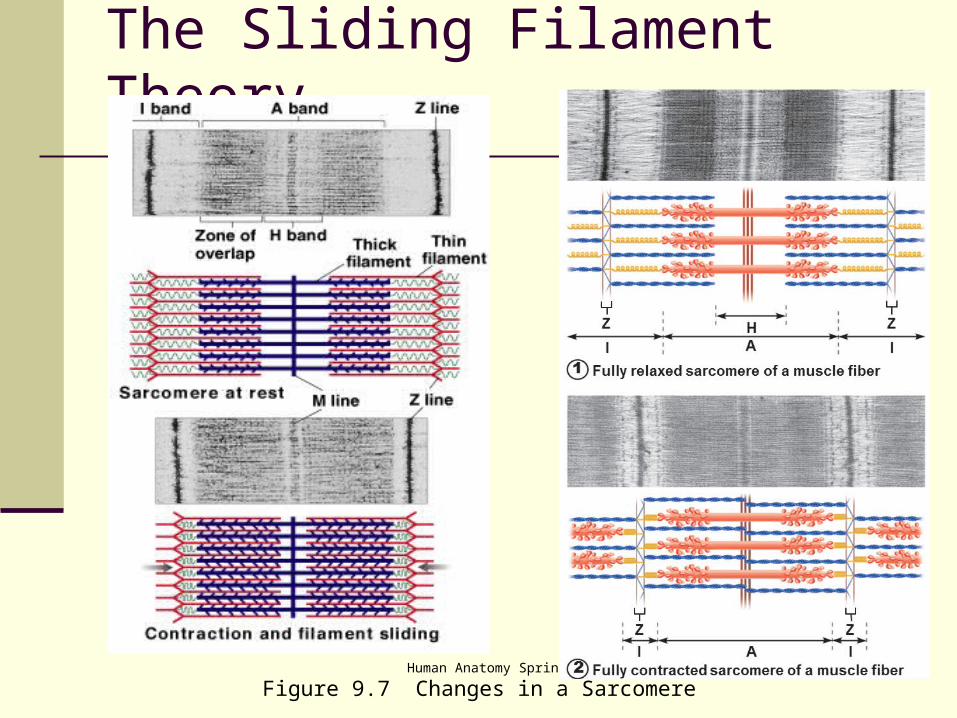
Sliding Filament Mechanism of Contraction Thin filaments slide past the thick ones so that the
actin and myosin filaments overlap to a greater degree
In the relaxed state, thin and thick filaments overlap only slightly
Upon stimulation, myosin heads bind to actin and sliding begins (interactive physiology page 17)
Each myosin head binds and detaches several times during contraction, acting like a ratchet to generate tension and propel the thin filaments to the center of the sarcomere
As this event occurs throughout the sarcomeres, the muscle shortensHuman Anatomy Spring 2012
Human Anatomy Spring 2012 23
Human Anatomy Spring 2012 24
Figure 9.7 Changes in a Sarcomere
The Sliding Filament Theory
Human Anatomy Spring 2012 25
Human Anatomy Spring 2012 26
Figure 9.4b Sarcomere Structure
Sarcomere Structure
Human Anatomy Spring 2012 27
Regulation of Contraction
In order to contract, a skeletal muscle must: Be stimulated by a nerve ending (NMJ) Propagate an electrical current, or action potential,
along its sarcolemma (T-tubules) Have a rise in intracellular Ca2+ levels, the final
trigger for contraction (SR)
Human Anatomy Spring 2012 28
Nerve Stimulus of Skeletal Muscle
Skeletal muscles are stimulated by motor neurons of the somatic nervous system
Axons of these neurons travel in nerves to muscle cells
Axons of motor neurons branch profusely as they enter muscles
Each axonal branch forms a neuromuscular junction with a single muscle fiber
Motorneuron
Axon
Muscle fiber
Path of actionpotential
Neuromuscularsynapse
Motorend plate
Myofibril
Human Anatomy Spring 2012 29
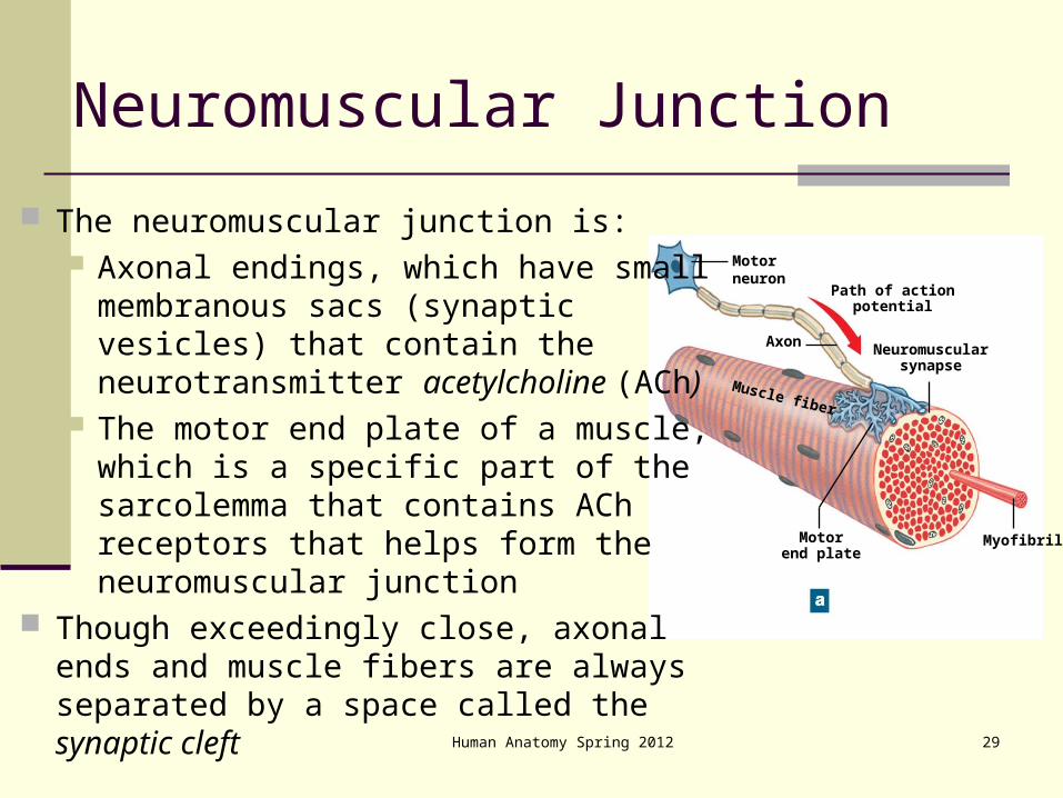
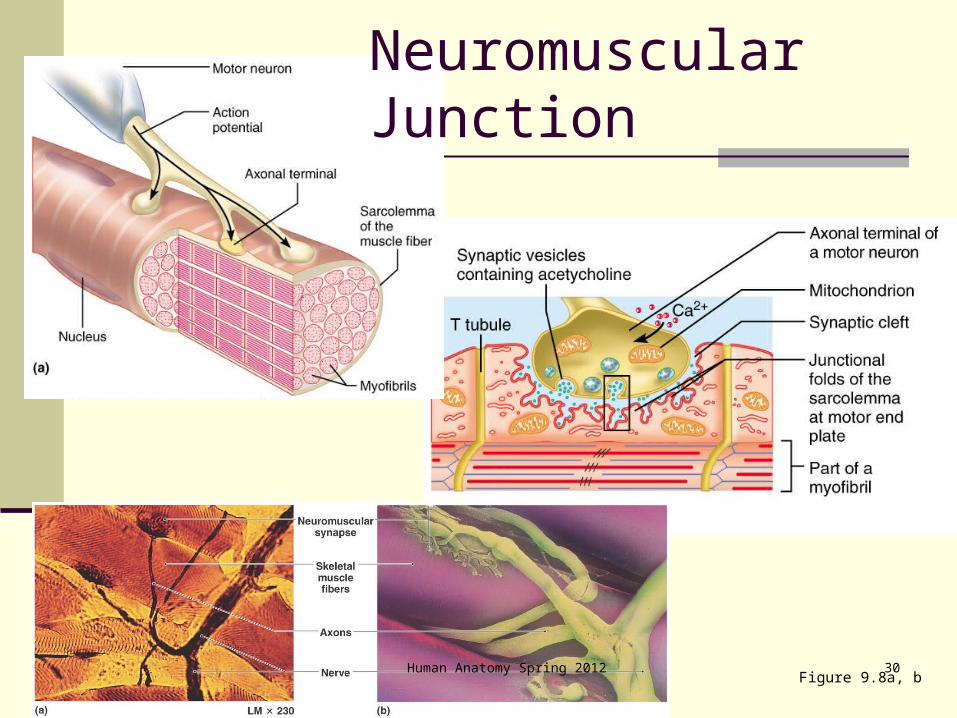
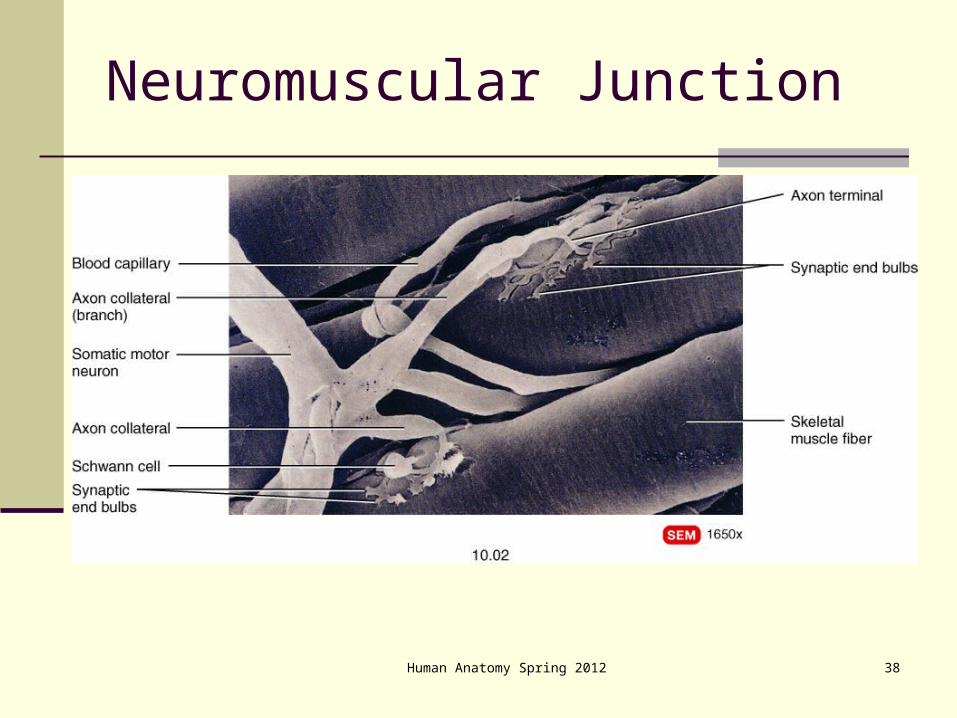
Neuromuscular Junction
The neuromuscular junction is: Axonal endings, which have small
membranous sacs (synaptic vesicles) that contain the neurotransmitter acetylcholine (ACh)
The motor end plate of a muscle, which is a specific part of the sarcolemma that contains ACh receptors that helps form the neuromuscular junction
Though exceedingly close, axonal ends and muscle fibers are always separated by a space called the synaptic cleft
30Figure 9.8a, b
Neuromuscular Junction
Human Anatomy Spring 2012

Human Anatomy Spring 2012 31
Neuromuscular Junction
When a nerve impulse reaches the end of an axon at the neuromuscular junction: Voltage-regulated calcium channels open and
allow Ca2+ to enter the axon Ca2+ inside the axon terminal causes axonal
vesicles to fuse with the axonal membrane This fusion releases ACh into the synaptic cleft via
exocytosis ACh diffuses across the synaptic cleft to ACh
receptors on the sarcolemma Binding of ACh to its receptors initiates an action
potential in the muscle

Human Anatomy Spring 2012 32
Neuromuscular Junction
Figure 9.8c

Human Anatomy Spring 2012 33
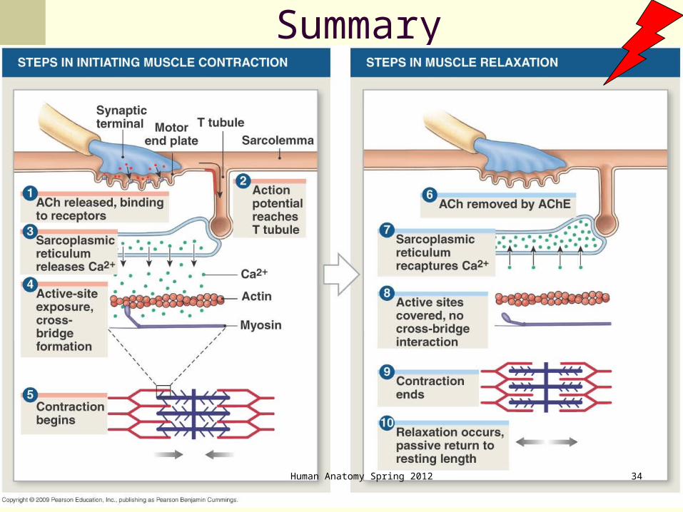
Summary
Human Anatomy Spring 2012 34

Human Anatomy Spring 2012 35
Excitation-Contraction Coupling
Spinal cord
Motor neuroncell body
Muscle
Nerve
Motorunit 1
Motorunit 2
Musclefibers
Motorneuronaxon
Axon terminals atneuromuscular junctions
36
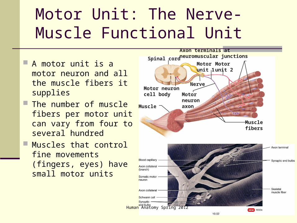
Motor Unit: The Nerve-Muscle Functional Unit
A motor unit is a motor neuron and all the muscle fibers it supplies
The number of muscle fibers per motor unit can vary from four to several hundred
Muscles that control fine movements (fingers, eyes) have small motor units
Human Anatomy Spring 2012

37
Motor Unit: The Nerve-Muscle Functional Unit
Large weight-bearing muscles (thighs, hips) have large motor units
Muscle fibers from a motor unit are spread throughout the muscle; therefore, contraction of a single motor unit causes weak contraction of the entire muscle
Spinal cord
Motor neuroncell body
Muscle
Nerve
Motorunit 1
Motorunit 2
Musclefibers
Motorneuronaxon
Axon terminals atneuromuscular junctions
Human Anatomy Spring 2012
Human Anatomy Spring 2012 38
Neuromuscular Junction
Human Anatomy Spring 2012 39
Muscle Tone
Muscle tone: The constant, slightly contracted state of all
muscles, which does not produce active movements
Keeps the muscles firm, healthy, and ready to respond to stimulus
Spinal reflexes account for muscle tone by: Activating one motor unit and then another Responding to activation of stretch receptors in
muscles and tendons

Human Anatomy Spring 2012 40
Force of Contraction The force of contraction is affected by:
The number of muscle fibers contracting – the more motor fibers in a muscle, the stronger the contraction
The relative size of the muscle – the bulkier the muscle, the greater its strength
Figure 9.19a

Human Anatomy Spring 2012 41
Force of Contraction
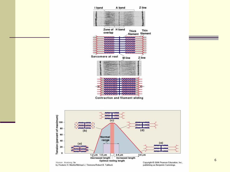
Frequency of stimulation Length-tension
relationships Series-elastic elements –
the noncontractile structures in a muscle
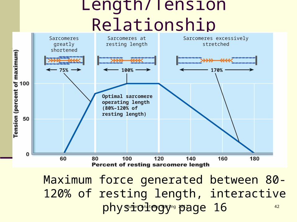
Degree of muscle stretch – muscles contract strongest when muscle fibers are 80-120% of their normal resting length
Figure 9.19a
42
Length/Tension Relationship
Maximum force generated between 80-120% of resting length, interactive physiology page 16
Think of cross-bridge mechanism for explanation
Sarcomeresgreatly
shortened
Sarcomeres atresting length
Sarcomeres excessivelystretched
170%
Optimal sarcomereoperating length(80%–120% ofresting length)
100%75%
Human Anatomy Spring 2012

43Human Anatomy Spring 2012
Human Anatomy Spring 2012 44
Muscle Fiber Type: Functional Characteristics
Speed of contraction – determined by speed in which ATPases split ATP The three types of fibers are slow and fast and
intermediate ATP-forming pathways
Oxidative fibers – use aerobic pathways Glycolytic fibers – use anaerobic glycolysis
These two criteria define three categories – slow oxidative fibers, fast oxidative fibers, and fast glycolytic fibers

Human Anatomy Spring 2012 45

Human Anatomy Spring 2012 46
Human Anatomy Spring 2012 47
Training
Slow, oxidative respond to endurance training. Diameter changes little.
Fast, oxidative respond to strength and power training. Diameter increases.
Intermediate can take on characteristics of fast or slow, depending on type of training.
In what birds do you expect to find FT? And ST?

Exercise causes: An increase in the number of mitochondria An increase in the activity of muscle spindles An increase in the concentration of glycolytic enzymes An increase in the glycogen reserves An increase in the number of myofibrils The net effect is an enlargement of the muscle
(hypertrophy) Disuse causes atrophy:
A decrease in muscle size A decrease in muscle tone
Muscle Hypertrophy
Human Anatomy Spring 2012 49
Developmental Aspects
Muscle tissue develops from embryonic mesoderm called myoblasts
Multinucleated skeletal muscles form by fusion of myoblasts forming a syncytium
The growth factor agrin stimulates the clustering of ACh receptors at newly forming motor end plates
As muscles are brought under the control of the somatic nervous system, the numbers of fast and slow fibers are also determined

Human Anatomy Spring 2012 50
Developmental Aspects
Cardiac myoblasts do not fuse but develop gap junctions at an early embryonic stage
Most smooth muscle follows the same pattern of gap junctions rather than fusion
Human Anatomy Spring 2012 51
Developmental Aspects: After Birth
Muscular development reflects neuromuscular coordination
Development occurs head-to-toe, and proximal-to-distal
Peak natural neural control of muscles is achieved by midadolescence
Athletics and training can improve neuromuscular control

Human Anatomy Spring 2012 52
Developmental Aspects: Male and Female
There is a biological basis for greater strength in men than in women
Women’s skeletal muscle makes up 36% of their body mass
Men’s skeletal muscle makes up 42% of their body mass
These differences are due primarily to the male sex hormone testosterone
With more muscle mass, men are generally stronger than women
Body strength per unit muscle mass, however, is the same

Human Anatomy Spring 2012 53
Homeostatic Imbalance: Age Related
With age, connective tissue increases and myofibrils, glycogen and myoglobin decrease
Muscles become stringier and more sinewy By age 80, 50% of muscle mass is lost
(sarcopenia), and myosatellite cells decrease Regular exercise reverses sarcopenia Aging of the cardiovascular system affects every
organ in the body Atherosclerosis may block distal arteries, leading to
intermittent claudication and causing severe pain in leg muscles

Human Anatomy Spring 2012 54
Developmental Aspects: Regeneration
Cardiac and skeletal muscle become amitotic, but can lengthen and thicken (hypertrophy)
Myoblastlike satellite cells of skeletal muscle show very limited regenerative ability (Cardiac tissue lacks satellite cells)
Smooth muscle has good regenerative ability (hyperplasia)

Levers
F1L1 = F2L2
Mass=Force L=length
There are several ways to increase the force efficiency of a lever: increasing the length of the in-lever arm decreasing the length of the out-lever or doing both of the above
Human Anatomy Spring 2012 55

Human Anatomy Spring 2012 56
See-saw
WheelbarrowLess distance
Hotdog tongsmost common, least mechanical advantage, more force, more speed/distance

Figure 12-21b
The Arm is a Lever and Fulcrum System
57
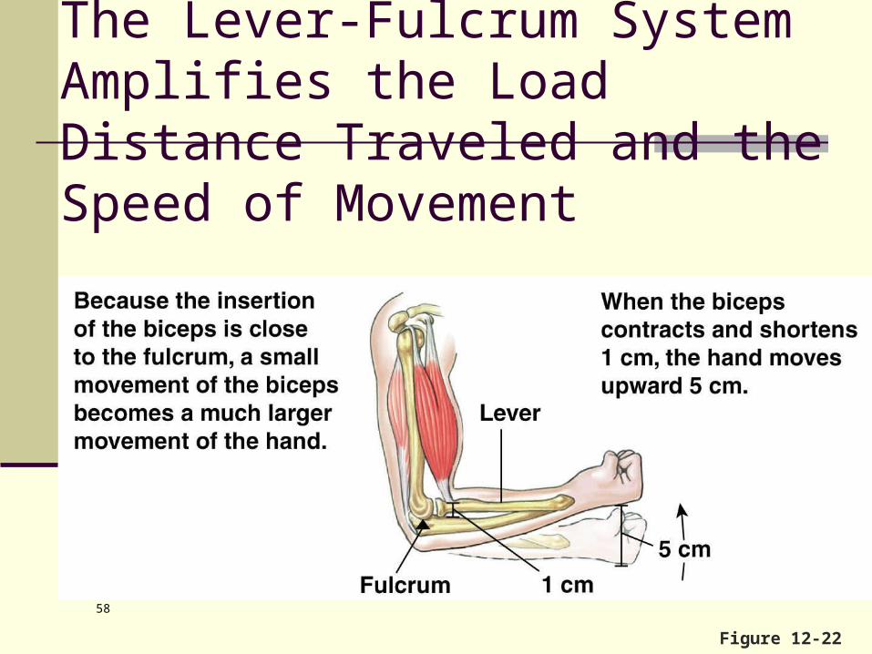
Figure 12-22
The Lever-Fulcrum System Amplifies the Load Distance Traveled and the Speed of Movement
58

Human Anatomy Spring 2012 59
Smooth Muscle
Composed of spindle-shaped fibers diameter of 2-10 m and lengths of several hundred m
Lack the coarse CT sheaths of skeletal muscle, but have fine endomysium
Figure 9.23

Human Anatomy Spring 2012 60
Smooth Muscle
Are generally organized into two layers (longitudinal and circular) of closely apposed fibers
Found in walls of hollow organs (except the heart)
Figure 9.23
Human Anatomy Spring 2012 61
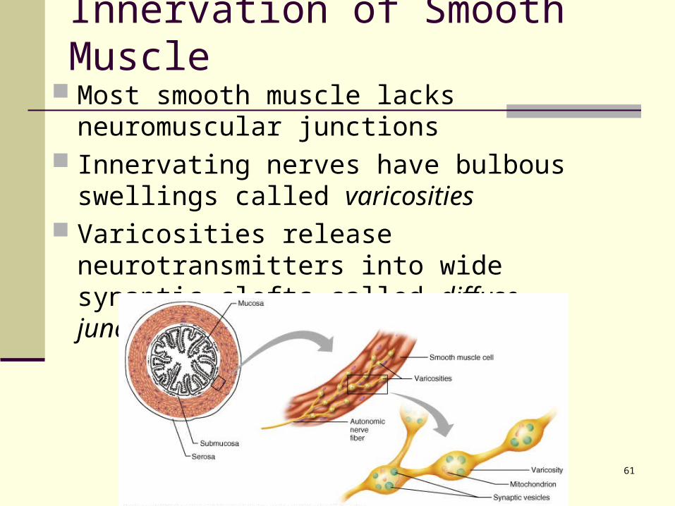
Innervation of Smooth Muscle Most smooth muscle lacks neuromuscular
junctions Innervating nerves have bulbous swellings
called varicosities Varicosities release neurotransmitters into
wide synaptic clefts called diffuse junctions

Human Anatomy Spring 2012 62
Microscopic Anatomy of Smooth Muscle
SR is less developed than in skeletal muscle and lacks a specific pattern (no cisterns)
T tubules are absent Plasma membranes have pouchlike infoldings
called caveoli Ca2+ is sequestered in the extracellular space
near the caveoli, allowing rapid influx when channels are opened
There are no visible striations and no sarcomeres Thin and thick filaments are present
Human Anatomy Spring 2012 63
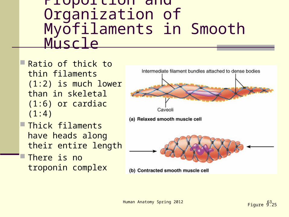
Ratio of thick to thin filaments (1:2) is much lower than in skeletal (1:6) or cardiac (1:4)
Thick filaments have heads along their entire length
There is no troponin complex
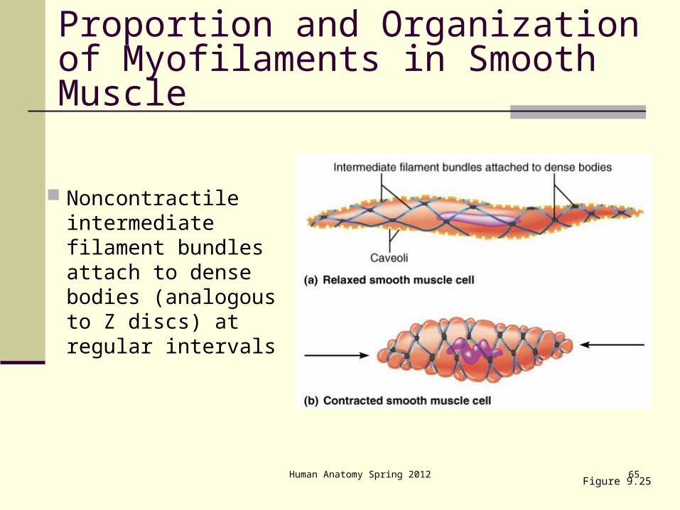
Figure 9.25
Proportion and Organization of Myofilaments in Smooth Muscle

Human Anatomy Spring 2012 64
Thick and thin filaments are arranged diagonally, causing smooth muscle to contract in a corkscrew manner
Figure 9.25
Proportion and Organization of Myofilaments in Smooth Muscle
Human Anatomy Spring 2012 65
Noncontractile intermediate filament bundles attach to dense bodies (analogous to Z discs) at regular intervals
Figure 9.25
Proportion and Organization of Myofilaments in Smooth Muscle

Human Anatomy Spring 2012 66
Contraction of Smooth Muscle
Whole sheets of smooth muscle exhibit slow, synchronized contraction
They contract in unison, reflecting their electrical coupling with gap junctions
Action potentials are transmitted from cell to cell Some smooth muscle cells:
Act as pacemakers and set the contractile pace for whole sheets of muscle
Are self-excitatory and depolarize without external stimuli

Human Anatomy Spring 2012 67
Contractile Mechanism
Actin and myosin interact according to the sliding filament mechanism
The final trigger for contractions is a rise in intracellular Ca2+
Ca2+ is released from the SR and from the extracellular space
Ca2+ interacts with calmodulin and myosin light chain kinase to activate myosin

Human Anatomy Spring 2012 68
Special Features of Smooth Muscle Contraction Unique characteristics of smooth muscle
include: Smooth muscle tone Slow, prolonged contractile activity Low energy requirements Response to stretch
Human Anatomy Spring 2012 69
Response to Stretch
Smooth muscles exhibits a phenomenon called stress-relaxation response in which: Smooth muscle responds to stretch only briefly,
and then adapts to its new length The new length, however, retains its ability to
contract This enables organs such as the stomach and
bladder to temporarily store contents
Human Anatomy Spring 2012 70
Types of Smooth Muscle: Single Unit
The cells of single unit smooth muscle, commonly called visceral muscle: Contract rhythmically as a unit Are electrically coupled to one another via gap
junctions Often exhibit spontaneous action potentials Are arranged in opposing sheets and exhibit
stress-relaxation response
Human Anatomy Spring 2012 71
Types of Smooth Muscle: Multiunit
Multiunit smooth muscles are found: In large airways to the lungs In large arteries In arrector pili muscles In the internal eye muscles
Characteristics include: Rare gap junctions Infrequent spontaneous depolarizations Structurally independent muscle fibers A rich nerve supply, which, with a number of muscle
fibers, forms motor units Graded contractions in response to neural stimuli
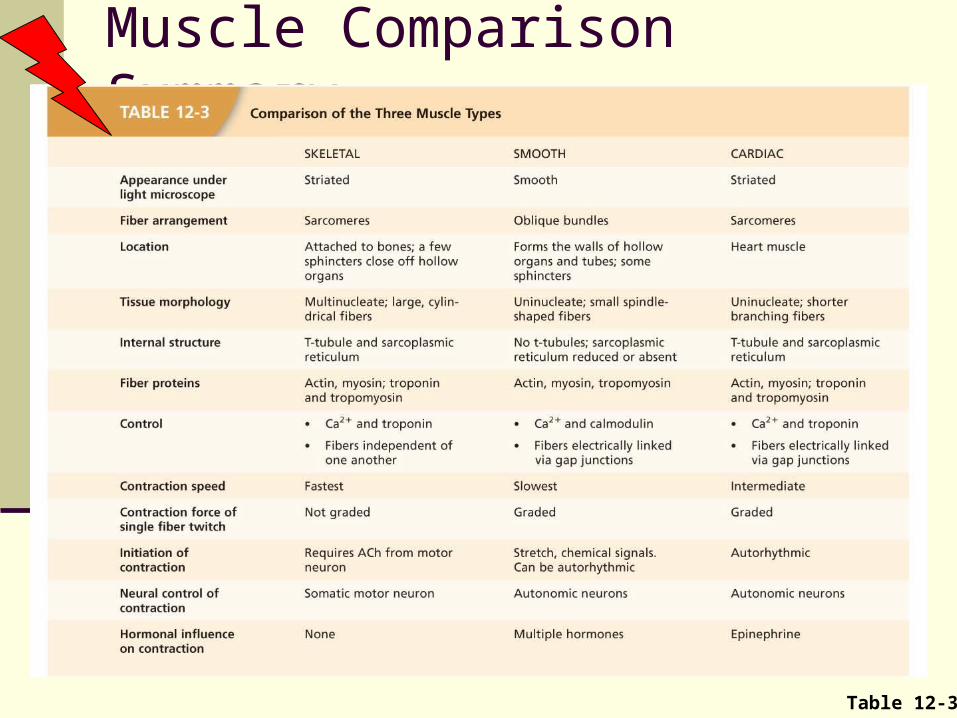
Muscle Comparison Summary
Table 12-3