Embed Size (px)
Citation preview
Chapter 9 Proteins
Mr. Kevin A. Boudreaux Angelo State University
CHEM 2353 Fundamentals of Organic Chemistry Organic and Biochemistry for Today (Seager & Slabaugh)
www.angelo.edu/faculty/kboudrea
Chapter Objectives:
• Learn about amino acid structure and classification.
• Learn about the formation of zwitterions and isoelectric points.
• Learn about reactions of amino acids, including the formation of disulfides, peptides and proteins.
• Learn about the characteristics, classification structure, and functions of proteins.
• Learn about the structures and characteristics that give rise to the primary, secondary, tertiary, and quaternary structure of proteins.
• Learn about protein hydrolysis and denaturation.
Chapter 9 Proteins
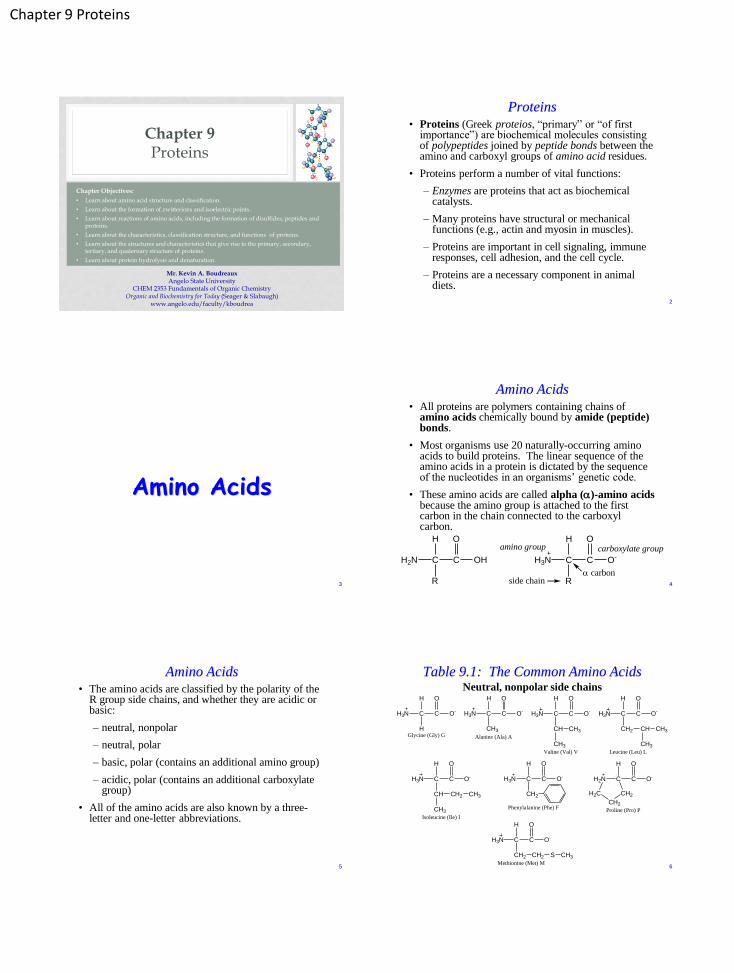
Proteins
• Proteins (Greek proteios, “primary” or “of first importance”) are biochemical molecules consisting of polypeptides joined by peptide bonds between the amino and carboxyl groups of amino acid residues.
• Proteins perform a number of vital functions:
– Enzymes are proteins that act as biochemical catalysts.
– Many proteins have structural or mechanical functions (e.g., actin and myosin in muscles).
– Proteins are important in cell signaling, immune responses, cell adhesion, and the cell cycle.
– Proteins are a necessary component in animal diets.
2
Amino Acids
3
Amino Acids • All proteins are polymers containing chains of
amino acids chemically bound by amide (peptide) bonds.
• Most organisms use 20 naturally-occurring amino acids to build proteins. The linear sequence of the amino acids in a protein is dictated by the sequence of the nucleotides in an organisms’ genetic code.
• These amino acids are called alpha (a)-amino acids because the amino group is attached to the first carbon in the chain connected to the carboxyl carbon.
4
C
H
H3N C
R
O
O-
a carbon
carboxylate groupamino group
side chain
C
H
H2N C
R
O
OH
Amino Acids • The amino acids are classified by the polarity of the
R group side chains, and whether they are acidic or basic:
– neutral, nonpolar
– neutral, polar
– basic, polar (contains an additional amino group)
– acidic, polar (contains an additional carboxylate group)
• All of the amino acids are also known by a three-letter and one-letter abbreviations.
5
Table 9.1: The Common Amino Acids
6
C
H
H3N C
H
O
O-
Glycine (Gly) G
Neutral, nonpolar side chains
C
H
H3N C
CH3
O
O-
Alanine (Ala) A
C
H
H3N C
CH
O
O-
Valine (Val) V
CH3
CH3
C
H
H3N C
CH2
O
O-
Leucine (Leu) L
CH CH3
CH3
C
H
H3N C
CH
O
O-
Isoleucine (Ile) I
CH2 CH3
CH3
C
H
H3N C
CH2
O
O-
Phenylalanine (Phe) F
C
H
H2N C
O
O-
Proline (Pro) P
H2C
CH2
CH2
C
H
H3N C
CH2
O
O-
Methionine (Met) M
CH2 S CH3
Chapter 9 Proteins
Table 9.1: The Common Amino Acids
7
Neutral, polar side chains
C
H
H3N C
CH2
O
O-
Serine (Ser) S
OH
C
H
H3N C
CH
O
O-
Threonine (Thr) T
OH
CH3
C
H
H3N C
CH2
O
O-
Tyrosine (Tyr)Y
OH
C
H
H3N C
CH2
O
O-
Tryptophan (Trp) W
N
H
C
H
H3N C
CH2
O
O-
Cysteine (Cys) C
SH
C
H
H3N C
CH2
O
O-
Asparagine (Asn) N
C
O
NH2
C
H
H3N C
CH2
O
O-
Glutamine (Gln) Q
C
O
NH2CH2
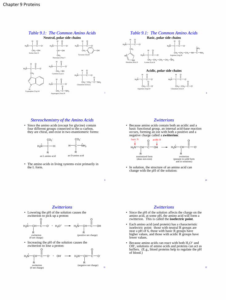
Table 9.1: The Common Amino Acids
8
Basic, polar side chains
C
H
H3N C
CH2
O
O-
Histidine (His) H
HN
NH
C
H
H3N C
CH2
O
O-
Lysine (Lys) K
CH2 CH2 CH2 NH3
C
H
H3N C
CH2
O
O-
Arginine (Arg) R
C
NH2
NH2CH2 CH2 NH
Acidic, polar side chains
C
H
H3N C
CH2
O
O-
Aspartate (Asp) D
C
O
O-
C
H
H3N C
CH2
O
O-
Glutamate (Glu) E
C
O
O-CH2
Stereochemistry of the Amino Acids • Since the amino acids (except for glycine) contain
four different groups connected to the a-carbon, they are chiral, and exist in two enantiomeric forms:
• The amino acids in living systems exist primarily in the L form.
9
CH3N
R
CO2-
H
an L-amino acid
CH
R
CO2-
NH3
an D-amino acid
Zwitterions • Because amino acids contain both an acidic and a
basic functional group, an internal acid-base reaction occurs, forming an ion with both a positive and a negative charge called a zwitterion:
• In solution, the structure of an amino acid can change with the pH of the solution:
10
CHH2N C
R
O
OH CHH3N C
R
O
O-
nonionized form(does not exist)
zwitterion(present in solid form
and in solutions)
acidic Hbasic N
Zwitterions • Lowering the pH of the solution causes the
zwitterion to pick up a proton:
• Increasing the pH of the solution causes the zwitterion to lose a proton:
11
CHH3N C
R
O
OHCHH3N C
R
O
O-
(positive net charge)zwitterion(0 net charge)
+ H3O+
CHH2N C
R
O
O-CHH3N C
R
O
O-
(negative net charge)zwitterion(0 net charge)
+ OH-
Zwitterions • Since the pH of the solution affects the charge on the
amino acid, at some pH, the amino acid will form a zwitterion. This is called the isoelectric point.
• Each amino acid (and protein) has a characteristic isoelectric point: those with neutral R groups are near a pH of 6, those with basic R groups have higher values, and those with acidic R groups have lower values.
• Because amino acids can react with both H3O+ and
OH-, solutions of amino acids and proteins can act as buffers. (E.g., blood proteins help to regulate the pH of blood.)
12
Chapter 9 Proteins
Examples: Amino Acid Structures
• Identify the following amino acid R groups as being polar or nonpolar, and acidic or basic:
• Draw the structure of the amino acid leucine (a) in acidic solution at a pH below the isoelectric point, and (b) in basic solution at a pH above the isoelectric point.
13
C
H
H3N C
CH2
O
O-
Leucine (Leu) L
CH CH3
CH3
C
H
H3N C
CH2
O
O- C
H
H3N C
CH2
O
O-
OH
C
H
H3N C
CH2
O
O-
CH2 CH2 CH2 NH3
Examples: Amino Acid Structures
• Draw Fischer projections representing the D and L form of aspartate and cysteine.
14
Reactions of Amino Acids
15
Oxidation of Cysteine • Amino acids can undergo any of the reactions
characteristic of the functional groups in the structure.
• Cysteine is the only amino acid that contains a sulfhydryl (thiol, R—SH) group. Thiols are easily oxidized to form disulfide bonds (R—S—S—R). This allows cysteine to dimerize to form cystine:
16
CHH3N C
CH2
O
O-
Cysteine
SH
CHH3N C
CH2
O
O-
SH
CHH3N C
CH2
O
O-
Cystine
S
CHH3N C
CH2
O
O-
S
[O]
oxidizing agent
reducing agent
Peptide Formation • Amides can be thought of as forming from the
reaction of an amine and a carboxylic acid:
• In the same way, two amino acids can combine to form a dipeptide, held together by a peptide bond:
17
R C OH NR' H R C N R' ++
H HO O
HOH
carboxylic acid amine amide linkage
++ HOHC
H
H3N C
H
O
O-
glycine
C
H
H3N C
CH3
O
O-
alanine
C
H
H3N C
H
O
glycylalanine(a dipeptide)
C
H
NH C
CH3
O
O-
peptide linkage
Peptide Formation • The two amino acids can connect the other way as
well, forming a structural isomer of the dipeptide, with a unique set of physical properties:
• A third amino acid can join the chain to form a tripeptide:
18
++ HOHC
H
H3N C
CH3
O
O-
alanine
C
H
H3N C
H
O
O-
glycine
C
H
H3N C
CH3
O
alanylglycine(a dipeptide)
C
H
NH C
H
O
O-
C
H
H3N C
CH3
O
alanylglycylvaline(a tripeptide)
C
H
NH C
H
O
NH C
CH CH3
CH3
C
O
O-
H

Chapter 9 Proteins
Peptides
• A fourth amino acid would form a tetrapeptide, a fifth would form a pentapeptide, and so on.
• Short chains are referred to as peptides, chains of up to about 50 amino acids are polypeptides, and chains of more than 50 amino acids are proteins. (The terms protein and polypeptide are often used interchangeably.)
• Amino acids in peptide chains are called amino acid residues.
– The residue with a free amino group is called the N-terminal residue, and is written on the left end of the chain.
– The residue with a free carboxylate group is called the C-terminal residue, and is written on the right end of the chain. 19
Peptides
• Peptides are named by starting at the N-terminal end and listing the amino acid residues from left to right.
• Large amino acid chains are unwieldy to draw in their complete forms, so they are usually represented by their three-letter abbreviations, separated by dashes:
– Gly-Ala (Gly = N-terminal, Ala = C-terminal)
– Ala-Gly (Ala = N-terminal, Gly = C-terminal)
• The tripeptide alanylglycylvaline can be written as Ala-Gly-Val. (There are five other arrangements of these amino acids that are possible.)
• Insulin has 51 amino acids, with 1.551066 different possible arrangements, but the body produces only one.
20
Examples: Reactions of Amino Acids
• Write two reactions to represent the formation of the two dipeptides that form when valine and serine react.
• Write a complete structural formula and an abbreviated formula for the tripeptide formed from aspartate, cysteine, and valine in which the C-terminal residue is cysteine and the N-terminal residue is valine.
21
Examples: Reactions of Amino Acids
• How many tripeptide isomers that contain one residue each of valine, phenylalanine, and lysine are possible? Write the abbreviated formulas for these peptides.
22
Important Peptides
23
Vasopressin and Oxytocin • More than 200 peptides have been identified as being essential to
the body’s proper functioning.
• Vasopressin and oxytocin are nonapeptide hormones secreted by the pituitary gland. Six of the amino acid residues are held in a loop by disulfide bridges formed by the oxidation of two cysteine residues. Even though the molecules are very similar, their biological functions are quite different:
– Vassopressin is known as antidiuretic hormone (ADH) because it reduces the amount of urine formed, which causes the body to conserve water. It also raises blood pressure.
– Oxytocin causes the smooth muscles of the uterus to contract, and is administered to induce labor. It also stimulates the smooth muscles of mammary glands to stimulate milk ejection.
24
Chapter 9 Proteins
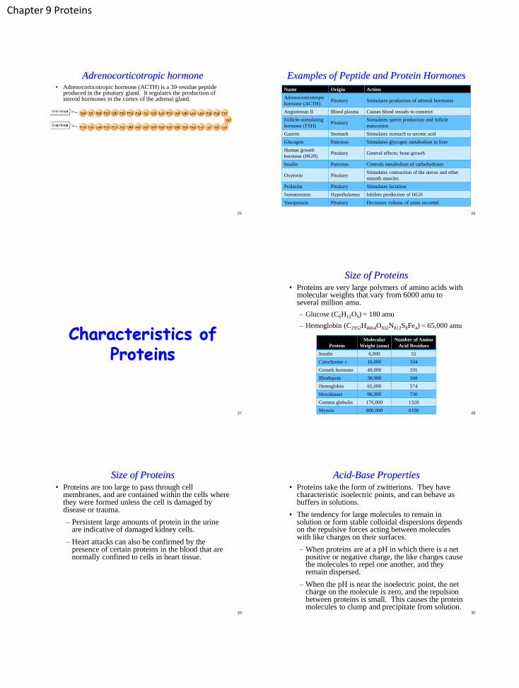
Adrenocorticotropic hormone • Adrenocorticotropic hormone (ACTH) is a 39-residue peptide
produced in the pituitary gland. It regulates the production of steroid hormones in the cortex of the adrenal gland.
25
Examples of Peptide and Protein Hormones
Name Origin Action
Adrenocorticotropic
hormone (ACTH) Pituitary Stimulates production of adrenal hormones
Angiotensin II Blood plasma Causes blood vessels to constrict
Follicle-stimulating
hormone (FSH) Pituitary
Stimulates sperm production and follicle
maturation
Gastrin Stomach Stimulates stomach to secrete acid
Glucagon Pancreas Stimulates glycogen metabolism in liver
Human growth
hormone (HGH) Pituitary General effects; bone growth
Insulin Pancreas Controls metabolism of carbohydrates
Oxytocin Pituitary Stimulates contraction of the uterus and other
smooth muscles
Prolactin Pituitary Stimulates lactation
Somatostatin Hypothalamus Inhibits production of HGH
Vasopressin Pituitary Decreases volume of urine excreted
26
Characteristics of Proteins
27
Size of Proteins • Proteins are very large polymers of amino acids with
molecular weights that vary from 6000 amu to several million amu.
– Glucose (C6H12O6) = 180 amu
– Hemoglobin (C2952H4664O832N812S8Fe4) = 65,000 amu
28
Protein
Molecular
Weight (amu)
Number of Amino
Acid Residues
Insulin 6,000 51
Cytochrome c 16,000 104
Growth hormone 49,000 191
Rhodopsin 38,900 348
Hemoglobin 65,000 574
Hexokinase 96,000 730
Gamma globulin 176,000 1320
Myosin 800,000 6100
Size of Proteins • Proteins are too large to pass through cell
membranes, and are contained within the cells where they were formed unless the cell is damaged by disease or trauma.
– Persistent large amounts of protein in the urine are indicative of damaged kidney cells.
– Heart attacks can also be confirmed by the presence of certain proteins in the blood that are normally confined to cells in heart tissue.
29
Acid-Base Properties • Proteins take the form of zwitterions. They have
characteristic isoelectric points, and can behave as buffers in solutions.
• The tendency for large molecules to remain in solution or form stable colloidal dispersions depends on the repulsive forces acting between molecules with like charges on their surfaces.
– When proteins are at a pH in which there is a net positive or negative charge, the like charges cause the molecules to repel one another, and they remain dispersed.
– When the pH is near the isoelectric point, the net charge on the molecule is zero, and the repulsion between proteins is small. This causes the protein molecules to clump and precipitate from solution.
30

Chapter 9 Proteins
Protein Function
• Proteins perform crucial roles in all biological processes.
1. Catalytic function: Nearly all reactions in living organisms are catalyzed by proteins functioning as enzymes. Without these catalysts, biological reactions would proceed much more slowly.
2. Structural function: In animals structural materials other than inorganic components of the skeleton are proteins, such as collagen (mechanical strength of skin and bone) and keratin (hair, skin, fingernails).
3. Storage function: Some proteins provide a way to store small molecules or ions, e.g., ovalbumin (used by embryos developing in bird eggs), casein (a milk protein) and gliadin (wheat seeds), and ferritin (a liver protein which complexes with iron ions).
31
Protein Function
4. Protective function: Antibodies are proteins that protect the body from disease by combining with and destroying viruses, bacteria, and other foreign substances. Another protective function is blood clotting, carried out by thrombin and fibrinogen.
5. Regulatory function: Body processes regulated by proteins include growth (growth hormone) and thyroid functions (thyrotropin).
6. Nerve impulse transmission: Some proteins act as receptors for small molecules that transmit impulses across the synapses that separate nerve cells (e.g., rhodopsin in vision).
7. Movement function: The proteins actin and myosin are important in muscle activity, regulating the contraction of muscle fibers.
32
Protein Function 8. Transport function: Some proteins bind small
molecules or ions and transport them through the body.
– Serum albumin is a blood protein that carries fatty acids between fat (adipose) tissue and other organs.
– Hemoglobin carries oxygen from the lungs to other body tissues.
– Transferrin is a carrier of iron in blood plasma.
• A typical human cell contains 9000 different proteins; the human body contains about 100,000 different proteins.
33
Table 19.4 Biological Functions of Proteins
34
Function Examples Occurrence or role
Catalysis lactate dehydrogenase Oxidizes lactic acid
cyctochrome c Transfers electrons
DNA polymerase Replicates and repairs DNA
Structure viral-coat proteins Sheath around nucleic acid of virus
glycoproteins Cell coats and walls
a-keratin Skin, hair, feathers, nails, and hooves
b-keratin Silk of cocoons and spiderwebs
collagen Fibrous connective tissue
elastin Elastic connective tissue
Storage ovalbumin Egg-white protein
casein A milk protein
ferritin Stores iron in the spleen
gliadin Stores amino acids in wheat
zein Stores amino acids in corn
Table 19.4 Biological Functions of Proteins
35
Function Examples Occurrence or role
Protection antibodies Form complexes with foreign proteins
fibrinogen Involved in blood clotting
thrombin Involved in blood clotting
Regulation insulin Regulates glucose metabolism
growth hormone Stimulates growth of bone
Nerve impulse
transmission
rhodopsin Involved in vision
acetylcholine receptor protein Impulse transmission in nerve cells
Movement myosin Thick filaments in muscle fiber
actin Thin filaments in muscle fiber
dynein Movement of cilia and flagella
Transport hemoglobin Transports O2 in blood
myoglobin Transports O2 in muscle cells
serum albumin Transports fatty acids in blood
transferrin Transports iron in blood
ceruloplasmin Transports copper in blood
Classification by Structural Shape Proteins can be classified on the basis of their
structural shapes:
• Fibrous proteins are made up of long rod-shaped or stringlike molecules that can intertwine with one another and form strong fibers.
– insoluble in water
– major components of connective tissue, elastic tissue,
hair, and skin
– e.g., collagen, elastin, and keratin.
• Globular proteins are more spherical in shape – dissolve in water or form stable suspensions.
– not found in structural tissue but are transport proteins, or proteins that may be moved easily through the body by the circularoty system
– e.g., hemoglobin and transferrin. 36

Chapter 9 Proteins
Classification by Composition
Proteins can also be classified by composition:
• Simple proteins contain only amino acid residues.
• Conjugated proteins also contain other organic or inorganic components, called prosthetic groups.
– nucleoproteins — nucleic acids (viruses).
– lipoproteins — lipids (fibrin in blood, serum lipoproteins)
– glycoproteins — carbohydrates (gamma globulin in blood, mucin in saliva)
– phosphoproteins — phosphate groups (casein in milk)
– hemoproteins — heme (hemoglobin, myoglobin, cytochromes)
– metalloproteins — iron (feritin, hemoglobin) or zinc (alcohol dehydrogenase)
37
38
Protein Structure
39
Protein Structure • The structure of proteins is much more complex than
that of simple organic molecules.
– Many protein molecules consist of a chain of amino acids twisted and folded into a complex three-dimensional structure
– The complex 3D structures of proteins impart unique features to proteins that allow them to function in diverse ways.
• There are four levels of organization in proteins structure: primary, secondary, tertiary, and quaternary.
40
Primary Structure of Proteins • The primary structure of a protein is the linear
sequence of the side chains that are connected to the protein backbone:
• Each protein has a unique sequence of amino acid residues that cause it to fold into a distinctive shape that allows the protein to function properly.
• Primary structure of human insulin:
41
CHNH C
R
O
protein backbone
CHNH C
R'
O
NH CH
R''
C
O
CHNH C
R'''
O
CHNH C
R''''
O
Secondary Structure — The a Helix • Hydrogen bonding causes protein chains to fold and
align to produce orderly patterns called secondary structures.
42
• The a-helix is a single protein chain twisted to resemble a coiled helical spring.

Chapter 9 Proteins
Secondary Structure — The a Helix • The a-helix is held in this shape
by hydrogen bonding interactions between amide groups, with the side chains extending outward from the coil.
• The amount of a-helix coiling in proteins is highly variable.
43
C
O
N
H
C N
H
O
• In a-keratin (hair, pictured below), myosin (muscles), epidermin (skin), and fibrin (blood clots), two or more helices coil together (supracoiling) to form cables. These cables make up bundles of fibers that strengthen tissues in which they are found:
Secondary Structure — The b-Pleated Sheet • Another secondary structure is the b-pleated sheet,
in which several protein chains lie side by side, held by hydrogen bonds between adjacent chains:
44
Secondary Structure — The b-Pleated Sheet • The b-pleated sheet structure is less common than
the a-helix; it is found extensively only in the protein of silk.
• The figure below shows both types of secondary structures in a single protein.
45
Tertiary Structure of Proteins • The tertiary structure of a protein refers to the
bending and folding of the protein into a specific three-dimensional shape.
• These structures result from four types of interactions between the R side chains of the amino acids residues:
1. Disulfide bridges can form between two cysteine residues that are close to each other in the same chain, or between cysteine residues in different chains. These bridges hold the protein chain in a loop or some other 3D shape.
2. Salt bridges are attractions between ions that result from the interactions of the ionized side chains of acidic amino acids (—COO-) and the side chains of basic amino acids (—NH3
+). 46
Tertiary Structure of Proteins 3. Hydrogen bonds can form between a variety of side
chains, especially those that contain:
Hydrogen bonding also influences the secondary structure, but here the hydrogen bonding is between R groups, while in secondary structures it is between the C=O and NH portions of the backbone.
4. Hydrophobic interactions result from the attraction of nonpolar groups, or when they are forced together by their mutual repulsion of the aqueous solvent. These interactions are particularly important between the benzene rings in phenylalanine or tryptophan. This type of interaction is relatively weak, but since it acts over large surface areas, the net effect is a strong interaction.
47
C
O
NH2OH NH2
Tertiary Structure of Proteins 4. cont. The compact structure of globular proteins in
aqueous solution, in which the nonpolar groups are pointed inward, away from the water molecules.
48

Chapter 9 Proteins
Tertiary Structure of Proteins — Summary
49
Examples: R-Group Interactions
• What kind of R-group interaction might be expected if the following side chains were in close proximity?
50
CH2 CH CH3
CH3
CH CH2 CH3
CH3
CH2 OHCH2 C
O
NH2
CH2 CH2 CH2 CH2 NH3CH2 C
O
O-
Visualizing Protein Structure
51
Quaternary Structure of Proteins • When two or more polypeptide chains are held
together by disulfide bridges, salt bridges, hydrogen bond, or hydrophobic interactions, forming a larger protein complex.
• Each of the polypeptide subunits has its own primary, secondary, and tertiary structure.
• The arrangement of the subunits to form a larger protein is the quaternary structure of the protein.
52
Hemoglobin • Hemoglobin is made of four subunits: two identical
alpha chains containing 141 AA’s and two identical beta chains containing 146 AA’s. Each subunit contains a heme group located in crevices near the exterior of the molecule.
53
Hemoglobin • A hemoglobin molecule in a person suffering from
sickle-cell anemia has a one-amino acid difference in the sixth position of the two b-chains of normal HbA (a glutamate is replaced with a valine).
• This changes the shape of red blood cells that carry this mutation to a characteristic sickle shape, which causes the cells to clump together and wedge in capillaries, particularly in the spleen, and cause excruciating pain.
54
• Cells blocking capillaries are rapidly destroyed, and the loss of these red blood cells causes anemia.
Chapter 9 Proteins
Protein Hydrolysis and Denaturation
55
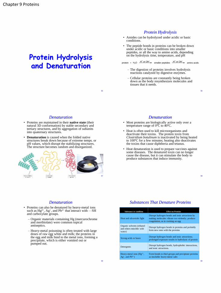
Protein Hydrolysis
• Amides can be hydrolyzed under acidic or basic conditions.
• The peptide bonds in proteins can be broken down under acidic or basic conditions into smaller peptides, or all the way to amino acids, depending on the hydrolysis time, temperature, and pH
– The digestion of proteins involves hydrolysis
reactions catalyzed by digestive enzymes.
– Cellular proteins are constantly being broken down as the body resynthesizes molecules and tissues that it needs.
56
protein + H2OH+ or OH-
smaller peptidesH+ or OH-
amino acids
Denaturation • Proteins are maintained in their native state (their
natural 3D conformation) by stable secondary and tertiary structures, and by aggregation of subunits into quaternary structures.
• Denaturation is caused when the folded native structures break down because of extreme temps. or pH values, which disrupt the stabilizing structures. The structure becomes random and disorganized.
57
Denaturation • Most proteins are biologically active only over a
temperature range of 0ºC to 40ºC.
• Heat is often used to kill microorganisms and deactivate their toxins. The protein toxin from Clostridium botulinum is inactivated by being heated to 100ºC for a few minutes; heating also deactivates the toxins that cause diphtheria and tetanus.
• Heat denaturation is used to prepare vaccines against some diseases. The denatured toxin can no longer cause the disease, but it can stimulate the body to produce substances that induce immunity.
58
Denaturation • Proteins can also be denatured by heavy-metal ions
such as Hg2+, Ag+, and Pb2+ that interact with —SH and carboxylate groups.
– Organic materials containing Hg (mercurochrome and merthiolate) were common topical antiseptics.
– Heavy-metal poisoning is often treated with large doses of raw egg white and milk; the proteins in the egg and milk bind to the metal ions, forming a precipitate, which is either vomited out or pumped out.
59
Substances That Denature Proteins
60
Substance or condition Effect on Proteins
Heat and ultraviolet light
Disrupt hydrogen bonds and ionic attractions by
making molecules vibrate too violently; produce
coagulation, as in cooking an egg
Organic solvents (ethanol
and others miscible with
water)
Disrupt hydrogen bonds in proteins and probably
form new ones with the proteins
Strong acids or bases Disrupt hydrogen bonds and ionic attractions;
prolonged exposure results in hydrolysis of protein
Detergents Disrupt hydrogen bonds, hydrophobic interactions,
and ionic attractions.
Heavy-metal ions (Hg2+,
Ag+, and Pb2+)
Form bonds to thiol groups and precipitate proteins
as insoluble heavy-metal salts


![Mass Spectrometric Analysis of l-Cysteine Metabolism: … · tion of [U-13C3, 15N]L-cysteine to the culture, the levels of [13C3,15N]L-cysteine increased, and [13C3, 15N]L-cysteine](https://img.pdfslide.net/doc/110x75/5fe663421198753c202620ce/mass-spectrometric-analysis-of-l-cysteine-metabolism-tion-of-u-13c3-15nl-cysteine.jpg)
![CU report m15 - Windeire · I.C. MacDonald, ITCA Health and Research Chair Cystine [sis -teen, -tin] Cystine, an amino acid (240.29 g/mol), is one of the building blocks of proteins](https://img.pdfslide.net/doc/110x75/5f1e528529afc2734b5876e6/cu-report-m15-windeire-ic-macdonald-itca-health-and-research-chair-cystine.jpg)