Embed Size (px)
Citation preview

In this chapter, you will learn that atoms are the fundamental building blocks of matter. You will then develop a detailed picture of the structure of atoms, which is the foundation for all chemistry. You will also learn that different types of atom are called elements, and that different atoms of the same element are called isotopes. Some isotopes, known as radioisotopes, are unstable and release radiation when they decay. To determine the relative masses of isotopes and atoms, chemists use an instrument called a mass spectrometer.
By the end of this chapter, you will also have a greater understanding of the different models used to describe the arrangement of electrons in atoms.
ContentBy the end of this chapter you will be able to:
• investigate the basic structure of stable and unstable isotopes by examining: - their position in the periodic table - the distribution of electrons, protons and neutrons in the atom - representation of the symbol, atomic number and mass number (nucleon number) ICT
• model the atom’s discrete energy levels, including electronic configuration and spdf notation (ACSCH017, ACSCH018, ACSCH020, ACSCH022) ICT
• calculate the relative atomic mass from isotopic composition (ACSCH024) ICT N
• investigate energy levels in atoms and ions through: - collecting primary data from a flame test using different ionic solutions of metals (ACSCH019) ICT
- examining spectral evidence for the Bohr model and introducing to the Schrödinger model
• investigate the properties of unstable isotopes using natural and human-made radioisotopes as examples, including but not limited to: - types of radiation - types of balanced nuclear reactions.
Chemistry Stage 6 Syllabus © NSW Education Standards Authority for and on behalf of the Crown in right of the State of NSW, 2017.
Atomic structure and atomic mass
CHAPTER
DRAFT

MODULE 1 | PROPERTIES AND STRUCTURE OF MATTER78
3.1 Inside atoms
CHEMISTRY INQUIRY CCT
Why are atoms of elements different from one another?COLLECT THIS …
• marbles and ball bearings of various sizes
• toy car plastic race track with a 90° curve
• stopwatch
• top-loading weighing balance
• camera
DO THIS …
1 Set up the race track as shown, with a raised starting position and a 90° curve at the base of the track.
2 Predict what will happen when you roll balls of various masses down the ramp and around the curved track.
3 Draw up a table in your workbook to record the results of your experiment.
4 Roll the balls down the track one at a time. Record the time each ball takes to reach the end of the track and the path each ball follows when it exits the track.
5 If possible, take a video of your experiment.
RECORD THIS …
1 Describe what happened when you rolled the different balls down the track.
2 Present a table of your results.
REFLECT ON THIS …
1 What makes the balls different to one another?
2 Identify any patterns in your results and explain why you think you observed them.
3 What could you do next time to improve your experiment?
ATOMIC MODELSUntil the mid-19th century, scientists believed that atoms were hard spheres that couldn’t be broken down into smaller parts. By the end of the 19th century, there was increasing evidence to suggest that atoms are made up of smaller particles. This led to a series of atomic models that attempted to explain the structure of atoms.
The current model of the atom is based on the work of many scientists. All atoms are made up of a small, positively charged nucleus surrounded by a much larger cloud of negatively charged electrons as shown in Figure 3.1.1. The nucleus is made up of two types of subatomic particle—protons and neutrons. The protons are positively charged and the neutrons have no charge.
ElectronsElectrons are negatively charged particles. You can imagine them forming a cloud of negative charge around the nucleus. This cloud gives the atom its size and volume.
An electron is approximately 1800 times smaller than a proton or neutron. Therefore, electrons contribute very little to the total mass of an atom. However, the space occupied by the cloud of electrons is 10 000–100 000 times larger than the nucleus.
Negative particles attract positive particles. This is called electrostatic attraction. Electrons are bound to the nucleus by the electrostatic attraction to the protons within the nucleus. The charge of an electron is equal but opposite to the charge of a proton. Electrons are said to have a charge of −1 and protons have a charge of +1.
In some circumstances, electrons can be easily removed from an atom. For example, when you rub a rubber balloon on a woollen jumper or dry hair, electrons
FIGURE 3.1.1 A simplified model of the atom. The central glow represents the nucleus where the protons and neutrons are housed. The nucleus is surrounded by a cloud of electrons that orbit the nucleus.
DRAFT
79CHAPTER 3 | ATOMIC STRUCTURE AND ATOMIC MASS
are transferred to the balloon and the balloon develops a negative charge. The negative charge is observed as an electrostatic force that can stick the balloon to a wall or even move an aluminium can. You will look more closely at the removal of electrons from atoms when looking at ionic compounds in Chapter 5.
The electricity that powers lights and appliances is the result of electrons moving in a current through wires. Sparks and lightning are also caused by electrons moving through air.
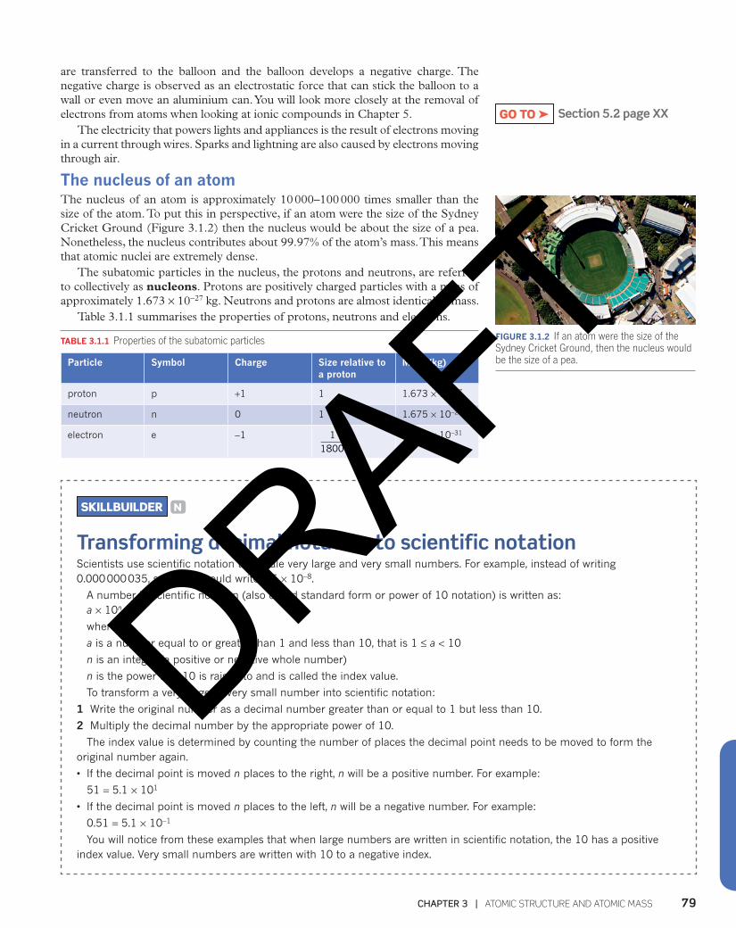
The nucleus of an atomThe nucleus of an atom is approximately 10 000–100 000 times smaller than the size of the atom. To put this in perspective, if an atom were the size of the Sydney Cricket Ground (Figure 3.1.2) then the nucleus would be about the size of a pea. Nonetheless, the nucleus contributes about 99.97% of the atom’s mass. This means that atomic nuclei are extremely dense.
The subatomic particles in the nucleus, the protons and neutrons, are referred to collectively as nucleons. Protons are positively charged particles with a mass of approximately 1.673 × 10⁻²⁷ kg. Neutrons and protons are almost identical in mass.
Table 3.1.1 summarises the properties of protons, neutrons and electrons.
TABLE 3.1.1 Properties of the subatomic particles
Particle Symbol Charge Size relative to a proton
Mass (kg)
proton p +1 1 1.673 × 10⁻²⁷
neutron n 0 1 1.675 × 10⁻²⁷
electron e −1 11800
9.109 × 10⁻³¹
Section 5.2 page XXGO TO ➤
FIGURE 3.1.2 If an atom were the size of the Sydney Cricket Ground, then the nucleus would be the size of a pea.
SKILLBUILDER N
Transforming decimal notation to scientific notationScientists use scientific notation to handle very large and very small numbers. For example, instead of writing 0.000 000 035, scientists would write 3.5 × 10⁻⁸.
A number in scientific notation (also called standard form or power of 10 notation) is written as: a × 10n
where
a is a number equal to or greater than 1 and less than 10, that is 1 ≤ a < 10
n is an integer (a positive or negative whole number)
n is the power that 10 is raised to and is called the index value.
To transform a very large or very small number into scientific notation:
1 Write the original number as a decimal number greater than or equal to 1 but less than 10.
2 Multiply the decimal number by the appropriate power of 10.
The index value is determined by counting the number of places the decimal point needs to be moved to form the original number again.
• If the decimal point is moved n places to the right, n will be a positive number. For example:
51 = 5.1 × 101
• If the decimal point is moved n places to the left, n will be a negative number. For example:
0.51 = 5.1 × 10⁻¹
You will notice from these examples that when large numbers are written in scientific notation, the 10 has a positive index value. Very small numbers are written with 10 to a negative index.
DRAFT

MODULE 1 | PROPERTIES AND STRUCTURE OF MATTER80
3.1 Review
SUMMARY
• All atoms are composed of a small, positively charged nucleus surrounded by a negatively charged cloud of electrons.
• The mass of an atom is mostly determined by the mass of the nucleus, while the size of an atom is determined by the cloud of electrons.
• The nucleus is made up of two types of subatomic particle—protons and neutrons. These particles are referred to as nucleons.
• Protons have a positive charge, electrons have a negative charge and neutrons have no charge.
• Protons and neutrons are similar in size and mass while electrons are approximately 1800 times smaller.
• The charges on protons and electrons are equal but opposite.
KEY QUESTIONS
1 How many times larger is the atom compared to its nucleus?
2 What subatomic particles make up the most mass of an atom and where are they found?
3 How are electrons held within the cloud surrounding the nucleus?
4 State the charge of:a an electronb a protonc a neutron.
DRAFT
81CHAPTER 3 | ATOMIC STRUCTURE AND ATOMIC MASS
3.2 Classifying atoms
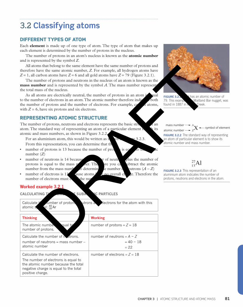
DIFFERENT TYPES OF ATOMEach element is made up of one type of atom. The type of atom that makes up each element is determined by the number of protons in the nucleus.
The number of protons in an atom’s nucleus is known as the atomic number and is represented by the symbol Z.
All atoms that belong to the same element have the same number of protons and therefore have the same atomic number, Z. For example, all hydrogen atoms have Z = 1, all carbon atoms have Z = 6 and all gold atoms have Z = 79 (Figure 3.2.1).
The number of protons and neutrons in the nucleus of an atom is known as the mass number and is represented by the symbol A. The mass number represents the total mass of the nucleus.
As all atoms are electrically neutral, the number of protons in an atom is equal to the number of electrons in an atom. The atomic number therefore indicates both the number of protons and the number of electrons. For example, carbon atoms, with Z = 6, have six protons and six electrons.
REPRESENTING ATOMIC STRUCTURE The number of protons, neutrons and electrons represents the basic structure of an atom. The standard way of representing an atom of a particular element is with its atomic and mass numbers, as shown in Figure 3.2.2.
For an aluminium atom, this would be written as shown in Figure 3.2.3. From this representation, you can determine that the:
• number of protons is 13 because the number of protons is equal to the atomic number (Z)
• number of neutrons is 14 because the number of neutrons plus the number of protons is equal to the mass number. Therefore you can subtract the atomic number from the mass number to determine the number of neutrons (A − Z)
• number of electrons is 13 because atoms have no overall charge. Therefore the number of electrons must equal the number of protons.
Worked example 3.2.1
CALCULATING THE NUMBER OF SUBATOMIC PARTICLES
Calculate the number of protons, neutrons and electrons for the atom with this atomic symbol: Ar18
40
Thinking Working
The atomic number is equal to the number of protons.
number of protons = Z = 18
Calculate the number of neutrons.
number of neutrons = mass number − atomic number
number of neutrons = A − Z
= 40 − 18
= 22
Calculate the number of electrons.
The number of electrons is equal to the atomic number because the total negative charge is equal to the total positive charge.
number of electrons = Z = 18
FIGURE 3.2.1 Gold has an atomic number of 79. This example, the Maitland Bar nugget, was found in 1887 in Meroo Creek.
mass number A
ZXatomic numbersymbol of element
FIGURE 3.2.2 The standard way of representing an atom of particular element is to show its atomic number and mass number.
FIGURE 3.2.3 This representation of an aluminium atom indicates the number of protons, neutrons and electrons in the atom.
27Al13
DRAFT

MODULE 1 | PROPERTIES AND STRUCTURE OF MATTER82
Worked example: Try yourself 3.2.1
CALCULATING THE NUMBER OF SUBATOMIC PARTICLES
Calculate the number of protons, neutrons and electrons for the atom with this atomic symbol: U192
235
ISOTOPES All atoms that belong to the same element have the same number of protons in the nucleus and therefore the same atomic number, Z. However, not all atoms that belong to the same element have the same mass number, A. For example, hydrogen atoms can have mass numbers of 1, 2 or 3. In other words, hydrogen atoms may contain just a single proton, a proton and a neutron, or a proton and two neutrons as shown in Figure 3.2.4. Atoms that have the same number of protons (atomic number) but different numbers of neutrons (and therefore different mass numbers) are known as isotopes.
electron
protonneutron
electron
protonneutron
electron
proton
hydrogen (protium)hydrogen-1, 11H
deuteriumhydrogen-2, 21H
tritiumhydrogen-3, 31H
FIGURE 3.2.4 The three isotopes of hydrogen are given special names. A hydrogen atom with just one proton in its nucleus is known as hydrogen or protium. A hydrogen atom with one proton and one neutron is known as deuterium. A hydrogen atom with one proton and two neutrons is known as tritium.
Carbon has three naturally occurring isotopes. These three isotopes are known as carbon-12, carbon-13 and carbon-14. Carbon-12 atoms have a mass number of 12, carbon-13 atoms have a mass number of 13 and carbon-14 atoms have a mass number of 14. In the 1950s and 1960s, nuclear weapons testing caused a spike in carbon-14 in the atmosphere. This has been declining in the last 50 years. These three carbon isotopes can be represented as shown in Figure 3.2.5.
126 C
carbon–12
136 C
carbon–13
146 C
carbon–14 FIGURE 3.2.5 Scientific representation of the three isotopes of carbon: carbon-12, carbon-13 and carbon-14.
RADIOISOTOPES Isotopes have identical chemical properties but different physical properties such as mass and density. In particular, some isotopes are radioactive so they are referred to as radioisotopes.
Figure 3.2.6 on page xx shows a plot of the number of neutrons versus the number of protons in various nuclei. It shows that there is a region called the band of stability where the nuclei are stable. Nuclei that lie outside of this region are unstable so they are radioactive.
DRAFT

83CHAPTER 3 | ATOMIC STRUCTURE AND ATOMIC MASS
Radioisotopes will decay (break down) to form stable nuclei. During the decay process, radiation is emitted in the form of alpha particles, beta particles or gamma radiation.
band ofstability
20
20 40 60 80 100
40
60
Num
ber
of n
eutr
ons,
(N)
Number of protons (Z)
Plot of element nuclei based on the numberof neutrons versus the number of protons
alpha emitters–these nuclei havetoo few neutronsto be stable
stable nuclei
beta emitters–these nucleihave too manyneutrons to bestable
80
100
120
140
N = Z
FIGURE 3.2.6 A plot of number of neutrons versus number of protons in various nuclei. The region of stable nuclei is called the band of stability (white). Nuclei that lie outside of this region are unstable so they are radioactive (pink and blue).
Alpha particles are made up of two protons and two neutrons which is the same as a helium nucleus. They are emitted by nuclei that have too few neutrons to be stable. Alpha particles are a lower energy form of radiation, travelling no further than a few centimetres from their source. They can be stopped by a sheet of paper or an equivalent material (Figure 3.2.7a).
Beta particles are high-energy electrons formed from the decay of nuclei with too many neutrons. They have more penetrating power than alpha particles and can be blocked by, for example, an aluminium plate several centimetres thick (Figure 3.2.7b).
Gamma radiation is a type of high-energy electromagnetic radiation. It has great penetrating power which can only be stopped by something as dense as lead plate, several centimetres thick (Figure 3.2.7c).
Natural and artificial radioisotopes and balanced nuclear reactions The decay of radioisotopes to produce different types of radiation can be represented by equations of balanced nuclear reactions.
For example, the decay of naturally occurring uranium-238 produces thorium-234, an alpha particle and gamma radiation. This decay is represented by the following equation:
γ→ + +U Th He92238
90238
24
The Greek letter γ is used to represent gamma radiation. Notice that when the decay produces beta particles, a new element is also formed.
aluminium
alpha
lead
(a)
aluminium
beta
lead
(b)
aluminium lead
gamma
(c)
FIGURE 3.2.7 Alpha particles (a), beta particles (b) and gamma particles (c) have different penetrating powers.
DRAFT

MODULE 1 | PROPERTIES AND STRUCTURE OF MATTER84
Carbon-14, mentioned earlier in this section, is a radioisotope that decays to produce beta particles (electrons). Its decay can be represented by the following equation:
→ + −C N e614
714
10
where
− e10 represents a beta particle (an electron).
The decay of carbon-14 can be used to determine the age of a once-living organism, such as a fossil, in a technique known as carbon dating. Living organisms take up carbon-14 from the environment so the ratio of carbon-12 to carbon-14 in the organism remains fairly constant over its lifetime. After the organism dies, it can no longer take up carbon-14 and so the amount of carbon-14 in the organism decreases as the isotope decays. By looking at the ratio of carbon-12 to carbon-14, the age of the fossilised object can be determined.
CHEMISTRY IN ACTION S
Carbon-14 and the illegal ivory tradeIsotopes are important in combatting ivory poaching in Africa. By performing carbon dating on the elephant tusks and ivory (Figure 3.2.8), scientists can determine how old the tusks are.
If the authorities know the age of the ivory, then legal action can be taken against the poachers and sellers of the product. Ivory products prior to 1989 are allowed to be traded. Since 1989, the trade on ivory has been made illegal worldwide.
When writing an equation for a balanced nuclear reaction, check that the atomic numbers add up to the same value on both sides of the equation. You will also need to do the same check for mass numbers.
FIGURE 3.2.8 These tusks are from African elephants killed for their ivory.
Technetium, which was discovered in 1936, is an artificial radioisotope that is routinely used in medicine. The medical radioisotope technetium-99m (the ‘m’ representing a nuclear isomer) is produced in nuclear reactors from the decay of uranium-238 to molybdenum-99. The molybdenum-99 is isolated and, through further decay, technetium-99m is formed according to the following equation:
Mo Tc e4299
4399m
10→ + −
Compounds containing technetium-99m can be injected into various body organs or a cancerous tumour in a patient. For example, a type of bone scintigraphy scan involves injecting the technetium-99m compound into a vein. When the technetium-99m decays, it releases gamma radiation, which is then detected by nuclear imaging equipment (Figure 3.2.9).FIGURE 3.2.9 A doctor reviews the results of a bone scintigraphy scan.
DRAFT

85CHAPTER 3 | ATOMIC STRUCTURE AND ATOMIC MASS
3.2 ReviewSUMMARY
• The number of subatomic particles in an atom can be determined from an element’s atomic number (Z) and mass number (A).
• The atomic number indicates how many protons or electrons an atom has.
• The mass number indicates how many protons and neutrons are in the nucleus of the atom.
• Isotopes are atoms with the same atomic number but different mass numbers, i.e. they have the same number of protons but different numbers of neutrons.
• Isotopes have the same chemical properties but different physical properties such as mass, density and radioactivity.
• Unstable isotopes that decay and release radiation are called radioisotopes.
• The three types of radiation released are alpha particles, beta particles and gamma radiation.
• The decay of a radioisotope can be represented by equations of balanced nuclear reactions.
KEY QUESTIONS
1 What term is given to the number of protons and neutrons in the nucleus of an atom?
2 Calculate the numbers of protons, neutrons and electrons in the atom P15
31 .
3 What is the correct name of the element that has an atom with seven protons and eight neutrons?
4 Explain the similarities and differences between isotopes of the same element.
5 How many more neutrons does an atom of carbon-14 have than an atom of the carbon-12 isotope?
6 Describe the difference between an alpha and beta particle and gamma radiation.
7 Balance the equations for the following nuclear reactions.
a → +Th Pa ___90234
91234
b → +Pa ___ He91234
24
c → +Rn Po ___86220
84216
d → + −Po ___ e84216
10
DRAFT
MODULE 1 | PROPERTIES AND STRUCTURE OF MATTER86
3.3 Masses of particles Atoms are particles so small that they cannot be counted individually or even in groups of thousands or millions. The mass of one atom is incredibly small. For example, one atom of carbon has a mass of approximately 2 × 10⁻²³ g.
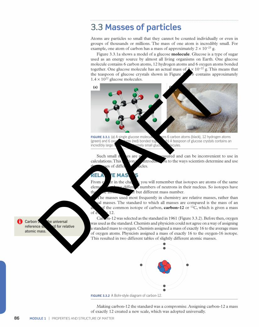
Figure 3.3.1a shows a model of a glucose molecule. Glucose is a type of sugar used as an energy source by almost all living organisms on Earth. One glucose molecule contains 6 carbon atoms, 12 hydrogen atoms and 6 oxygen atoms bonded together. One glucose molecule has an actual mass of 3 × 10⁻²² g. This means that the teaspoon of glucose crystals shown in Figure 3.3.1b contains approximately 1.4 × 10²² glucose molecules.
(a) (b)
FIGURE 3.3.1 (a) A single glucose molecule contains 6 carbon atoms (black), 12 hydrogen atoms (green) and 6 oxygen atoms (red) bonded together. (b) A teaspoon of glucose crystals contains an incredibly large number of extremely small glucose molecules.
Such small masses are not easily measured and can be inconvenient to use in calculations. This section will introduce you to the ways scientists determine and use the masses of different particles.
RELATIVE MASSES From earlier in the chapter, you will remember that isotopes are atoms of the same element that have different numbers of neutrons in their nucleus. So isotopes have the same atomic number but different mass number.
The masses used most frequently in chemistry are relative masses, rather than actual masses. The standard to which all masses are compared is the mass of an atom of the common isotope of carbon, carbon-12 or 12C, which is given a mass of exactly 12.
Carbon-12 was selected as the standard in 1961 (Figure 3.3.2). Before then, oxygen was used as the standard. Chemists and physicists could not agree on a way of assigning a standard mass to oxygen. Chemists assigned a mass of exactly 16 to the average mass of oxygen atoms. Physicists assigned a mass of exactly 16 to the oxygen-16 isotope. This resulted in two different tables of slightly different atomic masses.
FIGURE 3.3.2 A Bohr-style diagram of carbon-12.
Making carbon-12 the standard was a compromise. Assigning carbon-12 a mass of exactly 12 created a new scale, which was adopted universally.
Carbon-12 is the universal reference standard for relative atomic mass. DRAFT
87CHAPTER 3 | ATOMIC STRUCTURE AND ATOMIC MASS
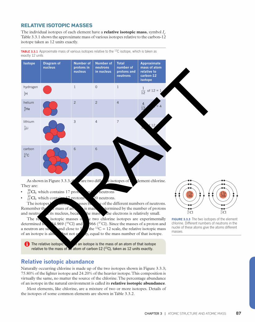
RELATIVE ISOTOPIC MASSESThe individual isotopes of each element have a relative isotopic mass, symbol Ir. Table 3.3.1 shows the approximate mass of various isotopes relative to the carbon-12 isotope taken as 12 units exactly.
TABLE 3.3.1 Approximate mass of various isotopes relative to the 12C isotope, which is taken as exactly 12 units
Isotope Diagram of nucleus
Number of protons in nucleus
Number of neutrons in nucleus
Total number of protons and neutrons
Approximate mass of atom relative to carbon-12 isotope
hydrogen
H11
1 0 1 112
of 12 = 1
helium
He24
2 2 4 412
of 12 = 4
lithium
Li37
3 4 7 712
of 12 = 7
carbon
C612
6 6 12 12
As shown in Figure 3.3.3, there are two different isotopes of the element chlorine. They are:
• Cl,1735 which contains 17 protons and 18 neutrons
• Cl,1737 which contains 17 protons and 20 neutrons.The isotopes have different masses because of the different numbers of neutrons.
Remember that the mass of an atom is mainly determined by the number of protons and neutrons in its nucleus, because the mass of the electrons is relatively small.
The relative isotopic masses of the two chlorine isotopes are experimentally determined to be 34.969 (35Cl) and 36.966 (37Cl). Since the masses of a proton and a neutron are similar and close to 1 on the 12C = 12 scale, the relative isotopic mass of an isotope is almost, but not exactly, equal to the mass number of that isotope.
The relative isotopic mass of an isotope is the mass of an atom of that isotope relative to the mass of an atom of carbon-12 (12C), taken as 12 units exactly.
Relative isotopic abundanceNaturally occurring chlorine is made up of the two isotopes shown in Figure 3.3.3; 75.80% of the lighter isotope and 24.20% of the heavier isotope. This composition is virtually the same, no matter the source of the chlorine. The percentage abundance of an isotope in the natural environment is called its relative isotopic abundance.
Most elements, like chlorine, are a mixture of two or more isotopes. Details of the isotopes of some common elements are shown in Table 3.3.2.
17p18n
17p20n
3517 Cl 37
17Cl
FIGURE 3.3.3 The two isotopes of the element chlorine. Different numbers of neutrons in the nuclei of these atoms give the atoms different masses.DRAFT

MODULE 1 | PROPERTIES AND STRUCTURE OF MATTER88
TABLE 3.3.2 Isotopic composition of some common elements
Element Isotopes Relative isotopic mass Relative isotopic abundance (%)
hydrogen 1H 1.008 99.986
2H 2.014 0.014
3H 3.016 0.0001
oxygen 16O 15.995 99.76
17O 16.999 0.04
18O 17.999 0.20
silver 107Ag 106.9 51.8
109Ag 108.9 48.2
The mass spectrometer Relative isotopic masses of elements and their isotopic abundances are determined by using an instrument called a mass spectrometer, which was invented by Francis Aston in 1919.
A mass spectrometer, as seen in Figure 3.3.4, separates the individual isotopes in a sample of an element and determines the mass of each isotope, relative to the carbon-12 isotope, and the relative abundances of the isotopes.
FIGURE 3.3.4 A mass spectrometer is used to determine the relative masses and abundances of different isotopes.
CHEMISTRY IN ACTION ICT
Mass spectrometry reveals historyScientists can use mass spectrometry to determine the relative abundance of one isotope to another in tissues taken from a dead organism. This can help scientists determine important features about that organism’s life.
For example, archaeologists can analyse the fossil jawbone shown in Figure 3.3.5 to calculate how long ago the short-necked giraffe lived, to study its diet, or to determine the habitat of the animal. Isotope analysis of a number of related fossils can help track the evolution of this species.
FIGURE 3.3.5 An ancient fossil jawbone from an extinct short-necked giraffe. Isotopic analysis using a mass spectrometer provides archaeologists with information about the organism’s life.
DRAFT
89CHAPTER 3 | ATOMIC STRUCTURE AND ATOMIC MASS
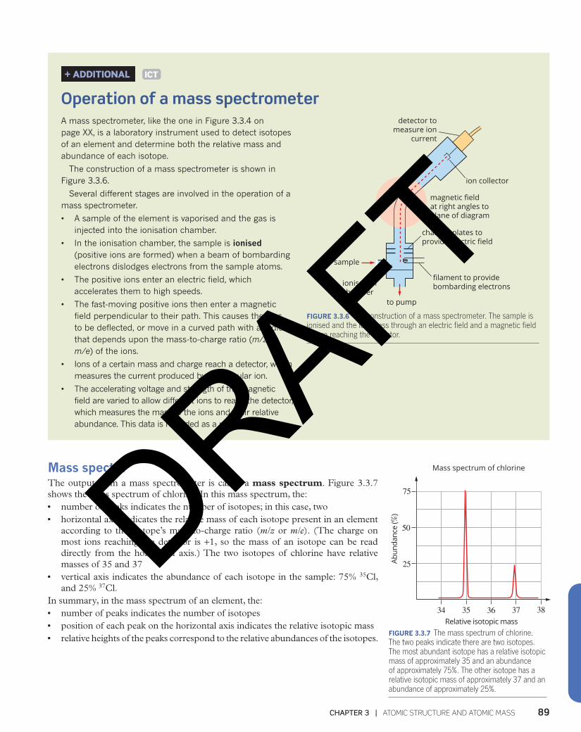
Mass spectra The output from a mass spectrometer is called a mass spectrum. Figure 3.3.7 shows the mass spectrum of chlorine. In this mass spectrum, the:• number of peaks indicates the number of isotopes; in this case, two• horizontal axis indicates the relative mass of each isotope present in an element
according to the isotope’s mass-to-charge ratio (m/z or m/e). (The charge on most ions reaching the detector is +1, so the mass of an isotope can be read directly from the horizontal axis.) The two isotopes of chlorine have relative masses of 35 and 37
• vertical axis indicates the abundance of each isotope in the sample: 75% 35Cl, and 25% 37Cl.
In summary, in the mass spectrum of an element, the:• number of peaks indicates the number of isotopes• position of each peak on the horizontal axis indicates the relative isotopic mass• relative heights of the peaks correspond to the relative abundances of the isotopes.
+ ADDITIONAL ICT
Operation of a mass spectrometerA mass spectrometer, like the one in Figure 3.3.4 on page XX, is a laboratory instrument used to detect isotopes of an element and determine both the relative mass and abundance of each isotope.
The construction of a mass spectrometer is shown in Figure 3.3.6.
Several different stages are involved in the operation of a mass spectrometer.
• A sample of the element is vaporised and the gas is injected into the ionisation chamber.
• In the ionisation chamber, the sample is ionised (positive ions are formed) when a beam of bombarding electrons dislodges electrons from the sample atoms.
• The positive ions enter an electric field, which accelerates them to high speeds.
• The fast-moving positive ions then enter a magnetic field perpendicular to their path. This causes the ions to be deflected, or move in a curved path with a radius that depends upon the mass-to-charge ratio (m/z or m/e) of the ions.
• Ions of a certain mass and charge reach a detector, which measures the current produced by a particular ion.
• The accelerating voltage and strength of the magnetic field are varied to allow different ions to reach the detector, which measures the mass of the ions and their relative abundance. This data is recorded as a mass spectrum.
detector tomeasure ion
current
ion collector
magnetic fieldat right angles toplane of diagram
charged plates toprovide electric field
filament to providebombarding electrons
to pump
ionisationchamber
sample
FIGURE 3.3.6 The construction of a mass spectrometer. The sample is ionised and the ions pass through an electric field and a magnetic field before reaching the detector.
34 35 36 37 38
75
Mass spectrum of chlorine
50
25Abun
danc
e ( %
)
Relative isotopic massFIGURE 3.3.7 The mass spectrum of chlorine. The two peaks indicate there are two isotopes. The most abundant isotope has a relative isotopic mass of approximately 35 and an abundance of approximately 75%. The other isotope has a relative isotopic mass of approximately 37 and an abundance of approximately 25%.
DRAFT
MODULE 1 | PROPERTIES AND STRUCTURE OF MATTER90
RELATIVE ATOMIC MASS Most elements consist of a mixture of isotopes. For the purpose of the calculations you will be doing later in this chapter, it is convenient to know the average relative mass of an atom in this mixture. This average is called the relative atomic mass of an element, and given the symbol Ar.
To calculate the average of the relative masses of the isotopes that exist in a naturally occurring mixture of an element, you must consider the relative abundances of each isotope.
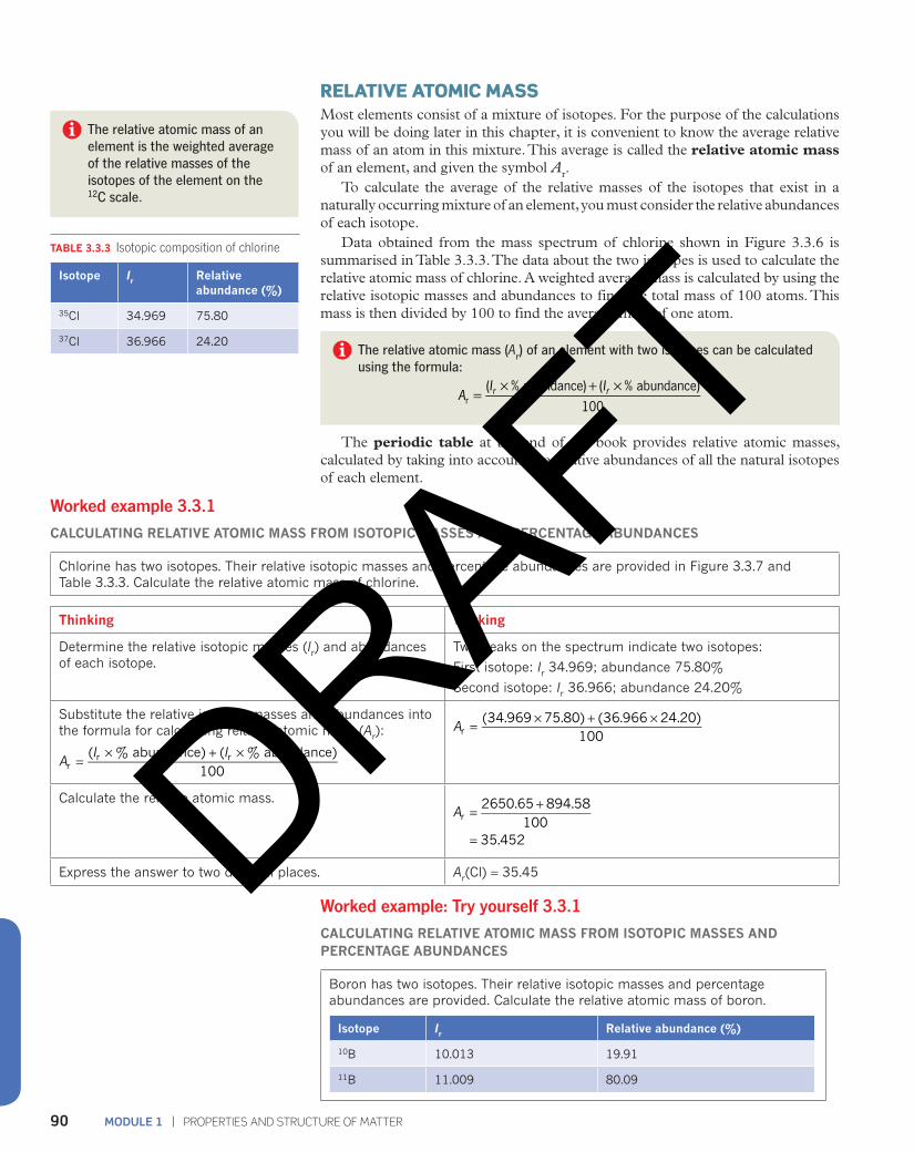
Data obtained from the mass spectrum of chlorine shown in Figure 3.3.6 is summarised in Table 3.3.3. The data about the two isotopes is used to calculate the relative atomic mass of chlorine. A weighted average mass is calculated by using the relative isotopic masses and abundances to find the total mass of 100 atoms. This mass is then divided by 100 to find the average mass of one atom.
The relative atomic mass (Ar) of an element with two isotopes can be calculated using the formula:
AI I( % abundance) ( % abundance)
100r r
r = × + ×
The periodic table at the end of the book provides relative atomic masses, calculated by taking into account the relative abundances of all the natural isotopes of each element.
TABLE 3.3.3 Isotopic composition of chlorine
Isotope Ir Relative abundance (%)
35Cl 34.969 75.80
37Cl 36.966 24.20
Worked example 3.3.1
CALCULATING RELATIVE ATOMIC MASS FROM ISOTOPIC MASSES AND PERCENTAGE ABUNDANCES
Chlorine has two isotopes. Their relative isotopic masses and percentage abundances are provided in Figure 3.3.7 and Table 3.3.3. Calculate the relative atomic mass of chlorine.
Thinking Working
Determine the relative isotopic masses (Ir) and abundances of each isotope.
Two peaks on the spectrum indicate two isotopes:
First isotope: Ir 34.969; abundance 75.80%
Second isotope: Ir 36.966; abundance 24.20%
Substitute the relative isotopic masses and abundances into the formula for calculating relative atomic mass (Ar):
AI I( % abundance) ( % abundance)
100rr r=
× + ×
A(34.969 75.80) (36.966 24.20)
100r = × + ×
Calculate the relative atomic mass.= +
=
A2650.65 894.58
10035.452
r
Express the answer to two decimal places. Ar(Cl) = 35.45
The relative atomic mass of an element is the weighted average of the relative masses of the isotopes of the element on the 12C scale.
Worked example: Try yourself 3.3.1
CALCULATING RELATIVE ATOMIC MASS FROM ISOTOPIC MASSES AND PERCENTAGE ABUNDANCES
Boron has two isotopes. Their relative isotopic masses and percentage abundances are provided. Calculate the relative atomic mass of boron.
Isotope Ir Relative abundance (%)
10B 10.013 19.91
11B 11.009 80.09
DRAFT
91CHAPTER 3 | ATOMIC STRUCTURE AND ATOMIC MASS
Worked example 3.3.2
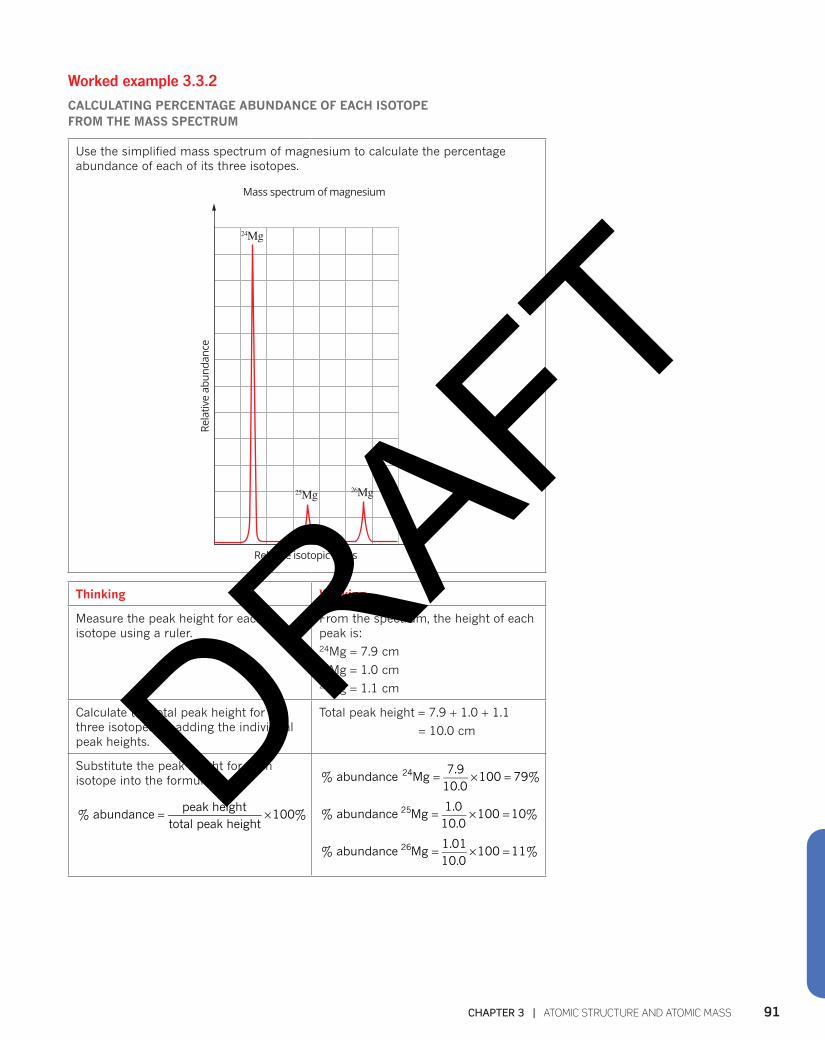
CALCULATING PERCENTAGE ABUNDANCE OF EACH ISOTOPE FROM THE MASS SPECTRUM
Use the simplified mass spectrum of magnesium to calculate the percentage abundance of each of its three isotopes.
Relative isotopic mass
Rela
tive
abun
danc
eMass spectrum of magnesium
24Mg
25Mg 26Mg
Thinking Working
Measure the peak height for each isotope using a ruler.
From the spectrum, the height of each peak is:24Mg = 7.9 cm25Mg = 1.0 cm26Mg = 1.1 cm
Calculate the total peak height for the three isotopes by adding the individual peak heights.
Total peak height = 7.9 + 1.0 + 1.1
= 10.0 cm
Substitute the peak height for each isotope into the formula:
= ×% abundancepeak height
total peak height100%
= × =% abundance Mg7.9
10.0100 79%24
= × =% abundance Mg1.010.0
100 10%25
= × =% abundance Mg1.0110.0
100 11%26
DRAFT
MODULE 1 | PROPERTIES AND STRUCTURE OF MATTER92
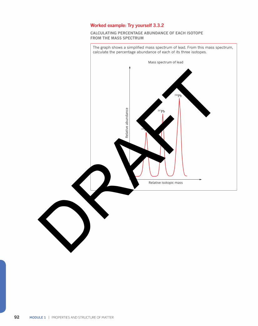
Worked example: Try yourself 3.3.2
CALCULATING PERCENTAGE ABUNDANCE OF EACH ISOTOPE FROM THE MASS SPECTRUM
The graph shows a simplified mass spectrum of lead. From this mass spectrum, calculate the percentage abundance of each of its three isotopes.
Relative isotopic mass
Mass spectrum of lead
206Pb
207Pb
208Pb
Rela
tive
abun
danc
e
DRAFT
93CHAPTER 3 | ATOMIC STRUCTURE AND ATOMIC MASS
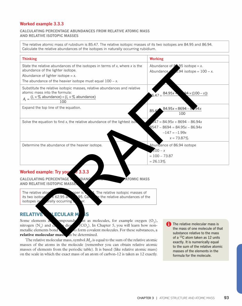
Worked example 3.3.3
CALCULATING PERCENTAGE ABUNDANCES FROM RELATIVE ATOMIC MASS AND RELATIVE ISOTOPIC MASSES
The relative atomic mass of rubidium is 85.47. The relative isotopic masses of its two isotopes are 84.95 and 86.94. Calculate the relative abundances of the isotopes in naturally occurring rubidium.
Thinking Working
State the relative abundances of the isotopes in terms of x, where x is the abundance of the lighter isotope.
Abundance of lighter isotope = x.
The abundance of the heavier isotope must equal 100 − x.
Abundance of 84.95 isotope = x.
Abundance of 86.94 isotope = 100 − x.
Substitute the relative isotopic masses, relative abundances and relative atomic mass into the formula:
AI I( % abundance) ( % abundance)
100rr r= × + × = + × −x x
85.4784.95 (86.94 (100 ))
100
Expand the top line of the equation. = + −x x
85.4784.95 8694 86.94
100
Solve the equation to find x, the relative abundance of the lightest isotope. 8547 = 84.95x + 8694 − 86.94x
8547 − 8694 = 84.95x − 86.94x
−147 = −1.99x
x = 73.87%
Determine the abundance of the heavier isotope. Abundance of 86.94 isotope
= 100 − x
= 100 − 73.87
= 26.13%
Worked example: Try yourself 3.3.3
CALCULATING PERCENTAGE ABUNDANCES FROM RELATIVE ATOMIC MASS AND RELATIVE ISOTOPIC MASSES
The relative atomic mass of copper is 63.54. The relative isotopic masses of its two isotopes are 62.95 and 64.95. Calculate the relative abundances of the isotopes in naturally occurring copper.
RELATIVE MOLECULAR MASS Some elements and compounds exist as molecules, for example oxygen (O2), nitrogen (N2) and carbon dioxide (CO2). In Chapter 5, you will learn how non-metallic elements bond together to form covalent molecules. For these substances, a relative molecular mass can be determined.
The relative molecular mass, symbol Mr, is equal to the sum of the relative atomic masses of the atoms in the molecule (remember you can obtain relative atomic masses of elements from the periodic table). It is based (like relative atomic mass) on the scale in which the exact mass of an atom of carbon-12 is taken as 12 exactly.
The relative molecular mass is the mass of one molecule of that substance relative to the mass of a 12C atom taken as 12 units exactly. It is numerically equal to the sum of the relative atomic masses of the elements in the formula for the molecule.
DRAFT
MODULE 1 | PROPERTIES AND STRUCTURE OF MATTER94
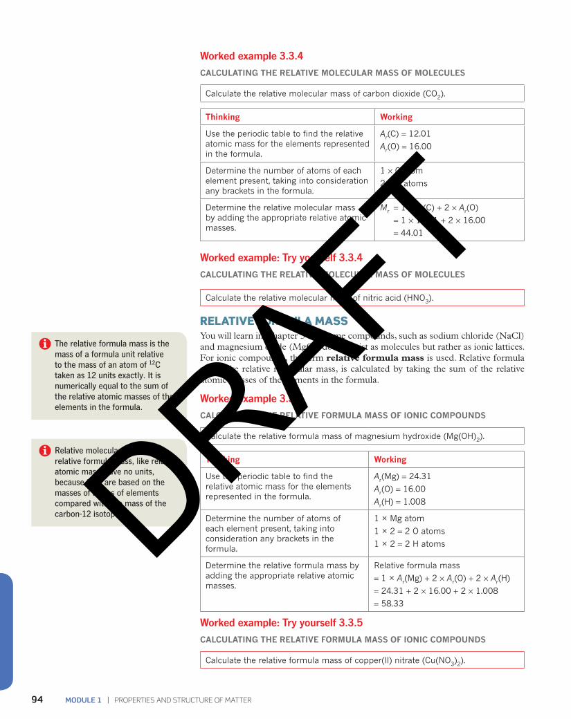
Worked example 3.3.4
CALCULATING THE RELATIVE MOLECULAR MASS OF MOLECULES
Calculate the relative molecular mass of carbon dioxide (CO2).
Thinking Working
Use the periodic table to find the relative atomic mass for the elements represented in the formula.
Ar(C) = 12.01
Ar(O) = 16.00
Determine the number of atoms of each element present, taking into consideration any brackets in the formula.
1 × C atom
2 × O atoms
Determine the relative molecular mass by adding the appropriate relative atomic masses.
Mr = 1 × Ar(C) + 2 × Ar(O)
= 1 × 12.01 + 2 × 16.00
= 44.01
Worked example: Try yourself 3.3.4
CALCULATING THE RELATIVE MOLECULAR MASS OF MOLECULES
Calculate the relative molecular mass of nitric acid (HNO3).
RELATIVE FORMULA MASS You will learn in Chapter 5 that some compounds, such as sodium chloride (NaCl) and magnesium oxide (MgO), do not exist as molecules but rather as ionic lattices. For ionic compounds, the term relative formula mass is used. Relative formula mass, like relative molecular mass, is calculated by taking the sum of the relative atomic masses of the elements in the formula.
Worked example 3.3.5
CALCULATING THE RELATIVE FORMULA MASS OF IONIC COMPOUNDS
Calculate the relative formula mass of magnesium hydroxide (Mg(OH)2).
Thinking Working
Use the periodic table to find the relative atomic mass for the elements represented in the formula.
Ar(Mg) = 24.31
Ar(O) = 16.00
Ar(H) = 1.008
Determine the number of atoms of each element present, taking into consideration any brackets in the formula.
1 × Mg atom
1 × 2 = 2 O atoms
1 × 2 = 2 H atoms
Determine the relative formula mass by adding the appropriate relative atomic masses.
Relative formula mass
= 1 × Ar(Mg) + 2 × Ar(O) + 2 × Ar(H)
= 24.31 + 2 × 16.00 + 2 × 1.008
= 58.33
Worked example: Try yourself 3.3.5
CALCULATING THE RELATIVE FORMULA MASS OF IONIC COMPOUNDS
Calculate the relative formula mass of copper(II) nitrate (Cu(NO3)2).
The relative formula mass is the mass of a formula unit relative to the mass of an atom of 12C taken as 12 units exactly. It is numerically equal to the sum of the relative atomic masses of the elements in the formula.
Relative molecular mass and relative formula mass, like relative atomic mass, have no units, because they are based on the masses of atoms of elements compared with the mass of the carbon-12 isotope. DRAFT
95CHAPTER 3 | ATOMIC STRUCTURE AND ATOMIC MASS
3.3 Review
SUMMARY
• Most elements consist of a mixture of isotopes.
• The most common isotope of carbon, carbon-12, is used as the reference standard to compare the masses of atoms.
• The carbon-12 isotope is assigned a mass of exactly 12 units.
• The relative isotopic mass, Ir, of an isotope is the mass of an atom of the isotope relative to the mass of an atom of carbon-12 taken as 12 units exactly.
• The relative isotopic mass and relative abundance of isotopes can be measured using a mass spectrometer.
• The relative atomic mass, Ar, of an element is a weighted average of its isotopic masses.
• The relative molecular mass of molecules, or formula mass of ionic compounds, is calculated from the sum of the relative atomic masses of its constituent elements.
KEY QUESTIONS
1 The isotopic composition of chlorine is shown in Table 3.3.3 on page xx. Select the correct statement about the relative atomic mass of chlorine.A The precise relative atomic mass of chlorine can
be determined by adding the two relative isotopic masses and dividing by two.
B The relative atomic mass of chlorine is based on the scale where carbon-12 has a mass of exactly 12.
C The lighter isotope is less abundant.D The isotopes have different masses because they
have different numbers of protons.
2 Use the data in Table 3.3.2 on page xx to calculate the relative atomic mass of:a oxygenb silverc hydrogen.
3 The element lithium has two isotopes:6Li with a relative isotopic mass of 6.027Li with a relative isotopic mass of 7.02.The relative atomic mass of lithium is 6.94. Calculate the percentage abundance of the lighter isotope.
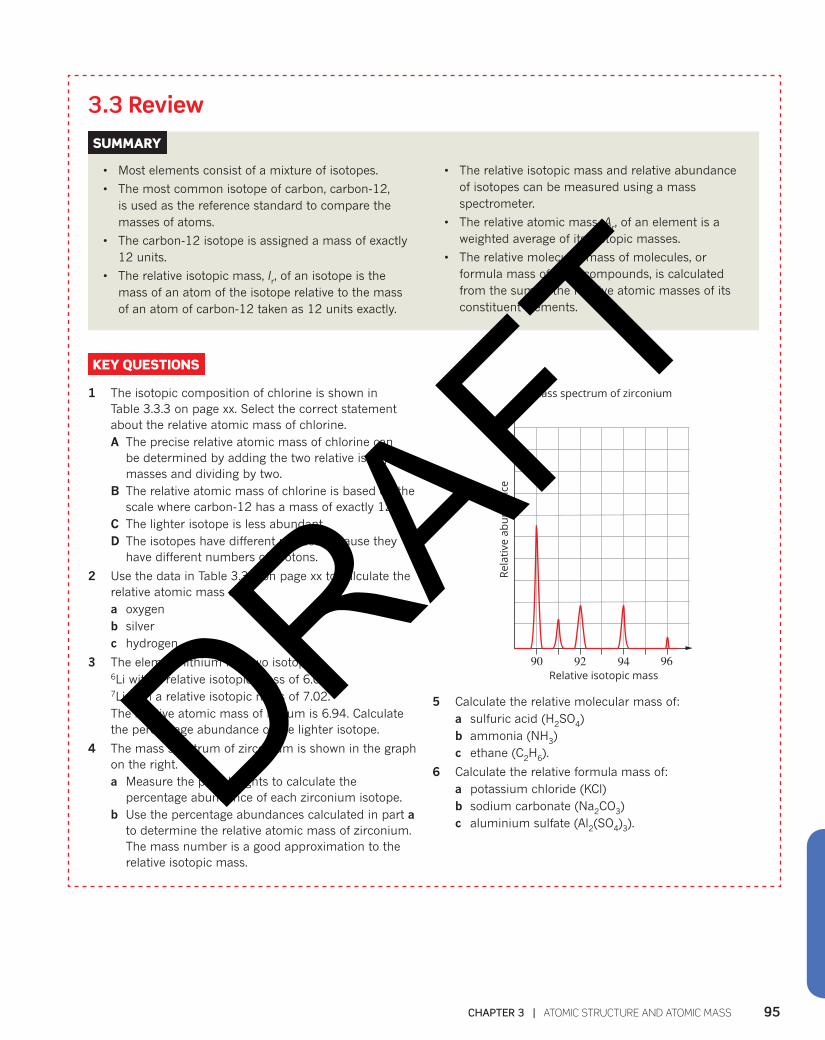
4 The mass spectrum of zirconium is shown in the graph on the right.a Measure the peak heights to calculate the
percentage abundance of each zirconium isotope. b Use the percentage abundances calculated in part a
to determine the relative atomic mass of zirconium. The mass number is a good approximation to the relative isotopic mass.
90 92 94
Mass spectrum of zirconium
Relative isotopic mass96
Rela
tive
abun
danc
e
5 Calculate the relative molecular mass of:a sulfuric acid (H2SO4)b ammonia (NH3)c ethane (C2H6).
6 Calculate the relative formula mass of:a potassium chloride (KCl)b sodium carbonate (Na2CO3)c aluminium sulfate (Al2(SO4)3).
DRAFT

MODULE 1 | PROPERTIES AND STRUCTURE OF MATTER96
3.4 Electronic structure of atoms When fireworks explode, they create a spectacular show of coloured lights (Figure 3.4.1). The light is produced by metal atoms that have been heated by the explosion. These coloured lights posed a significant problem for early scientists. The models the scientists were using could not explain the source of the light. However, the light was a clue that ultimately led to a better understanding of the arrangement of electrons in atoms.
FIGURE 3.4.1 The spectacular colours in this New Year’s Eve fireworks display are emitted by metal atoms that have been heated to very high temperatures.
FLAME TESTSA flame test is a simple method that can be used to determine the identity of a metal in a sample. A sample of the compound to be identified is inserted into a non-luminous Bunsen burner flame, as shown in Figure 3.4.2.
When the metal atoms are heated, they give off light of a characteristic colour. This means that the metal in an unknown sample can be identified by comparing the flame colour with the known characteristic colours produced by metals. Some examples of the flame colours produced by metals can be seen in Figure 3.4.3.
FIGURE 3.4.3 The colour of each flame is caused by the different metal compounds present and can be used to identify these metals. The flame colours shown here are for (from left to right): barium (yellow–green), lithium (crimson), strontium (scarlet), sodium (yellow), copper (green) and potassium (lilac).
flame colour
sample adheringto wire
Bunsen burner
FIGURE 3.4.2 Performing a flame test. A moist wire has been dipped in the sample and then placed in the flame. A fine spray of solution from a spray bottle could be used instead.
DRAFT
97CHAPTER 3 | ATOMIC STRUCTURE AND ATOMIC MASS
CHEMFILE IU
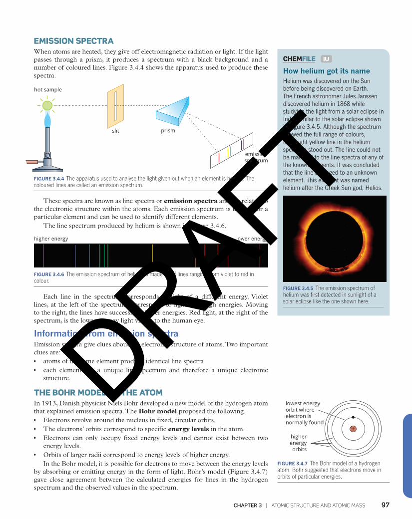
How helium got its name Helium was discovered on the Sun before being discovered on Earth. The French astronomer Jules Janssen discovered helium in 1868 while studying the light from a solar eclipse in India similar to the solar eclipse shown in Figure 3.4.5. Although the spectrum showed the full range of colours, the bright yellow line in the helium spectrum stood out. The line could not be matched to the line spectra of any of the known elements. It was concluded that the line belonged to an unknown element. This element was named helium after the Greek Sun god, Helios.
FIGURE 3.4.5 The emission spectrum of helium was first detected in sunlight of a solar eclipse like the one shown here.
EMISSION SPECTRAWhen atoms are heated, they give off electromagnetic radiation or light. If the light passes through a prism, it produces a spectrum with a black background and a number of coloured lines. Figure 3.4.4 shows the apparatus used to produce these spectra.
slit prism
emissionspectrum
hot sample
FIGURE 3.4.4 The apparatus used to analyse the light given out when an element is heated. The coloured lines are called an emission spectrum.
These spectra are known as line spectra or emission spectra and are related to the electronic structure within the atoms. Each emission spectrum is unique for a particular element and can be used to identify different elements.
The line spectrum produced by helium is shown in Figure 3.4.6.
higher energy lower energy
FIGURE 3.4.6 The emission spectrum of helium is made up of lines ranging from violet to red in colour.
Each line in the spectrum corresponds to light of a different energy. Violet lines, at the left of the spectrum, correspond to light with high energies. Moving to the right, the lines have successively lower energies. Red light, at the right of the spectrum, is the lowest energy light visible to the human eye.
Information from emission spectra Emission spectra give clues about the electronic structure of atoms. Two important clues are:• atoms of the same element produce identical line spectra• each element has a unique line spectrum and therefore a unique electronic
structure.
THE BOHR MODEL OF THE ATOMIn 1913, Danish physicist Niels Bohr developed a new model of the hydrogen atom that explained emission spectra. The Bohr model proposed the following.• Electrons revolve around the nucleus in fixed, circular orbits. • The electrons’ orbits correspond to specific energy levels in the atom. • Electrons can only occupy fixed energy levels and cannot exist between two
energy levels.• Orbits of larger radii correspond to energy levels of higher energy.
In the Bohr model, it is possible for electrons to move between the energy levels by absorbing or emitting energy in the form of light. Bohr’s model (Figure 3.4.7) gave close agreement between the calculated energies for lines in the hydrogen spectrum and the observed values in the spectrum.
+
lowest energyorbit whereelectron isnormally found
higherenergyorbits
FIGURE 3.4.7 The Bohr model of a hydrogen atom. Bohr suggested that electrons move in orbits of particular energies.
DRAFT
MODULE 1 | PROPERTIES AND STRUCTURE OF MATTER98
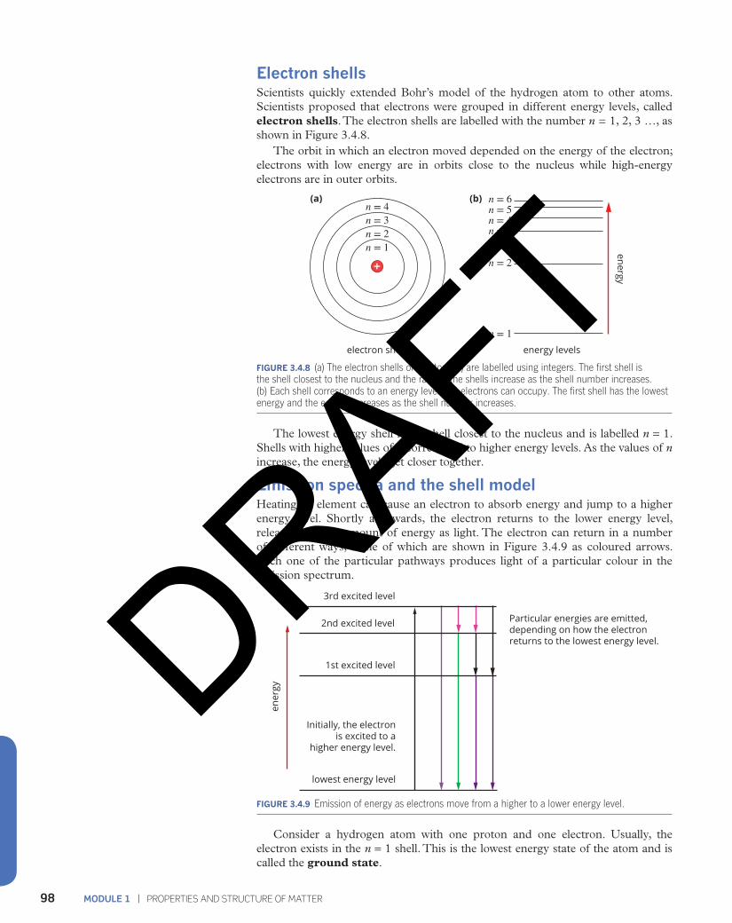
Electron shells Scientists quickly extended Bohr’s model of the hydrogen atom to other atoms. Scientists proposed that electrons were grouped in different energy levels, called electron shells. The electron shells are labelled with the number n = 1, 2, 3 …, as shown in Figure 3.4.8.
The orbit in which an electron moved depended on the energy of the electron; electrons with low energy are in orbits close to the nucleus while high-energy electrons are in outer orbits.
n = 4n = 3n = 2n = 1
n = 6n = 5n = 4n = 3
n = 2
n = 1
energy
electron shells energy levels
(a) (b)
FIGURE 3.4.8 (a) The electron shells of an atom (n) are labelled using integers. The first shell is the shell closest to the nucleus and the radii of the shells increase as the shell number increases. (b) Each shell corresponds to an energy level that electrons can occupy. The first shell has the lowest energy and the energy increases as the shell number increases.
The lowest energy shell is the shell closest to the nucleus and is labelled n = 1. Shells with higher values of n correspond to higher energy levels. As the values of n increase, the energy levels get closer together.
Emission spectra and the shell model Heating an element can cause an electron to absorb energy and jump to a higher energy level. Shortly afterwards, the electron returns to the lower energy level, releasing a fixed amount of energy as light. The electron can return in a number of different ways, some of which are shown in Figure 3.4.9 as coloured arrows. Each one of the particular pathways produces light of a particular colour in the emission spectrum.
3rd excited level
2nd excited level
1st excited level
lowest energy level
Initially, the electron is excited to a
higher energy level.
Particular energies are emitted,depending on how the electronreturns to the lowest energy level.
ener
gy
FIGURE 3.4.9 Emission of energy as electrons move from a higher to a lower energy level.
Consider a hydrogen atom with one proton and one electron. Usually, the electron exists in the n = 1 shell. This is the lowest energy state of the atom and is called the ground state.
DRAFT

99CHAPTER 3 | ATOMIC STRUCTURE AND ATOMIC MASS
When the hydrogen atom is heated, the electron absorbs energy and jumps to a higher energy level. This is known as an excited state.
Shortly afterwards, the electron returns to the ground state. The electron may return directly to the ground state or may move to other energy levels before returning to the ground state. For example, an electron in the n = 4 shell may move to the n = 2 shell before returning to the n = 1 ground state.
As the electron falls to a lower energy shell, it emits energy in the form of light. This energy is exactly equal to the energy difference between the two energy levels. Each transition corresponds to a specific energy of light and therefore one line in the line spectrum of hydrogen shown in Figure 3.4.10.
FIGURE 3.4.10 The emission spectrum of the hydrogen atom has four lines in the visible range: violet, blue, green and red.
When an electron falls from a high energy shell to a lower energy shell it emits energy in the form of light.
3.4 Review
SUMMARY
• When atoms are heated, they emit electromagnetic radiation or light.
• Flame tests are a simple technique that can be used to identify the metal in a sample.
• When light is passed through a prism, it produces a spectrum made up of lines of different colours. These spectra are known as emission or line spectra.
• The emission spectra produced by atoms of the same element are identical.
• Each element has a unique emission spectrum.
• The Bohr model of the atom was the first atomic model to explain the origin of emission spectra.
• The Bohr model assumes that electrons can only exist in fixed, circular orbits of specific energies. These orbits later came to be known as energy levels or shells.
• When an electron absorbs energy (e.g. heat or light), the electron can jump from one shell to a higher energy shell.
• When an electron falls from a higher energy shell to a lower energy shell, it emits energy in the form of light.
• Each line in the emission spectrum corresponds to a specific electron transition between two shells.
KEY QUESTIONS
1 Select words from the following list to complete the sentences below. Not all of the words provided are required.
protons, higher, transition, electrons, lower, let out, emit, excited
When a sample containing copper is heated in the flame of a Bunsen burner, the flame turns a green colour. This is because the __________ in the copper atoms absorb energy and move to _________ energy levels and then _______ light that corresponds to a green colour as they return to ___________ energy levels.
2 Explain what an emission spectrum is.
3 Explain how emission spectra are evidence for electron shells in the Bohr model.
4 What four assumptions did Bohr make about the electronic structure of the atom?
5 What form of energy is emitted when an electron moves from a higher energy shell to a lower energy shell?
DRAFT
MODULE 1 | PROPERTIES AND STRUCTURE OF MATTER100
3.5 Electronic configuration and the shell model Bohr’s model of the hydrogen atom gave close agreement between his calculated energies for lines in hydrogen’s emission spectra and the observed values. In Bohr’s model, electrons are confined to specific energy levels or shells. In this section, you will look at how scientists were able to determine the electronic arrangement in other atoms.
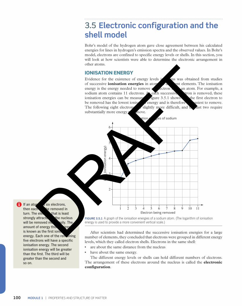
IONISATION ENERGYEvidence for the existence of energy levels in atoms was obtained from studies of successive ionisation energies in atoms of different elements. The ionisation energy is the energy needed to remove an electron from an atom. For example, a sodium atom contains 11 electrons. As each successive electron is removed, these ionisation energies can be measured. Figure 3.5.1 shows that the first electron to be removed has the lowest ionisation energy and is therefore the easiest to remove. The following eight electrons are slightly more difficult, and the last two require substantially more energy to remove.
2
2 31 4 5 6 7 8 9 10 11
3
4
5
6
Log 10
(ion
isat
ion
ener
gy)
Electron being removed
Ionisation energies of sodium
FIGURE 3.5.1 A graph of the ionisation energies of a sodium atom. (The logarithm of ionisation energy is used to provide a more convenient vertical scale.)
After scientists had determined the successive ionisation energies for a large number of elements, they concluded that electrons were grouped in different energy levels, which they called electron shells. Electrons in the same shell:• are about the same distance from the nucleus• have about the same energy.
The different energy levels or shells can hold different numbers of electrons. The arrangement of these electrons around the nucleus is called the electronic configuration.
If an atom has six electrons, then each can be removed in turn. The electron that is least strongly attracted to the nucleus will be removed most easily. The amount of energy this requires is known as the first ionisation energy. Each one of the remaining five electrons will have a specific ionisation energy. The second ionisation energy will be greater than the first. The third will be greater than the second and so on.
DRAFT
101CHAPTER 3 | ATOMIC STRUCTURE AND ATOMIC MASS
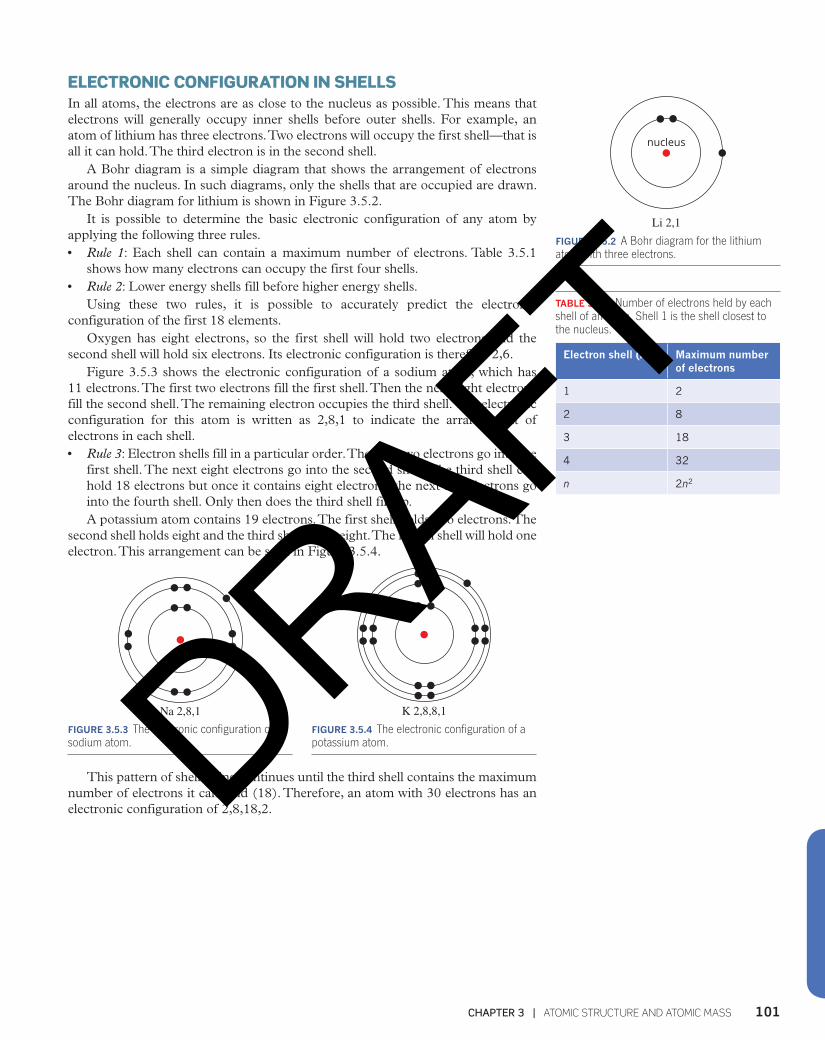
ELECTRONIC CONFIGURATION IN SHELLSIn all atoms, the electrons are as close to the nucleus as possible. This means that electrons will generally occupy inner shells before outer shells. For example, an atom of lithium has three electrons. Two electrons will occupy the first shell—that is all it can hold. The third electron is in the second shell.
A Bohr diagram is a simple diagram that shows the arrangement of electrons around the nucleus. In such diagrams, only the shells that are occupied are drawn. The Bohr diagram for lithium is shown in Figure 3.5.2.
It is possible to determine the basic electronic configuration of any atom by applying the following three rules. • Rule 1: Each shell can contain a maximum number of electrons. Table 3.5.1
shows how many electrons can occupy the first four shells.• Rule 2: Lower energy shells fill before higher energy shells.
Using these two rules, it is possible to accurately predict the electronic configuration of the first 18 elements.
Oxygen has eight electrons, so the first shell will hold two electrons and the second shell will hold six electrons. Its electronic configuration is therefore 2,6.
Figure 3.5.3 shows the electronic configuration of a sodium atom, which has 11 electrons. The first two electrons fill the first shell. Then the next eight electrons fill the second shell. The remaining electron occupies the third shell. The electronic configuration for this atom is written as 2,8,1 to indicate the arrangement of electrons in each shell.• Rule 3: Electron shells fill in a particular order. The first two electrons go into the
first shell. The next eight electrons go into the second shell. The third shell can hold 18 electrons but once it contains eight electrons, the next two electrons go into the fourth shell. Only then does the third shell fill up.A potassium atom contains 19 electrons. The first shell holds two electrons. The
second shell holds eight and the third shell holds eight. The fourth shell will hold one electron. This arrangement can be seen in Figure 3.5.4.
FIGURE 3.5.3 The electronic configuration of a sodium atom.
Na 2,8,1 K 2,8,8,1FIGURE 3.5.4 The electronic configuration of a potassium atom.
This pattern of shell filling continues until the third shell contains the maximum number of electrons it can hold (18). Therefore, an atom with 30 electrons has an electronic configuration of 2,8,18,2.
nucleus
Li 2,1FIGURE 3.5.2 A Bohr diagram for the lithium atom with three electrons.
TABLE 3.5.1 Number of electrons held by each shell of an atom. Shell 1 is the shell closest to the nucleus.
Electron shell (n) Maximum number of electrons
1 2
2 8
3 18
4 32
n 2n2
DRAFT
MODULE 1 | PROPERTIES AND STRUCTURE OF MATTER102
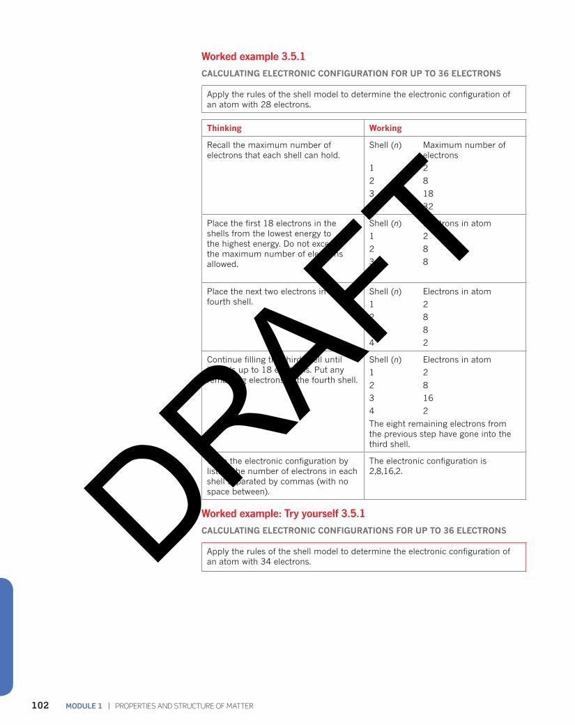
Worked example 3.5.1
CALCULATING ELECTRONIC CONFIGURATION FOR UP TO 36 ELECTRONS
Apply the rules of the shell model to determine the electronic configuration of an atom with 28 electrons.
Thinking Working
Recall the maximum number of electrons that each shell can hold.
Shell (n) Maximum number of electrons
1 2
2 8
3 18
4 32
Place the first 18 electrons in the shells from the lowest energy to the highest energy. Do not exceed the maximum number of electrons allowed.
Shell (n) Electrons in atom
1 2
2 8
3 8
4
Place the next two electrons in the fourth shell.
Shell (n) Electrons in atom
1 2
2 8
3 8
4 2
Continue filling the third shell until it holds up to 18 electrons. Put any remaining electrons in the fourth shell.
Shell (n) Electrons in atom
1 2
2 8
3 16
4 2
The eight remaining electrons from the previous step have gone into the third shell.
Write the electronic configuration by listing the number of electrons in each shell separated by commas (with no space between).
The electronic configuration is 2,8,16,2.
Worked example: Try yourself 3.5.1
CALCULATING ELECTRONIC CONFIGURATIONS FOR UP TO 36 ELECTRONS
Apply the rules of the shell model to determine the electronic configuration of an atom with 34 electrons.DRAFT
103CHAPTER 3 | ATOMIC STRUCTURE AND ATOMIC MASS
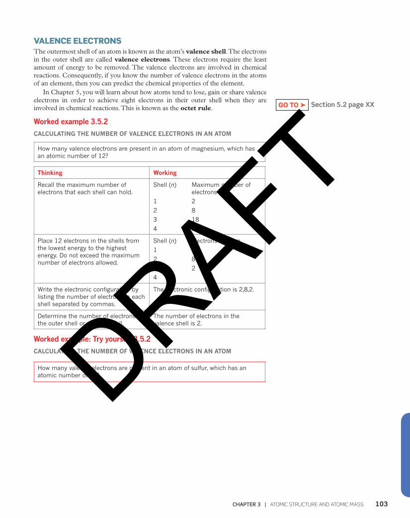
VALENCE ELECTRONS The outermost shell of an atom is known as the atom’s valence shell. The electrons in the outer shell are called valence electrons. These electrons require the least amount of energy to be removed. The valence electrons are involved in chemical reactions. Consequently, if you know the number of valence electrons in the atoms of an element, then you can predict the chemical properties of the element.
In Chapter 5, you will learn about how atoms tend to lose, gain or share valence electrons in order to achieve eight electrons in their outer shell when they are involved in chemical reactions. This is known as the octet rule.
Worked example 3.5.2
CALCULATING THE NUMBER OF VALENCE ELECTRONS IN AN ATOM
How many valence electrons are present in an atom of magnesium, which has an atomic number of 12?
Thinking Working
Recall the maximum number of electrons that each shell can hold.
Shell (n) Maximum number of electrons
1 2
2 8
3 18
4 32
Place 12 electrons in the shells from the lowest energy to the highest energy. Do not exceed the maximum number of electrons allowed.
Shell (n) Electrons in atom
1 2
2 8
3 2
4
Write the electronic configuration by listing the number of electrons in each shell separated by commas.
The electronic configuration is 2,8,2.
Determine the number of electrons in the outer shell or valence shell.
The number of electrons in the valence shell is 2.
Worked example: Try yourself 3.5.2
CALCULATING THE NUMBER OF VALENCE ELECTRONS IN AN ATOM
How many valence electrons are present in an atom of sulfur, which has an atomic number of 16?
Section 5.2 page XXGO TO ➤
DRAFT

MODULE 1 | PROPERTIES AND STRUCTURE OF MATTER104
3.5 Review
SUMMARY
• The electronic configuration of an atom indicates the arrangement of electrons in each shell.
• Bohr diagrams can be used to show the electronic configurations of atoms.
• Each shell can have a maximum number of electrons. This is summarised in the table.
Electron shell (n) Maximum number of electrons
1 2
2 8
3 18
4 32
n 2n2
• The lowest energy shells fill first until they are full or have eight electrons. At that point, the next energy shell accepts two electrons before the unfilled shell with eight electrons continues filling.
• The outer shell cannot contain more than eight electrons.
• The outer shell is called the valence shell.
• Electrons in the outer shell are called valence electrons.
KEY QUESTIONS
1 Write the electronic configurations for atoms with:a 5 electronsb 12 electronsc 20 electronsd 35 electrons.
2 Write the electronic configuration of:a Beb Sc Ard Mge Ne.
3 Draw the Bohr diagram for one atom of scandium.
4 Write the name and symbol of the element with the electronic configuration:a 2b 2,7c 2,8,3d 2,5e 2,8,7.
5 What is the number of valence electrons for an atom with 35 electrons?
DRAFT
105CHAPTER 3 | ATOMIC STRUCTURE AND ATOMIC MASS
3.6 The Schrödinger model of the atomThe shell model of the atom that developed from the work of Niels Bohr mathematically explained the lines in the emission spectrum of hydrogen atoms. However, there were some things that the model could not explain. The shell model:• cannot accurately predict the emission spectra of atoms with more than one
electron• is unable to explain why electron shells can only hold 2n2 electrons• does not explain why the fourth shell accepts two electrons before the third shell
is completely filled.These failings of the shell model indicate that the model is incomplete. Obtaining
a better model of the atom required scientists to think about electrons in an entirely different way.
A QUANTUM MECHANICAL VIEW OF ATOMS The Bohr model of the atom was revolutionary when it was proposed. Before Bohr’s model, Rutherford and other scientists believed that electrons could orbit the nucleus at any distance from the nucleus. This picture of the electrons was based on how scientists observed the world around them. For example, planets can revolve at any distance around the Sun. However, Bohr’s theory stated that electrons only occupy specific, circular orbits. This was the first suggestion that the physics inside atoms might be very different from the physics we experience in our daily lives.
Quantum mechanicsThe word ‘quantum’ means ‘a specific amount’. In the Bohr model, the electrons can only have specific amounts of energy depending on which shell they are in. The energy of the electrons is said to be quantised.
In 1926, the German scientist Erwin Schrödinger proposed that electrons behaved as waves around the nucleus. Using a mathematical approach and this wave theory, Schrödinger developed a model of the atom called quantum mechanics. This is the model of the atom that scientists use today.
Quantum mechanics describes the behaviour of extremely small particles like electrons. You rarely experience quantum mechanics in your everyday life. As a result, the predictions of quantum mechanics are often difficult to imagine. Nonetheless, quantum mechanics accurately predicts the behaviour of electrons in atoms.
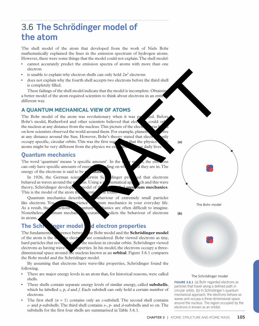
The Schrödinger model and electron propertiesThe fundamental difference between the Bohr model and the Schrödinger model of the atom is the way the electrons are considered. Bohr viewed electrons as tiny, hard particles that revolve around the nucleus in circular orbits. Schrödinger viewed electrons as having wave-like properties. In his model, the electrons occupy a three-dimensional space around the nucleus known as an orbital. Figure 3.6.1 compares the Bohr model and the Schrödinger model.
By assuming that electrons have wave-like properties, Schrödinger found the following.• There are major energy levels in an atom that, for historical reasons, were called
shells.• These shells contain separate energy levels of similar energy, called subshells,
which he labelled s, p, d and f. Each subshell can only hold a certain number of electrons.
• The first shell (n = 1) contains only an s-subshell. The second shell contains s- and p-subshells. The third shell contains s-, p- and d-subshells and so on. The subshells for the first four shells are summarised in Table 3.6.1.
(a) (b)
The Schrӧdinger modelThe Bohr model
(a) (b)
The Schrӧdinger modelThe Bohr model
FIGURE 3.6.1 (a) Bohr regarded electrons as particles that travel along a defined path in circular orbits. (b) In Schrödinger’s quantum mechanical approach, the electrons behave as waves and occupy a three-dimensional space around the nucleus. The region occupied by the electrons is known as an orbital.
DRAFT
MODULE 1 | PROPERTIES AND STRUCTURE OF MATTER106
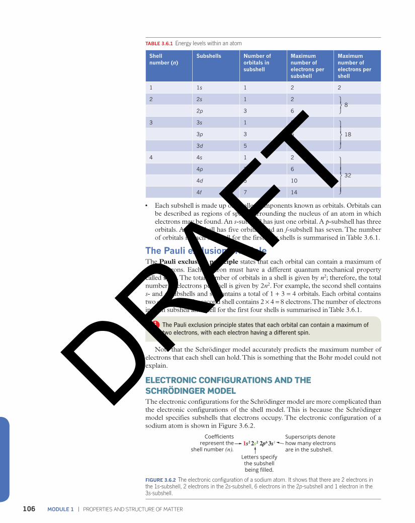
TABLE 3.6.1 Energy levels within an atom
Shell number (n)
Subshells Number of orbitals in subshell
Maximum number of electrons per subshell
Maximum number of electrons per shell
1 1s 1 2 2
2 2s 1 2
2p 3 6 8
3 3s 1 2
3p 3 6
3d 5 10
18
4 4s 1 2
4p 3 6
4d 5 10 32
4f 7 14
• Each subshell is made up of smaller components known as orbitals. Orbitals can be described as regions of space surrounding the nucleus of an atom in which electrons may be found. An s-subshell has just one orbital. A p-subshell has three orbitals. A d-subshell has five orbitals and an f-subshell has seven. The number of orbitals in each subshell for the first four shells is summarised in Table 3.6.1.
The Pauli exclusion principleThe Pauli exclusion principle states that each orbital can contain a maximum of two electrons. Each electron must have a different quantum mechanical property called spin. The total number of orbitals in a shell is given by n2; therefore, the total number of electrons per shell is given by 2n2. For example, the second shell contains s- and p-subshells and so contains a total of 1 + 3 = 4 orbitals. Each orbital contains two electrons so the second shell contains 2 × 4 = 8 electrons. The number of electrons in each subshell and shell for the first four shells is summarised in Table 3.6.1.
The Pauli exclusion principle states that each orbital can contain a maximum of two electrons, with each electron having a different spin.
Note that the Schrödinger model accurately predicts the maximum number of electrons that each shell can hold. This is something that the Bohr model could not explain.
ELECTRONIC CONFIGURATIONS AND THE SCHRÖDINGER MODELThe electronic configurations for the Schrödinger model are more complicated than the electronic configurations of the shell model. This is because the Schrödinger model specifies subshells that electrons occupy. The electronic configuration of a sodium atom is shown in Figure 3.6.2.
Coefficientsrepresent the
shell number (n).Letters specify
the subshellbeing filled.
1s2 2s2 2p6 3s1Superscripts denotehow many electronsare in the subshell.
FIGURE 3.6.2 The electronic configuration of a sodium atom. It shows that there are 2 electrons in the 1s-subshell, 2 electrons in the 2s-subshell, 6 electrons in the 2p-subshell and 1 electron in the 3s-subshell.
⎫⎬⎭
⎫⎬⎪
⎭⎪
⎫
⎬⎪⎪
⎭⎪⎪
DRAFT
107CHAPTER 3 | ATOMIC STRUCTURE AND ATOMIC MASS
Sodium has 11 electrons. The electronic configuration in Figure 3.6.2 indicates that, for sodium, there:• are two electrons in the s-subshell in the first shell• are two electrons in the s-subshell of the second shell• are six electrons in the p-subshell of the second shell• is one electron in the s-subshell of the third shell.
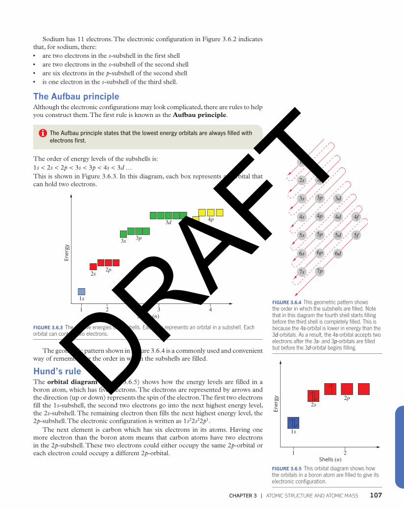
The Aufbau principleAlthough the electronic configurations may look complicated, there are rules to help you construct them. The first rule is known as the Aufbau principle.
The Aufbau principle states that the lowest energy orbitals are always filled with electrons first.
The order of energy levels of the subshells is:1s < 2s < 2p < 3s < 3p < 4s < 3d …This is shown in Figure 3.6.3. In this diagram, each box represents an orbital that can hold two electrons.
1
1s
2s2p
3s 3p
3d4s
4p
2 3 4
Ener
gy
Shells (n)
FIGURE 3.6.3 The relative energies of subshells. Each box represents an orbital in a subshell. Each orbital can contain two electrons.
The geometric pattern shown in Figure 3.6.4 is a commonly used and convenient way of remembering the order in which the subshells are filled.
Hund’s rule The orbital diagram (Figure 3.6.5) shows how the energy levels are filled in a boron atom, which has five electrons. The electrons are represented by arrows and the direction (up or down) represents the spin of the electron. The first two electrons fill the 1s-subshell, the second two electrons go into the next highest energy level, the 2s-subshell. The remaining electron then fills the next highest energy level, the 2p-subshell. The electronic configuration is written as 1s22s22p1.
The next element is carbon which has six electrons in its atoms. Having one more electron than the boron atom means that carbon atoms have two electrons in the 2p-subshell. These two electrons could either occupy the same 2p-orbital or each electron could occupy a different 2p-orbital. 1
1s
2s2p
2
Ener
gy
Shells (n)FIGURE 3.6.5 This orbital diagram shows how the orbitals in a boron atom are filled to give its electronic configuration.
1s
7s
6s
5s
4s
3s
2s
7p
6p
5p
4p
3p
6d
5d 5f
4f4d
3d
2p
FIGURE 3.6.4 This geometric pattern shows the order in which the subshells are filled. Note that in this diagram the fourth shell starts filling before the third shell is completely filled. This is because the 4s-orbital is lower in energy than the 3d-orbitals. As a result, the 4s-orbital accepts two electrons after the 3s- and 3p-orbitals are filled but before the 3d-orbital begins filling.DRAFT
MODULE 1 | PROPERTIES AND STRUCTURE OF MATTER108
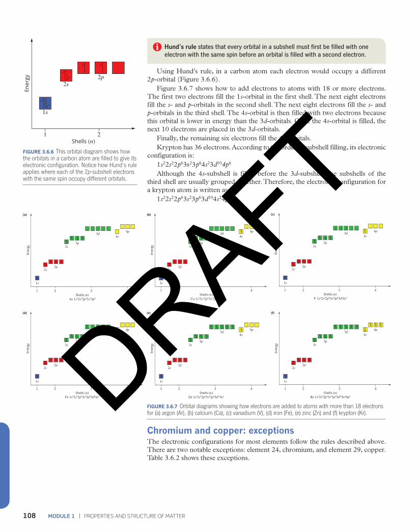
Hund’s rule states that every orbital in a subshell must first be filled with one electron with the same spin before an orbital is filled with a second electron.
Using Hund’s rule, in a carbon atom each electron would occupy a different 2p-orbital (Figure 3.6.6).
Figure 3.6.7 shows how to add electrons to atoms with 18 or more electrons. The first two electrons fill the 1s-orbital in the first shell. The next eight electrons fill the s- and p-orbitals in the second shell. The next eight electrons fill the s- and p-orbitals in the third shell. The 4s-orbital is then filled with two electrons because this orbital is lower in energy than the 3d-orbitals. Once the 4s-orbital is filled, the next 10 electrons are placed in the 3d-orbitals.
Finally, the remaining six electrons fill the 4p-orbitals.Krypton has 36 electrons. According to the order of subshell filling, its electronic
configuration is:1s22s22p63s23p64s23d104p6
Although the 4s-subshell is filled before the 3d-subshell, the subshells of the third shell are usually grouped together. Therefore, the electronic configuration for a krypton atom is written as:
1s22s22p63s23p63d104s24p6
1
1s
2s2p
2
Ener
gy
Shells (n)
FIGURE 3.6.6 This orbital diagram shows how the orbitals in a carbon atom are filled to give its electronic configuration. Notice how Hund’s rule applies where each of the 2p-subshell electrons with the same spin occupy different orbitals.
FIGURE 3.6.7 Orbital diagrams showing how electrons are added to atoms with more than 18 electrons for (a) argon (Ar), (b) calcium (Ca), (c) vanadium (V), (d) iron (Fe), (e) zinc (Zn) and (f) krypton (Kr).
Chromium and copper: exceptions The electronic configurations for most elements follow the rules described above. There are two notable exceptions: element 24, chromium, and element 29, copper. Table 3.6.2 shows these exceptions.
1
1s
2s2p
3s3p
3d4s
4p
2 3 4
Ener
gy
Ar 1s22s22p63s23p6Shells (n)
(a)
1
1s
2s2p
3s3p
3d4s
4p
2 3 4
Ener
gy
Fe 1s22s22p63s23p63d64s2Shells (n)
(d)
1
1s
2s2p
3s3p
3d4s
4p
2 3 4
Ener
gy
Ca 1s22s22p63s23p64s2Shells (n)
(b)
1
1s
2s2p
3s3p
3d4s
4p
2 3 4
Ener
gy
Zn 1s22s22p63s23p63d104s2Shells (n)
(e)
1
1s
2s2p
3s3p
3d4s
4p
2 3 4
Ener
gy
V 1s22s22p63s23p63d34s2Shells (n)
(c)
1
1s
2s2p
3s3p
3d4s
4p
2 3 4
Ener
gy
Kr 1s22s22p63s23p63d104s24p6Shells (n)
(f)
DRAFT

109CHAPTER 3 | ATOMIC STRUCTURE AND ATOMIC MASS
TABLE 3.6.2 The electronic configurations for chromium and copper
Element Electronic configuration predicted according to the rules
Actual electronic configuration
chromium (Cr) 1s22s22p63s23p63d44s2 1s22s22p63s23p63d54s1
copper (Cu) 1s22s22p63s23p63d94s2 1s22s22p63s23p63d104s1
Each orbital can hold two electrons. In the d-subshell there are five orbitals and therefore 10 electrons that can be held within them. According to Hund’s rule, as a subshell fills, a single electron is placed in each orbital first. Then a second electron is entered into the orbitals until the filling process is complete.
Chemists calculate that there is very little difference in energy between 3d- and 4s-orbitals, and the 3d 54s1 configuration for chromium is slightly more stable than the 3d 44s2 configuration. This is because each of the five d-orbitals is exactly half filled with one d-orbital empty.
Similarly for copper, the 3d104s1 arrangement with five completely filled d-orbitals is more stable than the 3d 94s2 configuration with a partially filled d-orbital.
Worked example 3.6.1
WRITING ELECTRONIC CONFIGURATIONS USING THE SCHRÖDINGER MODEL
Write the Schrödinger model of electronic configuration for a manganese atom with 25 electrons.
Thinking Working
Recall the order in which the subshells fill by listing them from lowest energy to highest energy and the number of orbitals in each.
1s, 1 orbital 3p, 3 orbitals
2s, 1 orbital 4s, 1 orbital
2p, 3 orbitals 3d, 5 orbitals
3s, 1 orbital 4p, 3 orbitals
Fill the subshells by assigning two electrons per orbital, starting from the lowest energy subshells until you have reached the total number of electrons in your atom.
Subshell Electrons Progressive in subshell total of electrons
1s 2 2
2s 2 4
2p 6 10
3s 2 12
3p 6 18
4s 2 20
3d 5 25
4p
Write the electronic configuration by writing each subshell with the number of electrons as a superscript. Remember to group subshells from the same shell.
1s22s22p63s23p63d54s2
Worked example: Try yourself 3.6.1
WRITING ELECTRONIC CONFIGURATIONS USING THE SCHRÖDINGER MODEL
Write the Schrödinger model of electronic configuration for a vanadium atom with 23 electrons.
DRAFT

MODULE 1 | PROPERTIES AND STRUCTURE OF MATTER110
3.6 Review
SUMMARY
• The shell model of the atom was unable to fully explain the properties of atoms and a new model was needed to describe the electron behaviour in atoms.
• The Schrödinger model proposed that electrons behave as waves and occupy a three-dimensional space around the nucleus.
• The Schrödinger model predicted that subshells are energy levels within the major shells. The subshells consist of orbitals.
• An orbital can be regarded as a region of space surrounding the nucleus in which an electron may be found.
• Orbitals of similar energy are grouped in subshells that are labelled s, p, d and f.
• The Pauli exclusion principle states that each orbital can hold a maximum of two electrons. Each electron must have a different quantum mechanical property called spin.
• Each subshell has a different energy in an atom.
• The Aufbau principle states that electrons fill the subshells from the lowest energy subshell to the highest energy subshell.
• The 4s-subshell is lower in energy than the 3d-subshell, so the fourth shell accepts two electrons before the third shell is completely filled.
• Electronic configurations of atoms in the Schrödinger model specify the number of electrons in each subshell.
• Hund’s rule states that every orbital in a subshell must first be filled with one electron with the same spin before an orbital is filled with a second electron.
KEY QUESTIONS
1 Which subshell has the lowest energy?A 3dB 4sC 4pD 5s
2 Copy and complete the table to write the electronic configuration of each of the atoms listed.
Element (atomic number)
Electronic configuration using the shell model
Electronic configuration using the subshell model
boron (5) 2,3 1s22s22p1
lithium (3)
chlorine (17)
sodium (11)
neon (10)
potassium (19)
scandium (21)
iron (26)
bromine (35)
3 In terms of energy levels, what is the essential difference between the shell model and the subshell model of the atom?
4 Draw orbital diagrams showing the electronic configuration of each of the following atoms.a Beb Nc Sid Ni (only show the 3d and 4s-subshells for
simplicity).DRAFT
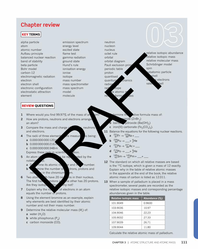
111CHAPTER 3 | ATOMIC STRUCTURE AND ATOMIC MASS
Chapter review
KEY TERMS
alpha particleatomatomic numberAufbau principle balanced nuclear reactionband of stabilitybeta particleBohr modelcarbon-12electromagnetic radiationelectronelectron shellelectronic configuration electrostatic attractionelement
emission spectrumenergy levelexcited stateflame test gamma radiation ground stateHund’s rule ionisation energyionise isotopemass numbermass spectrometermass spectrummodelmolecule
neutronnucleonnucleusoctet ruleorbitalorbital diagram Pauli exclusion principle periodic tableprotonquantisedquantum mechanicsradioactiveradioisotoperelative atomic massrelative formula mass
relative isotopic abundancerelative isotopic massrelative molecular mass Schrödinger modelspin subatomic particlesubshellvalence electronsvalence shell
REVIEW QUESTIONS
1 Where would you find 99.97% of the mass of an atom?
2 How are protons, neutrons and electrons arranged in an atom?
3 Compare the mass and charge of protons, neutrons and electrons.
4 The radii of three elements were measured as being:a 0.000 000 000 120 mb 0.000 000 000 216 m c 0.000 000 000 348 mExpress these radii in scientific notation.
5 An atom of chromium can be represented by the notation Cr.24
52
a Determine its atomic number and mass number. b Determine the number of electrons, protons and
neutrons in the chromium atom.
6 Two atoms both have 20 neutrons in their nucleus. The first has 19 protons and the other has 20 protons. Are they isotopes? Why or why not?
7 Explain why the number of electrons in an atom equals the number of protons.
8 Using the element bromine as an example, explain why elements are best identified by their atomic number and not their mass number.
9 Determine the relative molecular mass (Mr) of:a water (H2O)b white phosphorus (P4)c carbon monoxide (CO).
10 Determine the relative formula mass of:a zinc bromide (ZnBr2)b barium hydroxide (Ba(OH)2)c iron(III) carbonate (Fe2(CO3)3).
11 Balance the equations for the following nuclear reactions.a → +Fr Ra ___87
23188
223
b → +Pu ___ He94239
24
c → +Pa Ac ___91231
89227
d → +Po ___ He84214
24
e → + +Sm Pm ___ ___62149
61145
12 The standard on which all relative masses are based is the 12C isotope, which is given a mass of 12 exactly. Explain why in the table of relative atomic masses in the appendix at the end of the book, the relative atomic mass of carbon is listed as 12.011.
13 When a sample of palladium is placed in a mass spectrometer, several peaks are recorded as the relative isotopic masses and corresponding percentage abundances given in the table.
Relative isotopic mass Abundance (%)
101.9049 0.9600
103.9036 10.97
104.9046 22.23
105.9032 27.33
107.9039 26.71
109.9044 11.80
Calculate the relative atomic mass of palladium.
DRAFT
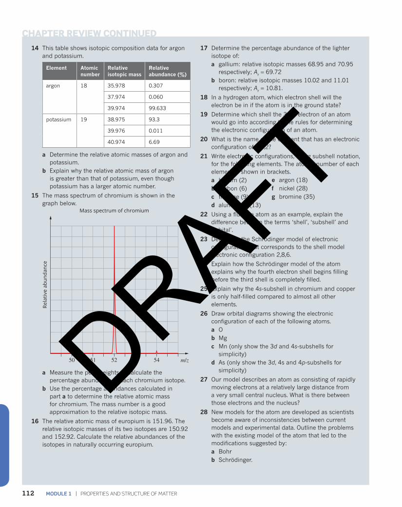
MODULE 1 | PROPERTIES AND STRUCTURE OF MATTER112
14 This table shows isotopic composition data for argon and potassium.
Element Atomic number
Relative isotopic mass
Relative abundance (%)
argon 18 35.978 0.307
37.974 0.060
39.974 99.633
potassium 19 38.975 93.3
39.976 0.011
40.974 6.69
a Determine the relative atomic masses of argon and potassium.
b Explain why the relative atomic mass of argon is greater than that of potassium, even though potassium has a larger atomic number.
15 The mass spectrum of chromium is shown in the graph below.
Rela
tive
abun
danc
e
Mass spectrum of chromium
m/z50 51 52 53 54
a Measure the peak heights to calculate the percentage abundance of each chromium isotope.
b Use the percentage abundances calculated in part a to determine the relative atomic mass for chromium. The mass number is a good approximation to the relative isotopic mass.
16 The relative atomic mass of europium is 151.96. The relative isotopic masses of its two isotopes are 150.92 and 152.92. Calculate the relative abundances of the isotopes in naturally occurring europium.
17 Determine the percentage abundance of the lighter isotope of:a gallium: relative isotopic masses 68.95 and 70.95
respectively; Ar = 69.72b boron: relative isotopic masses 10.02 and 11.01
respectively; Ar = 10.81.
18 In a hydrogen atom, which electron shell will the electron be in if the atom is in the ground state?
19 Determine which shell the 30th electron of an atom would go into according to the rules for determining the electronic configuration of an atom.
20 What is the name of the element that has an electronic configuration of 2,8,2?
21 Write electronic configurations, using subshell notation, for the following elements. The atomic number of each element is shown in brackets.a helium (2) b carbon (6)c fluorine (9)d aluminium (13)
22 Using a fluorine atom as an example, explain the difference between the terms ‘shell’, ‘subshell’ and ‘orbital’.
23 Determine the Schrödinger model of electronic configuration that corresponds to the shell model electronic configuration 2,8,6.
24 Explain how the Schrödinger model of the atom explains why the fourth electron shell begins filling before the third shell is completely filled.
25 Explain why the 4s-subshell in chromium and copper is only half-filled compared to almost all other elements.
26 Draw orbital diagrams showing the electronic configuration of each of the following atoms.a Ob Mgc Mn (only show the 3d and 4s-subshells for
simplicity)d As (only show the 3d, 4s and 4p-subshells for
simplicity)
27 Our model describes an atom as consisting of rapidly moving electrons at a relatively large distance from a very small central nucleus. What is there between those electrons and the nucleus?
28 New models for the atom are developed as scientists become aware of inconsistencies between current models and experimental data. Outline the problems with the existing model of the atom that led to the modifications suggested by:a Bohrb Schrödinger.
e argon (18)f nickel (28)g bromine (35)
CHAPTER REVIEW CONTINUED
DRAFT