Embed Size (px)
Citation preview

Chapter-I
Review of literature and genesis of the Thesis
1

1. Cataract and its Etiology
Cataract can be simply defined as loss of transparency of the crystallin lens, in
which the refractive index of the lens differ significantly over distances approximating
the wavelength of the transmitted light. Variation in the refractive index over these
distances can result from changes in lens cell structure, changes in lens protein
constituents, or both. Cataracts are generally associated with alteration of the lens
micro-architecture. When mutations in crystallin proteins are enough to cause
aggregation they usually lead to congenital cataract, however if they merely make
them susceptible to environmental insults such as light, hyperglycemic or oxidative
damage they might attribute to age-related cataract (1). The ongoing epidemiological
studies have figured out few risk factors such as UV-B exposure, low antioxidant
intake, certain medications, cigarette smoking, diabetes, gout as well as family history
in the development of cataracts (2). All these mechanisms result in the structural
changes in the α-crystallins. From αA-crystallins, Cys 131 and Cys 142 are present in
transparent human lenses as a mixture of cysteine sulfhydryl and half-cysteine
disulfide groups, whereas cataractous lenses were lacking cysteine sulfahydryl group
(3).
In contrast to age related forms of cataracts, early childhood cataracts occur in
developed countries with a frequency of 30 cases in 100, 000 births; with further 10
cases being detected by the age of 15 years (mainly as dominant forms). In developing
countries these rates are likely to be higher because of rubella infections and
consanguinity (for the recessive forms; 4)
2. Classification of Cataracts
Cataracts are broadly divisible into two major groups based on etiological factors.
1. Developmental or congenital cataract in which the normal development of the lens
is affected by genetic, nutritional or inflammatory changes.
2. Degenerative cataract includes senile cataract and are associated with radiation or
systemic diseases.
2

3. Subtypes of Cataracts
Cataracts are also classified according to the location of opacity (Fig.1) in the affected
lens and to the specific factors involved in their formation. The cataracts are further
subdivided into seven subtypes.
Fig: 1.1. Schematic illustration of mouse eye lens showing locations of the lens opacity at different parts of the lens. The cataracts are named as per their location in different parts of the lens. Anterior cataract (AC), Posterior cataract (PC), Nuclear cataract (N), and Equatorial cataract (EC).
1. The opacities are located at the anterior or posterior pole of the lens in anterior-
polar and posterior-polar cataract respectively.
2. In Zonular nuclear cataract, opaque cells are deposited in the lens surrounded by
clear zones. This type of cataract may lead to slight or significant impairment of the
vision depending on the density of the opacity.
3. Nuclear or central cataract is a rare type in which the opacity affects the embryonic
nucleus of the lens and cause little damage to the vision.
4. Small round opacities scattered irregularly throughout the nucleus and cortex in the
punctuate type of cataract. Occasionally these opacities appear light blue in color and
consequently, this type is sometimes called caerulean cataract.
3

5. The galactosemic cataract is associated with a hereditary anomaly of lactose
metabolism. In this type a central portion of the lens is visible as a transparent portion
within a largely opacified lens.
6. The development of cataract is associated with a mutation of the L-ferritin gene
which codes for an iron storage protein in hereditary hyperferrintinaemia cataract
syndrome (HHCS).
7. Senile cataract is the commonest type to develop after the age of 50. This type of
cataract is usually bilateral and begins either in the superficial cortex or close to the
nucleus of the lens.
4. Therapeutic Measures of Cataracts
Medical remedies have been inaccurately advertised as effective for the treatment of
cataracts. In fact there is no proven medical treatment available to reverse or slow the
progression of, or prevent the formation of a cataract. Surgical removal of the opaque
lens is the only possible remedy both in animals and humans, and often provides a
return of functional vision. Various surgical procedures available require meticulous
and precise microsurgical techniques. Surgery is performed using an operating
microscope and sophisticated microsurgical instruments. Two techniques are currently
used to remove cataract from the eye. One method involves making a large incision
into the eye and removing the affected lens. A newer advance method i.e.
phacoemulsification, allow for small incision surgery, and may prove a better result.
This technique involves placing a small ultrasonic instrument into the eye. This
instrument generates about 40,000 tiny vibrations per second to fragment the cataract.
The lens material is then aspirated out of the eye, much like a small intraocular
vacuum (5). This sophisticated technique has been associated with a higher success
rate for many cataract surgery patients.
4

5. Mutant Mice Models in Cataract study
Significant progress in understanding the genetics of human congenital cataracts could
be possible due to the intense cellular and molecular analysis of the animal models
with congenital eye defects. Murine cataract mutants are excellent models for detailed
studies on the process of lens opacification. These mouse models allow one to study
all stages of the embryonic development. Hereditary mouse cataract models have great
relevance to humans because it is estimated that congenital cataracts comprise
approximately 10% of visual loss in humans.
For decades the target of the developmental biologists has been to focus the
development of ocular structures. With the advent of modern genetics, now it becomes
possible to characterize mutation that causes human disorder and their comparison
with mutations in animal models. The outcome of this comparative study revealed that
there is common genetic network that underlies eye development in flies, mice and
human.
Over past decades few spontaneous mutant animal models with congenital eye
diseases have been identified and studied for their phenotypic and genotypic details.
With the availability of recombinant DNA technology several targeted mutations were
created in mice to study the function of genes in the mammalian eye development.
Extensive characterization of these models has contributed significantly towards the
understanding of vertebrate lens development, lens physiology and mechanism that
underlie the developments of cataracts in humans and animals.
A variety of mouse mutagenesis technologies, both gene- and phenotype-driven, are
being used to explore systematic and comprehensive approaches to mammalian gene
function studies (6). One of the major groups at the GSF - National Research Center
for Environment and Health, Institute of Developmental Genetics, Germany, has been
actively involved in mutagenesis program for past several years and used
5

ethylnotrosourea (ENU) as a mutagenic agent to generate mutation in mice (7). They
initiated a systematic approach to identify murine cataract mutations (8) and collected
about 150 lines of independent origin and distinct phenotypes (9, 10). Some of them
have been characterized for the molecular alterations. The Cat2 mutant family
represents the largest group among Neuherberg cataract collection center.
Table-1.1. Mutant alleles coding for α-Crystallin proteins involved in congenital cataracts. Name Allele
Symbol Phenotype Molecular
Changes Protein Functions Ref.
Cryaa CryaaL Recessive progressive
opacity
Targeted deletion Loss of function 11
Lop18 Recessive nuclear & cortical
cataract
R54H n.d. 12
Aey7 Nuclear cataract V124E n.d. 13
CRYAA Recessive cataract W9X Loss of function 14
CRYAA Dominant nuclear cataract R49C Abnormal nuclear cataract 15
CRYAA Dominant nuclear cataract R116C Increased membrane
binding capacity
16
Cryab CryaaL No ocular phenotype Targeted deletion Loss of function 17
CRYAB Myopathy with cataract R120G Irregular structure 18,19
Posterior polar cataract 450delA Aberrant protein 20
6

Table-1.2. Mutant alleles coding for β-Crystallin proteins involved in congenital cataracts. Name Allele
Symbol Phenotype Molecular
Changes Protein Functions Ref.
Cryba1 Po Progressive cataract Splicing intron6
W168R;∆W168
4TH Greek key motif 21
CRYBA1 Zonular cataract Splicing intron 3 n.d. 22
CRYBA1 Pulverulent cataract Splicing intron 3 n.d. 23
CRYBA1 Lamellar cataract G91del Reduced solubility 24,25
CRYBA1 Cataract Splicing intrin n.d. 26
CRYBB1 Pulverulent cataract G220X Reduced solubility 27
Crybb2 Philly Progressive cataract 12-bp deletion in
exon 6
4TH Greek key motif 28
Aey2 Progressive cataract V187E 4TH Greek key motif 29
CRYBB2 Cerulean cataract Q155X n.d. 30
CRYBB2 Coppock-like cataract Q155X n.d. 31
CRYBB2 Suture cataract & Cerulean
opacity
Q155X n.d. 32
CRYBB2 Central nuclear cataract W151C Loss of solubility? 33
7

Table-1.3. Mutant alleles coding for γ-Crystallin proteins involved in congenital cataracts. Name Allele
Symbol Phenotype Molecular
Changes Protein Functions Ref.
Cryga-f Several
alleles in
mice
Various types of cataract Missense,nonsense
,deletion/insertion
Mainly Greek key motif
affected
34
CRYGC Coppock-like cataract T5P Folding Impairment 35
CRYGC Zonular-Pulverulent cataract Insertion, 52 new
amino acids
Hybrid protein 36
CRYGC Lamellar cataract R168W 4TH Greek key motif 37
CRYGD Punctate Progressive cataract R14C Alter surface protein 38
CRYGD Lamellar cataract P23T n.d. 37
CRYGD Cerulean cataract P23T 1st Greek Key motif, altered
folding & solubility
39
CRYGD Cataract P23T n.d. 26
CRYGD Coral-shape cataract P23T Less soluble 40
CRYGD Fasciculiform cataract P23T n.d. 41
CRYGD Prismatic crystals R36C crystallization 42
CRYGD Aculeliform cataract R58H Folding Impairment 35
CRYGD Central-nuclear W158X 4TH Greek key motif 37
n.d., not detected. (Courtesy, Graw, J. 2004. Int. J. Dev. Biol. 48: 1031-1044).
8

6. The Eye Development in Vertebrate Animals
The eye is a very complex structure that originates from primordial tissues derived
from a number of sources, including the wall of diencephalon, the overlying surface
ectoderm and immigrating neural crest cells. Normal eye development begins with an
evagination of neural ectoderm to form the early optic vesicle. The optic vesicle
approaches the surface ectoderm leading to the neural tissue and head ectoderm
appears to be in close contact. Lens development starts by the invagination of the lens
pit at the places of the lens placode at both sides of the prospective forebrain. This
initial process takes place in the mouse embryo at day 9.5 of embryonic development.
The lens placode begins to form the lens pit and subsequently the lens vesicle (LV) at
E11.5. From the posterior wall of the LV primary lens fibers (PLF) cells grow into
lumen and fill over the lens cavity by the end of 13th day. From that time on, a life
long process of formation of secondary fiber cells is initiated. The equatorial zone has
been established and secondary lens fibers (SLF) are being laid down. As long as the
lens grows, new SLF move in from the equator onto the outer cortex of the lens. The
lens continues to develop throughout life. During the process of terminal
differentiation of the lens fiber cells, all cell organelles are degraded, resulted finally
to cells without nuclei and mitochondria in the center of the lens. The development
process is completed for the mouse lens two weeks after birth, when eye lids are
separated (43).
9

A B
Fig. 1.2: Histological view of vertebrate eye lens and lens fiber cells. The lens nucleus is located at center place, surrounded by lens cortex. The single layer of epithelial cells is present at the anterior site of the lens which is mitotically active (A). The primary lens fibers are arranged in the center of the lens and surrounded by secondary lens fiber cells (B).
7. Molecular Biology of Eye and Lens Development in Mice
Lens-cell differentiation occurs at a fairly early stage of embryogenesis and results in
very simple tissue architecture. These features allow the embryonic lens to provide a
paradigm of tissue development starting from tissue induction to tissue maturation
(44). Over the past decade, numbers of genes and transcription factors participating
during the various stages of lens development, along with their actual functions have
identified by using modern genetic and tissue manipulating tools.
7. 1. Transcription Factors in Eye Development and their Mutations
The eye develops through a temporally and spatially regulated pattern of
differentiation, coordinated by several growth and transcription factors. Several
transcription factors are implicated in the regulation of these processes. Some are
discovered as regulators of crystallin expression whereas others are found in different
context.
10

PAX6
PAX6 is a transcriptional protein, crucial for early eye determination (45), the
specification of ocular tissue and normal eye development in vertebrates (46). Pax6,
mutation affects various alleles of Small eye (Sey) in mouse and rat as well as in the
human inherited diseases known as Aniridia (45, 47, 48) and Peter’s anomaly (49).
Walter Gehring’s group in 1995, showed ectopic expression of the mouse Pax6 that
induces formation of functional ommatidal eye in Drosophila antennae or legs (50).
This suggests that the homologous genes, i.e. eyeless from Drosophila and Pax6 from
the mouse share the same functions. Pax6 function seems fairly well conserved
through the animal kingdom as Pax6 can rescue genetic defect of eyeless, a
Drosophila homologue of small eye (pax6), required for development of the
compound eyes which are analogous to retinas of vertebrates (45, 50). Pax6 is
expressed both in the optic vesicle/retina and in the lateral head ectoderm to receive
inductive signals from the optic vesicle. Expression of Pax6 in the latter is
independent of the former, initiating far earlier than the optic vesicle apposition, and is
not affected by the removal of the optic vesicle (51). Pax6 continue to be expressed in
the entire lens vesicle and when the lens mature, its expression is largely confined to
the lens epithelium (51, 52), arguing that the pax6 function in the lens is related
primarily to the nature of the epithelial cells.
Pitx3
The second important transcription factor involved in early eye development is Pitx3.
In the mouse mutant aphakia (53), the promoter of the Pitx3 is affected by two
deletions (54, 55). The phenotype is characterized by a small lens vesicle with a stable
contact to the cornea, progressed with degradation of lens vesicle leading to eye
without lens. In contrast to the mouse condition, mutation of pitx3 gene caused
congenital cataract in the human counterpart (56). Pitx3 is strongly expressed in the
developing lens vesicle at day 11 of embryonic mouse eye development, and later in
11

the anterior epithelium and equator region (57). There are recent reports that Pitx3 is
also expressed in the domaminergic neurons of the substatia nigra in the brain (58).
Maf, Sox, Fox and Eya
Some other genes coding for transcription factors involved in the eye and lens
development are Maf, Sox1, Sox2, FoxC1 and FoxE3. Particularly, Maf and Sox 1 act
as transcription factors on the promoters of the γ-crystallin encoding genes. It was
demonstrated by several authors that L-Maf is involved in the regulation of crystallin
expression in chicken and Xenopus. L-Maf is first expressed at the lens placode and is
maintained in specifically in lens cells (59). The targeted deletion of c-Maf in the
mouse, the third mammalian member of the Maf family, stops primary lens fiber
elongation at the lens vesicle stage (60).
Sox
The Sox family of transcription factors has a high mobility group (HMG) domain in
common. The Sox1, Sox2 and Sox3 genes belong to subgroup B of Sry gene (61). Sox2
is expressed during early eye development in the lens placode; a portion of ectoderm
invaginates to form the lens vesicle. This invagination coincides with the onset of
Sox1 expression in the mouse lens placode. At latter stages, Sox2 is regulated and Sox1
come into action (62). A targeted mutation of Sox1 caused microphthalmia and
cataract in mice. Mutant phenotype is characterized by the failure of fiber cells
elongation associated with complete absence of Cryg transcripts (63).
Eya
Another mammalian family of genes has close relationship to a Drosophila gene: eyes
absent. In mouse and man four members belong to this family (Eya1-4). Only one
mutation reported in human Eya gene leads to formation of cataract together with
Peter’s anomaly and nystagmus (64).
12

Six
This factor is encoded by the vertebrate homologue of Drosophila’s Sine oculis which
is expressed from the lens placode stage till the development of lens and it’s
expression decreases as lens develops (65,66).
In addition, other several genes, e.g. Shh, rx/eyeless, Lhx, Bmp4 or Bmp7, though to be
involved in the early stages of eye development, however, the phenotype produced in
targeted deletion are showing anophthalmia or microphthalmia but no cataracts.
Fibroblast growth factors
The presence of transcripts of three subtypes of fibroblast growth factors (FGF1,2,3)
in the developing optic cup and vesicle is critical for the lens fiber differentiation and
their subsequent survival (67).
7. 2. Crystallin Genes in Eye Development and their Mutations
The vertebrate eye lens is a unique, highly transparent and flexible organ, derived
from only one type of cell. The eye lens contains large amount of proteins (~30-35%),
reducing the water content to ~60-70% which is usually approximately 95% in the
cells. Lens cells synthesize abundant amount of crystallin proteins. In the adult lens,
crystallins are accumulated at high concentration in soluble form which keeps the
transparency and high reflectivity of the lens. The crystallins are classified into major
sub classes, i.e. α, β, γ and δ-crystallins, according to their decreasing molecular
weight and increasing isoelectric point of the native proteins. Delta crystallin is
present in the lens of avian and reptilian animals replacing γ-crystallins found in other
vertebrate species. These crystallins are very tissue specific and expression of most of
the crystallins is limited to lens and few other tissues and expression of the genes
coding for crystallins is good indication of lens differentiation (68). The crystallins
have been characterized with respect to their genetic organization, the regulation of
their expression pattern and their participation in several diseases. Evolutionary
13

analysis has demonstrated the relationship of crystallins to proteins, implicated in the
protection against stress.
Expression of α--crystallin genes begins at the lens vesicle stage, whereas β and γ-
crystallin usually expressed with the lens fiber differentiation. Expression of Delta
crystallin is initiated at the placode stage and, hence, serves as a marker for
transcriptional regulations occurring early in the lens cell differentiation in response to
the induction by the optic vesicle. The expression of β, γ and δ–crystallin genes is
presumably augmented through the same mechanism in the lens fiber cells.
Table-1.4. Major Crystallin genes in the vertebrate eye lens. Gene Mouse
Chr.
Proteins Mol.
weight
Expression in
Tissue
Functions
Cryaa 17 αA-Crystallin
αAins-Cryst.
20 kDa
25 kDa
lens, spleen Structural proteins,
Chaperone, Autokinase &
gene activator
Cryab 9 αB-Crystallin 22 kDa lens, heart, brain,
muscle & kidney
Heat shock proteins
Cryba1 11 (46) βA1/A3-crystallin 23/25
kDa
Lens Structural proteins
Cryba2 1 (41) βA2-crystallin 22 kDa Lens Structural proteins
Cryba4 5 (59) βA4-crystallin 22kDa Lens Structural proteins
Crybb1 5 (59) βB1-crystallin 28kDa Lens Structural proteins
Crybb2 5 (60) βB2-crystallin 23 kea Lens Structural proteins
Crybb3 5 (60) βB3-crystallin 24 kDa Lens Structural proteins
Cry b2 Pseudogene Lens Structural proteins
Crygs ? γS-crystallin 20 kDa Lens Structural proteins
Cryga-
Crygf
1 (32) γA-γF-crystallin 20 kDa Lens Structural proteins
*cM position according to the Mouse Genome Database (1997) in brackets.
14

α-Crystallins
The α-Crystallin represent the major class of water soluble proteins in the lens
(~30%), which is composed of two related proteins, αA and αB crystallins and are
encoded by two genes, Cryaa and Cryab. The molecular mass ranges between
approximately 800-1000 kDa; the isolated subunits have molecular masses of 20 and
22 kDa (αA and αB crystallin respectively) and isoelectric points of the native
proteins were known ranging from 4.5 to 5.0 (69,70). The α-Crystallins are widely
accepted as the structural proteins and the object of a variety of post-translational
modifications, e.g. truncation, glycosylation, glycation, carbamylation and acetylation
(71).
Sequence comparison revealed homology between the small heat shock proteins from
Drosophila and the α-Crystallin proteins (72). The most exciting finding of (73)
concerns the function of α-crystallin as a molecular chaperone and demonstrated that
α-crystallin prevents thermal aggregation of several enzymes and β/γ-crystallins. It
binds denatured protein and keeps it in solution. The chaperone activity is necessary
for the lens because degradation and extrusion of defective proteins is not possible as
it is in other tissue. Moreover the lens is exposed to variety of hazardous agents, in
particular light of various wavelengths, which leads to perioxidative effects of quite a
number of lens proteins.
Variations in the response of Cryab expression were observed against heat-shock and
oxidative stress, in ocular trabecular meshwork. A transient change in mRNA
mobility was observed only after heat-shock (74). The other important aspect of α-
crystallin concerns its participation in the intracellular architecture via cellular
filaments. The lens fiber cells exhibit a unique filamentous polymer, called beaded
filaments, when analyzed by electron microscopy. It is formed by a lens specific
cytoskeletal protein, referred to as CP49, and α-crystallin (75). αA-crystallin also
interacts with tubulin (76) and actin (77). They encoded by two genes Cryaa and
Cryab, which are located on different chromosomes. Cryaa and Cryab are expressed
15

in a variety of species, e.g. mouse, man, hamster, rat, chicken and rabbit (Table 1).
Both genes contain three exons of similar size.
It is now widely accepted that several diseases are accompanied by alterations in α-
crystallin. Brady et. al. 1997 (78) reported an opacifiction of the lens in mouse
knocked out for α- A-crystallin gene. The targeted disruption of α-A gene produced
lens opacities, resulting from the inclusion bodies containing α-B crystallin. This
finding was supported by reporting new lens opacity gene (lop18). In the homozygous
mutant animals, tiny vacuoles were visible in the lens cortex at E14; these vacuoles
become more prominent at E16, and continued the cataract development even after
birth. There was prominent degeneration of the cortex, migration of the lens epithelial
nuclei and formation of abnormal lens fiber at posterior pole of the lens at the final
stage 4 months after the birth (79). Alterations in α- B crystallin, which was also
expressed outside the lens are known to be associated with a broad variety of
degenerative neurological diseases. Duguid, et. al. (80) reported accumulation of α-B
crystallin in scrapie-infected hamster brain cells and latter in human brain cells having
Creutzfed-Jacob disease (CJD).
β-Crystallin
The β-crystallin polypeptide is a member of β-crystallin superfamily. According to
original finding of three main members of the lens crystallin proteins, the β-crystallin
were characterized as oligomers (the molecular mass of the monomers is between 22
and 28 kDa) with native molecular masses ranging up to 200 kDa for octameric forms
and general isoelectric point ranging between 5.7 and 7.0. Biochemically, β-crystallin
are also characterized by blocked N-termini. The β-crystallin is composed of two
domains built up by two Greek key motifs each and individual Greek key motifs are
encoded by separate exons (Fig.1.3). The β-crystallin can be divided into two
subgroup such as βA and βB-crystallin. Each subgroup is encoded by three genes
16

Cryba1, -2, -4; Crybb1, -2, -3. Cryba1 encodes two proteins such βA1- and βA3-
crystallin.
The expression of the β-crystallin starts from the early eye development and continue
to increase after birth to accumulate at high concentration in the lens cortex. However,
the pattern of expression among the individual β-crystallin varies. Northern blot
analysis showed that all Cryb genes in rats were expressed from E13 onward; the
Crybb3 reaches it’s highest level around birth and drop down to background level
after 6 months; the Cryba1 transcript are maximally present two months after birth
reach back ground level at the age of 8 months (81). Recently βB2-crystallin was
detected outside of the lens in mouse and cat neural and pigmented retinas as well as
in cat iris (82).
Philly mouse is one of the well studied inherited cataract mutant mice which lack a
functional mRNA responsible for the Crybb2 (83). The corresponding cDNA from
Philly lenses contains an in-frame deletion of 12 bp resulting in a loss of 4 amino
acids. The deleted region is very close to carboxy-terminus and is responsible for the
formation of the tertiary structure of the βB2- crystallin (84) suggesting that the
morphological alterations produced in the Philly mouse are the consequences of
mutation in Crybb2 gene.
In human two cataract mutations are currently thought to be associated to the β-
crystallin encoding genes. A linkage of gene causing autosomal dominant zonular
cataracts and Y sutural opacities to human chromosome 17q11-12 is reported in three
generation family (85). The CRYBA1 gene is localized in this region and therefore
serve good candidate gene for this diseases. The second one, a cerulean blue cataract
is described in a large family as a dominant inherited disorder. This disease was
closely linked to the region of human chromosome 22, where two gene encoding β-
crystallin CRYBB 2 & 3, and a pseudo gene CRYBB2 I (86). In addition, βB2 –
crystallin is also known to be involved in age-dependent cataract formation. The
17

formation of disulfide bonds between Cys37 and Cys66, only in human senile nuclear
cataract (Gade IV), but not in normal lenses have been demonstrated (87).
γ -Crystallins
The γ-crystallin polypeptide is a member of γ-crystallin superfamily. The native γ-
crystallins protein are characterized as monomers with a molecular weights of 20 kDa
and a free N-terminus. They are the most basic crystallins in mammals, with
isoelectric points ranging from 7.1 to 8.6 (88). A γ-crystallin protein is composed of
two domains built up by two Greek key motifs each, encoding two motifs by one exon
only (Fig. 1.3). The γ-crystallin encoding genes are (Cryg) are localized as a cluster of
6 genes (γA-γF-crystallin: Cryga-Crygf). This gene cluster is found on mouse
chromosome 1 (89) and on human chromosome 2 (90). The six genes are very similar
and the protein sequences, which are deduced from the mouse Cryge and Crygf genes,
are even identical (91). A Cryg gene is composed of three exons; the first exon
contains only 9 bp, followed by a short intron of about 100 bp. The second exon (243
bp) and the third exon (273-276 bp) are separated by a large intron of about 1-2 kb
(Fig.1.4). In mouse lens Cryg genes are expressed from E 13.5 onwards in primary
lens fibers and latter on in the secondary lens fiber cells (92, 93). At the first weeks
after birth, the expression of Cryg genes reaches to maximum level (94). The decrease
of Cryg genes expression could be related with an increase in methylation of the 5’
regions of various rat Cryg genes (95). In human, the expression of Cryg genes
observed during the prenatal development, because different time period of mouse and
human intra-uterine life.
18

Domain I Domain II
MIotif I Motif II MIotif I Motif II
Fig.1.3. Greek Key motifs of β/γ-Crystallins. The general structure of the 4 Greek Key motifs in the β/γ-Crystallin superfamily is schematically indicated. The motifs are built up by symmetrically, twisted antiparallel β-sheets. The N- and C-terminal extension varies among the subfamilies. In the β-crystallin, each motif is encoded by one exon, whereas in the γ-crystallin each exon encodes one domain consisting of two motifs (Courtesy, Graw, J. 1997. The Crystallins: Genes, Proteins and Diseases. Biol. Chem.378:1331-1348).
19

Exon 1 Intron Exon 2 Intron Exon 3
9 bp 100 bp 243 bp 1.2 Kb 275 bp
Fig.1.4. Structure of mouse Cryg gene. A typical Cryg is composed of three exons; the first exon contains only 9 bp and is followed by a short intron of 100bp. The second exon (243 bp) and the third exon (275 bp) are separated by a large intron of about 1.2 kb.
20

Various mutations in the Cryg genes have been reported by mammalian lens research.
Currently, in mice eight independent mutations were known to be linked with the Cryg
gene cluster (Table 1). The Eye lens obsolescence (Elo) mutant represent with a single
nucleotide deletion in the Cryge gene. The reading frame of the gene and fourth Greek
key motif of the protein is destroyed by the mutation (96). Seven independently
observed and phenotypically distinct mutant lines under Neuheberg Cat2 mutant series
were demonstrated to be closely associated with the Cryg gene cluster (97, 98, 99).
The molecular analysis of Cat2nop have demonstrated a combined deletion and
insertion of 11 and 4 bp, respectively in the Crygb gene and Cat2ns as a deletion of the
entire 3’ end of Cryge gene. The maturation of primary lens fibers and differentiation
of secondary lens fiber cell are affected in the Elo and Cat2 mutants. The elongation
of the secondary fiber cells is not mature and they do not reach the poles of the lens.
In the case of the Cat2ns mutants the phenotype is appeared as a suture cataract in the
heterozygotes (100) and in the case of the Cat2nop mutants as a nuclear cataract. The
Elo mutant was first manifested with change as impaired elongation of the central
fibers at the basal cytoplasm on E12.5 day. Necrotic cells were found among the
central lens fibers, which never attained full maturation length and progressively
degenerated thereafter (101). This phenotype seems to be more severe than the Cat2nop
phenotype. In this mutant expression of Cryge genes decreased from E12, however,
morphological alterations can be observed by E15 (93). In humans, a mutation re-
activating the human Cryge pseudo gene was identified leading to the so-called
Coppock-like cataract. The Coppock-like cataract is nonprogressive and affects only
embryonic nucleus of the lens resulting in a very mild phenotype. A cluster of
sequence changes was found within and around the TATA box leading to its
reactivation (102).
Other than initiation of the crystallin expression, it is known that invagination and
separation of the lens vesicle from ectodermal layer is associated with expression of
the cell adhesion molecule N- cadherin, which eventually supplants E- cadherin
expressed in the ectoderm (103). Cell elongation in the lens fiber compartment is
associated with expression of intermediate filament proteins filensin CP49 (104).
21

Entry into the post-mitotic phase of the fiber cells is dependent on the expression of
Cdk inhibitors p27 and p57, and mutant mice minus p27 and /or p57 affects the lens
fiber maturation (105,106).
7. 3. Lens Membrane Genes and their Mutations
Membrane Intrinsic Protein (MIP)
MIP form specialized junctions between the fiber cells and is expressed first in the
primary fiber cells of the early lens vesicle. In situ hybridization demonstrated highest
MIP expression in the elongating fiber cells in the bow region. MIP antiserum
specifically decorates fiber cell membranes (107,108). In Cataract Fraser (CatF r)
mutant, the cell nuclei in the deep cortext become abnormally pycnotic, degeneration
of cytoplasm and destruction of the lenticular nucleus follow (109).
Lim:
Mice heterozygous or homozygous for the Total opacity (To3) is mutation that display
a total opacity of the lens with a dense cataract. Additionally homozygotes display
microphthalmia and abnormally small eyes. Histological examination indicated
vacuolization of the lens and gross disorganization of the lens with posterior lens
rupture only in homozygotes. The molecular analysis of To3 revealed a single G-T
transversion of the Lim 2 gene coding for a lens specific integral membrane protein,
MP 19 (110). In human, a missense mutation in the LIM2 gene resulted into autosomal
recessive presenile cataract (111).
Connexins:
Connexins (Cx) are gap junction proteins that permit intercellular exchange of both
ionic and biochemical molecules. The connexin gene family encodes at least 20
different proteins, of which three are expressed in the lens (112, 113) that facilitate the
22

gap junctional coupling necessary for homeostasis and growth. The lens epithelium
predominantly expresses Cx43 (114, 115), however its expression is down regulated
and replaced by Cx46 and Cx50 during epithelium-to-fiber cell differentiation (116,
117, 118, 119).
The deletion of these connexin proteins demonstrated their diverse role in the lens
homeostasis. Cx43 knockout phenotype exhibits cardiac malformation and neonatal
death (120). Targeted mutation of Cx50 in mice resulted to milder nuclear cataracts
and significant reduction in the lens growth (121, 122), in contrast, deletion of Cx46
produced severe cataracts without altering ocular growth (123).
The other group at The Jackson Laboratory (TJL), USA, is involved in the
identification of new ocular phenotypes in mutagenesis programs at the Neuroscience
Mutagenesis Facility. Eye investigators at TJL are particularly interested in glaucoma,
ocular development, retinal degeneration, corneal disease, age-related macular
degeneration, and ocular neovascularization. Dr. Smith works with other staff
scientists at TJL to define ocular clinical and morphologic phenotypes in mice and to
compare them to similar human diseases. Here we attempted to establish and
characterize spontaneous congenital cataract mutant mice of the kind we have
observed. We therefore believe that our study will provide novel information
regarding genes implicated in eye development.
To our knowledge this is the first attempt to establish and characterize a mutant mouse
model for the congenital cataract and microphthalmia in this country. From the
morphological observations of dcm phenotypes, our studies have indicated that there
are four developmental anomalies such as cataract, microphthalmia, microphakia and
aniridia, in dcm mice. Histological examination of the mutant mice eyes during the
fetal life have shown normal formation of eyes in early part of the embryonic eye
development, however the major structural changes noticed in lens fiber cells (LFC)
were degeneration, loss of elongation and destruction of microarchitecture that
subsequently affect the survivability of the LFC. In high resolution 2 D separation of
23

the lens proteins, we have noticed few missed and over expressed proteins in cataract
mutant lenses when compared with that of wild type counterparts. The attempt
towards the identification of gene (s) responsible for these abnormalities in dcm
phenotype is conceptually logical that the identified gene (s) may probably have a
crucial role in the process of mammalian eye development.
The preliminary findings from the morphological, histological and breeding studies
revealed that appearance of dcm phenotype in mutant mice may be produced due to
genetic disorder and further explorations of the relationship between dcm phenotype
and its genetic origin could be highly informative in unraveling the mechanism
implicated in the development of mutant phenotype.
The eye is a very complex structure that originates from primordial tissues derived
from a number of sources and is formed via a number of developmental stages. Lens
formation is the result of a series of inductive processes. One of the most important
events in the eye development is communication between the lens placode and the
overlying surface ectoderm. Alterations in these processes lead to phenotypes that
primarily affect vision, and therefore provide an excellent model system in
developmental biology research for extrapolating the eye defects in humans.
In dcm mutation, it may be possible that some point mutation or truncation in a
crystallin may affect its solubility and therefore cause the cataract, or it may also be
able to arise due to a defective transcription factor that controls the expression of
many genes important for lens development. Hence to arrive at this, an extensive
characterization of this mutant mouse model at cellular and molecular level is
essential. The information generated from the molecular analysis would probably
explain the mechanism that underlies the formation of lens opacity in dcm phenotypes.
Upon characterization, the present mutant mouse model with congenital eye
abnormalities could be an excellent animal model in developmental biology research
for investigating the eye defects in humans and would enhance our understanding of
the pathophysiology of congenital cataractogenesis in humans.
24
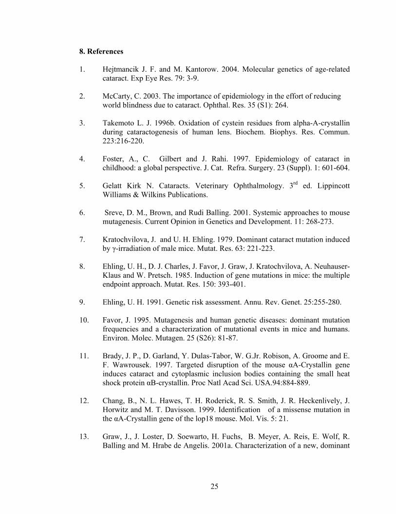
8. References
1. Hejtmancik J. F. and M. Kantorow. 2004. Molecular genetics of age-related cataract. Exp Eye Res. 79: 3-9.
2. McCarty, C. 2003. The importance of epidemiology in the effort of reducing
world blindness due to cataract. Ophthal. Res. 35 (S1): 264. 3. Takemoto L. J. 1996b. Oxidation of cystein residues from alpha-A-crystallin
during cataractogenesis of human lens. Biochem. Biophys. Res. Commun. 223:216-220.
4. Foster, A., C. Gilbert and J. Rahi. 1997. Epidemiology of cataract in
childhood: a global perspective. J. Cat. Refra. Surgery. 23 (Suppl). 1: 601-604.
5. Gelatt Kirk N. Cataracts. Veterinary Ophthalmology. 3rd ed. Lippincott Williams & Wilkins Publications.
6. Sreve, D. M., Brown, and Rudi Balling. 2001. Systemic approaches to mouse
mutagenesis. Current Opinion in Genetics and Development. 11: 268-273.
7. Kratochvilova, J. and U. H. Ehling. 1979. Dominant cataract mutation induced by γ-irradiation of male mice. Mutat. Res. 63: 221-223.
8. Ehling, U. H., D. J. Charles, J. Favor, J. Graw, J. Kratochvilova, A. Neuhauser-
Klaus and W. Pretsch. 1985. Induction of gene mutations in mice: the multiple endpoint approach. Mutat. Res. 150: 393-401.
9. Ehling, U. H. 1991. Genetic risk assessment. Annu. Rev. Genet. 25:255-280. 10. Favor, J. 1995. Mutagenesis and human genetic diseases: dominant mutation
frequencies and a characterization of mutational events in mice and humans. Environ. Molec. Mutagen. 25 (S26): 81-87.
11. Brady, J. P., D. Garland, Y. Dulas-Tabor, W. G.Jr. Robison, A. Groome and E.
F. Wawrousek. 1997. Targeted disruption of the mouse αΑ-Crystallin gene induces cataract and cytoplasmic inclusion bodies containing the small heat shock protein αB-crystallin. Proc Natl Acad Sci. USA.94:884-889.
12. Chang, B., N. L. Hawes, T. H. Roderick, R. S. Smith, J. R. Heckenlively, J.
Horwitz and M. T. Davisson. 1999. Identification of a missense mutation in the αΑ-Crystallin gene of the lop18 mouse. Mol. Vis. 5: 21.
13. Graw, J., J. Loster, D. Soewarto, H. Fuchs, B. Meyer, A. Reis, E. Wolf, R.
Balling and M. Hrabe de Angelis. 2001a. Characterization of a new, dominant
25

V124E mutation in the mouse αA-crystallin encoding gene. Invest. Ophthalmol. Vis. Sic. 42: 2909-2915.
14. Pras, E., M. L. Frydman, E. Levy-nissenbaum, T. Bakhan, J. Raz, E. I. Assia,
B. Goldman, and E. Pras. 2000. A nonsense mutation (W9X) in CRYAA causes autosomal recessive cataract in an inbred Jewish Persian family. Invest. Ophthalmol. Vis Sci. 41: 3511-3515.
15. Mackay,D. S., U. P. Andley, and A. Shiels. 2003. Cell death triggered by a
novel mutation in the αA-crystallin gene underlies autosomal dominant cataract linked to chromosome 21q. Eur. J. Hum. Genet. 11: 784-793.
16. Cobb, B. A. and J. M. Petrash. 2000. Structural and functional changes in the
αA-crystallin R116C mutant in hereditary cataracts. Biochemistry 39: 15791-15798.
17. Brady, J. P., D. L. Garland, D. E. Green, E. R. Tamm, F. J. Giblin, and E. F.
Wawrousek. 2001. αB-crystallin in lens development and muscle integrity: a gene knockout approach. Invest. Ophthalmol. Vis Sic. 42: 2924-2934.
18. Vicart,P., A.Caron, P.Guicheney, Z. Li, M.C. Prevost, A.Faure D.Chateau, F.
Chapon, F.Tome, J.M. Dupret, D.Paulin, and M. Fardeau. 1998. A missense mutation in the άB-crystallin chaperone gene causes a desmin-related myopathy. Nat. Genet. 20: 92-95.
19. Bova, M.P., O.Yaron, Q. Huang, L. Ding, D.A. Haley, P.L.Stewart, and J.
Howitzj. 1999. Mutation R120G in αB-crystallin, which is linked to a desmin-related myopathy, results in an irregular structure and defective chaperone-like function. Proc. Natl. Acad. Sci. USA. 96:6137-6142.
20. Berry, V., P. Francis, M.A. Reddy, D. Collyer, E. Vithana, I. Mackay, G.
Dawson, A.H. Carey, A. Moore, S.S Bhattacharya, and R. A. Quinlan. 2001. Alpha-B- crystallin gene (CRYAB) mutation causes dominant congenital posterior polar cataract in humans. Am. J. Hum. Genet. 69: 1141-1145.
21. Graw, J., M. Jung, J.Loster, N. Klopp, D. Soewarto, C. Fella, H. Fuchs, A.
Reis, E. Wolf, R. Balling and M. Hrabe de Angelis. 1999. Mutation in the βA3/A1-Crystallin encoding gene Cryba1 causes a dominant cataract in the mouse. Genomics. 62: 67-73.
22. Kannabiran, C., P. K. Rogan, L. Olmos, S. Basti, G. N. Rao, M. Kaiser-
Kupfer, and J. F. Hejtmancik. 1998. Autosomal dominant zonular cataract with sutural opacities is associated with a splice mutation in the βA3/A1-crystallin gene. Mol. Vis. 4: 18.
26

23. Bateman, J.B., D.D. Geyer, P. Flodman, M. Johannes, J. Sikela, N. Walter, A.T. Moreira, K. Clancy, and M. A. Spence. 2002. A new βA1-crystallin splice junction mutation in autosomal dominant cataract. Invest. Ophthalmol. Vis Sci. 41: 3278-3285.
24. Qi, Y., H. Jia, S. Huang, H. Lin, J. Gu, H. Su, T. Zhang, Y. Gao, L.Qu, D. Li.
and Y. Li. 2004. A deletion mutation in the βA1/A3 crystallin gene (CRYBA1/A3) is associated with autosomal dominant congenital nuclear cataract in a Chinese Family. Hum. Genet. 114: 192-197.
25. Reddy,M.A., O. A. Bateman, C. Chakarova, J. Ferris, V. Berry, E. Lomas, R.
Sarra, M. A. Smith, A. T. Moore, S. S. Bhattacharya, and C. Slingsby. 2004. Characterization of the G91del CRYBA1/3-crystillin protein: a cause by human inherited cataract. Hum. Mol. Genet. 13: 945-953.
26. Burdon, K.P., M.G. Wirth, D.A. Mackay, I. M. Russell-Eggitt, J. E. Craig,
J.E., Elder, J. L., Dickinson, and M.M. Sale. 2004. Investigation of crystallin genes in familial cataract, and report of two disease associated mutation. Br. J. Ophthalmol. 88: 79-83.
27. Mackay, D. S., O. B. Boskovska, H. L. S. Knopf, K. J. Lampi, and A. Shiels.
2002. A nonsense mutation in CRYBB1 associated with autosomal dominant cataract linked to human chromosome 22q. Am. J. Hum. Genet. 71: 1216-1221.
28. Chambers, C. and P. Russell. 1991. Deletion mutation in an eye lens β-
crystallin. J. Biol. Chem. 266: 6742-6746.
29. Graw, J., J. Loster, D. Soewarto, H. Fuchs, A. Reis, E. Wolf, R. Balling and M. Hrabe de Angelis. 2001c. Aey2, a new mutation in the βB2-crystallin encoding gene in the mouse. Invest. Ophthalmol. Vis. Sic. 42: 1574-1580.
30. Litt, M., R. Carrero-Valenzuela, D. Lamorticella, D. W. Schultz, T. N.
Mitchell, P. Kramer, and I. H. Maumenee. 1997. Autosomal dominant cerulean cataract is associated with a chain termination mutation in the human β-crystallin gene CRYBB2. Hum. Mol. Genet. 6: 665-668.
31. Gill, D., R. Klose, F. L. Munier, M. Mcfadden, M. Priston, G. Billingsley, N.
Ducrey, D. F. Schorderet and E. Heon. 2000. Genetic hetero-geneity of the Coppock-like cataract: a mutation in CRYBB2 on chromosome 22q11.2. Invest. Ophthalmol. Vis. Sci. 41: 159-165.
32. Vanita, V. Sarhadi, A.Reis, M.Jung, D.Singh, K.Sperling, J.R.Singh, and
J.Burger. 2001. A unique form of autosomal dominant cataract explained by gene conversion between β-crystallin B2 and its pseudogene. J. Med. Genet. 38: 392-396.
27
33. Santhiya,S.T., S.M.Manisastry, D,Rawlley, R.Malathi, S. Anishetty, P.M.
Gopinath, P. Vijalakshmi, P. Namperumalsamy, and J.Graw. 2004. Mutation analysis in Indian families suffering from congenital cataracts: identification of several SNPs and new causative allele in the CRYBB2 gene. Invest. Ophthalmol. Vis. Sci. 45: 3577-3607.
34. Graw, J. 2004. Congenital hereditary cataracts. Int. J. Dev. Biol. 48: 1031-1044.
35. Heon, E., M. Priston, D. F. Schorderet, G. D. Billingsley, P. O. Girard, N.
Lubsen, and F. L. Munier. 1999. The γ-crystallin and human cataracts: A puzzle made clearer. Am. J. Hum. Mol. Genet. 65: 1261-1267.
36. Ren, Z., A. Li, B. S. Shastry, T. Padma, R. Ayyangari, M. H. Scott, M. M.
Parks,M. I. Kaiser-Kupfer, and J. F. Hejtmancik. 2000. A5-base insertion in the γC-crystillin gene is associated with autosomal dominant variable zonular pulverulent cataracts. Hum. Genet. 106: 531-537.
37. Santhiya,S.T., M.S.Manohar, D.Rawlley, P. Vijayalakshmi, P.
Namperumalsamy, P.M.Gopinath, J. Joster, and J.Graw. 2002. Novel mutation in the γ-crystallin genes causes autosomal dominant congenital cataracts. J.Med. Genet. 39: 352-358.
38. Stephan, D.A., E. Gillanders, D. Vanderveen, D. Freas-lutz, G. Wistow, A.D.
Baxevanis, C.M.Robbins, A.Vanauken, I.M. Quesenberry, J.Bailey-wilson, S.H. Juo, J.M.Trent, L.Smiyh, and M.J. Brownstein. 1999. Progressive juvenile-onset punctuate cataracts caused by mutation in the γD-crystallin gene. Proc. Natl. Acad. Sci. USA. 96: 1008-1012.
39. Nandrot,E., C. Slingsby, A. Basak, M. Chedrif-chefchaouni, B. Benazzouz, Y.
Hajaji, S. Boutayeb, O. Gribouval, L. Arbogast, A. Berraho, M. Abitbol, and L. Hilal. 2003. Gamma-D crystallin gene (CRYGD) mutation causes autosomal dominant congenital cerulean cataracts. J. Med. Genet. 40: 262-267.
40. Mackay, D.S., U. P. Andley, and A. Shiels. 2004. A missense mutation in the
γD-crystallin gene (CRYGD) associated with autosomal dominant “coral-like” cataract linked to chromosome 2q. Mol. Vis. 10: 155-162.
41. Shentu, X., K. Yao, W. Xu, S. Zheng, S. Hu, and X. Gong. 2004. Special
fasciculiform cataract caused by a mutation in the the γD-crystallin gene. Mol. Vis. 10: 233-239.
42. Kmoch, S., J. Brynda, B. Asfaw, K. Bezouska, P. Novak, P. Rezakova, L.
Ondrova, M. Filipec, J. Sedlacek and M. Elleder. 2000. Link between a novel,
28

human γD-crystallin allele and a unique cataract phenotype explained by protein crystallography. Hum. Mol. Genet. 9: 1779-1786.
43. Graw, J. 1997. The Crystallins: Genes, Proteins and Diseases. Biol.
Chem.378:1331-1348. 44. Hisato Kondoh. 1999. Transcription factors for lens development assessed in
vivo. Current opinion in Genetics & Development. 9:301-308. 45. Quiring R., U. Walidorf, U. Kloter, and W. J. Gehring. 1994. Homology of the
eyeless genes of Drosophila to the small eye gene in mice and Aniridia in humans. Science. 265: 785-789.
46. Hill, R. E., J. Favor, B. L. M. Hogan, C. C. T. Ton, G. F. Saunders, I. M.
Hanson, J. Prosser, T. Jordan, N. D. Hastie, and V. van Heyningen. 1991. Mouse small eye results from mutations in a paired-like homeobox-containing gene. Nature. 354:522-525.
47. Glaser T., D.S. Walton, and R.L. Maas.1992. Genomic structure, evolutionary conservation and aniridia mutations in the human Pax6 gene. Nature Genet. 2:232-238.
48. Glaser T., L. Jepeal J. G. Edwards, S. R .Young, J. Favor, and R.L. Maas.1994. Pax6 gene dosage effect in family with a congenital cataracts, aniridia, anophthalmia and central nervous system defects. Nature Genet. 7:463-471.
49. Hanson I.M., J.M. Fletcher, T. Jordan, A. Brown, D. Taylor, R. J.Adams, H.H.Punnett, and R. van Heyningen.1994. Mutation at the PAX6 locus is found in the heterogenous anterior segment malformations including Peter’s anomaly. Nature Genet.6: 168-173.
50. Halder, G., P. Callaerts, and W. J. Gehring. 1995. Induction of ectopic eyes by
targeted expression of the eyeless genes in Drosophila. Science. 267: 1788-1792.
51. Yang, L H. S., J. M. Jacobean, R.D. D. Pasko, and O. Sundin. 1994. Pax6 is
first expressed in a region of ectoderm anterior to the early neural plate: implications for stepwise determination of the lens. Dev. Biol.162:181-194.
52. Grindley, J.C., D. R. Davidson, and R. E. Hill. 1995. The role of pax6 in eye
and nasal development. Development. 121: 1433-1442. 53. Varnum D. S. and L. C. Stevens. 1968. Aphakia, a new mutation in the mouse.
J. Hered. 59:147-150.
29

54. Semina, E., J. C. Murray, R. Reiter, R. F. Hrstka, and J. Graw. 2000. Deletion in the promoter region and altered expression of Pitx3 homeobox gene in aphakia mice. Hum. Mol. Genet. 9: 1575-1585.
55. Reiger, D. K., E. Reichenberger, W. Mclean, A. Sidow, and B. R. Olsen. 2001.
A double-deletion mutation in the Pitx3 gene causes arrested lens development in aphakia mice. Genomics. 72: 61-72.
56. Semina, E. V., R. E. Ferrell, H. A. Mintz-Hittner, P. Bitoun, W. L. M. Alward,
R. S. Reiter, C. Funkhauser, S. Daack-Hirsch, and J. C. Murray. 1998. A novel homeobox gene PITX3 is mutated in families with autosomal dominant cataracts and ASMD. Nat. Genet. 19:167-170.
57. Semina, E. V., R. S. Reiter, and J. C. Murray. 1997. Isolation of new
homeobox gene belonging to the Pitxl Rieg family: expression during lens development and mapping to the aphakia region on mouse chromosome 19. Hum. Mol. Genet. 6: 2109-2116.
58. Nunes, I., L. T.Tovmasian, R. M. Silva, R. E. Burke, and S. P. Goff. 2003.
Pitx3 is required for development of substantia nigra dopam inergic neurons. Proc. Natl. Acad. Sci. USA. 100: 4245-4250.
59. Shimada, N., T. Aya-Murata, H. M. Reza, and K. Yasuda. 2003. Cooperative
action between L-Maf and Sox2 on δ-crystallin gene expression during chick lens development. Mech. Dev. 120: 455-465.
60. Ring,B. Z.,S. P. Cordes, P. A. Overbeek, and G. S. Barsh. 2000. Regulation of
mouse lens fiber cell development and differentiation by the Maf gene. Development. 127: 307-317.
61. Kamachi,Y., M. Uchikawa, and H. Kondoh. 2000. Pairing SOX off with
partners in the regulation of embryonic development. Trends Genet.16: 182-187.
62. Kamachi,Y., M. Uchikawa, J. Collignon, R. Lovell-Badge and H.
Kondoh.1998. Involvement of Sox1, 2 and 3 in the early and subsequent molecular events of lens induction. Development. 125:2521-2532.
63. Nishiguchi, S., H. Wood, H. Kondoh, R. Lovell-Badge, and V. Episkopou.
1998. Sox1 directly regulates the γ-crystallin gene and is essential for lens development in mice. Genes Dev. 12: 776.781.
64. Azuma, N., A. Hirakyama, T. Inoue, A. Asaka, and M. Yamada. 2000.
Mutations of a human homologue to the Drosophila eyes absent gene (EYA1) detected in patient with congenital cataracts and ocular anterior segment anomalies. Hum. Mol. Genet. 9: 363-366.
30

65. Oliver,G., A. Mailhos, R. Wehr, N. G. Copeland, N. A. Jenkins, and P. Gruss. 1995. Six3, a murine homologue of sine oculis gene demarcates the most anterior border of the developing neural plate and is expressed during eye development. Development. (Cambridge, UK), 121:4045-4055.
66. Bovolenta, P., A.Mallamaci, L. Puelles, and E. Boncinelli. 1998. Expression
pattern of cSix3, a member of Six/sine oculis family of transcription factors. Mech. Dev. 70: 201-203.
67. Robinson, M. L., C. Ohtaka-Maruyama, C. Chan, S. Jamieson, C. Dickson,
P.A.Overbeek, and A.B. Chepelinsky. 1998. Disregulation of ocular morphogenesis by lens-specific expression of FGF-3/int-2 in transgenic mice. Dev. Bio. 198.13-31.
68. Piatigorsky, J. 1981. Lens differentiation in vertebrates. Differentiation. 19:
134-153. 69. Rink, H., J. Bours, and H. J. Hoenders.1982. Guidelines for the classification
of lenses and the characterization of lens proteins. Ophthalmic Res. 14: 284-291.
70. de Jong, W. W. 1982. Eye lens proteins and vertebrate phylogeny. In:
Macromolecular Sequences in Systematic and Evolutionary Biology, M. Goodman, ed. (New York and London:Plenum-Press), pp. 75-114.
71. Groenen,P. J. T. A., K. B. Merck, W. W. de Jong, and H. Bloemendal. 1994.
Structure and modifications of the junior chaperone α—crystallin. From lens transparency to molecular pathology. Eur. J. Biochem. 225: 1-19.
72. Ingolia, T. D. and E. A. Craig. 1982. Four small Drosophila heat shock
proteins are related to each other and to mammalian α-crystallin. Proc. Natl. Acad. Sci. USA. 79: 2360-2364.
73. Horwitz, J. 1992. α-crystallin can function as a molecular chaperone. Proc.
Natl. Acad. Sci. USA. 89: 10449-10453. 74. Tamm, E. R., P. Russell, D. H. Johnson, and J. Piatigorsky. 1996. Human and
monkey trabecular meshwork accumulate αB-crystallin in response to heat shock and oxidative stress. Invest. Ophthalmol. Vis. Sci. 37:2402-2413.
75. Carter J. M., A. M. hutcheson, and R. A. Quinlan.1995. In vitro studies on the
assembly properties of the lens proteins CP49, CP115: assembly with ά-crysallin but not with vimentin. Exp. Eye Res. 60: 181-192.
31

76. Kato, K., H. Ito, Y. Inaguma, K. Okamoto, and S. Saga. 1996. Synthesis and accumulation of αB-crystallin in C6 glioma cells is induced by agents that promote the disassembly of microtubules. J. Biol. Chem. 271: 26989-26994.
77. Wang, K., and A. Spector. 1996. ά-Crystallin stabilizes actin filaments and
prevents cytochalasin-induced depolymerization in a phosphorylation dependent manner. Eur. J. Biochem. 242: 56-66.
78. Brady, J.P., D. Garland, Y. Duglas-Tabor, W. G, Jr Robinson, A. Groome, and
E. F. Wawrousek. 1997. Targeted disruption of the mouse άA-crystallin gene induces cataract and cytoplasmic inclusion bodies containing the small heat shock protein άB-crystallin. Proc. Natl. Acad. Sci. USA. 94: 884-889.
79. Chang, B., L. N. Hawes, R.S. SMITH, J.R. Heckenlively, M. T. Davisson and
H. H. Roderick. 1996. Chromosomal localization of a new mouse lens opacity gene (lop18). Genomics. 36: 171-173.
80. Duguid, J. R., R. G. Rohwer and B. Seed.1988. Isolation of cDNA of scrapie-
modulated RNAs by subtractive hybridization of a cDNA library. Proc. Natl. Acad. Sci. USA. 85: 5738-5742.
81. Aarts,H. M. J., N. H. Lubsen, and J. G. G. Schoenmakers. 1989. Crystallin
gene expression during lens development. Eur. J. Biochem.183: 31-36. 82. Head, M.W., K. Sedowofia, and R. M. Clayton. 1995. Βb2-crystllin in the
mammalian retina. Exp. Eye. Res. 61: 423-428. 83. Kador , P. F., H.N. Fukui, S. Fulushi, H.M. Jernigan, Jr., and J.H. Kinoshita.
1980. Philly mouse: a new model of hereditary cataract. Exp Eye. Res. 30: 59-68.
84. Chambers, C. and P. Russell. 1991. Deletion mutation in an eye lens β-
crystallin. J. Biol. Chem. 266: 6742-6746. 85. Padma, T., R. Ayyagari, J. S. Murty, S. Basti, T. Fletcher, G. N. Rao, M.
Kaiser-Kupfer , and J.F. Hejtmancik. 1995. Autosomal dominant zonular cataract with sutural opacities localized to chromosome 17q11-12. Am. J. Hum. Genet. 57: 850-845.
86. Kramer, P., J. Yount, T. Mitchell, D. LaMorticella, R. Carrero-Valenzuela, E.
Lovrien, I. Maumenee, and M. Litt. 1996. A second gene for cerulean cataracts maps to the β-crystally region on chromosome 22 Genomics. 35: 539-542.
87. Takemoto, L. J. 1997. Disulfide bond formation of cysteine-37 and cysteine 66
of BetaB2crystallin during cataractogenesis of the human lens Exp. Eye Res. 64: 609-614.
32

88. Rink, H., J. Bours, and H. J. Hoenders.1982. Guidelines for the classification of lenses and the characterization of lens proteins. Ophthalmic Res. 14: 284-291.
89. Skow, L. C. M. JE. Donner, S. M. Huang, J. M. Garder. B. A. Taylor, W. G.
Beamer, and P. A. Lalley. 1988. Mapping of mouse gamma crystalline genes on chromosome 1. Biochem. Genet. 26: 557-570.
90. den Dunnen, J. T. R. J. E. Jongbloed, A. H. M. Geurts ven Kessel, and J.G.G.
Schoenmakers. 1985. Human lens γ-crystallin sequences are located in the p12-qter region of chromosome 2. Hum. Genet. 70: 217-227.
91. Graw, J., L. Coban, A. Liebstein, and T. Werner. 1991. Murine γE-crystallin is
distinct from murine γ2-crystallin. Gene. 104: 265-270. 92. Van Leen, R. W. M. L. Breuer, N. H. Lubsen, and J. G. G. Schoenmakers.
1987. Developmental expression of crystalline genes. In situ hybridization reveals a differential localization of specific mRNAs. Dev. Biol. 123: 338-345.
93. Santhiya, S. T., S. M. Abd-alla, J. Loster, and J. Graw. 1995. Reduced levels of
γ-Crystallin transcripts during embryonic development of murine Cat2nop
mutant mice.Graefe’s Arch. Clin. Exp. Ophthalmol. 233:795-800. 94. Goring, D. R., M. L.Breitman, and L. C. Tsui.1992. Temporal regulation of six
crystalline transcripts during mouse lens development. Exp. Eye. Res. 54: 785-795.
95. Peek, R., R.W.L. M.Niessen, J. G. G. Schoenmakers, and N. H. Lubsen.1991.
DNA methylation as a regulatory mechanism in rat γ-Crystallin gene expression. Nuc. Acids Res. 19: 77-83.
96. Cartier, M., L. M. Breitman, and L. C. Tsui. 1992. A frameshift mutation in
the γE-crystallin gene of the Elo mouse. Nature Genet. 2: 42-45. 97. Loster, J., W. Pretsch, R. Sandulache, T. Schmitt-John, M. F. Lyon, and J.
Graw. 1994. Close linkage of the dominant cataract mutations (Cat-2) with ldh-1 and Cryge on mouse chromosome 1. Genomics. 23: 240-242.
98. Kratochvilova, J. and J. Favor. 1992. Allelism tests of 15 dominant cataract
mutations in mice. Genet. Res. Camb. 59: 199-203. 99. Everett, C. A., P. H. Glenister, D. M. Taylor, M. F. Lyon, J. Kratochvilova-
Loster, and J. Favor. 1994. Mapping of six dominant cataract genes in the mouse. Genomics. 20: 429-434.
33

100. Graw, J., J. Kratochvilova, A. Lobke, P. Reitmeir, E. Schaffer and A. Wulff. 1989. Characterization of Scat (Suture cataract), a dominant cataract mutation in mice. Exp. Eye. Res. 49: 469-477.
101. Oda, S. I., K. Watanabe, H. Fujisawa, and Y. Kameyama. 1980. Impaired
development of lens fibers in genetic microphthalmia, eye lens obsolescence, Elo, of the mouse. Exp. Eye. Res. 31:673-681.
102. Brakenhoff, R. H., H. A. M. Henskens, M. W. P. C. van Rossum, N. H.
lubsen, and J. G. G. Schoenmakers. 1994. Activation of the γE-crystallin pseudgene in the human hereditary Coppocklike cataract. Hum. Mol. Genet. 3: 279-283.
103. Hatta, K. and M. Takeichi. 1986. Expression of N-cadherin adhesion molecules associated with early morphogenetic events in chick development. Nature. 320:447-449.
104. Sandilands, A. A. R. Prescott, J. M. Carter, A. M. Hutcheson, R. A. Quinian, J. Richards, and P. G. FitzGerald. 1995. Vimentin and CP49/ filensin form distinct networks in the lens which are independently modulated during lens fiber cell differentiation. J CELL SCI. 108: 1397-1406.
105. Zhang, P., N. J. Liegeos, C. Wong, M. Finegold, H. Hou, J. C. Thomson, A. Silverman, J. W. Harper, R. A. DePinho, and S. J. Elledge. 1997. Altered cell differentiation and proliferation in mice lacking p57KIP2 indicates a role in Beckwith-Wielerman syndrome. Nature. 387:151-158.
106. Zhang, P., C. Wong, R. A. DePinho, J. W. Harper, and S. J. Elledge. 1998. Cooperation between Cdk inhibitors p27 KIP1 and p57KIP2 in the control of tissue growth and development. Genes Dev. 12: 3162-3167.
107. Shiels, A. and C. S. Griffin. 1993. Aberrant expression of the gene for lens
major intrinsic protein in the CAT Mouse. Curr. Eye. Res. 12:212-215.
108. Zhou, L., T. Chen, and R. L. Church. 2002. Temporal expression of three mouse lens fiber cell membrane protein genes during early eye development. Mol. Vis. 8: 143-148.
109. Zwaan, J. and R. M. Williams. 1969. Cataracts and abnormal proliferation of
the lens epithelium in mice carrying the cat-fr gene. Exp. Eye. Res. 8:161-167.
34

110. Steele, E. C. JR, S. Kerscher, M. F. Lyon, P. H. Glenister, J. Favor, J. Wang and R. L. Church. 1997. Identification of a mutation in the MP19 gene, Lim2 in the cataractous mouse mutant To3. Mol. Vis. 3:5.
111. Pras, E., E. Levy-Nissenbaum, T. Bakhan H. Lahat, E. Assia, N. Geffencarmi, M. Frydman, B. Goldman, and E. Pras. 2002. A. missense mutation in the LIM2 gene is associated with autosomal recessive presenile cataract in an inbred Iraqi Jewish family. Am. J. Human. Genet. 70: 1363-1367.
112. Willecke, K., J. Eiberger, J. Degen, D. Eckhardt, A. Romualdi, M. Geldenagel,
J. Deutsch and G. Sohl. 2002. Structural and functional diversity of connexin genes in the mouse and human genome. J. Biol. Chem. 383:725-737.
113. White T.W. and R. Bruzzone. 2000. Intercellular communication in the eye:
clarifying the need for connexin diversity. Brain Res. Rev. 32:130-137.
114. Beyer E.C., Kistler J., Paul D. L. and D. A. Goodenough. 1989. Antisera directed against connexin43 peptides react with a 43-kD protein localized to gap junctions in myocardium and other tissues. J. Cell. Biol. 108: 595-605.
115. Musil L. S. Beyer E. C. and D. A. Goodenough. 1990. Expression of the gap
junction protein connexin43 in embryonic chick lens; molecular cloning, ultra structural localization and post-translational phosphorylation. J. Membr. Biol. 116;163-175.
116. Kistler J. B. Kirkland and S. Bullivant. 1985. Identification of a 70,000-D protein in lens membrane junctional domains. J. Cell Biol. 101;28-35.
117. Paul D. L. I. Ebihara I. J. Talemoto, K. I Swenson and D. A. Goodenough. 1991. Connexin46, a novel lens gap junction protein, induces voltage-gated currents in nonjunctional plasma membrane of xenopus oocytes. J. Cell Biol. 115: 1077-1089.
118. White T.W., R. Bruzzone., D. A. Goodenough and D. L. Paul. 1992. Mouse
Cx50, a functional member of the connexin family of gap junction proteins, is the lens fiber protein Mp70. Mol Biol Cell. 3: 711-720.
119. Evans C. W., S. Eastwood, J. Rains, W. T. Gruijters, S. Bullivant and J. B.
Kistler. 1993. Gap junction formation during development of the mouse lens. Eur J Cell Biol. 60;243-249.
120. Reaume A.G., P. A. de Sousa, S. Kulkarni, B.L. Langille, D. Zhu, T.C. Davies,
S.C. Juneja, G.M.Kidder and J. Rossant. 1995. Cardiac malformation in neonatal mice lacking connexins 43. Science. 267: 1831-1834.
35

121. White T. W., Goodenough D.A., and D. L. Paul. 1998. Targeted ablation of Connexins50 in mice results in microphthalmia and zonular pulverulent cataracts. 143 (3) 815-825.
122. Rong P., X. Wang, I. NIESMAN, y. Wu, L. E. Benedetti, I. Dunia, E. Levy
and X. Gong. 2002. Disruption of Gja8 (alpha8 connexin) in mice leads to microphthalmia associated with retardation of lens growth and lens fiber maturation. Development. 129: 176-174.
123. Gong, X., E. Li, G. Klier, Q. Huang, Y. Wu, H. Lei, N. M. Kumar, J. Horwitz,
and N. B. Gilula. 1997. Disruption of alpha3 connexin gene leads to proteolysis and cataractogenesis in mice. Cell. 91:833-843.
36