Embed Size (px)
Citation preview

1
Chapter One: Introduction
2
1. INTRODUCTION
Prevalence is a measurement of the proportion of population actually having the disease
at a specific period of time; in other word the prevalence tells us of the number of
people with the disease divided by the number of population at a specific time (Webb et
al., 2005).
The prevalence provides an estimation of probability that an individual will have oral
disease at a specific period of time and also identifying risk groups within the
population studied (Heinekens and Buring. 1987).
Globally, studies providing a wide spectrum of oral lesion includes a study reporting on
the prevalence of oral lesions among 20,000 adult Swedish population by Axell (1976)
and another study carried out in the United States of American (USA) which reported
the common oral lesions among the USA population (Bouquot., 1986). In Asia the
survey provided the prevalence of oral lesions was found to be in Indian population
(Smith et al., 1975; Metha et al., 1972).
Tobacco smoking and alcohol consumption has long been associated and indicated
worldwide as the major factors in the development of cancer and other systemic
diseases in developed countries (Peto et al., 1992; Jaber et al., 1999). Smoking habit has
a great impact on oral carcinogenesis prior to malignant transformation and the alcohol
drinking in high level has also been shown to have strong association with oral cancer in
the American Population (Morse et al., 2007).
In Asian countries particularly in India, oral cancer and precancer has been associated
with betel quid chewing (Nair et al., 1999). Betel quid chewing is a common habit in
many Asian countries and this habit spread to other regions of the world through
emigration (Reichart et al., 1987).
3
Similar studies have been also conducted Malaysia had been able to identify the high
risk groups for oral precancerous lesions where strong causal relationship with quid
chewing was reported (Zain and Ghazali 2001). In Malaysia approximately 25% of all
causes of death in Malaysia are due to tobacco usage (Ministry of Health Malaysia,
1997). A nationwide survey on oral mucosal lesions in Malaysia in 1993/1994 (Zain et
al., 1997) showed high prevalence of oral precancerous lesions among the Indians and
Indigenous people of Sabah and Sarawak who practiced betel quid chewing. In Yemen
Scheifele et al (2007) reported a significant association between oral leukoplakia and
shammah usage (tobacco quid form of quid). The prevalence of oral cancer was 1%
among Shammah users. Among the qat chewer‘s there are different of oral keratotic
white lesions with different degrees of underlying pathology depending on the
frequency and duration of qat chewing (Aiman et al., 2004). Histopathologic alteration
in the oral mucosa such as acanthosis, orthokeratosis, epithelial hyperplasia with
irregular rete redges have been described in qat chewers. Qat chewing with cigarette or
water-pipe smoking may increase the risk of developing such pathologic changes in the
oral mucosa (Aiman., 2007). Although prevalence of oral mucosal lesions has been
reported in many countries, these prevalence data are usually restricted to very few
lesions in each study. There is thus a need to obtain data from different countries with a
large random sample which can be tedious and thus appropriate information on oral
mucosal lesion prevalence can still be obtained from small low budget studies on
selected population (Axell et a1., 1990).

4
Chapter Two: Literature Review
5
2. LITERATURE REVIEW
2.1. Prevalence of oral mucosal lesions
Reports on prevalence of oral mucosal lesions showed variations in the prevalence rate
which may be related to methodology, difference in diagnostic criteria used, selection of
participants and the risk habits practiced among the population.
2.1.1. Worldwide distribution
A study by Axell (1976) where he conducted oral examination on 22033 partciptants,
reported the prevalence of oral mucosal lesions in adult Swedish population. In this
study; the prevalence of about 60 oral mucosal lesions were recorded and compared
with previous findings. Prevalence of lesions detected were of focal epithelial
hyperplasia (0.11%), leukoedema (49.07%), geographic tongue (8.45%) and lichen
planus (1.85%). Some lesions which was found in this study are directly or indirectly
related to local etiologic factors such as denture status and tobacco habits.
Bouqout in 1986 reported on the prevalence of common oral lesions during mass
screening of American population. The most oral mucosal lesions were white lesions
which accounted for 37.6% however, the most common clinical appearance of oral
lesions was that of a single, exophytic mass which accounted for 37.4% of all recorded
lesions. In this study the leukoplakia reached to more than 26% and the prevalence of
other lesions were listed as traumatic ulcer, aphthus ulcer, leukoedema, glossitis, ranula
and candidiasis.
6
In Thailand the prevalence of oral mucosal lesions such as chewers mucosa was 13.1%.
Leukoedema was12.4% and slightly more common among women, preleukoplakia was
1.8%, more among men and the leukoplakia was 1.1% which more frequent among
men. High prevalence of smoking cigarrette was obseved among the middle age
however, betel chewing was more prevalent among the old age. There was a postive
correlation between some oral mucosal lesion and the risk habits (smoking ,quid
chewing) (Reichart et al.,1987).
In a study conducted on adults Southern Chinese; the prevalence of oral mucosal lesion
was found to be 13% in urban men, 6% in urban women, 15% in rural men,
and 4% in
rural women. Tongue lesions and white lesions were relatively common, this study
showed there is a positive relationship between risk habits (smoking and alcohol
consumption) and prevalence of oral mucosal lesions (some white lesions and tongue
lesions) ( Lin et al., 2001).
From a study conducted in 2004 in London among alcohol misusers, the prevalence of
oral mucosal lesion was found to be 28.1% (n=227). The high prevalence of oral
mucosal lesion was frictional lesions (8.8%), scar tissue of lip 4.8%, candidosis 3.8%
and angular cheiltis 3.0%. The alcohol related lesion was white patch similar to the
diagnosis of leukoplakia. The study also that 56% were alcohol users and 46% were
alcohol and substance abuse users. The prevalence of tobacco smoking was 85% among
only alcohol users and 95% among the other group (alcohol with substance abuse).
There was no significant relationship between the prevalence of oral mucosal lesion and
smokers (Harris et al., 2004).
The prevalence of oral mucosal lesions in South India was 4.1%. The prevalence of
leukoplakia, oral submucous fibrosis and oral lichen planus was 0.59%, 0.55%, and
7
0.15% respectively. The prevalence of smoking, alcohol drinking and quid chewing
was 15.02%, 8.78% and 6.99% respectively. Smoking and quid chewing were
significant predictors of leukoplakia in this population (Saraswathi et al., 2006).
In Taiwan the prevalence of leukoplakia, erythroplakia, oral lichen planus, oral
submucous fibrosis and verrucous lesions were 7.44%, 1.95%, 2.98%, 1.58% and
0.84% respectively. The prevalence of smoking habit was 20.4%; areca nut chewing
was 7.16% while high prevalence of alcohol consumption which was 18.14%. There is
a statistically significant association between leukoplakia, oral submucous fibrosis,
verrucous lesions and the risk habit, areca quid chewing (Chung et al., 2005).
8
2.1.2. Malaysian prevalence of oral mucosal lesions
The first epidemiology study in Malaysia was a dental survey conducted in 1962 where
the interdepartmental committee on National defence (ICCND) comprising a joint
United States – Malaysia team conducted in a Federation of Malaysia Nutrition
Survey, quoted from Zain et al (1997). The next population–based dental survey was
conducted by the Ministry of Health Malaysia in 1974/75. This study was confined to
peninsular Malaysia where the precancerous lesion was found (1.3%). However, the
other lesions was (0.4%) where put under smoker keratosis.
From the early study was that study done by Ramanathan et al (1973(a)) and reported
the prevalence of Oral cancer and precancer was found (1.5%) where the prevalence of
oral cancer was (0.5%). However, the precancerous lesions included smoker‘s keratosis
was found ( 0.12%). Other study by Ramanathan et al (1973(b)) which conducted on
407 medical attendants and health workers and reported 55 (13.55%) subjects had oral
precancerous lesions. Also the smoker‘s keratosis included in this study of which 6
subjects (12.0%) had oral precancerous lesions.
The other study in 1978 carried out by Dental Division by Ministry of Health on total
of 9073 Malaysian subjects and reported the prevalence of leukoplakia was (1.3%),
erthroplakia (0.2%) and oral cancer as (0.01%).
Many studies in Malaysia reported that the quid chewing is a risk factor like other
countries in the spread of oral mucosal lesion particularly oral precancerous and
cancerous lesions, the Indian and Indigenous people were high risk group especially the
women in both group due to using the tobacco in their quid (Gupta et al., 1997).
The positive association of oral mucosal lesion and cigarette smoking such as
leukoedema as well as denture stomatitis. There was no relationship between the
9
cigarette smoking and prevalence of aphthous ulcer and coated tongue. There was no
statistically significant differences between the cigarettes smokers and non smokers in
prevalence of pre-leukoplakia (Zain and Razak.,1989).
The prevalence of oral soft tissue lesions in Malaysia was recorded from examination of
dental outpatients in Thailand and Malaysia where three cases of leukoplakia (1.3%),
one case of betel quid related lesion and one case squamous cell carcinoma (0.4%) was
detected in Malaysians. The was a high prevalence of lichen planus (2.1%) in Malaysian
oupatients. The prevalence of tobacco in some form was 27.5% where the cigerrate
smoking was the predominant habit and the prevalence of quid chewing among the
Malaysian out patients was 2.6% (n=6). Three tobacco associated leukoplakia were
found and also three betel quid lesions (Axell et al., 1990).
The prevalence of oral mucosal lesions among elderly Malaysians was found to be
22.8% (n=111). A total of 145 oral lesions were detected. The prevalence of oral
mucosal lesions was highest among Indians and least among the Chinese. The most
common finding was tongue lesions which was found to be 10.7%, followed by oral
pigmentation (4.9%) and white lesions (4.3%). Denture related lesions were
comparatively low at 2.5%. Two cases of oral cancer was detected giving a relatively
high prevalence of 0.4 % ( Taiyeb et al., 1995).
A nationwide Malaysian dental survey showed the prevalence of oral mucosal was 9.7%
with no predictable difference between males (9.1%) and females (10.1%). The most
common lesion was denture stomatitis; leukoplakia, an oral precancerous lesion was the
most common oral lesion where the males and females ratio for leukoplakia was 3:1.
The smokers palate was more among male while betel chewers mucosa was more
among female. Five cases of oral cancer was reported in three male and two in female.
10
One humdred sixty five (165) subjects had oral lesions which includes precancerous
lesions and 187 (1.6%) had betel chewers mucosa. The prevalence of oral precancer
lesions in decreasing order was firstly the Indians (4.0%) followed by the in Other
Bumiputras who are mainly the indigenous people of Sabah & Sarawak (2.5%). The
lowest prevalence was among the Chinese(0.05%). The prevalence risk habits among
Malaysian was found to be 19.2% smokers, 4.87% betel–quid chewers and 1.7% were
alcohol consumers (Zain et al., 1997).
In a study by reviewing different types of studies that proved the importance in
making comparisons between studies such as the incidence of data for oral cancer in
Malaysia was reported by Hirayama in 1966, 35 years ago which estimated that 3.1 new
cases per 100,000 population was diagnosed for the year 1963 (Zain and Ghazali.,
2001).
11
2.1.3. Yemeni prevalence of oral mucosal lesions
From earliest epidemiology studies in Yemen which deal about with the oral lesion was
that study which was carried out in 1987 by Hill and Gibson. This study reported that
keratosis of buccal mucosa was related to gat chewing.
The other study conducted in 2004 showed oral white lesions (oral kerstosis ) in 342
(22.4%) Yemeni subjects with a mean age of 27 years old with 87.4% being. The white
lesion was graded from mild whitening in appearance to homogenous-like lesions. The
prevalence of qat chewing in this study was 61.12% while the pevalence of smoking
habit was found to be 26.36%. There was a significant relationship beween risk habits
(qat chewing, smoking, and shammah usage) and the prevalence of oral white lesions.
(Aiman et al., 2004).
In a study carried out among the Yemeni shammah users, the prevalence of oral
squamous carcinoma (OSCC) among the shammah users was 1% (n=2). The prevalence
of mucosal burn (MB) was 31%, oral leukoplakia was 27%. No shammah users was
diagnosed with either mucosal burn and or leukoplakia. When shammah associated
lesiosn was combined, the prevalence of shammah-associated lesions was found to be
58%. The prevalence of lichen planus was 0.5% and oral lichenoid reaction was 4.0%
while the prevalence of other lesions such as frictional lesion was 4.0%,
pseudomemebranous candidosis was 2.5%, mosrsicatio buccarum was 0.5% and white
sponge nevous was 0.5%. All the participtants in this study were shammah users. There
was a significant association between of the prevalence of oral leukoplakia and the daily
duration of the contact of shammah with the oral mucosa (Scheifele et al., 2007).
In another study the possible synergistic effect of qat in the development of OSCC of
the floor of the mouth was reported (Kennedy et al., 1983). For another case report, it
12
was shown that of plasma cell gingivitis can be induced by qat, where the lesion
disappeared after discontinuation of qat chewing (El-Shoura et al., 1995).
2.2. Characterstics of oral mucosal lesions
2.2. 1. Normal Oral mucosa
Oral mucosa is the lining of the oral cavity which has a variety of functions, such as
protection, sensation and secretion, and histologically adapted to the unique
environment inside the mouth. Oral mucosa lacks the appendages seen in skin, but
sebaceous glands can be found in the upper lip and buccal mucosa. The mobile part of
oral mucosa which lined the vestibule and floor of mouth joins the tightly adherent
gingiva of the dental alveolus and is easily visible in normal mucosa. Gingiva appears
paler pink secondary to decreased visibility of underlying blood vessels through the
relatively opaque keratin layer. The gingival margin should be is usually well defined
with slightly rolled margin. The interdentally papillae is pointed and the texture of the
attached gingiva exhibits stippling, representing collagen fibres attaching the gingiva to
the underlying periosteum (Bruch and Treister., 2009).
2.2.2. Definition of oral mucosal lesions
Oral mucosal lesion is defined as any change in oral mucosal surface and these changes
may present as red, white, ulcerative and pigmented or as any swelling or as variants of
developmental defects (Epinoza et al., 2003). The oral mucosal lesions have many
causes which include infection from bacteria, viruses, fungi, parasites; other influences
such as physical and thermal causes; changes in immune system; the systemic diseases;
13
neoplasia; trauma and other factors including aging and chronic habits such as the use of
tobacco and alcohol (Reichart., 2000).
2.2.3. Types of oral mucosal lesions
The oral mucosal lesion can be classified into broad categories namely: oral malignant
lesions, oral potentially malignant disorder and the other oral mucosal lesions which are
not malignant and not potentially malignant disorders.
2.2.3.1. Oral malignant lesions (OML)
Malignant epithelial lesions include squamous cell carcinoma, verrucous carcinoma,
basaloid squamous cell carcinoma, papillary squamous cell carcinoma, spindle cell
carcinoma, acantholytic squamous cell carcinoma, adenosquamous carcinoma,
carcinoma cuniculatum and lymphoepithelial carcinoma (Barnes et al., 2005).
The most prevalent of oral malignant lesions in the world is oral squamous cell
carcinoma which is one of the 10 common causes of death (Baum, 2007; Bruch and
Treister., 2009) :
Squamous cell carcinoma (SCC):
This lesion may appear a flat raised exophytic growing or ulcerated (showing
surface erosion). The surface texture can range from smooth to irregular with
induration, firmness or hardness and fixation immobility or palpable adherence
to underlying structures indicating infiltration of cancer cells into deeper tissue
a. Verrucous carcinoma
14
It is a low-grade variant of SCC with a distinctive exophytic and papillary, or
warty, appearance atypically whitish or gray color and common sites are the
buccal mucosa, gingiva, and vestibule.
2.2.3.2. Oral potentially malignant disorders (OPMD)
Malignant transformations have been discussed in a World Health Organization
workshop held in 2005, the potentially malignant disorders were recommended in
reference to precancerous lesions as not all disorders described under this term may
transform to cancerous lesions (Warnakulasuriya et al., 2007). Leukoplakia and
erythroplakia are the most common ones potentially premalignant disorders. The
diagnosis of these lesions with exclusion of the other red and white lesions in addition
to the lichen planus seemed to be accepted in the literature as being a potentially
malignant disorder. However, the risk of malignant transformation for the other red and
white lesions is lower than leukoplakia (Van Der Waal., 2009).
a. Leukoplakia
Leukoplakia was defined in 1877 by Schwimmer as a white lesion in the tongue that
was probably syphilitic glossitis for a long time leukoplakia has been used to describe
white plaque or patches.
WHO in 1978 defined the leukoplakia as a white patch or plaque that cannot be
characterized clinically or histopathologically as any other disease which is based on the
exclusion of other conditions to get the diagnosis of leukoplakia and described it as a
protective reaction against a chronic irritation. In 1980 WHO described the leukoplakia
as white patches which vary from quite small to an extensive lesion involving large area
15
of oral mucosa and the surface of this lesion maybe smooth, wrinkled with shallow
small crack.
From the international seminar hold in 1983 recommended that the use of the term
leukoplakia should be avoided if the cause is known except in those cases where it was
believed that the cause was tobacco (Axell et al., 1984). Leukoplakia was then, defined
as a predominantly white lesions of oral mucosa which cannot be characterized as any
other definable disease (Axell et al., 1996). Recently, Warnakulasuriya et al (2007)
recommended that the term leukoplakia should be used to recognize white plaques of
questionable risk having excluded other known disease or disorders that carry no
increased risk for oral cancer.
b. Erythroplakia
Erythroplakia is a fiery red patch of the oral mucosa that cannot be characterized
clinically or microscopically as any other definable entity, which would exclude all
the inflammatory condition which may cause red appearance of oral mucosa.
Erythroplakia is precancerous lesion and some cases of erythroplakia showed
different degrees of dysplasia histologically (Shafer and Waldron., 1975). The
common sites in the oral cavity affected by erythroplakia are soft palate, floor of the
mouth and buccal mucosa (Scully, 2004).
c. Oral Lichen planus (LP)
The oral lichen planus (OLP) presents as reticular, erythematous and erosive lesions
with distinct white mucosal changes called Wickham‘s striae. Women are more affected
16
than men, with most patients diagnosed at ages of 40-50 years old (Bruch and Treister.,
2009). Lichen planus may contain both red and white appearance with different texture
such as reticular, papules, plaque; bullous, erythematous and ulcerative forms
(Greenberg and Glick, 2009).
The oral lichen planus affects from 1- 4% of the adult population (Bougout and Gorlin,
1986, Axell, 1987; Axell and Rundquist, 1987; Axell et al., 1990; Salonen et al., 1990;
Banoczy and Rigo, 1991; Albrecht et al‘ 1992). Oral lichen planus may affect the
middle aged, elderly and also affects the childern and young adults (Silverman and
Griffith,1972).
There are two types of OLP according to the site of the lesion namely the extra-oral and
intra-oral type. Typically 90% intra oral lesion affects the posterior buccal mucosa 30%
the tongue, 13% the alveolar ridge /gingiva and rarely on the lip vermillion or palate
(Axell and Rundquist, 1987).
d. Oral submucous fibrosis(OSF)
Oral submucous fibrosis has been conservatively diagnosed only on the basis of
palpable fibrous bands. The palpable fibrous bands are not always present, in several
instances a tough leathery mucosa with all the associated symptomatic, clinical and
histopathological characteristics of OSF is seen (Pindborg et al., 1980; Seedat et al.,
1988). Areca nut is the principle aetiological agent, also the gentic traits play rule in
occurrence of this type of disease in some cases (Pindborg et al., 1997).
OSF can be diagnosed on the basis of the presence of one or more of the following
characteristics:
1. Palpable fibrous bands
17
2. The mucosal texture feels tough and leathery
3. Blanching of the mucosa Blanching is further defined as a persistent, white, marble-
like appearance. This blanching needs to be distinguished from the pale appearance of
the mucosa due to vascular or haematological disorders, or from the loss of normal
pigmentation (Zain et al., 1999).
2.2.3.3. Other lesions (not OML /OPMD)
Clinically the oral mucosal lesions may be seen as according to the disorder of oral
mucosa to red and white and it may appear white or red appearance and white red in the
same time (Greenberg and Glick, 2008) :- these lesions can be discuss as white, red,
white and red, ulcerated and swelling /pigmented lesion.
a. White lesions
i. Fordyce’s granules
Clinical features of Fordyce‘s granules are yellow spots beneath the oral
mucosa as a result of ectopic sebaceous glands which are more common in the
buccal mucosa and also in retro molar area. The spots may be seen in the lips
and in vermillion border (scully, 2004).
ii. Lina Alba
Lina Alba is a common oral finding that appears as a raise wavy line located in
the occlusal line of buccal mucosa bilaterally extends from the canine area to
retromolar area which cannot be rubbed off (Langlais et al., 2009).
18
iii. Leukoedema
Leukoedema is a common mucosal alteration which represents the variation of
normal condition in the buccal mucosa bilaterally and it may be seen rarely on
the labial mucosa, soft palate, and floor of the mouth. It usually has a faint,
white, diffuse, and filmy appearance, with numerous surface folds resulting in
wrinkling of the mucosa. It cannot be scraped off, and it disappears or fades
upon stretching the mucosa most common in black adult (Greenberg and Glick.
2008).
iv. White sponge naevous
White sponge nevus presents as bilateral symmetric white, soft, ―spongy,‖ or
velvety thick plaques in the buccal mucosa and may be the ventral tongue, floor
of the mouth, labial mucosa, soft palate, and alveolar mucosa (Greenberg and
Glick, 2008).
v. Frictional white lesion
Frictional white lesions can be caused by a variety of physical and chemical irritants
such as frictional trauma, heat, prolonged aspirin contact and excessive use of
mouthwash or other caustic liquids. Frictional trauma is often noted on the attached
gingiva. It is cause by excessive tooth brushing, movement of oral prostheses and
chewing on the edentulous ridge. With time the mucosa becomes thickened with a
roughened white surface (Langlais et al., 2009).
Any friction in oral mucosa may result in hyperkeratosis that means a thickening of the
keratin on the surface which has an opaque white appearance of the tissue. There are
main lead to frictional keratosis is trauma and the diagnosis will be identified by know
19
the trauma causing the lesion and it will be recovery after elimination of the cause.
(Ibsen,and Phelasn., 2009).
b. Red lesions
i. Erythematous candidosis (EC)
EC is present in three forms (acute EC, chronic EC and chronic nodular / hyperplasic
form as (Greenberg and Glick, 2008).
1. Acute form of EC
This type of EC presents as red painful areas of oral mucosa sometime may be seen as
circumscribed multifocal erythematous patches.
2. Chronic EC
The chronic EC appears as erythematous area of mucosa with or without irregular
white patches in the centre of the lesion.
3. Chronic nodular/hyperplasic form
This form of candidosis is presents as an erythematous area with white pinhead –sized
nodules surrounded by whitish margin and cannot be rubbed off.
ii. Median rhomboid glossitis (MRG)
This lesion appears as a red smooth and sometimes slightly elevated and lobulated of
tongue mucosa anterior to the foramen caecum which mostly appears in adults. Candida
albicans plays a role in its aetiology (Pinborg et al., 1997).
20
C. Ulcerated lesion
(i). Aphthous ulcer
Aphthous ulcers (aphthae or canker sores) are painful solitary or multiple erosions of
the oral mucous membrane. Aphthous ulcer is the most common condition of the oral
mucosa in developed countries, affecting around 20% of the general population, mostly
young adults. Diagnosis is based on history and examination .Recurrence of aphthous
ulcerations is idiopathic in most patients. However, in a minority of patients, recurrent
aphthae can be an oral manifestation of systemic diseases or vitamin deficiencies.
Minor aphthae which comprises of 80-85% of cases often cause minimal symptoms
will heal spontaneously without scarring within one to two weeks and recur at intervals
of one to four months. However Major aphthae <10% of cases are often more painful
and usually heal within one to two months with scarring and recur frequently.
The other ulcer which are called herpiform aphthous ulcers comprise of 5% of the cases
and is very painful and can be recovered from within one month (Bischoff et al.,
2009).
(ii). Traumatic ulcer
This type ulcer may be burns from chemicals of various kinds of heat (cold, or ionizing
radiation or factitious ulceration, especially of the maxillary gingivae or palate. At any
age, trauma, hard foods, appliances may also cause ulceration. The lingual fraenum may
be traumatized by repeated rubbing over the lower incisor teeth in cunnilingus, in
recurrent coughing as in whooping cough, or in self-mutilating conditions. Most ulcers
of local cause have an obvious aetiology, are acute, usually single ulcers and last less
21
than three weeks and heal spontaneously. Chronic trauma may produce an ulcer with a
keratotic margin (Scully and Felix, 2005).
d. Swelling and pigmented lesions
i. Pyogenic granuloma
Pyogenic granuloma is a pedunculated hemorrhagic nodule that occurs most frequently
on the gingiva and has a strong tendency to recur after simple excision. Chronic
irritation is a causative factor for these lesions may sometimes be hard to identify, but
the fact that they are usually located close to the gingival margin suggests that calculus
(Greenberg and Click, 2008). It is rapidly growing lesion that develops as a response to
local irritation, poor hygiene, overhanging dental fillings, trauma, or increased hormone
levels in pregnancy (Demir et al., 2004).
ii. Fibro epithelial polyp
Fibro epithelial polyps tend to form smooth nodules or swellings that may be soft or
firm and usually covered by normal, pink mucosa unless ulcerated. The polypoid
swellings may be sessile or pedunculated (Cardesa and Slootweg., 2006).
E. Quid /Shammah/Qat related lesions (Not.ML and not PML)
1. Quid related lesion
Oral mucosal lesions may result in mechanical or chemical trauma of quid chewing
which are categorized according to the affected area by betel quid such as overleaf.
22
i. Betel Chewer‘s mucosa (BCM)
Betel Chewer‗s mucosa is related to betel quid use and this condition is induced by
either direct chemical effect of quid substance or due to action of chewing as a
traumatic effect.
The clinical finding of Betel chewer‘s mucosa is a red-brownsih discolouration of the
affected oral mucosa in the buccal mucosa where the habitual chewing of betel quid.
The coloured material stems from the betel quid which is composed of calcium
hydroxide and poly phenols that make the teeth black in colour due to polymerisation (
Reichart et al, 1985 ).
Some cases of (BCM) showed desquamate or peel where loose detached white tags of
tissues which can be seen and felt,wrinkled appearance of oral mucosa with evidence of
incoorportion of the quid ingredients in the form of yelowish or reddishbrown peel
(Gupta, 1980).
ii. Areca nut related lesion
The oral mucosa of areca nut chewers appears healthy mucosa from the clinical
appearance with no textural and color changes.but Buccal mucosa, both sides and one
side of oral cavity may show an ill-defined whitish gray discoloration that cannot be
rubbed off. The mucosa also may show rough linen-like texture .
Rarely, typical localized leukoplakia, erythroplakia, erythroplakialike lesions (possibly
due to chronic trauma) and frank malignancies may be seen among areca nut chewers so
these lesions need to be identified from other lesions induced from other habits (Seedat
,1985).
23
iii. Betel Quid lichenoid lesion
This lesion resembles oral lichen planus as a result of using the quid however, there are
specific differences. It is characterized by the presence of fine, white, wavy, parallel
lines that do not overlap or criss-cross, are non-elevated, and in some instances radiate
from a central erythematous area. The lesion generally occurs at the site of placement of
the quid. This lesion was described as a lichen planus-like lesion but it is now termed a
‗‗betel-quid lichenoid lesion‘‘. This lesion may regress with decrease in the frequency
or duration of quid use or a change in the site of placement of the quid. There may be
complete regression if the quid habit is given up (Zain et al., 1999).
2. Shammah related lesions
A study carried out in Saudi Arabia by Salem et al, 1984 showed the prevalence of oral
lesion especially the pre malignant lesion and malignant lesion and relatively high
incidence of oral cancer between shammah users and The oral leukoplakia associated
with shammah chewing. The white lesion which look like oral leukoplakia caused by
shammah was extended from the labial frenum in the mandible to the canine region
(Zhang et al., 2001).
Cases with oral leukoplakia (OL) or mucosal burns (MB) were compared with users
without any lesion. MB was detected in 31%, of which 46.8% were located on the
tongue or floor of the mouth, and OL in 27%,Oral mucosal burns (MB) were defined as:
Clinically; white or white-yellow lesions that could not or only partly be wiped of a
history of burning sensation during 48 h before examination, and (3) an individual
experience where is comparable lesions normally quickly disappeared, when shammah
had been placed elsewhere or the use had been temporarily stopped (Scheifele et al.,
2007).

24
3. Qat related lesions
Keratotic white lesions associated with qat chewing reported by Airman et al.
(2004) as a result of the mechanical friction during chewing, the chemical
constituents or additives to qat or both of these mechanisms vary in their clinical
features and graded as the following:
Grade I: mild whitening at the site of qat chewing that is similar to leukoedema
as defined by (Lynch et al., 2003).
Grade III: a very clear oral keratotic white lesion at the site of chewing that is
similar to homogenous leukoplakia as defined in Malmo¨ International
Seminar for oral white lesions ( Axell et al .,1984).
Grade II: oral white lesions at the site of chewing which are defined more than
grade I and less than grade III.
White lesions on the oral mucosa were most common on the lower buccal attached
gingival mucosa, the alveolar mucosa and the lower mucobuccal fold at the and non-
homogenous were noted at the chewing site of qat chewers (Gorsky et.al.,2004).
2.3. Risk habits
2.3. 1.Tobacco smoking
2.3. 1.1. Types of tobacco smoking
Tobacco smoking is the most popular smoking particularly in the developed countries;
this habit is increasing rapidly in the developing countries. The cigarette consists of very
small pieces of tobacco wrapped in paper with different grades or blends of tobacco.
25
There are many brands of cigarettes with many changes in cigarette design during the
last decades due to a demand of cigarettes in many countries (WHO, 1985).
Cigarettes are shreds of tobacco wrapped in paper as compared to cigars, where the
shredded tobacco is wrapped in tobacco leaf also, there are variation of cigars and
cigarettes which exist such as; bidis (tobacco hand –rolled in dried leaf of various
plants) chuttas (small cigars smoked with the burning end held in the mouth) all of these
often have high nictotine and tar substance. Other form of tobacco smoking is pipe
smoking (Kupper et al., 2002).
The smoking of tobacco in different forms such as cigars or cheroots, loose tobacco in
pipe and loose tobacco rolled into hand-made cigarettes is familiar in many countries.
There is wide Variation in the tar, nicotine and nitrosamine contents according to spices,
curing, additives and the way of combustion (Jonshon, 2001).
The smoking device (cigarettes, cigars. pipe, etc) determine the intensity of exposure to
tobacco in addition to the method, may be determined by the depth inhalation.
Filtered cigarettes have low risk for the most tobacco-related oral lesions than unfiltered
and high tar cigarettes (WHO, 2003).
2.3. 1.2. Prevalence of tobacco smoking and relation to oral disease
Globally, there are currently 1.3 billon smokers with 900 of smokers in the developing
countries as reported by the World Health Organization (WHO) and Federation Dental
international (FDI). The prevalence of smoking in the world is 29% (57% of males and
10.3% of females) from the age above 15 years old of age (WHO, 2004b). In India the
prevalence of smoking tobacco was 16.2% and was high among men (Neufeld et al.,
2005).
26
In Malaysia as reported by Abu Bakar (2006) the smokers over 15 years old are five
million. According to the second national health morbidity survey which reported that
one in every four Malaysians was smokers. Smoking habits indicated 27.9% in Malays,
19.2% in Chinese and16.2% in Indians (Haniza et al., 1999).
Yemen ranks the second country from the Arabic countries in the number of smokers
after Tunisia. Yemeni smoke 604 billion cigarettes per year according to a study
conducted by World Health Organization (Alaya‘a., 2009)
There are 11 compounds find in cigarettes such as 2-napthylamine, vinyl chloride,
arsenic and chromium are group1human carcinogenic (WHO, 2004(b)).
Among smokers were found benign oral mucosa alterations in the palate, tongue and
any part of oral cavity such as:
a- Nicotine stomatitis
This oral mucosal lesion appears in those who smoke pipe and cigar due to the
trauma of heat or chemical irritants in tobacco. Clinically presents as multiple
discrete keratotic papules with depressed red centres which represent dilute and
inflammation of minor salivary glands (Langlais et al., 1998).
b- Reverse smoking and palatal mucosal change
The reverse smoking is a habitual practice in many parts of the world such as
India and Philippines in this habit the lit end is held inside the mouth, this habit
leads to palatal change including leukoplakia, fissuring, mucosal thickening,
pigmentation, erythema and ulceration (Silverman and Shillitoe , 1990).
c. Hairy tongue
This lesion present as hyper trophy of the filiform papilla of dorsal surface of
tongue produce hairy like appearance(hairy tongue, black hairy tongue )these
lesions have been associated with heavy smokers (Regezi et al., 2008).
27
2.3.2. Quid chewing
2.3.2.1. Types of quids
Quid is defined as a substance or mixture of components placed in the oral cavity and
chewed thus, remaining in contact to oral mucosa which varies in composition. Usually
composing from one or both of two substances tobacco or areca nut in raw form or any
manufactured or processed form (proceeding workshop, 1997).The composition of quid
mixture can be divided into three categories namely: areca nut quid (quid without
tobacco), tobacco quid (quid without areca nut) quid with areca nut and tobacco called
areca nut quid (Zain et al.1999) the further termed ―betel quid that means tobacco quid
included betel leaf therefore any form of quid mixture when using betel leaf should be
―betel quid‘. This habit comes as tobacco chewing (tobacco quid) and snuff (ground or
powdered tobacco, either moist or dry which inhaled or placed in the oral cavity.
Tobacco quid chewing habit is common in South and South East Asia where the
tobacco is usually chewed together with another substance such as areca nut, betel leaf,
ash, lime and cotton or sesame oil as termed ‗betel–quid. The average of consumption
of betel quid in these regions is 10-15 g/day in regular users and kept in oral cavity in
contact with oral mucosa for several hours per day (Kupper et al., 2002).
Betel quid chewing is a practice in many countries in Asia like India, Thailand, Sri
Lanka, Malaysia, Myanmar, Taiwan and China .betel quid is a combination of betel
leaf, areca nut and slaked lime (Gupta et al., 2004).
The dried ripe areca nut with slaked lime is used In India, Sir Lanka and Malaysia
(Reichert, 2006). The Taiwanese quid chewers used unripe areca nut with slaked lime
and betel inflorescence which is wrapped in betel without tobacco (WHO, 2004 (a)).
28
In Thailand, Cambodia and Myanmar the quid contains cloves, cinnamon and roots of
certain local plant to their quid however, the Cambodian betel quid includes tobacco
where it used to rub their gum after chewing (Gupta et al., 2004).
There are various types of tobacco quid in many forms which can be chewed, sucked or
applied to the teeth and gum in India (Gupta and Ray, 2003).
There is another form of tobacco quid snuff type called shammah which is a native
name for a mixture of powdered tobacco leaves, carbonate, lime, ash and other
substance (Yousif and Hashash.., 1983).
Some species of shammah including black pepper and flavouring agents the different of
additives result in characteristics of colour and /or different brands of shammah which is
greenish yellow powder or paste that is placed in the lower buccal sulcus or sometimes
in upper labial vestibule. Shammah is practiced in Yemen, south Saudi Arabia, Algeria
(Salem et.al.1984; Stirling et al., 1981; Amer et.al., 1985; El-Alkkad et al., 1986).
2.3.2.2. Prevalence of quid chewing
Around 600 million betel chewers in the world with commonly practice quid chewing in
Asia –pacific region (Gupta et al., 2002). In Northern Mariana Island were found 64.3%
betel quid chewers from 309 school children (Oakley et al., 2005).
According to population based survey carried out in India, Nepal and Pakistan by Gupta
et al (2004). It was found that 20%-40% of age people 15years old were quid chewers.
The World Health Organization in 2004 reported that many people in the Asian regions
chewed areca nut with a higher percentage among women who also added tobacco to
the quid.
In Taiwan 2million from 20 million were betel chewers or ex-chewers (Lin et al., 2006).
29
In Malaysia betel chewing is a dying habit particularly in the younger generation and
urban communities, and this habit is widely practiced among the Indian and Malay
communities (Reichart., 2006). A nationwide survey conducted in 1993/1994 reported
the overall betel quid prevalence as 6.9%, the betel quid habit was found more among
Indians, Malays and other Bumiputras (Zain et al., 1997).
2.3.3.Al cohol drinking habit
2.3.3.1Type of alcohol beverages
Alcholol beverages containing alcohol (common name for ethanol ) and can be termed
as beers (typically containing 5% of alchohol), wine (containing 12% of alchohol)
spirits (40% alcohol),other less common beverages include cider, fortifed wine and
flavoured wine which are limited in particular regions. The distribution between each
type of beverage is different from region to region where there is decrease in alcohol
consumption in developed countries and a corresponding increase in cosumption in less
developed countries.(WHO, 2003).
On global scale the consumption of alcohol beverage by an adult is 9g/per day against
roughly 3% of calories (WHO, 1999).
In Malaysia beer and stout are the most common types of alcohol drinking habit by
many ethnic groups. However, homemade are also widely used in some ethnic groups
such as rice alcohol ‗tuak which is popular in the indigenous people of Sabah and
Sarawak (WHO, 2004) but ― toddy‖ (alcohol that is tapped from conconut palm with
varying degrees of fermentation fall within proof spirit range from 3.8%-15.1%)among
the Indians, another type is consumed called ―samsu ―(locally brewed Chinese alcohol
that can reach up 169.1% proof spirit (Ramanathan et al., 1976).
30
2.3.3.2. Prevalence of alcohol drinking and relation to oral disease
The United Nation Food & Agriculture Organization (FAO) reported that Thailand
ranked fifth worldwide in alcohol consumption with 15.3 million drinkers in 2001. The
prevalence of alcohol consumption among the Korean population was 51.5% for
moderate, 12.5% for excessive and 8.0%for heavy consumers. The prevalence of
alcohol consumption in Malaysia was 4.2% with high prevalence among Indians (13%)
followed by indigenous Sabah and Sarawak (10%) followed by Chinese (7.8%) (Zain et
al., 1995). In 1999 the World Health Organization reported that the recorded adult per
capita consumption for Malaysia was 1.06 litres of pure alcohol.
Approximately 75% of all cancer arise in association with alcohol and tobacco use (La
vecchie et al .,2004 ; Llewellyn et al .,2004). For almost half a century alcohol has been
recognized as an important risk factor for oral cancer (Wynder and Bross .1957).
Oral cancer rate in United Kingdom was more than double in 20 years where 7%of
population are dependent alcohol with an increase of oral cancer incidence Europe and
United States (la vecchia et al., 2004 Schantz and Yu, 2002).
There was strong evidence that high alcohol intake is related to carcinogenesis
especially cancer of oral cavity pharynx, larynx and liver (Gerhauser, 2005). Alcohol
beverage is causally related to cancers of oral cavity and other parts of the human body
(Baan et al., 2007).
2.3.4. Qat chewing
Qat is a green-leaved plant that has been chewed for its stimulant effect for centuries the
most active ingredients of qat are alkaloids such as cathinone and cathine. Cathinone is
the main psychoactive constituent of qat, and has a similar action to amphetamine,
inducing the release of dopamine, a neurotransmitter, from pre-synaptic storage (Kalix,
31
1992; Patel, 2000). This type of plant is known by different names in different
countries: chat in Ethiopia, qat in Yemen, mirra in Kenya and qaad or jaad in Somalia,
but in most of the literature it is known as qat. In qat-growing countries, the chewing of
qat leaves for social and psychological reasons has been practised for many centuries.
Its use has gradually expanded to neighbouring countries and beyond through
commercial routes,recently, increasing numbers of immigrants have spread the practice
to Europe and the United States( Nencini et al., 1988)
The leave of this plant elevate and produce stimulant effect unlike the chewing of
tobacco. The first qat was found in 1237 and the production of this plant is more
especially in Yemen. The other countries producing this type of plant is Ethiopia and
Kenya (McKee, 1987). This is a destructive habit and has effects similar to those of
amphetamines with mild euphoria, energy. The active substance in the fresh qat leave is
cathinone which causes sympato-mimetic effects and induces symptoms such as
euphoria and hyperactivity. Cathinone has analogous mechanisms of action with
pharmacological properties (Valterio & Kalix , 1982). Purified cathinone is a Class C
drug, and thus controlled by the Misuse of Drugs Act 1971, but when present in the
form of qat, it has no legal implications in the UK, whereas certain European countries
and the United States consider qat to be a controlled substance (El-Wajeh, Thornhill.,
2009).
2.3.4.1. Prevalence qat chewing and relation to oral disease
In study carried out in 2500 Yemeni subjects showed 1528 of them (61.12%) were qat
chewers; 342 cases (22.4%) had oral keratotic white lesions at the site of qat chewing,
while only 6 (0.6%) non-chewer cases had white lesions in their oral caviy and the
32
relation between qat chewer and oral white lesion was significant (p-value=0.00)
(Aiman et al., 2004).
In other study done by Gorsky et.al (2004) which conducted on 1500 Yemenite Israeli
Jews qat chewers. This study presented white lesions on the oral mucosa which was
most common on the lower buccal attached gingival mucosa, the alveolar mucosa and
the lower mucobuccal fold at the second premolar and molar areas. White lesions were
identified in 39 subjects (83%) of the Qat chewers compared to only 9 individuals
(16.3%) of the control group (p < 0.001). White lesions were identified in 48
individuals, and in 41 (85.4%) subjects were completely homogenous. Five of the
seven non-homogenous lesions (71.4%) in qat chewers. These findings indicate an
approximately threefold higher risk of developing non-homogenous white changes in
Qat chewers compared to non-chewers. A significantly higher occurrence of white
lesions was seen on the chewing side (37 subjects (100%) versus 3 lesions (7.7%) on the
non-chewing side. Although 3 subjects (8.1%), were smokers, had white lesions on the
non-chewing side. White lesions were noted at the chewing site of all chewers. Two
patients chewed on both sides, and white lesions were identified in these patients on
both sides of their oral cavities.
From study in Kenya carried out by Fasanmade et al (2007) was found saquamous cell
carcinoma in A 42-year-old African woman who chewed qat for along time and
preferred placing chewed residues under the tongue on the same side as the subsequent
lesion.
Qat chewing is a widely practised in Southern Arabia and Eastern Africa as socio-
cultural habit (El-Wajeh1 and Thornhill., 2009). Adverse effects of qat chewing have
been studied by (Halbach,1972; Luqman and Danowski,1976), and provied that chronic
qat chewing caused stomatitis followed by secondary infections. These effects were due
33
to the mechanical action on the oral tissues in additional to the chemical irritation on the
oral mucosal surfaces.
Other study showed the occurance of oral cavity tumor among the qat chewers
(Kennedy et al., 1983). A study carried out Kenyain population indicated the association
between oral leukoplakia and cigarettes smoking, alcohol consumption and qat chewing.
There is no significant association between the qat chewing and leukoplakia compared
to tobacco and alcohol consumption (Macigo et al., 1995).

34
Chapter Three: Purpose of Study
35
3. PURPOSE OF STUDY
3.1. Rationale of this study
The prevalence of oral mucosal lesions and related risk habits differs from region to
region in the world. The prevalence of oral mucosal lesions in Malaysia and Yemen has
been reported. However, there are few data on the prevalence of oral mucosal lesions in
Yemen and data on association of oral mucosal lesions and related risk habits in Yemen
is also still not well established. Thus, the need to compare the prevalence data and their
related habits of the Yemen population with a well established oral mucosal lesions
prevalence and related risk habits of the Malaysian population.
3.2. Aim of study
To determine the prevalence of oral mucosal lesions and their related risk habits
(smoking, quid chewing, alcohol consumption, and qat chewing) in out-patients of two
dental clinics in Malaysia and Yemen. It is also the aim of this study to investigate the
influence of these risk habits on the occurrence of oral mucosal lesions.
3.3. Specific objectives
1- To determine and compare the prevalence of oral mucosal lesions in outpatients
attending dental clinics in Malaysia and Yemen.
2- To determine and compare the prevalence of risk habits (smoking, quid/qat chewing
and alcohol consumption) in outpatients attending dental clinics in Malaysia and
Yemen.
3- To determine the relationship between risk habits and the prevalence of oral mucosal
lesions in both countries.
36
Chapter Four: Methodology
37
4. METHODOLOGY
4.1 .Study design and study population
This is a cross-sectional study conducted from May 2009 to October 2009. It was
carried out on adult outpatients aged 18 years and above who were attending dental
clinics at the Faculty of Dentistry, University of Malaya (UM) in Kuala Lumpur,
Malaysia and the Al-Thawra Modern General Hospital, Sana‘a in Yemen.
4.2. Sample size estimation
This study was a comparative study between Malaysia and Yemen. Using the PS -
Power and Sample Size Calculation Software (Dupont and Plummer, 1998), the sample
size was then estimated for each objective of the study. The highest number of sample
was yielded for the objective to compare the prevalence of oral mucosal lesion between
Malaysia and Yemen, thus the number was used as estimation of sample size needed in
this study. It would be able to detect a difference of 8% in the prevalence of oral mucosa
lesion between these 2 countries if the different exists. Other parameters used are as
below:
Level of significance (α) = 5 %.
Power of study = 95 %.
Prevalence of oral white lesions in Malaysia = 14 %. (Axell et al., 1990)
Prevalence of white lesion in Yemen = 22.4 %. (Aiman et al., 2004))
Ratio = 1: 1
Sample size = 546 for each group.
38
4.3. Inclusion and exclusion criteria
Inclusion criteria
New patients seeking dental treatment at the Faculty of Dentistry, University of Malaya
in Kuala Lumpur, Malaysia from May to July 2009 and Althawra Modern General
Hospital in Sana‘a, Yemen from August to October 2009 were included in this study.
Exclusion criteria
Patients who have had treatment for oral mucosal lesions were excluded from the study.
4.4 Training and calibration
The author of this thesis was trained by Professor Dr. Rosnah Binti Zain (RB Zain)
who has been the trainer and consultant for oral cancer screening programme for the
Oral Health Division, Ministry of Health, Malaysia since 1993. The sequences of the
process of training and calibration were as follows:
Pictorial manual of oral mucosal lesion (Zain et al., 2002) was presented to the trainee
prior to the training. The pictorial manual included written clinical criteria for oral
mucosal lesions.
1. A series of lectures were given on the definition, aetiology and clinical
appearance of oral cancer and potentially malignant disorders; biological
aspects of oral cancer; oral cancer related risk habits; criteria and differential
diagnoses of oral mucosal lesions.
2. The trainee was subjected to lesion identification/recognition via 3 spot
diagnoses sessions using digital images where the final percentage of
accuracy of lesion diagnoses achieved by the trainee against the trainer was
100% pre-survey, 95.2% post-survey.
39
3. The trainee was also given a clinical hands-on demonstration/examination of
10 patients having oral cancer, potentially malignant disorder and no oral
lesions. This clinical hands-on demonstration/examination was conducted at
the Hospital Tengku Ampuan Rahimah (HTAR), Ministry of Health
Malaysia (MOH) in the district of Klang, Selangor.
4. The intra-examination accuracy of diagnosing a lesion was 81.0%.
5. The post-survey sensitivity of trainee vs trainer was 95.2% and the
specificity was 81.2%
4.5. Conduct of study
4.5.1. Ethical approval and permission
Ethical approval to conduct the study was obtained from the Medical Ethics
Committee, Faculty of Dentistry, University of Malaya and the administration of Al-
Thawra Modern General Hospital in Sana'a, Yemen.
4.5.2. Structured questionnaire
The questionnaire in this study was prepared to obtain information on
sociodemographic characteristics of the participants (age, gender, and ethnicity) and
questions on oral risk habits commonly practiced in Malaysia and Yemen (smoking,
quid chewing, consumption of alcoholic beverages and qat chewing).
With regards to Yemen, questions on alcohol consumed were excluded because the
populations of Yemen are Muslims where the Islamic religion prohibited the
40
consumption of alcohol beverages. The qat chewing habit which is a common practice
in Yemen was included in the Yemen questionnaire.
4.5.2.1. Validation of the questionnaire
The questionnaire was translated from English into two languages (Bahasa Melayu and
Arabic language). These were then back-translated to English from the two languages
(retranslated back to original language) to detect any anomalies in the first translation.
4.5.2.2. Pre-testing the questionnaire (Appendix 9-12).
A pre-test of the questionnaire was conducted prior to actual data collection .It was
conducted on 20 participants who are outpatients in the waiting area of the registration
counter of the Faculty of Dentistry, UM , including some general staff of the faculty.
The questionnaire was pretested for ambiguity, clarity, sequencing and understanding of
the instructional questions. The pre-test interviews were carried out on over 2 days. The
time required for each participant to be interviewed was 3-5 minutes.
4.5.3 Data collection
4.5.3.1 Interview questionnaire
Informed consent was taken from the patients prior to the interview and mouth
examination. The participants were subjected to interviews in the waiting area of the
dental clinics.
41
4.5. 3.2. Identification of oral mucosal lesions
Criteria of oral mucosal lesion
The clinical criteria for oral mucosal lesions are as in table 4.1 which was based on Zain
et al (2002).For cases not in Zain et al (2002). The criteria was based on Seedat et al
(1985) for areca quid, Aiman et al (2004) for qat related lesions, Scheifele et al (2007)
for shammah related lesions, Bruch and Triester (2009) for hairy tongue, Laskaris
(2006) for hyperplastic gingivitis,Field and longman (2003) for coated tongue.
4.5.3.3. Clinical examination and recording the oral mucosal lesions
The mouth examination was done systemically using a dental mirror with the participant
seated in the dental chair. The mouth examination was carried out under standard dental
illumination. The systematic mouth examination procedures were as shown in Zain et al
( 2002).
All oral mucosal lesions were recorded in the clinical case sheet which contained a
topographic mouth map to register the location of oral mucosal lesions in the oral
cavity.
4.6 Data Entry and Statistical Analysis of Data
All the data collected from this study was entered into the SPSS (statistical software)
version 17.0. The chi-square statistical test was used to compare the prevalence of oral
mucosal lesions and related risk habits between Malaysian and Yemeni dental
outpatients. The same statistical test was used to evaluate the relationship between oral
risk habits and oral mucosal lesions. The alpha value was set at p=0.05.
42
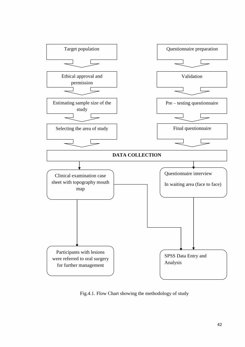
Fig.4.1. Flow Chart showing the methodology of study
Questionnaire preparation Target population
Ethical approval and
permission
Validation
Pre – testing questionnaire Estimating sample size of the
study
Final questionnaire Selecting the area of study
DATA COLLECTION
Questionnaire interview
In waiting area (face to face)
Clinical examination case
sheet with topography mouth
map
SPSS Data Entry and
Analysis
Participants with lesions
were referred to oral surgery
for further management

43
Chapter five: Result
44
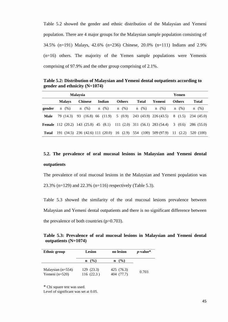
5. RESULT
5.1. Sociodemographic characteristics of the study population
At the dental clinics, the Faculty of Dentistry University of Malaya (UM) Kuala
Lumpur (KL); Malaysia and the Al-Thawra Modern General Hospital, Sana‘a (SAH);
Yemen, a total of 554 and 520 out-patients were interviewed and the oral mucosa
examined respectively during the period from May to October 2009. The mean age of
the Malaysian dental outpatients was 41.97±17.04 years with an age range of 18 - 89
years. The mean age of the Yemeni dental outpatients was 36 ±15.62 years old with an
age range of 18-95 years.
Table 5.1 shows the age and gender distribution of the Malaysian and Yemeni
populations. There were 43.9 % (n=243) males and 56.1 % (n=311) females in Malaysia
while in the Yemen sample, the male population was 45.0 % (n=234) and females was
55.0 % (n=286).
Table 5.1: Distribution of age and gender in Malaysians and Yemeni sample
population (N=1074)
Majority of the outpatients from both countries were from the 18-34 years age groups
with 42.6 % Malaysians and 53.5% for Yemenis.
Age
Group
Malaysian dental outpatients Yemeni dental outpatients
Male
n=243 (%)
Female
n=311 (%)
Total
n=554 (%)
Male
n=234 (%)
Female
n=286 (%)
Total
n=520 (%)
18-34 86 (15.5) 150 (27.1) 236 (42.6) 105 ( 20.2) 173 (33.3) 278 (53.5)
35-54 81(14.6) 93 (16.8) 174 (31.4) 73 (14.0) 86 (16.5) 159(30.6)
≥55 76 (13.7) 68 (12.3) 144 (26.0) 56 (10.8) 27 (5.2) 83(16.0)
Total 243(43.9) 311 (56.1) 554 (100.0) 234(45.0) 286 (55.0) 520(100.0)
45
Table 5.2 showed the gender and ethnic distribution of the Malaysian and Yemeni
population. There are 4 major groups for the Malaysian sample population consisting of
34.5% (n=191) Malays, 42.6% (n=236) Chinese, 20.0% (n=111) Indians and 2.9%
(n=16) others. The majority of the Yemen sample populations were Yemenis
comprising of 97.9% and the other group comprising of 2.1%.
Table 5.2: Distribution of Malaysian and Yemeni dental outpatients according to
gender and ethnicity (N=1074)
5.2. The prevalence of oral mucosal lesions in Malaysian and Yemeni dental
outpatients
The prevalence of oral mucosal lesions in the Malaysian and Yemeni population was
23.3% (n=129) and 22.3% (n=116) respectively (Table 5.3).
Table 5.3 showed the similarity of the oral mucosal lesions prevalence between
Malaysian and Yemeni dental outpatients and there is no significant difference between
the prevalence of both countries (p=0.703).
Table 5.3: Prevalence of oral mucosal lesions in Malaysian and Yemeni dental
outpatients (N=1074)
* Chi square test was used.
Level of significant was set at 0.05.
Malaysia Yemen
Malays Chinese Indian Others Total Yemeni Others Total
gender n (%) n (%) n (%) n (%) n (%) n (%) n (%) n (%)
Male 79 (14.3) 93 (16.8) 66 (11.9) 5 (0.9) 243 (43.9) 226 (43.5) 8 (1.5) 234 (45.0)
Female 112 (20.2) 143 (25.8) 45 (8.1) 111 (2.0) 311 (56.1) 283 (54.4) 3 (0.6) 286 (55.0)
Total 191 (34.5) 236 (42.6) 111 (20.0) 16 (2.9) 554 (100) 509 (97.9) 11 (2.2) 520 (100)
Ethnic group Lesion no lesion p-value*
n (%) n (%)
0.703 Malaysian (n=554)
129 (23.3) 425 (76.3)
Yemeni (n=520) 116 (22.3 ) 404 (77.7)
46
Table 5.4 showed that among those aged 18-34 years old there were more Yemeni with
oral mucosal lesions as compared to Malaysians. This relationship is statistically
significant (p=0.049). However, there were no statistically significant difference
between the oral mucosal lesions prevalence of Malaysian and Yemenis aged 35-54
years and those aged ≥ 55 years (p=0.173 and p=0.950 respectively).
Table 5.4: Comparison of Prevalence of oral mucosal lesion in Malaysian and
Yemeni outpatient according to age
Age group Lesion No. lesion p-value *
n (%) n (%)
Age group: 18-34 years:
Malaysian (n=236)
29 (12.3)
207 (87.7)
0.049 Yemeni (n=279) 52 (18.6) 227 (81.4)
Age group: 35-54 years:
Malaysian (n=174)
52 (29.9)
122 (70.1)
0.173 Yemeni (n=159) 37 (23.3) 122 (76.7)
Age group - ≥ 55 years
Malaysian (n=144)
48 (33.3)
96 (66.7)
0.950
Yemeni (n=82) 27 (32.9) 55 (67.1)
* Chi square test was used.
Level of significant was set at 0.05.
Table 5.5 showed that among the gender there were more Malaysian males with oral
mucosal lesions as compared to Yemeni males while there were more Yemeni females
with oral mucosal lesions compared with Malaysian females. However, there is no
statistically significant difference between the Malaysian and Yemeni males and
females in prevalence of oral mucosal lesions.
Table 5.5: Comparison of Prevalence of oral mucosal lesion in Malaysian and
Yemeni outpatient according to gender
Gender Lesion No. lesion p-value *
n (%) n (%)
Male Malaysian
84 (34.3)
161 (65.7) 0.081
Yemeni 63 (26.9) 171 (73.1)
Females
Malaysian
45 (14.6)
264 (85.4) 0.192
Yemeni 53 (18.5) 233 (81.5)
*chi square test was used.
Level of significant was set at 0.05.

47
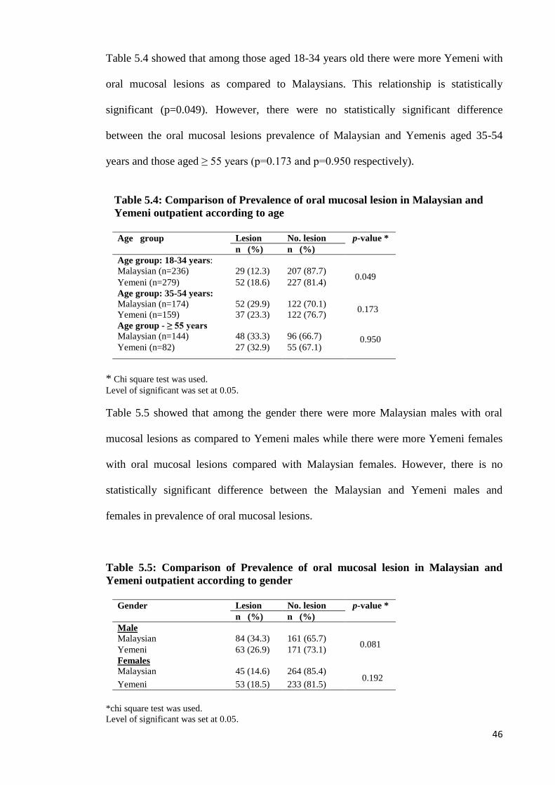
Table 5.6 showed the distribution of oral mucosal lesions in Malaysian and Yemenis
dental outpatients. From the Malaysian dental outpatients sample, only one subject (2%)
had oral cancer which was in an 86 years old Indian lady (Fig. 5.1) while nine Yemeni
subjects (1.7%) had oral cancer with all aged above 65 years where 6 cases were in
males and 3 cases were females (Fig. 5.2). The prevalence of potentially malignant
disorders (i.e. leukoplakia, and lichen planus) in the Malaysian outpatients was found to
be 0.8% (n= 4) as compared to 0.2% (n=1) in the Yemeni dental outpatients. Biopsies
for all the oral cancers confirmed to be oral squamous cell carcinoma.
The highest prevalence of oral mucosal lesions among Malaysian dental outpatients was
cheek biting (7.2%) while among the Yemenis; the highest prevalence was frictional
lesions (5.58%) (Fig.5.30). Among Malaysian patients, the prevalence of Fordyce‘s
spots was 3.4% while this condition was not identified in the Yemeni patients. The
prevalence of traumatic ulcers was similar in both countries (1.4% among Yemenis and
1.3% among Malaysians).
48
Table 5.6: Prevalence of oral mucosal lesions in outpatients from 2 dental clinics in
Malaysia and Yemen
Oral mucosal lesions
Malaysian Yemeni
Males Females Total Males Females Total
(n=243) (n=311) ( n=554) (n=234) (n=286) (n=520)
n % n % n % n % n % n %
1. Malignant lesions(ML)
Oral cancer 0 0.0 1 0.32 1 0.2 6 2.6 3 1.1 9 1.7
2. Potentially malignant disorders(PMD)
Leukoplakia 2 0.8 0 0.0 2 0.4 1 0.4 0 0.0 1 0.2
Lichen planus 0 0.0 2 0.6 2 0.4 0 0.0 0 0.0 0 0.0
3. Other lesions (Non-ML & Non-PMD)
white lesions
Cheek biting 23 9.5 17 5.5 40 7.2 11 4.7 13 4.6 24 4.6
Frictional lesions 13 5.4 5 1.6 18 3.2 16 6.8 13 4.6 27 5.2
Fordyce‘s spots 17 7.0 2 0.6 19 3.4 0 0.0 0 0.0 0 0.0
Geographic tongue 2 0.8 1 0.3 3 0.5 2 0.9 3 1.1 5 1.0
Lina alba 2 0.8 3 1.0 5 0.9 1 0.4 0 0.0 1 0.2
Leukoedema 0 0.0 0 0.0 0 0.0 1 0.4 0 0.0 1 0.2
Coated tongue 0 0.0 0 0.0 0 0.0 5 2.1 4 1.4 9 1.7
Red lesions
Acute erythematous
candidiasis
2 0.8 2 0.6 4 0.7 4 1.7 5 1.8 9 1.7
Denture stomatitis 5 2.0 6 1.9 11 2.0 0 0.0 0 0.0 0 0.0
Hyperplastic gingivitis 1 0.4 0 0.0 1 0.2 1 0.4 1 0.3 2 0.4
Ulcerated lesions
Angular cheilitis 0 0.0 1 0.3 1 0.2 0 0.0 0 0.0 0 0.0
Minor aphthous ulcer 2 0.8 6 1.9 8 1.4 2 0.9 1 0.3 3 0.6
Traumatic ulcer 5 2.1 2 0.6 7 1.3 3 1.3 4 1.4 7 1.4
Quid related lesion
Chewer‘s mucosa 0 0.0 2 0.6 2 0.4 0 0.0 0 0.0 0 0.0
shammah related lesion 0 0.0 0 0.0 0 0.0 1 0.4 0 0.0 1 0.2
Pigmented / swelling lesion
Pyogenic granuloma 1 0.4 0 0.0 1 0.2 0 0.0 1 0.3 1 0.2
Excessive melanin
pigmentation
0 0.0 0 0.0 0 0.0 0 0.0 1 0.3 1 0.2
Fibroepithelial polyp 2 0.8 1 0.3 3 0.5 0 0.0 3 1.1 3 0.6
Pericoronitis 1 0.4 0 0.0 1 0.2 0 0.0 5 1.8 5 1.0
Hairy tongue 0 0.0 0 0.0 0 0.0 3 1.3 1 0.3 4 0.8
49
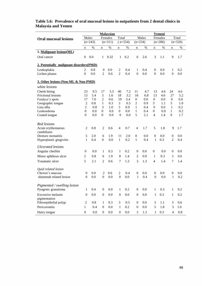
Fig.5.1 Oral carcinoma in the mouth of an 86 years old Indian lady who chewed
quid without tobacco for long time.
(a) (b)
(c)
(d)
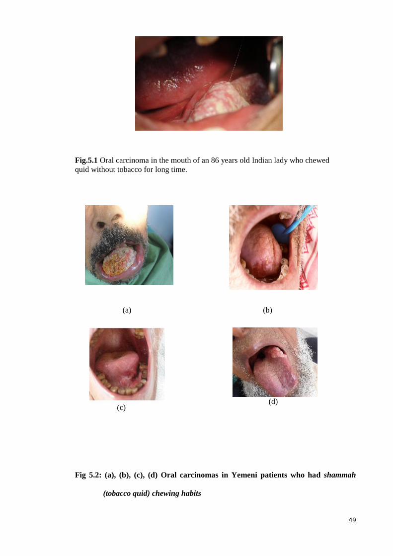
Fig 5.2: (a), (b), (c), (d) Oral carcinomas in Yemeni patients who had shammah
(tobacco quid) chewing habits
50
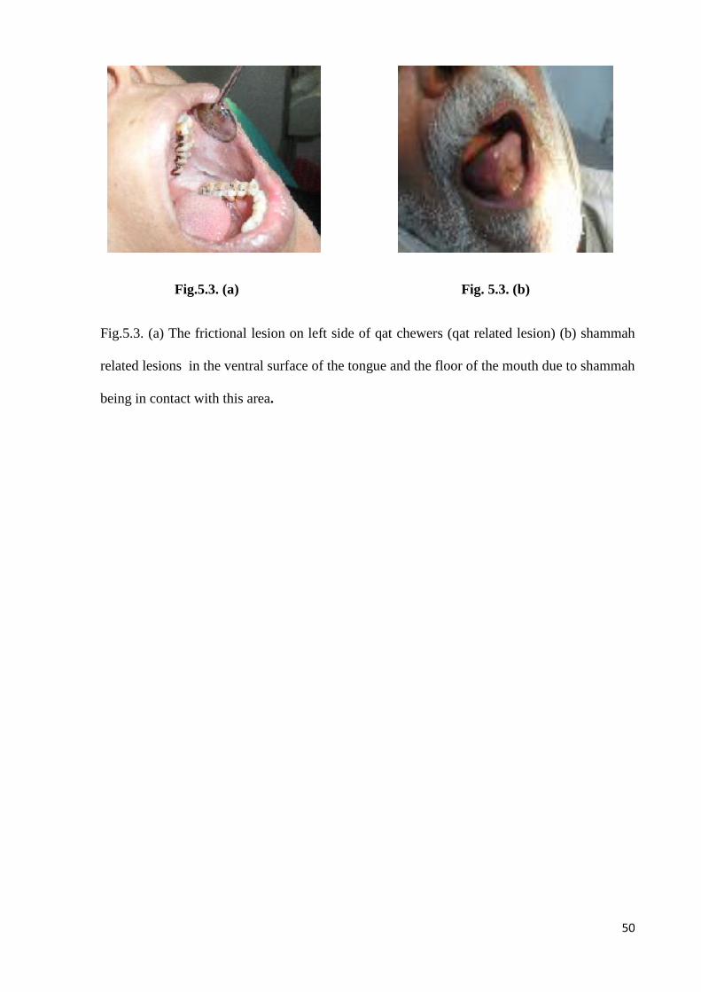
Fig.5.3. (a) Fig. 5.3. (b)
Fig.5.3. (a) The frictional lesion on left side of qat chewers (qat related lesion) (b) shammah
related lesions in the ventral surface of the tongue and the floor of the mouth due to shammah
being in contact with this area.
51
5.3. The prevalence of risk habits in outpatients attending dental clinics in
Malaysia and Yemen.
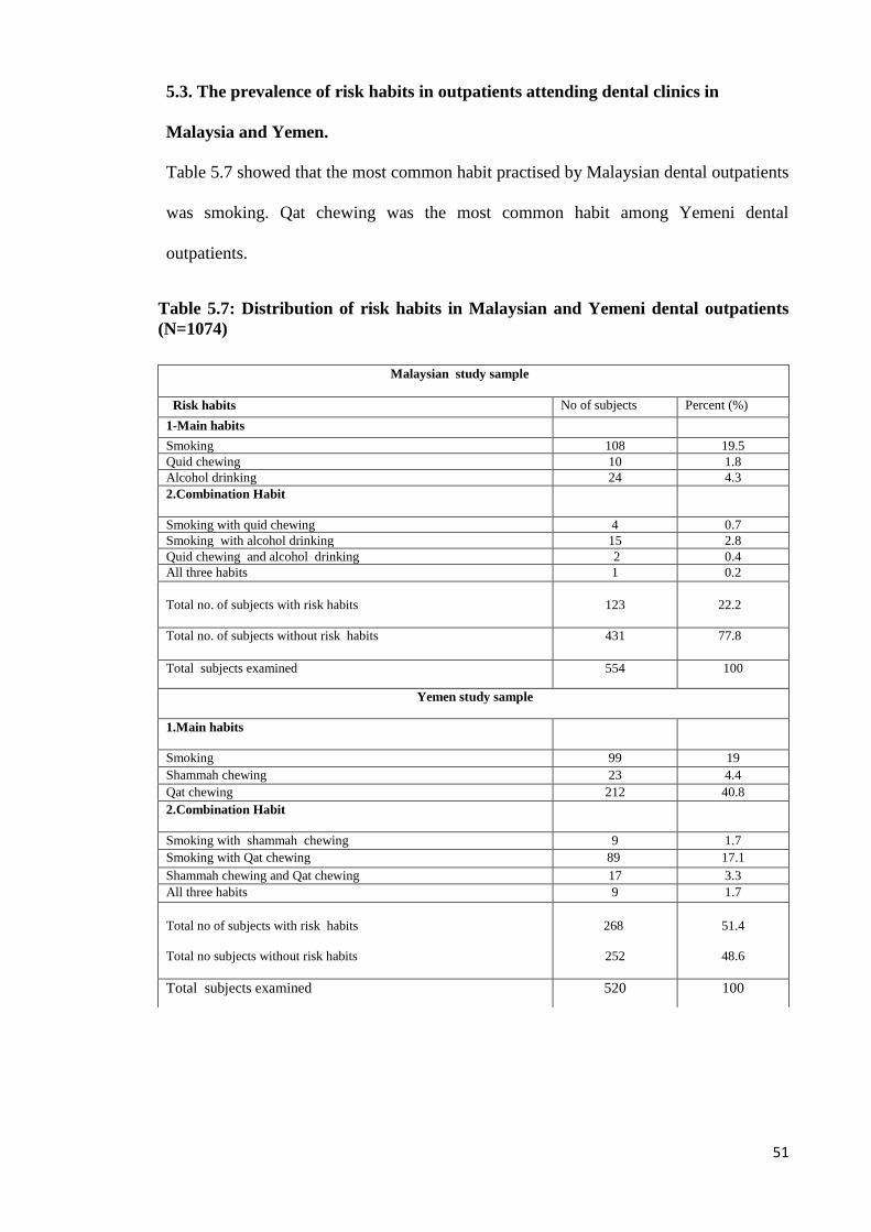
Table 5.7 showed that the most common habit practised by Malaysian dental outpatients
was smoking. Qat chewing was the most common habit among Yemeni dental
outpatients.
Table 5.7: Distribution of risk habits in Malaysian and Yemeni dental outpatients
(N=1074)
Malaysian study sample
Risk habits No of subjects Percent (%)
1-Main habits
Smoking 108 19.5
Quid chewing 10 1.8
Alcohol drinking 24 4.3
2.Combination Habit
Smoking with quid chewing 4 0.7
Smoking with alcohol drinking 15 2.8
Quid chewing and alcohol drinking 2 0.4
All three habits 1 0.2
Total no. of subjects with risk habits
123
22.2
Total no. of subjects without risk habits 431 77.8
Total subjects examined 554 100
Yemen study sample
1.Main habits
Smoking 99 19
Shammah chewing 23 4.4
Qat chewing 212 40.8
2.Combination Habit
Smoking with shammah chewing 9 1.7
Smoking with Qat chewing 89 17.1
Shammah chewing and Qat chewing 17 3.3
All three habits 9 1.7
Total no of subjects with risk habits
Total no subjects without risk habits
268
252
51.4
48.6
Total subjects examined 520 100
52
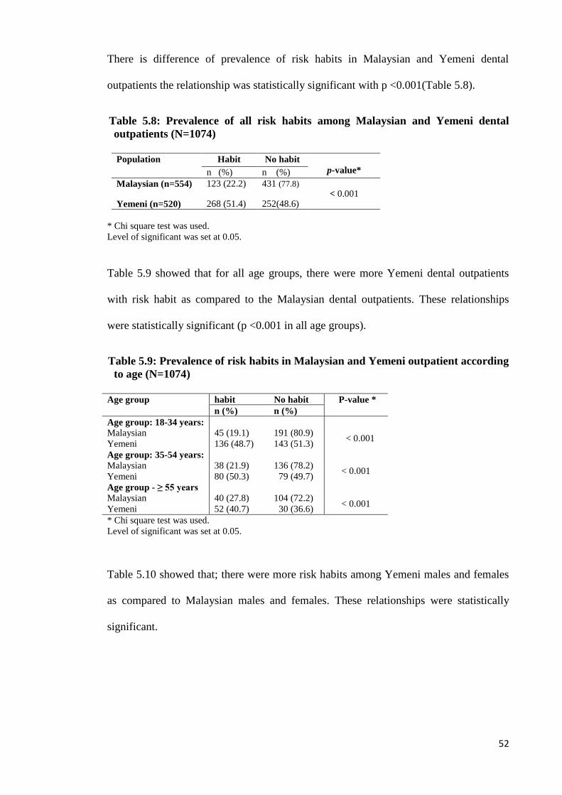
There is difference of prevalence of risk habits in Malaysian and Yemeni dental
outpatients the relationship was statistically significant with p <0.001(Table 5.8).
Table 5.8: Prevalence of all risk habits among Malaysian and Yemeni dental
outpatients (N=1074)
Population Habit No habit
p-value* n (%) n (%)
Malaysian (n=554) 123 (22.2) 431 (77.8) < 0.001
Yemeni (n=520) 268 (51.4) 252(48.6)
* Chi square test was used.
Level of significant was set at 0.05.
Table 5.9 showed that for all age groups, there were more Yemeni dental outpatients
with risk habit as compared to the Malaysian dental outpatients. These relationships
were statistically significant (p <0.001 in all age groups).
Table 5.9: Prevalence of risk habits in Malaysian and Yemeni outpatient according
to age (N=1074)
Age group habit No habit P-value *
n (%) n (%)
Age group: 18-34 years:
Malaysian
45 (19.1)
191 (80.9)
< 0.001 Yemeni 136 (48.7) 143 (51.3)
Age group: 35-54 years:
Malaysian
38 (21.9)
136 (78.2)
< 0.001 Yemeni 80 (50.3) 79 (49.7)
Age group - ≥ 55 years
Malaysian
40 (27.8)
104 (72.2) < 0.001
Yemeni 52 (40.7) 30 (36.6)
* Chi square test was used.
Level of significant was set at 0.05.
Table 5.10 showed that; there were more risk habits among Yemeni males and females
as compared to Malaysian males and females. These relationships were statistically
significant.
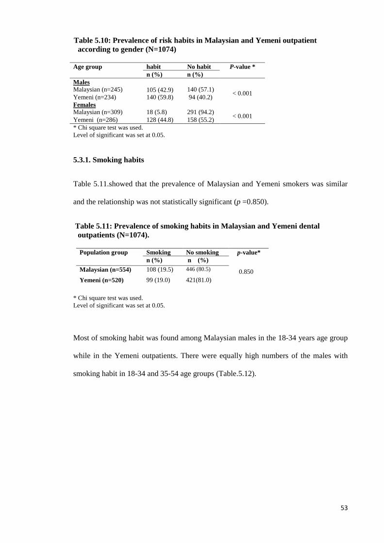
53
Table 5.10: Prevalence of risk habits in Malaysian and Yemeni outpatient
according to gender (N=1074)
Age group habit No habit P-value *
n (%) n (%)
Males
Malaysian (n=245) 105 (42.9)
140 (57.1)
< 0.001 Yemeni (n=234) 140 (59.8) 94 (40.2)
Females
Malaysian (n=309)
18 (5.8)
291 (94.2) < 0.001
Yemeni (n=286) 128 (44.8) 158 (55.2)
* Chi square test was used.
Level of significant was set at 0.05.
5.3.1. Smoking habits
Table 5.11.showed that the prevalence of Malaysian and Yemeni smokers was similar
and the relationship was not statistically significant (p =0.850).
Table 5.11: Prevalence of smoking habits in Malaysian and Yemeni dental
outpatients (N=1074).
* Chi square test was used.
Level of significant was set at 0.05.
Most of smoking habit was found among Malaysian males in the 18-34 years age group
while in the Yemeni outpatients. There were equally high numbers of the males with
smoking habit in 18-34 and 35-54 age groups (Table.5.12).
Population group Smoking No smoking p-value*
n (%) n (%)
Malaysian (n=554) 108 (19.5) 446 (80.5) 0.850
Yemeni (n=520) 99 (19.0) 421(81.0)
54
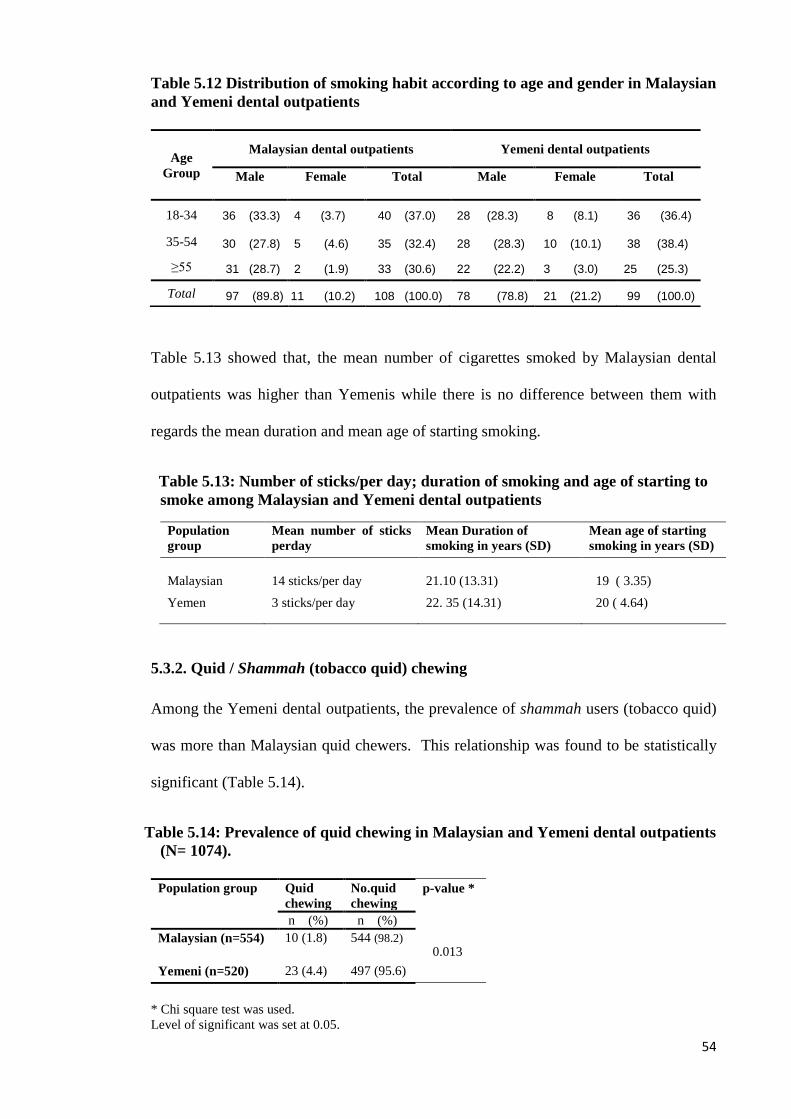
Table 5.12 Distribution of smoking habit according to age and gender in Malaysian
and Yemeni dental outpatients
Table 5.13 showed that, the mean number of cigarettes smoked by Malaysian dental
outpatients was higher than Yemenis while there is no difference between them with
regards the mean duration and mean age of starting smoking.
Table 5.13: Number of sticks/per day; duration of smoking and age of starting to
smoke among Malaysian and Yemeni dental outpatients
Population
group
Mean number of sticks
perday
Mean Duration of
smoking in years (SD)
Mean age of starting
smoking in years (SD)
Malaysian
14 sticks/per day
21.10 (13.31)
19 ( 3.35)
Yemen 3 sticks/per day 22. 35 (14.31) 20 ( 4.64)
5.3.2. Quid / Shammah (tobacco quid) chewing
Among the Yemeni dental outpatients, the prevalence of shammah users (tobacco quid)
was more than Malaysian quid chewers. This relationship was found to be statistically
significant (Table 5.14).
Table 5.14: Prevalence of quid chewing in Malaysian and Yemeni dental outpatients
(N= 1074).
* Chi square test was used.
Level of significant was set at 0.05.
Age
Group
Malaysian dental outpatients Yemeni dental outpatients
Male
Female
Total
Male
Female
Total
18-34 36 (33.3) 4 (3.7) 40 (37.0) 28 (28.3) 8 (8.1) 36 (36.4)
35-54 30 (27.8) 5 (4.6) 35 (32.4) 28 (28.3) 10 (10.1) 38 (38.4)
≥55 31 (28.7) 2 (1.9) 33 (30.6) 22 (22.2) 3 (3.0) 25 (25.3)
Total 97 (89.8) 11 (10.2) 108 (100.0) 78 (78.8) 21 (21.2) 99 (100.0)
Population group Quid
chewing
No.quid
chewing
p-value *
n (%) n (%)
0.013 Malaysian (n=554) 10 (1.8) 544 (98.2)
Yemeni (n=520) 23 (4.4) 497 (95.6)
55
Similarity of prevalence quid chewing without tobacco between males an d females in
Malaysian outpatients while most of quid chewing with tobacco among Yemeni males
particularly in aged group ≥ 55 (Table 5.15).
Table5.15.Distribution of quid chewing according to age and gender in Malaysian
and Yemeni dental outpatients
Table 5.16 showed that Malaysian quid chewers started chewing at an earlier age than
Yemenis. However, the Yemenis had longer duration of chewing and a higher
frequency than the Malaysian quid chewers.
Table 5.16: Frequency/per day, duration of quid chewing and age of starting to
chew (N=33)
Population Mean Frequency/per
day
Mean duration of quid
chewing in years (SD)
Mean Age of staring to
chew in years (SD)
Malaysian 2.2 times /per day 38.49 (23.02) 21.6 (18.2)
Yemeni 7.9 time/per day 54.81 (18.57) 28 (14.05)
Most of the Malaysian quid chewers kept the quid in the sulcus while the Yemeni quid
(shammah) chewers kept the quid under the tongue. The other site for Yemeni was the
lower sulcus (Table 5.17).
Age
Group
Malaysian dental outpatients Yemeni dental outpatients
Male
Female
Total
Male
Female
Total
18-34 1 (10.0) 1 (10.0) 2 (20.0) 4 (17.4) 0 (0.00) 4 (17.4)
35-54 0 (0.00) 0 (0.00) 0 (0.00) 2 (8.7) 1 (4.3) 3 (13.0)
≥55 4 (40.0) 4 (40.0) 8 (80.0) 12 (52.2) 4 (17.4) 16 (69.6)
Total 5 (50). 5 (50.0) 10 (100.0) 18 (78.3) 5 (21.7) 23 (100.0)
56
Table 5.17: Distribution of placement sites for quid /shammah (tobacco quid)
chewing among the Malaysian and Yemeni quid chewers
5.3.3. Qat chewing
Table 5.18 showed a very high difference of qat chewing in the males as compared to
females.
Table 5.18: Prevalence of qat chewing in relation to age among individuals
Yemeni dental patients (N=212)
Table 5.19 showed among the Yemeni qat chewers there is no big difference between
males and female of mean hours of qat chewing. The males qat chewers had longer
duration more than females.
Site of placement of quid/shammah Malaysian
n = 10
Yemeni
n=23
no (%) no (%)
Part of mouth kept the mixture was kept:
Left upper sulcus 2 (20) 0 (0.0)
Left lower sulcus 3 (30) 3 (13.04)
Right lower sulcus 3 (30) 1 (4.35)
Anterior lower sulcus 0 (0.0) 6 (2608)
Underneath the tongue 0 (0.0) 12 (53.6)
Others 2 (20) 1 (4.35)
Age
Group
Yemeni dental outpatients
Male
n= (%)
Female
n= (%)
Total
n= (%)
18-34 58 (69.1) 26 (31.1) 84 (100)
35-54 53 (61.6) 33 (38.4) 86 (100)
≥55 37 (88.1) 5 (11.9) 42 (100)
Total 148 (69.8) 64 ( 30.2) 212 (100)
57
Table 5.19: Mean hours, duration of qat chewing and age of starting to chew
Gender Mean hours of chewing
(SD)
Mean duration of qat
chewing in years (SD)
Mean Age of staring to chew in
years (SD)
Male
4.75 (1.93)
22.25 (14.7)
19.5 (4.2)
Female 3.92 (0.74) 14.99 (10.38) 21.44 (4.3)
The highest frequency of qat chewing was every day for both Yemeni males and
females (Table 5.20).
Table 5.20: Frequency of qat chewing and sites of placement qat among the
Yemeni outpatients
5.3.4 Alcohol drinking
From the whole Malaysian dental outpatients was found 4.3 % (n=24) were alcohol
drinkers with the mean age of starting drinking being 19.5±6.78 years.
Table 5.21 showed that the Malaysian males had a higher prevalence alcohol drinking
habit as compared to Malaysian females with high prevalence in the Chinese and Indian
males.
Frequency/ site placement of qat
chewing
Male
(n=148)
Female
(n=64)
Total
(n=212)
n (%) n (%) n (%)
Frequency of qat chewing
Every day 109 (73.65) 31 (48.44) 140 (66.04)
two -three times a week 19 (12.83) 8 (12.5) 27 (12.74)
one time a week 20 (13.51) 25 (39.06) 45 (21.23)
Sites of placement qat
Left 102 (68.92) 39 (60.94) 141 (66.51)
Right 34 (22.97) 21 (32.81) 55 (25.95)
Both 7 (4.73) 3 (4.69) 10 (4.72)
58
Table 5.21: Prevalence of alcohol drinking relation to ethnic, gender in Malaysian
dental outpatients (N=24)
Gender Malaya Chinese Indian Total
n (%) n (%) n (%) n (%)
Male (20) 2 (10.0) 9 (45.0) 9 (45.0) 20 (100)
Female (4) 1 (25.0) 3 (75.0) 0 (0.00) 4 (100)
Total 3 (12.5) 12 (50.0) 9 (37.5) 24 (100)
Most of the alcohol drinkers consumed alcoholic beverage only once/twice per week
while the most common drink was beer with high finding among Chinese follow by
Indian (table 5.22).
Table 5.22: Frequency and types of alcohol consumption by Malaysian dental
outpatients (N=24).
Alcohol consumption Malaya Chinese Indian Total
n (%) n (%) n (%) n (%)
Frequency
Almost daily 1(50.0) 0 (0.0) 1(50) 2(100)
Three to five a week 1(16.7) 3 (50) 2(33.3) 6 (100)
Once or twice a week 1(6.3) 9 (56.3) 6 (37.5) 16 (100)
Type of alcohol
beer 2 (9.5) 11(52.4) 8 (38.1) 21 (100)
wine 1(33.3) 1(33.3) 1(33.3 3 (100)
others 0 (0.00) 1(100) 0 (0.00) 1 (100)
5.4. The relationship between risk habits and the prevalence of oral mucosal
lesions in Malaysia and Yemen
Table 5.25 showed that there is a higher prevalence of oral mucosal lesions among the
patients with risk habits as compared to those without habits among Malaysian and
Yemeni dental outpatients. The relationship was statistically significant.
59
Table 5.23: Relationship between the risk habits and prevalence of oral mucosal
lesions in Malaysian and Yemeni dental outpatients (N=1074)
* Chi square test was used.
Level of significant was set at 0.05.
There were more smokers with oral mucosal lesions among Malaysian and Yemeni
dental outpatients. The relationship was found to be statistically significant (Table.5.24).
Table 5.24: Relationship between smoking habit and prevalence of oral mucosal
lesions in Malaysian and Yemeni dental outpatients (N=1074)
* Chi square test was used.
Level of significant was set at 0.05.
Table 5.25 showed that for the Malaysian and Yemeni dental outpatients, most of the
quid chewers and shammah users had oral mucosal lesions and the relationship is
statistically significant.
Risk habits Lesion No lesion p-value *
n (%) n (%)
< 0.001
Malaysian
Habit
60 (48.8)
63 (51.3)
Without habit 69(16.0) 362 (84.4)
Yemeni
0.003 Habit 74 (27.6) 194 (72.4)
Without habit 42 (16.7) 210 (83.3)
Risk habit Lesion No lesion p-value *
n (%) n (%)
Malaysian
< 0.001 Smoking 54(50) 54(50)
Not smoking 75(16.8) 371(83.2)
Yemeni
< 0.001 Smoking 46 (46.5) 53 (53.5)
Not smoking 70 (16.6) 351(83.4)
60
Table 5.25: Relationship between the quid/ tobacco quid (shammah) chewing and
prevalence of oral mucosal lesions among Malaysian and Yemeni dental
outpatients (N=1074)
* Chi square test was used.
Level of significant was set at 0.05.
The relationship for alcohol drinking and qat chewing was unique to the Malaysian and
Yemeni population respectively and thus the analysis for the relationship between the
respective habit and the prevalence of oral mucosal lesions was done only for the
respective population.
Table 5.26 showed that there are more oral mucosal lesions among alcohol drinkers as
compared to non-drinkers and the relationship was statistically significant.
Table 5.26: Relationship between alcohol drinking and prevalence of oral mucosal
lesions among Malaysian dental outpatients (N=554)
Alcohol drinking Lesion No.lesion p-value*
n (%) n (%)
0.029 Alcohol drinking (n=24) 10 (41.7) 14 (58.3)
Not. Alcohol drinking (n=530) 119(22.5) 425(77.5)
* Chi square test was used.
Level of significant was set at 0.05.
There is higher prevalence of oral mucosal lesions among the qat chewers as compared
to non qat chewers showing a statistically significant relationship (Table5.27).
Risk habit Lesion No lesion p-value *
n (%) n (%)
Malaysian
0.006
Quid chewers (n=10) 6 (60) 4 (40)
Non. quid chewers (n=544) 123(22.6) 421(77.4)
Yemeni
< 0.001 Sammah users (n=23) 14(60.9) 9 (39.1)
Non. Shammah users (n=497) 102(20.5) 395 (79.5)
61
Table 5.27 Relation between qat chewing and prevalence of oral mucosal lesions
among Yemeni dental outpatients (N=520)
Qat chewing Lesion No.lesion p-value *
n (%) n (%)
< 0.001 Qat chewing (n=212) 65 (30.7) 147(69.3)
Not. Qat chewing (n=308) 51 (16.6) 257(83.4)
* Chi square test was used.
Level of significant was set at 0.05.
62
Chapter Six: Discussion
63
6. DISCUSSION
6.1. Limitations of the study
The sample size estimation done for this study took into consideration the anticipated
difference to be detected in the outcome variable between the two populations groups.
Thus, estimation of the anticipated difference was obtained from the available literature
related to the study (Axell et al ., 1990 Aiman et al., 2004; Campbell,M and Machin, D.,
2005). The main limitation, there was not adequate information on the prevalence of
each oral mucosal lesions comparing Yemen and Malaysia. Therefore, among all the
oral mucosal lesions, a difference of 8% in the prevalence of oral white lesions in
Yemen (22.4%) and Malaysia (14%) was chosen as estimation. Furthermore, a
difference of 8% has yielded us the largest affordable sample size which was 546
patients.
Not all outpatients attending to the primary care unit at the Faculty of Dentistry,
University of Malaya, Malaysia, agreed to participate in this study as some of them had
intended to only seek dental treatment. They instead viewed this study as a waste of
their time. Due to this limitation, when we recruited the samples we have employed
convenient sampling method which may introduce sampling bias. In general, sampling
bias is important when the sample of cases is unrepresentative with respect to the risk
factor being studied where some of the outpatients did not agree to participate and
considered as a responsive bias (Hulley et al., 2007). However, the response rate in this
study was > 70% which was good and acceptable. In addition to that, it also indicates
small number of non respondent (<30%) whereby the characteristics of the non
respondent were also noted as not much different from the selected samples.

64
For those who had participated in the study, an explanation was given as to the
importance of the oral health screening. The participants were required to answer the
questionnaire while waiting to be called into the surgery room for a systematic clinical
examination
Due to the above-mentioned problem, there was difficulty in taking photographs for
some lesions. In dental clinics of Althawra Modern General Hospital in Sana‘a, the
registration department referred some outpatients suspected of having oral lesions to
nearby specialist clinics. Thus, although there were limitations in the present research
study due to its small sample size in the Yemen population, this has not compromised
the power of the study which stipulates a power of 94% through a software application.
6.2. Prevalence of oral mucosal lesions and comparison between Malaysia and
Yemen
Oral mucosal conditions may be caused by local factors (bacterial or viral), systemic
diseases, drug-related reactions or lifestyle factors such as the consumption of tobacco,
betel-quid and alcohol (Harris et al., 2004). The method to determine the prevalence of
oral mucosal lesions may vary. The majority of previous research studies correlate oral
mucosal disease with oral cancer and precancerous conditions, while some authors have
recorded overall oral mucosal lesions. Axell (1976) for instance, has reported 60
different oral mucosal lesions in Swedish populations. Although the prevalence of oral
mucosal lesions has been reported in many countries, these prevalence data are usually
restricted to very few lesions in each study. Thus, there is a need to obtain data from
different countries with large random samples though the process can be tedious.
However, appropriate information on oral mucosal lesion prevalence can still be
obtained from small low budget studies on selected population (Axell et a1., 1990).
Overall, the prevalence of oral mucosal lesions and related risk habits may differ from
65
region to region in the world. The prevalence of oral mucosal lesions in countries such
as Malaysia and Yemen has been reported. However, there are few data on the
prevalence of oral mucosal lesions in Yemen. Further, more data on the association of
oral mucosal lesions and related risk habits in Yemen is still not well-established. Thus,
there is a need to compare the prevalence of oral mucosal lesions and the related habits
of the Yemen population with a well-established data among the Malaysian population.
The current study is a hospital-based one where it showed that the prevalence of oral
mucosal lesions is similar in both Malaysia and Yemen at 23.3 % (n=129) and 22.3 %
(n=116) respectively. However, when comparing within the Malaysian population, the
prevalence was high as compared to 9.7 % (n=1131) reported in a previous study (Zain
et al., 1997). This may be due to the fact that the current study is a hospital-based study
where patients tend to prefer visiting the place. The prevalence of oral mucosal lesions
in the current study is similar to the finding in a previous study by Taiyeb et al., (1995)
where it was found that 22.8% (n=111) may be due to the participants in both studies
practising the same habits. The prevalence for Yemenis in this study was similar to an
earlier study in Yemen conducted by Aiman et al., (2004) where the prevalence was
found to be 22.4% where both the studies are also hospital-based. However, it is very
low compared to that found among the Yemeni subjects (slightly lower than 50%) in a
study conducted by Hill and Gibson (1987). This could be due to the latter study being
carried out only among the Yemeni qat-chewers. In comparing the finding of the present
study with that of worldwide studies such as a study in the USA carried out by Shulman
(2005), it was reported that the prevalence of oral mucosal lesions in children and
youths was 9.1%, which was very low compared to the finding of the present study.
This could relate to low prevalence risk habits among children and youths. In an Italian
study carried out by Campisi and Margiotta (2000), they reported the prevalence of oral
lesions at 81.3% which is very high compared to the present study. This may be the
66
result of several factors with the most common habit in the latter study being alcohol
drinking. The age of the participants was ≥40 years old and there was also another risk
habit of exposure to actinic radiation.
However, the statistical results in the present study is higher than that found in another
hospital based in Saudi Arabia which reported 15% (n=383) (Mobeeriek and AlDosari,
2009). This may be due to the age of the subjects in the latter study which was 15 years
old and above, which could indicate the difference of risk habit practice among the
population. Higher frequency of oral mucosal lesions is also found among Yemeni
dental outpatients aged 18-34. However, among Malaysian outpatients, most of the oral
mucosal lesions were found among those aged ≥55 years compared with recent findings
among patients ranging between the ages of 65 to 74 (Zain et al., 1997).
Among Malaysian dental outpatients, the prevalence of oral mucosal lesions in males
and females was 84 (34.3%) and 45 (14.6%) respectively. This could reflect that the
most risk habit was among Malaysian males. However, there is not much difference of
the prevalence of oral mucosal lesions in males and females among Yemeni dental
outpatients which is 63 (26.9%) to 53 (18.5%). Aiman et al., (2004) reported that the
prevalence of oral mucosal lesion among Yemeni females to males was 32.8% (20.8%)
That perhaps resulted to the latter study including many females with risk habit (qat
chewing).
The most common lesions among Malaysian outpatients found in the present study were
cheek biting which represented 7.2%, followed by Fordyce‘s spots 3.4%, frictional
lesions 3.2% and denture stomatitis 2.0%. However, the most common oral mucosal
lesion among Malaysian subjects in a study by Taiyeb et al., (1995) was tongue lesions,
10.7%, followed by oral pigmentation (4.9%) and white lesions (4.3%) in a the study
conducted on elderly Malaysians. The most common lesion among Yemenis was
frictional lesion, similar to that found among Yemeni subjects by Aiman et al., (2004).
67
Both studies showed that qat chewing was the most common practising habit among
them. The prevalence of oral mucosal lesions reported by Harris et al., (2004) was
28.1% (n=195). This is slightly higher than the findings in the present study. The
sample size of the study is close to the present study. However, all participants in the
study carried out in the UK were alcohol drinkers.
6.2.1. Oral malignant lesions and oral potentially malignant disorders
The prevalence of oral malignant lesions among Yemeni outpatients (1.7%) is found to
be higher than that of their Malaysian counterparts. This could be due to the fact that
more Yemeni dental outpatients chewed quid containing tobacco called shammah. In
addition, it is very high compared to the finding by Zain et al., (1997) that may be
associated with the latter being a nationwide study and the participants could have also
practised different habits. In the present study however, the prevalence of oral malignant
in the Malaysian outpatients is similar to the finding among Malaysian outpatients by
Axell et al (1990) since both studies are hospital based and the dental outpatients in both
studies had practised same habits (quid chewing). However, in the present study, the
prevalence of oral malignant lesions among Malaysian outpatients is low as compared
to that found in elderly Malaysian subjects (Ali et al., 1996) where 12.7% of subjects
had practised quid chewing. Another study among the Yemeni population carried out by
Sheilfe et al (2007) reported that the oral cancer rate among shammah users is low
compared to the finding of the Yemeni dental outpatients in the present study. This may
relate to the study being a case control one and the sample was lower than the present
study.
In a recent study in India carried out by Mehrota et al (2010), the prevalence of oral
squamous cell carcinoma was reported to be 0.5% (n=2) which is slightly higher than
the findings among Malaysian outpatients, even though the participants practised the
68
same habits in both studies. That may be due to the majority of the Indian subjects
indulging in risk habits. However in comparison to the Yemeni dental outpatients, it
was found to be very low and this maybe the result of the Yemeni dental outpatients
practicing different habits. Comparing the Yemeni dental outpatients in the present
study and in a recent study in India carried out by Mathew et al., (2008), the prevalence
of oral malignant lesions was 1.7% with a high prevalence among males as in the
present study. The latter study is a comparable study with the present study because
both studies are hospital-based.
The prevalence of potentially-malignant disorders (leukoplakia and lichen planus)
among Malaysian outpatients was 0.8% considerably very low compared to the findings
among elderly Malaysian subjects (3.3%) where the leukoplakias was the most
common potentially malignant disorders (Ali et al., 1996). All the participants in the
latter study were aged 60 and above, and most of them were in the risk group: Indians
who had chewed quid. The prevalence of potentially malignant disorder among Yemeni
outpatients was 0.2% (leukoplakia) very low as compared to (28%) (Leuokplakia,
erythroplakia and lichen planus) among Yemeni subjects (Sheilfe et al., 2007). The
prevalence of potentially malignant disorders (leukoplakia and lichen planus) in the
current study is low as compared to Malaysian subjects reported by Zain et al., (1997)
where the study was a population-based and the sampling also came from the
indigenous group. In the present study, lichen planus is figured 0.4 % similar to that
found among the Malaysian subjects with 0.38% (Zain et al., 1997). However, the
lichen planus in the present study was found among the Chinese group only, compared
to that found in the latter study in the Malay and Indian groups respectively.
In a study carried out by Chung et al., (2005) in Taiwan, it has been reported that the
prevalence of potential malignant disorders is 12.7%, which is high when compared to
the findings of the present study (0.89%). This could be attributed to the fact that the
69
study in Taiwan was a cross-sectional community survey (house to house) in a suburban
population where the people practised areca nut chewing in addition to smoking and
alcohol drinking. Another study in Thailand carried out by Reichart et al., (1987)
showed the prevalence of leukoplakia which was 1.1% high as compared to 0.4% in the
current study. This could be referred to the differences in habits practised by the
population group (betel and miang chewing). Among the American population, the
prevalence of malignant disorders (leukoplakia) was reported around 3% where the
leukoplakia was put under oral keratosis (Bouquot and Gorlin ,1986) higher than the
finding in the present study. The population in the USA study had practised tobacco in
snuff and they were aged 35 and above. The prevalence of leukoplakia among Indian
subjects reported by Saraswathi et al., (2006) was 0.6%, close to that found among
Malaysian dental outpatients in the present study. This could due to the same habits
practiced.
6.2.2. Other lesions (non malignant and non potentially malignant disorder)
i. White lesions
In the present study, the prevalence for cheek biting in Malaysian dental outpatients was
7.2% which is higher than the Yemeni outpatients (4.6%). Axell et al., (1990) reported
that 5.6% in Malaysian outpatients was presented with white lesions in comparison with
5.1% in the population study conducted among adult Swedish population (Axell, 1976).
The present finding for frictional lesions is 5.2% among Yemeni outpatients which is
slightly higher than in the Malaysian outpatients 3.2%. However, this finding is similar
to that found among Malaysian outpatients and adult Swedish population (Axell et al.,
1990; Axell., 1976). In the current study; the finding for leukoedema in the Yemeni
outpatients is 0.2% compared to 3.7% found in the Indian outpatients (Methew et al.,
70
2008). This percentage is also very low compared to 48% that was found in adult
Swedish population (Axell, 1976). The finding for geographic tongue in the Malaysian
outpatients was 0.5% while 1.0% was found in the Yemeni population, in comparison
with the findings reported among Thai and Malaysian population prevalence 5.1% and
6.4% respectively (Axell et al., 1990) and 8.5% among the Swedish population
(Axell.1976). Fordyce‘s spots were observed at 3.1% in the Malaysian outpatients
which is more prevalent among males (7%) compared to 0.6% among females.
However, Axell et al., (1990) had reported the prevalence of Fordyce‘s spots in
Malaysian outpatients which was 61.8% and 82.8% among the Swedish population
(Axell et atl, 1976) which was very different from the finding in the present study.
The frequency of coated tongue in Yemeni dental outpatients was (1.7%) (1.0% in
males, 0.8% in female). This finding is lower than that found in the Italian population
(47.45%) (Campisi and Margiotta, 2000). The occurrence of hairy tongue in the present
study was 0.8% in Yemen outpatients with more frequency in males where a similar
percentage of prevalence of 0.9% was reported in Malaysian outpatients (Axell et al.,
1990).
ii. Red oral lesions
The findings of denture stomatitis in Malaysian outpatients was (0.2%) without
predilection difference between males and females. This finding was lower than the
prevalence of 3.4% found in Malaysian subjects which is more frequent among females
(Zain 1995). However, it is close to that found among Indian population (0.84 %) with
more frequency among females than males (Mathewa et al., 2008). The prevalence of
denture stomatitis was very low compared to the finding in Slovenia population (14.7%)
with high prevalence among males (Marji, 2000).
71
iii. Ulcerated lesions
In the current study, the frequency of aphthous ulcer in Malaysian outpatients was 1.4%
with a high prevalence among females compared to 0.6% found among Yemen
outpatients with more frequency among males. Zain (2000) reported a prevalence of
0.5% among Malaysian subjects. The finding of the present study among Malaysian
outpatients was similar to that found among found in adult Chinese (Lin et al., 2001) but
lower than that reported by Mathewa et al., (2008) among Indian population with a
prevalence of 2.1%.
iv. Pigmented / swelling lesion
Fibro epithelial polyp in the present study figured 0.5% and 0. 6% in both the Malaysian
and Yemeni participants respectively. The prevalence is low compared to the one found
in 3.9% of the Malaysian outpatients (Axell et al., 1990). In the present study, the
findings of hairy tongue among Yemeni outpatients was 0.8% similar to that found
among the Malaysian outpatients (0.9%) (Axell et al., 1990). Similar observation for
pyogenic granuloma was noted in the present study among Malaysian and Yemeni
outpatients which was 0.2%. However, this percentage is low compared to that found
among Thai and Malaysian outpatients (Axell et al., 1990) but similar to that found in
adult Swedish population (Axell., 1976).
72
v. Quid related lesion
In the present study, the prevalence of Betel Chewer‘s Mucosa was 0.4% which was
predominant in Indian women compared to 4.9% that was found among the Malaysian
subjects with the most frequent being among the Indians and more among women ( Zain
et al., 1997). Zain et al., (1995) also reported that 1.6% of the Malaysian subjects were
presented with Betel Chewer‘s Mucosa and a majority are subjects of high risk groups
such as Indians and other Bumiputras. The prevalence for Shammah related lesion in
Yemeni outpatients was 0.2%. This percentage is very low compared to the finding in
Yemeni subjects with 31% prevalence of shammah relate lesion (Scheifele et al., 2007).
However, the latter study was conducted among the shammah users.
6.3. Prevalence of risk habits and comparison between Malaysia and Yemen
The common risk habits creating oral mucosal lesions are tobacco smoking, quid
chewing and alcohol drinking, In addition to the environmental and genetic factors
(Johnson, 2003b ) on the other hand, many studies showed the association between
some oral mucosal lesion and qat chewing (Alsharabi, 2002; Aiman et al., 2004;
Gorsky et al., 2004; Fasanmade, 2007). The prevalence for risk habits (predominately
smoking habit) in the Malaysian outpatients is 22.2%, close to that found in in
Malaysian outpatients ((Predominately smoking habit) (Axell et al., 1990). Among
Yemeni outpatients, the high prevalence of risk habit of 51.4% was qat chewing. This is
slightly lower compared to a report by Aiman et al., (2004). In the present study, the
most risk habits in Malaysia are among men (85.4%). However, there was no big
difference of risk habits between men and women among the Yemeni outpatients.
73
6.3.1 Smoking habits
In the present study, the similarity of prevalence for smoking habits in Malaysian and
Yemeni outpatients of 19.5 and 19.0% respectively was recorded. The prevalence is
lower than what was reported among Yemeni subjects (Aiman et al., 2004). The
smoking habit in the present study was the predominant habit among Malaysian
outpatients similar to that found by Axell, et al (1990). The prevalence of smoking
habits in American Indians/ Alaskan natives in a study carried out by Park et al (1997)
and other study conducted in rural China by Yang et al (2008) is very high compared to
the finding in present study. The current study showed that the smoking habits are more
prevalent in Yemeni in the age groups ranging from 18-34. However, the smoking habit
in the Malaysian population is more prevalent in ages between 34-54. Among the
Malaysian dental outpatients, the frequency of smoking habits among men was 17.3%
and this is similar to the other study by WHO (2004 (b)) which showed that the
percentage of women smokers was smaller than that of male smokers (women 7.2% and
men 49.8%). However, the smoking habits in Yemeni subjects were more pronounced
among males than in females at 32.8% and 9.4% respectively (Aiman et al., 2004).
6.3.2. Quid chewing
In a recent study, the prevalence of quid chewing without tobacco in Malaysia was 1.8
%, slightly lower compared to 2.6% reported by Axell et al., (1990). However, this is
very low compared to that found in the Asian regional study (WHO,2004(a) (Gupta and
Warnakulasuriya, 2002). The prevalence of tobacco quid (shammah) in this study
among the Yemeni participants was 4.4% which is higher than the that of the Malaysian
outpatients. The composition of shammah in this study was tobacco, lime and ash.
However the composition of the shammah in Algerians was tobacco, carbonate lime and
other substance (Zhang et al., 2001).
74
6.3.3 Alcohol drinking
The prevalence of alcohol consumption among Malaysian dental outpatients was found
to be at 4.3% which is higher compared to the findings among Singaporean Malays at
1.1%. However, the percentage is very low with that found among Thai population at
31.4% (WHO, 1999). The previous study carried out by Zain et al (1995) had found the
alcohol consumption to be 35.4% and more prevalent among the Chinese groups for
both studies.
6.3.4 .Qat chewing
In the present study, the prevalence of qat chewing in Yemeni outpatients was figured at
40.8% which is low in comparison with previous studies carried out among Yemeni
subjects (Aiman et al., 2004, Alsharabi. 2002). Most of the qat chewers in the present
study were males similar to that found in a study by Aiman et al., (2004). In the present
study, the males who practised qat chewing were more than the females at 69% (n=148)
and 32 .2% (n=64) respectively as compared to qat chewers among Yemeni subjects
reported by Aiman et al., (2004) (87.04%) and (12.9%) respectively. In comparison to
the frequency of qat chewing, the present study showed high frequency of qat chewing
everyday and most of the qat chewers chewed qat leaves in the left side of the mouth
similar to a previous study by Aiman et al., 2004.
75
6.4. The relationship between risk habit and prevalence of oral mucosal lesions in
Malaysia and Yemen
The findings in the present study showed that there is a higher prevalence of oral
mucosal lesions among patients with risk habits as compared to those without habits
among Malaysians and Yemenis dental outpatients The relationship was statistically
significant supporting the positive association between cigarette smoking and
leukoedema, as well as denture stomatitis (p<0.001) (Zain and Razak, 1989). The
current study shows more Malaysian and Yemeni smokers with oral lesions. The
relationship was found to be statistically significant (p<0.001) as found in a Spanish
population which indicated that there is a significant association between smoking and
prevalence of oral mucosal lesion (García-Pola et al., 2002). This study also showed that
most of the quid chewers among Malaysian and Yemeni outpatients presented with oral
mucosal lesions and the relationship was statistically significant in comparison with a
study by Axell et al., (1990) which reported that the oral malignant and the most of
potentially malignant disorders has an association with betel quid chewing. In another
study by William et al., (1999) in Saudi Arabia, it was reported that oral cancer had
appeared where shammah (tobacco quid) was commonly used. Among the Malaysian
outpatients, there were high oral mucosal lesions among alcohol drinkers similar to
another study in an adult Spanish population by García-Pola et al., (2002).
In the current study, there were found more oral mucosal lesions among the qat
chewers. There is a high statistically significant relationship between the prevalence of
oral mucosal lesions and qat chewing with a p-value <0.001 similar to that found in a
study by Aiman et al (2004) which reported that the oral white lesions were more
common among the qat chewers than the non- qat chewers. In the present study there is
similarity in the prevalence of oral mucosal lesions between Malaysia and Yemen even
though there is a big difference of the prevalence of risk habit between Malaysian and
76
Yemen where the risk habit among Yemeni outpatients was higher than the Malaysians.
These may be due to the most common habit among Yemeni was qat chewing. Qat is a
green leave. The most active ingredients of qat are alkaloids such as cathinone and
cathine. Cathinone is the main psychoactive constituent of qat, and has a similar action
to amphetamine, inducing the release of dopamine, a neurotransmitter, from pre-
synaptic storage (Kalix 1992 , Patel. ,2000).There are also small amounts of ethereal oil,
sterols and triterpenes, together with 5% protein which has insignificant nutritional
value. Ascorbic acid is also present in the leaves (Raman, 1983). Khat leaves also
contains tannin (7–14% by weight in dried leaves) and minute amount of thiamin,
niacin, riboflavin, iron and amino acids (Lugman and Danowski, 1976). Apart from
tannin, these substances are unlikely to contribute to the biological effect of khat (Kalix,
1992). The above composition of the qat leaves may be responsible to lesser effect in
oral mucosa

77
Chapter Seven: Conclusion
78
7. CONCLUSION
7.1. Conclusion
This study found that there is no difference in the prevalence of oral mucosal lesions
among Malaysian and Yemeni dental outpatients. However, there is a difference in the
prevalence of risk habits in Malaysian and Yemeni dental outpatients. The relationship
was statistically significant at p <0.001. The prevalence of smoking habits was similarat
both centres, and statistically significant difference was found in the prevalence of quid
chewing habits between Malaysian and Yemeni dental outpatients (p=0.013). There is a
low prevalence of alcohol drinking habits among Malaysians with no such habits found
among Yemenis; while there is a high prevalence of qat chewing habits among Yemenis
and the habits do not exist among Malaysians. There is a higher prevalence of oral
mucosal lesions among the dental outpatients who have risk habits as compared to those
without habits and this relationship was found to be statistically significant.
7.2. Recommendation
It is hoped that the results from this study will lead to similar works on a more
nationwide scale in Malaysia. In the context of Yemen, it is hoped that the findings will
act as a database for a nationwide survey with larger samples to better understand the
influence of oral risk habits in the prevalence of oral mucosal lesion especially the
precancerous and cancerous lesions. It is recommended that more hospital dental clinics
participate in such a research. In addition, this study will help to identify the risk groups
and aid programmes that can improve the oral health on a regular basis to promote oral
health care and awareness among the population. , Further studies need be conducted to
look into the outpatients of variable clinics and Yemen aid nationwide study to
determine the prevalence of oral mucosal lesions and related risk habits.
79
8. REFERENCES
Abu Bakar, S. (2006). Smoking Cessation Programme in Malaysia. Proceeding Oral
Cancer in Asia Pacific–A Regional Updated & Networking, Kuala Lumpur, Malaysia p.
49-50.
Aiman, A. A. (2007). Histopathologic changes in oral mucosa of Yemenis addicted to
water-pipe and cigarette smoking in addition to takhzeen al-qat. Oral Surg Oral Med
Oral Pathol Oral Radiol Endod; 103:e55-e59.
Aiman, A. A., Al-Sharabi, A. K. and Aguirre, J. M. (2006). Histopathological changes
in oral mucosa due to takhzeen al-qat: a study of 70 biopsies. J Oral Pathol Med; 35:81–
5.
Aiman, A. A., Al-Sharabi, A. K., Aguirre, J. M. and Nahas, R. (2004). A study of 342
oral keratotic white lesions induced by takhzeen al-qat among 2500 Yemeni. J Oral
Pathol Med; 3: 368–72.
Albrecht, M., Bánóczy, J., Dinya, E., Tamás, G. Jr. (1992). Occurrence of oral
leukoplakia and lichen planus in diabetes mellitus. J Oral Pathol Med; 21:364.
Al-Mobeeriek, A. and AlDosari, A. M. (2009). Prevalence of oral lesions among Saudi
dental patients. Ann Saudi Med; 29: 365-8.
Alaya‘a, Z. (2009). Yemen ranks second in the number of smokers in Arab
World Observer (ye); issued by World Health Organization.
Ali, T. B., Jalalludin R. L., Abdul Razak, I, I. A., Zain, R. B. (1996). Prevalence of oral
precancerous and cancerous lesions in the elderly Malaysians. Asia pacific Academic
Consortium for Public Health (ACPACPH) J; 9:24-27.
AlSharabi, A. K. (2002). Oral and para-oral lesions caused by takhzeen (chewing) al-
Qat. Thesis Doctoral, University of Khartoum: Al-Khartoum (Sudan). p. 122.
Amer, M., Bull, C. A., Daouk, M., McArthur, P. D., Lundmark, G. J. and EL-Sensoussi,
M. (1985). ‘Shamma ’ usage and oral cancer in Saudi Arabia Ann.Saudi Med 5:135-
41.
Axell, T. (1976). A Prevalence study of oral mucosal lesions in an adult Swedish
population .Odont Revy; 1-100.
Axell, T. (1987). Occurrence of leukoplakia and some other white lesions among 20333
adult Swedish people. Community Dent Oral Epidemiol; 15: 46-51.
Axell, T and Rundquist, L. (1987). Oral lichen planus – a demographic study.
Community Dent Oral Epidemiol; 15: 52.-56.
Axell, T., Holmstrup, P., Kramer, I. R. H., Pindborg, J. J.and Shear, M. (1984).
International seminar on leukoplakia and associated lesions related to tobacco
habits.Community Dent Oral Epidemiol; 12:145-154.
80
Axell, T., Pindborg., J. J., Smith, C. J., van der Waal, I. (1996). International
collaborative group on oral white lesions with special reference to precancerous and
tobacco related lesion. Conclusions of an international symposium held in Uppsala,
Sweden. J Oral Pathol Med; 25:49-54.
Axell, T., Zain, R. B., Siwamogstham, T. D and Thampipt, J. (1990). Prevalence of oral
soft tissue lesion in out-patients at two Malaysain and Thai dental schools. Community
Dent Oral Epidemiol; 18:95-9.
Baan, R., Straif, K., Grosse, Y., Secretan, B., EL Ghissassi, F., Bouvard, V. Altieri, A.,
Cogliano, V. (2007). Carcinogenicity of alcohol beverages. Lancet Oncol; 8: 292-293.
Banoczy, J. and Rigo, O. (1991). Prevalence study of oral precancerous lesions with a
complex screening system in Hungary. Community Dent Oral Epidemiol; 19: 265-267.
Barnes L,Eveson J.W., Reichart P., Sidransky D.(2005). World Health Organization
classification of tumors.Pathology and Gentics of Head and Neck Tumors. IARC Press.
Lyon p.166
Baum, B. J. (2007). Inadequate training in biological sciences and medicine for dental
students impending crisis for Dentistry. J Am Dent Assoc; 138:16-25.
Bischoff, E.W., Uijen, A., Van der wel, M. (2009). Aphthous ulcers. BMJ Publishing
Group Ltd .339, 25, p b2382.
Bouquot, J. E. Gorlin, R.J. (1986). Leukoplakia, lichen planus and other oral keratoses
in 23,616 white Americans over 35 years of age. Oral Surg Oral Med Oral Pathol;
61:373-381.
Bougout, J. E. (1986). Common oral lesions found during a mass screening
examination. J Am Dent Assoc; 112:50-57.
Bruch, J. M and Treister, N. S. (2009). Clinical oral medicine and pathology. Humana
Press. London. P: 11-23.
Cawson, R. A., Langdon, J. D. and Eveson, J. W. (1996). Erythroplasia
(‗erythroplakia‘).In: Surgical pathology of the mouth and jaws. Wright—an imprint of
Butterworth–Heinemann Ltd., Oxford, London, Boston. p.180.
Chung C. H., Yang Y.H., Wang T. Y., Shieh T.Y., Warnakulasuriya S. (2005). Oral
precancerous disorders associated with areca quid chewing, smoking, and alcohol
drinking in southern Taiwan. J Oral Pathol Med 34: 460–6.
Demir, Y., Demir, S. and Aktepe, F. (2004). Cutaneous lobular capillary hemangioma
induced by pregnancy. J Cutan Pathol; 31: 77-80.
Dental Division. Ministry of Health. Malaysia (1978). Dental epidemiological survey of
adults in peninsular Malaysia 1974-1975. Dental Division. Ministry of Health.
Malaysia; 36-37: 98-9.
Dupont, W. D.,Pulmmer,W.D.Jr. (1998). Power and sample size calculation for studies
involving linear regression. Contal Clin Trail; 19(6):589-601.
81
El- Akkad, S. M., Amer, M. H., Lin, G. S., Sabbah, R. S. and Godwin, J. T. (1986).
Pattern of cancer in Arabians referred to King Faisal Specialist Hospital Cancer.
58:1172-8.
El-Shoura, S. M., Abdel Aziz, M. and Ali, M. E. (1995). Deleterious effects of khat
addiction on sperm parameters and sperm ultra-structure. Hum Reprod; 10: 2295-2300.
El-Wajeh,Y. A. M. and Thornhill, M. H. (2009). Qat and its health effects; Br Dent J;
206:17–21.
Epinoza, I., Rojas, R., Aranda, W. and Gamonal, J. (2003). Prevalence of oral mucosal
lesion in elderly people in Santiago Chisle. J Oral Pathol Med; 32:571-5.
Fasanmade, A., Kwok, E. and Newman, L. (2007). Oral squamous cell carcinoma
associated with khat chewing. Oral Surg Oral Med Oral Pathol Oral Radio Endo;
104(1):e53-e55.
Field, A., Longman, l. (2003). Tyldesley‘s oral medicine fifth edition Oxford
University Press. UK. Chapter 6.p. 69.
Campisi, G. and Margiotta, V. (2001). Oral mucosal lesions and risk habits among men
in an Italian study population. J Oral Pathol Med; 30:22-8.
Cardesa A., Slootweg. P. J. (2006). Pathology of head and neck. Springer-Verlag
Berlin Heidelberg. p.108
Gerhauser, C. (2005). Beer constituents as potential cancer chemopreventive agents. Eur .J Cancer; 41(13): 1941-54.
García-Pola Vallejo, M. J., Martínez Díaz-Canel A. I, García MartínJ, .M., González
García M. (2002). Risk factors for oral soft tissue lesions in an adult Spanish
population Community Dent Oral Epidemiol; 30: 277–85.
Greenberg, M. S. and Michael Glick, M. (2008). Burket‘sOral MedicineDiagnosis &
Treatment 11th
edition Hamilton. Spain. p. 10, 77.
Gorsky, M., Epstein, J. B., Levi, H., Yarom, N. (2004). Oral white lesions associated
with chewing khat. Tobac induced dis; 2: 145–150.
Gupta, P. C., Mehta, F. S., Daftary, D. K., Pindborg, J. J., Bhonsle, R. B., Jalnawalla, P.
N.Sinor P.N,Pitkar V.K,Murti P. R., Irani R.R ,Shanh H.T., Kadam, P.M., Iyer, K.S,
Iyer H.M., Hegde, A. K, Chandrashekar, G.K, Shiroff B.C., Sahiar, B.E. and Meha,
M.N (1980). Incidence of oral cancer and natural history of oral precancerous lesions in
a 10-year follow-up study of Indian villagers. Community Dent Oral Epidemiol;
8(6):283–333.
Gupta, P. C. and Ray, C. S. (2003). Smokeless tobacco and health in India and South
Asia. Respirology; 8:419–31.
Gupa, P. C., Zain, R. B., Ikeda, N., Yaacob, M. and Metha, H. (1997). Prevalence of
tobacco use behavior in Malaysia-result of sample survey .Book of abstract for the 10th
World Conference on tobacco or health in Beijing ,China –August 24-28: p 60,abstract
#OS 110.
82
Gupta, P. C and Warnakulasuriya, S. (2002). Global epidemiology of areca nut usage
addict. Biol.7:77-83.
Halbach, H. (1972). Medical aspects of the chewing of khat leaves. Bull WHO; 47:21-
29.
Haniza, M. A., Maimunah, A. H., Rusilwati, J., Latipah, S. and Surya, A. (1999).
National Health and Mobility Survey: Smoking among adult. Institute of public health,
Ministry of Health Malaysia. Kaula Lumpur. 15:23.
Harris, C. K., Warnakulasuriya, K. A., Cooper D.J., Gelbier, S. (2004). Prevalence of
oral mucosal lesions in alcohol misusers in south London. J Oral Pathol Med.;
33(5):253-9.
Heinekens, C. H. and Buring, J. E. (1987). Epidiomlogy in Medicine Boston /Tornoto
.Little Brown and Company; PP.64-65.
Hill, C. M and Gibson, A. (1987). The oral and dental effect of the qat chewing. Oral
Surg Oral Pathol; 63:433-6.
Hulley, S, B, Cummings, S,R. Browner, W, S; Grady, D, G. Newman, T, B. (2007)
Designing Clinical Research, 3rd Edition Lippincott Williams & Wilkins. USA. P.116
Ibsen, O. Phelan, J, (2009). Oral Pathology for Dental Hygienist. , 5th edition.
Philadelphia,USA Elsevier Health Sciences. p.
Jaber, M. A., Porter, S. R., Gilthorpe, M. S., Bedi, R. and Scully, C. (1999). Risk
factors for oral epithelial dysplasia--the role of smoking and alcohol Oral Oncol 35 :2;
151-6.
Johnson, N. (2001). Tobacco use and oral cancer: a global perspective. J Dent
Educ.65:328-339.
Johnson., N.W. Aetiology and risk factors for oral cancer. In: Shah JP, Johnson NW,
Batsakis JG, eds. (2003) Oral Cancer. London: Martin Dunitz, an imprint of the Taylor
& Francis Group. Pp.33–75.
Kalix, P. (1992). Cathinone: a natural amphetamine. J Pharmacol Toxicol; 70:77–86.
Kennedy, J., Teague, J., Rokaw, W. and Cooney, E. (1983). A medical evaluation of
qat use in North Yemen .Social Science and Med; 11:783-793.
Kupper, H., Boffetta, P., Adami, H. O. (2002). Tobacco use and cancer causation:
association by tumor type. J Int Med.252:206-224.
Langlais, R. P., Miller, C. S., Nield-Gehrig, J.S. (2009). Color atlas of common oral
disease. Lippincott Williams & Wilkins. Philadelphia . p. 55–7.
Laskari, G. (2006). Pocket Atlas of oral diseases. Georg Thieme Verlag. Germany. 2nd
edition. P. 224
Llewellyn, C. D., Johnson, N. W. and Warnakulasuriya, K. A. A. S. (2004). Risk
factors for oral cancer in newly diagnosed patients aged 45 years and younger: a case–
control study in Southern England J Oral Patho.Med. 33; 9: 525-532.
83
Lin, C. F., Wang, J. D., Chen, P. H., Chang, S. J., Yang, H.Y. and Ko, Y. C. (2006).
Predictors of betel quid chewing behaviour and Cessation patterns in Singapore
pharmacogeneti; 5: 332-334.
Lin H. C., Corbet E.F., Lo E. C. (2001). Oral mucosal lesions in adult Chinese. J Dent.
Res.; 80(5): 1486-90
Lu, C. T., Yen, Y. Y., Ho, C., Ko, Y. C., Tsai, C. C., Hsieh, C. C. and Lan, S. J. (1996).
A case control study of oral cancer in Changhua County Taiwan. J Oral Pathol Med;
25:245-248.
Luqman, W. and Danowski, T. S. (1976). The use of qat in Yemen: Social and medical
observations .Annals of Internal Medicine 85:246-249.
Lynch, M. A., Brightman, V. J., Greenberg, M. S. (2003). Burkets OralMedicine, 9th
edn. New York: Lippincott-Raven.
La Vecchia, C., Lucchini, F., Negri, E., and Levi, F. (2004). Trends in oral cancer
mortality in Europe. Oral Oncol 40, 433–439
Macigo, F. G., Mwaniki, D. L. Gutha, S. W. (1995). The association between oral
leukoplakia and use tobacco, alcohol and khat .Euro J Oral Sci;103(5): 268-273.
Machin, D Campbell,M,J, (2005). Design of Studies for Medical Researchd John Wiley
& Sons Ltd, Englan. P.44,45
Mehrota., R, Thomas., S, Nair., P, Pandya., S, Singh.,M, Nigam., N.S., Shulka
P.(2010). Prevalence of oral soft tissue lesions in Vidisha. BMC Res Notes. 2010 Jan
25; 3:23.
Metha, F. S., Gupta, P. C., Daftary, D. K., Pindborg, J. J. Choski.k.S. (1972). An
Epidemiologic Study Of Oral Cancer And Precancerous Conditopn Among 101761
Villagers In Matharashtra, India. Int Jcancer;10:134-41.
Methew, A. L., Pai, K. M., Sholapurkar, A. A. Vengal, M. (2008). The prevalence of
oral mucosal lesions in patients visiting a dental school in southern india .Indian J Dent
Res; 9:99-103.
Marija Kovac-Kavcic Uros Skaleric (2000). The prevalence of oral mucosal lesions in a
population in Ljubljana, Slovenia J Oral Pathol Med; 29: 331–5.
McKee, C. M. (1987). Medical and social aspects of qat in Yemen: a review. Journal of
the Royal Society of Medicine; 80 (12): 762-5.
.
Morse, D. E., Psoter, W.J., Cleveland, D., Cohen D, Mohit-Tabatabai, M.,Kosis, D.L.,
Eisenberg, E. (2007). Smoking and drinking in relation to oral cancer and oral
epithelial dysplasia cancer causes control 18(9):919-929
Nair, U. J., Nair, J., Mathew, B. and Bartsh, H. (1999). Gluthathione S-tranferase MI
and T1null genotype as risk factorsfor oral leukoplakia in ethnic Indian betel/quid
chewers. Carcinogensis;20:743-748.
Nencini, P., Grasssi, M. C., Botan, A. A., Asseyr, A. F. and Paoli, E. (1988). Khat
chewing spread to the Somali community in Rome. Drug Alcohol Depend; 23: 255-258.
84
Neufeld, K. J., Peters,D. H., Rani, M., Bonu, S., Brooner, R. K. (2005). Regular use
of alcohol and tobacco in India and its association with age, gender and poverty. Drug
Alcohol Depend; 77(3):283-91.
Park, J.K., Muscat, J. E., Ren, Q., Schantz, S. P., Harwwhick, R. D., Stern, J. C., Pike,
V., Richie, J. P. Jr., and Lazarus, P. (1997). CYPIAI and CSTMI polymorphysims and
oral cancer risk. Cancer Epidemiol Biomakers PREV.6:791-797.
Patel, N. B. (2000). Mechanism of action of cathinone: the active ingredient of qat
(Catha edulis). East Afr Med J; 77:329–332.
Peto R, Lopez AD, Boreham J, Thun M, Heath C Jr (1992) Mortality from tobacco in
developed countries. The Lancet 339: 1268–1278.
Pindborg, J. J., Reichart, P. A., Smith, C. J. and Van der Waal, I. (1997). A denoid
squamous cell carcinoma. World Health Organization: Histology typing of cancer and
precancer of oral mucosal. 2. Springer, Berlin Heidelberg New York;. p. 15.
Proceeding of workshop on lesions associated with betel-quid & tobacco chewing habits
with special consideration on betel chewer s mucosa. Dent J Malaysia, 1997; 18:6-8.
Pindborg, J. J., Bhonsle, R. B., Murti, P. R., Gupta, P. C., Daftary, D. K. and Mehta, F.
S. (1980). Incidence rate and early forms of oral submucous fibrosis. Oral Surg Oral
Med Oral Pathol; 50: 40–4
Oakley, E., Demaine, L. and Warnakulasuriya, S. (2005). Areca (betel) nut chewing
habit among high-school children in the Commonwealth of the Northern Mariana
Islands (Micronesia). Bull World Health Organ 83:656–660.
Ramanthan K and Lakshami S. (1976). Oral carcinoma in peninsula Malaysia: racial
variation in the Indian, Malays, Chinese and Caucasian .G.A.N.N Monograph on
Cancer Res. 18:27-36.
Ramanthan, K., Keat, T. C., Retnanesan, A. and Canaganayagam, A. (1973a). Oral
precancerous condition frequency in 1648 Malaysians with correlation to oral habits.
Dent J Malaysia-Singapore; 13:11-12.
Ramanathan, K., Canayanayagam, A., Tan, C. K. and Ratnanesan, A. (1973b).
Freguency of oral precancerous conditions to oral habits Med J Malays; 27: 173-81
Raman R (1983). Catha Edulis Forskal. Geographical dispersal,botanical,ecological and
agronomical aspects with specialreference to Yemen Arab Republic.Gottingen:
University of Gottingenpublications,;30–119.
Reichart, P., Lenz, H., Beck-Er, J. and Mohr, U. (1985). The black layer on the teeth of
betel chewers:a light microscopic , microdiographic and electron-microscope study . J
Oral Pathology; 14: 466-75.
Reichart, P A. (2000). Oral mucosal lesions in a representative cross-sectional study of
aging Germans. Community Dent Oral Epidemiol; 28:390-8.
85
Reichart, P. A, Mohr, U., Srisuwan, S., Geerling, H., Theetranont, C. kangwanpong, T.
(1987). Precancerous and other oral mucosal lesions related to chewing, smoking ,and
drinking habit in Thailand, community Dent Oral Epidemiol .15:152-160.
Reichart, P. A. (2006). Risk habits and oral cancer in Asia-Pacific Region .oral cancer
in the Asia pacific – A Regional Update & Networking .Kuala Lumpur, Malaysia
(Pg19-23).
Regezi J.A., Sciubba,J.J., Jordan,C.K. ( 2008). Oral Pathology: Clinical Pathologic
Correlations. Philadelphia: WB Saunders Co. p.84
Salonen, L., Axell, T., Hellden, L. (1990). Occurance of oral mucosal lesion s, the
influence of tobacco habits and estimate of treatment time in an adult population. .J Oral
Pathol. Med.; 19: 170-6.
Salem, G., Juhl, R. and Schiodt, T. (1984). Oral malignant and premalignant change in
shammah‘-users from Gizan region , Saudi Arabia . Acta OdontolScand; 42 (1): 41-5.
Saraswathi,T. R., Ranganathan k., Sharmugam S., Sowmya R., Narasimhan P.D.,
Gunaseelan R. (2006). Prevalence of oral lesions in relation to habits:cross-sectional
study in South India. Indian J Dent.Res.17 (3):121-5 . Schantz, S. P. and Yu, G. P. (2002). Head and neck cancer incidence trendsin young
Americans, 1973-1997, with a special analysis for tonguecancer. Arch Otolaryngol
Head Neck Surg. 128: 268–274.
Scheifele, C., Nassar, A. Reichart, P.A. (2007). Prevalence of oral cancer and
potentially malignant lesions among shammah users in Yemen. Oral Ocol; 43 (1): 42-
50.
Schwimmer, E. (1877). Some rare clinical pictures of oral and lingual mucosa .Orv
Enyesult Evkonyve ; No48.
Scully, C. and Felix, D. H. (2005). Oral medicine—Update for the dental
practitionerAphthous and other common ulcers. British Dental Journal ; 199: 259 –264.
Scully, C. (2004). Oral and maxillofacial medicine. The basis of diagnosis and
treatment. Edinburgh, London, New York: Wright. Elsevier Science Ltd.; p. 289–90.
Seedat, H. (1985). Oral submucous fibrosis in Durban, Natal: a study of its
epidemiology, aetiology and morphological features. PhD Thesis.University of
Stellenbosch.
Seedat, H. A. and Van Wyk, C. W. (1988). The oral features of betel nut chewers
without submucous fibrosis. J Biol Buccale; 16:123–8.
Shafer, W. G. and Waldron, C. A. (1975). Erythroplakia of the oral cavity.
Cancer;36:1021-1028.
Shulman, J.D. (2005). Prevalence of oral mucosal lesions in children and youths in
USA. Int.J.Paediatr Dent.15;(2):89-97.
86
Silverman, S.J and Shillitoe, E. J. (1990). Oral cancer. Atlanta: American Cancer
Society; 2:12–15.
Silverman, Jr. S. and Griffith, M. (1972). Smoking characteristics of patients with oral
carcinoma and risk for second oral primary carcinoma. J Am Dent Assoc; 85: 637–640.
Smith, L.W., Bhargava, K., Mani, N. J., Malaowallla, A.M. and Silverman, S. (1975).
Oral cancer and precancerous lesions in 57518 industrial workers of Gujera. India.
Indian J Cancer ;12:118-23.
Stewart, B. W. and Kleihues, P. (edu) (2003). World Cancer Report. IRAC Press.lLyon.
p22,29.
Stirling G., Zaharan, F., Jamjoon, A and Eed, D. (1981). Cancer of the mouth in the
western region of saudia Arabia . A histopathological and experimental study. King
Abdulaziz Med J.1: 10-16.
Taiyeb Ali, T. B., Razak, I. A., Raja Latifah, R. J, Zain, R. B. (1995). An epidemiology
survey of oral mucosal conditions among eldely Malaysians. Gerodontology; 12:37-40.
Valterio, C. and Kalix, P. (1982). The effect of the alkaloid cathinone on motor activity
of mice. Arch Int Pharmacodyn; 255: 196-203.
Van der waal, I. (2009). Potentially malignant disorders of the oral and oropharyngeal
mucosa; terminology, classification and present concepts of management. Oral
Oncology 45 4-5:317-323.
Warnakulasuriya, S., Mak, V., Moller. H. (2007). Oral cancer survival in young people
in South East England. Oral Oncol; 43:982–986.
Wynder, E. L., Bross, I.J. (1957). Aetiological Factors in Mouth Cancer; An Approach
to its Prevention, British med. J; 5028:1137-43.
Webb, P., Bain, C. and Pirozzo, S. (2005). Essential of epidemiology: an introduction
for students and health professionals, University Press Cambridge. UK. P: 30-31.
William, F., Allard, Edward, B., DeVol and Ofelia, B. Te. (1999). Smokless tobacco
(shamma) and oral cancer in Saudi Arabia Community Dent. Oral.Epidemiology; 27:
398-405.
World Health Organization. (1980). Guide to epidemiology and diagnosis of oral
mucosal lesions and conditions. Community Dent Oral epidemiol; 8: 1- 26.
World Health Organization . (1978). Definition of leukoplakia and related lesions;an aid
to studies on oral per cancer. Oral Surg Oral Med Oral Pathol 46:518-539.
World Health Organization. (1999). Global status report on alcohol, Geneva. Malaysia.
west.htm.
World Health Organization (2003). World Cancer Report. Lyon: International Agency
for Research on Cancer, IRAC Press.lLyon . p22, 29.
87
World Health Organization (2004a) International Agency for research on cancer
(Monographs, on the evaluation of carcinogenic risks to Humans: betel quid and areca
nut chewing and some areca nut derived Nitrosamines –summary of data reported and
evaluation Vol .85.Lyon, France.
World Health Organization (2004b). International Agency for research on cancer
(IARC) (Monographs, on the evaluation of carcinogenic risks to Humans: Tobacco in
involuntary smoking Vol 83.Lyon, France.
World health organization. (1985). Tobacco habits other than smoking; betel quid and
areca nut chewing and some related nitrosamines. IRAC Monogr. Eval. of the Carcinog.
Risk of Chem Hum. ; 37:141-200.
Yang, T., Li, F., Yang, X., Wu, Z., Feng, X., Wang, Y., Wang, X. and Abudllah, A. S
(2008). Smoking patterns and socio-demographic factors associated with tobacco use
among Chinese rural males residents: a descriptive analysis .BMC Public health 8:284.
Yang, Y-H., Lien1, Y-C., Ho. P-S., Chen, C-H., Chang, JSF, Cheng, T-C. and Shieh, T-
Y. (2005). The effects of chewing areca/betel quid with and without cigarette smoking
on oralsubmucous fibrosis and oralmucosal lesionsOral Diseases;11:88–94.
Yousif, A. and Hashash, M. (1983). Common features and surgical interference in
prevalent oral cancer in Saudi Arabia (a preliminary report). J Laryngol Otolaryngol;
37(9):837-43.
Zain, R. B. (1995). A preliminary report on the prevalence of oral mucosal lesions in
army personnel, in Johor, Malaysia Dental J Malaysia; 16(2):40-43.
Zain, R. B. and Ghazali, M. (2001). A Review of Epidemiological Studies Of Oral
Cancer And Precancer In Malaysia.Annals of Dentistry, 8 (1). pp. 50-56. ISSN 0128-
7532.
Zain, R. B., Ikeda, N., Gupta, P. C., Warnakulasuriya, K. A. A. S., van Wyk, C. W. and
Shrestha, P. (1999). Oral mucosal lesions associated with betel quid, areca nut and
tobacco chewing habits: consensus from a workshop held In Kuala Lumpur, Malaysia,
November 25–27, 1996. J Oral Pathol Med; 28(1):1-4.
Zain, R. B., Ikeda, N., Razak, I. A., Axell, T., Majid, Z. A., Gupta, P. C. and Yaacob M.
(1997). Anational epidemiology survey of oral mucosal lesion in Malaysia Community
Dent Oral epidemiol; 25: 377-83.
Zain R.B. (2000). Oral recurrent aphthous ulcers/stomatitis: prevalence in Malaysia and
epidemiological update J Oral Sci; (42) (1 ): 15-9.
Zain, R. B., Ikeda, N., Reichart, P. Axell, T. (2002). Clinical criteria for diagnosis of
oral mucosal lesion published by Faculty of Dentistry, University of Malay Kuala
Lumpur. Malaysia;10-13.
Zain, R. B., Ikeda, N. and Yaacob, M. (1995). Oral mucosal lesions survey of adults in
Malaysia. Kuala Lumpur, Ministry of health, Malaysia University of Malaya, Malaysia
and Aichingakuin University. Japan; 1-61.
88
Zain, R. B and Razak, I. A. (1989). Association between cigarette smoking and
prevalence of oral mucosal lesions among Malaysian army personnel. Community Dent
Oral Epidemiol; 17: 148-9.
Zhang, X., Schmitz, W., Gelderblom, H. R. and Reichart, P. (2001). Shammah induced
oral leukoplakia like – lesions. Oral Oncology; 37: 609-612.
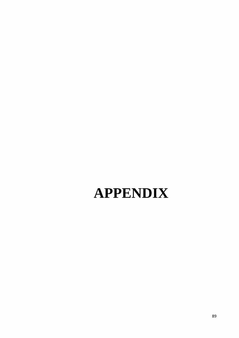
89
APPENDIX
90
STUDY POPULATION
91
APPENDIX 1
A.1. Fig. Overview of field work (interview questionnaire and clinical
examination)
92
CONSENT &APPROVAL
93
APPENDIX 2
A.2. Patient information sheet (English version)
94
APPENDIX 3
A.3. Patient information sheet (Bahasa melayu verion)
95
96
APPENDIX 4
A.4. Patient information sheet (Arabic version)
97
APPENDIX 5
A.5. Consent form (English version)
98
APPENDIX 6
A.6. Consent form (Bahasa melayu verion)
99
APPENDIX 7
A.7. Consent form (Arabic verion)
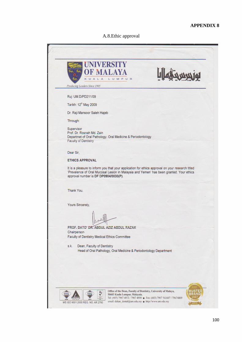
100
APPENDIX 8
A.8.Ethic approval
101
-
QUESTIONNAIRE
102
APPENDIX 9
A.9. Questionnaire (English version)
103
104
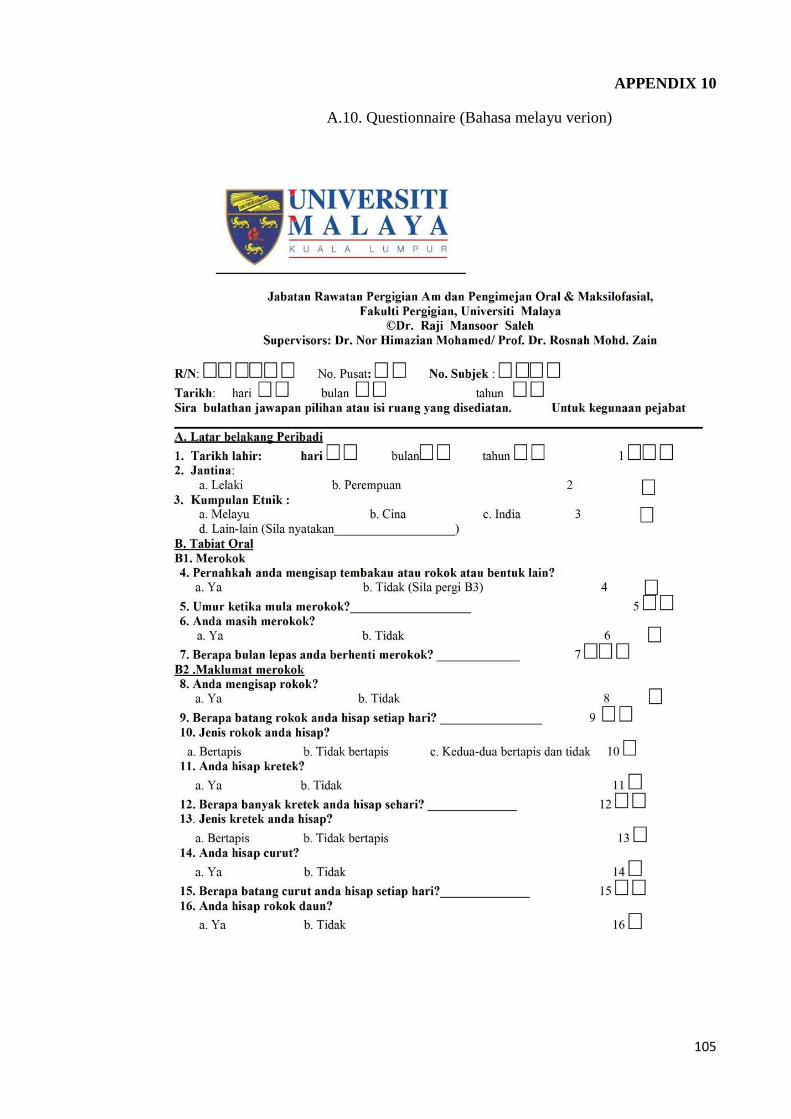
105
APPENDIX 10
A.10. Questionnaire (Bahasa melayu verion)
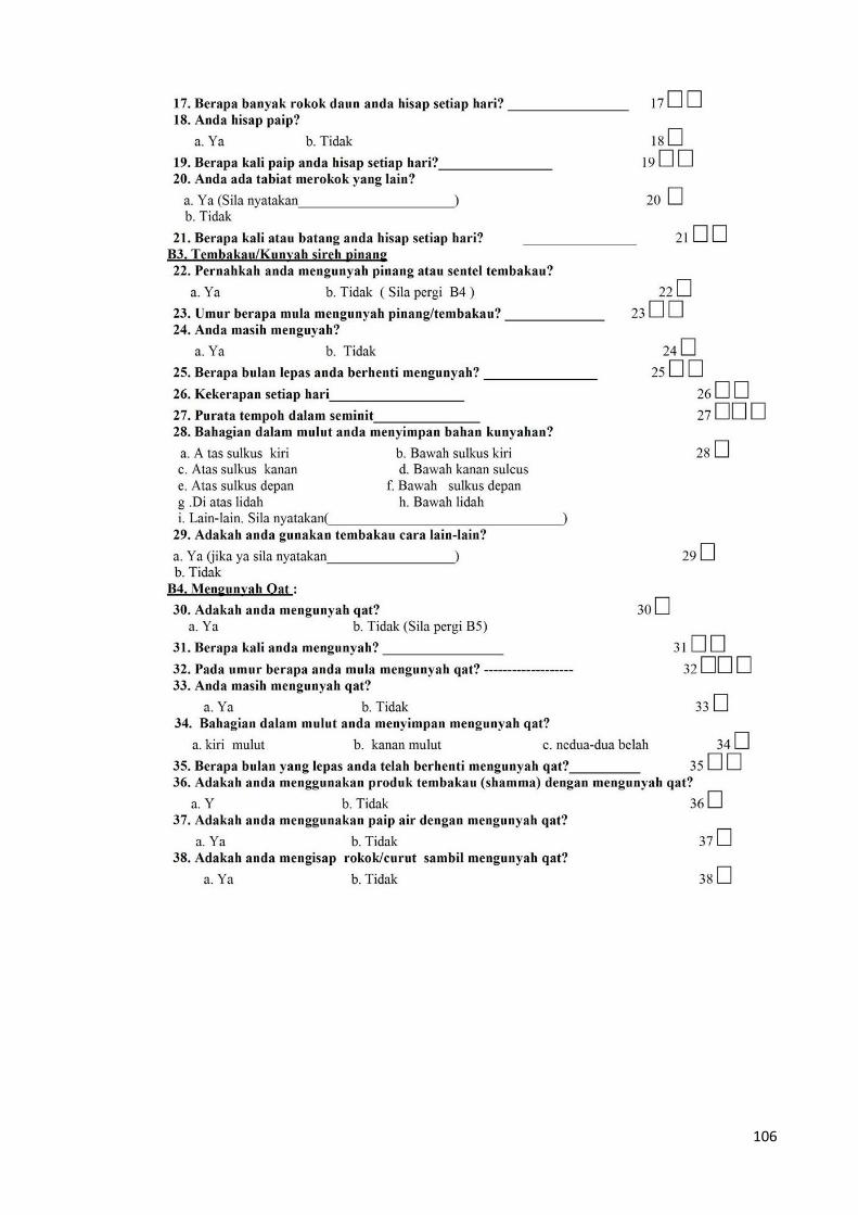
106
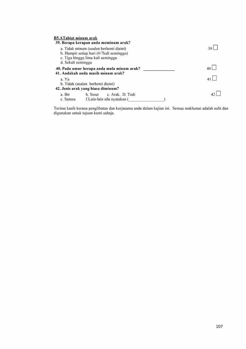
107
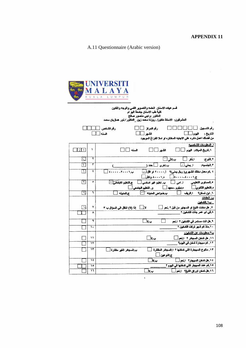
108
APPENDIX 11
A.11 Questionnaire (Arabic version)
109
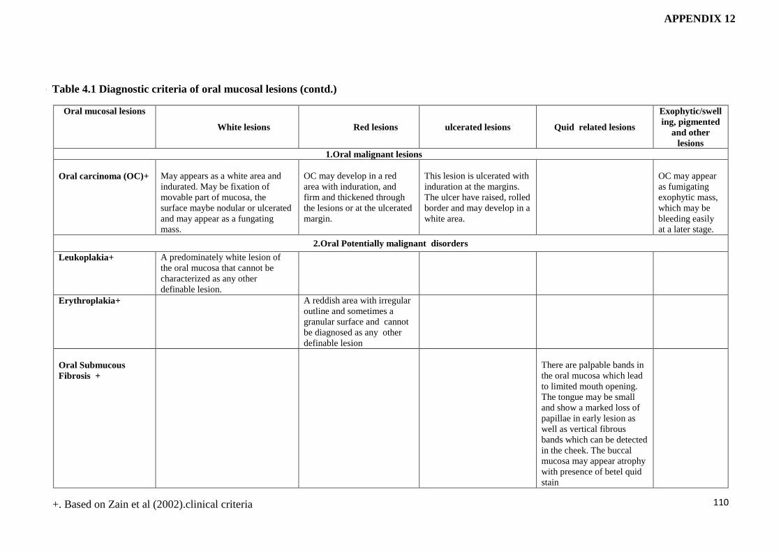
110
Oral mucosal lesions
White lesions Red lesions ulcerated lesions Quid related lesions
Exophytic/swell
ing, pigmented
and other
lesions
1.Oral malignant lesions
Oral carcinoma (OC)+
May appears as a white area and
indurated. May be fixation of
movable part of mucosa, the
surface maybe nodular or ulcerated
and may appear as a fungating
mass.
OC may develop in a red
area with induration, and
firm and thickened through
the lesions or at the ulcerated
margin.
This lesion is ulcerated with
induration at the margins.
The ulcer have raised, rolled
border and may develop in a
white area.
OC may appear
as fumigating
exophytic mass,
which may be
bleeding easily
at a later stage.
2.Oral Potentially malignant disorders
Leukoplakia+ A predominately white lesion of
the oral mucosa that cannot be
characterized as any other
definable lesion.
Erythroplakia+ A reddish area with irregular
outline and sometimes a
granular surface and cannot
be diagnosed as any other
definable lesion
Oral Submucous
Fibrosis +
There are palpable bands in
the oral mucosa which lead
to limited mouth opening.
The tongue may be small
and show a marked loss of
papillae in early lesion as
well as vertical fibrous
bands which can be detected
in the cheek. The buccal
mucosa may appear atrophy
with presence of betel quid
stain
+. Based on Zain et al (2002).clinical criteria
Table Table 4.1 Diagnostic criteria of oral mucosal lesions (contd.)
APPENDIX 12
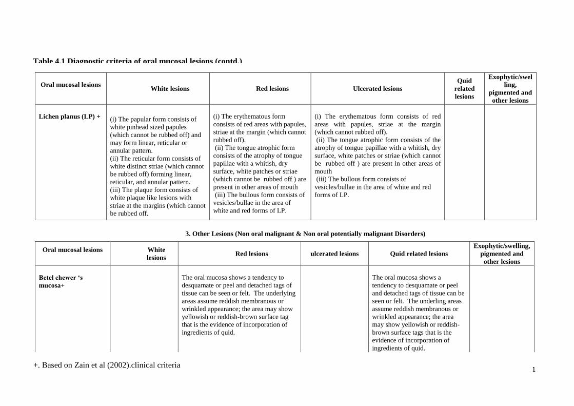
111
3. Other Lesions (Non oral malignant & Non oral potentially malignant Disorders)
Oral mucosal lesions
White lesions Red lesions Ulcerated lesions
Quid
related
lesions
Exophytic/swel
ling,
pigmented and
other lesions
Lichen planus (LP) +
(i) The papular form consists of
white pinhead sized papules
(which cannot be rubbed off) and
may form linear, reticular or
annular pattern.
(ii) The reticular form consists of
white distinct striae (which cannot
be rubbed off) forming linear,
reticular, and annular pattern.
(iii) The plaque form consists of
white plaque like lesions with
striae at the margins (which cannot
be rubbed off.
(i) The erythematous form
consists of red areas with papules,
striae at the margin (which cannot
rubbed off).
(ii) The tongue atrophic form
consists of the atrophy of tongue
papillae with a whitish, dry
surface, white patches or striae
(which cannot be rubbed off ) are
present in other areas of mouth
(iii) The bullous form consists of
vesicles/bullae in the area of
white and red forms of LP.
(i) The erythematous form consists of red
areas with papules, striae at the margin
(which cannot rubbed off).
(ii) The tongue atrophic form consists of the
atrophy of tongue papillae with a whitish, dry
surface, white patches or striae (which cannot
be rubbed off ) are present in other areas of
mouth
(iii) The bullous form consists of
vesicles/bullae in the area of white and red
forms of LP.
Oral mucosal lesions
White
lesions Red lesions ulcerated lesions Quid related lesions
Exophytic/swelling,
pigmented and
other lesions
Betel chewer ‘s
mucosa+
The oral mucosa shows a tendency to
desquamate or peel and detached tags of
tissue can be seen or felt. The underlying
areas assume reddish membranous or
wrinkled appearance; the area may show
yellowish or reddish-brown surface tag
that is the evidence of incorporation of
ingredients of quid.
The oral mucosa shows a
tendency to desquamate or peel
and detached tags of tissue can be
seen or felt. The underling areas
assume reddish membranous or
wrinkled appearance; the area
may show yellowish or reddish-
brown surface tags that is the
evidence of incorporation of
ingredients of quid.
Tabl Table 4.1 Diagnostic criteria of oral mucosal lesions (contd.)
+. Based on Zain et al (2002).clinical criteria
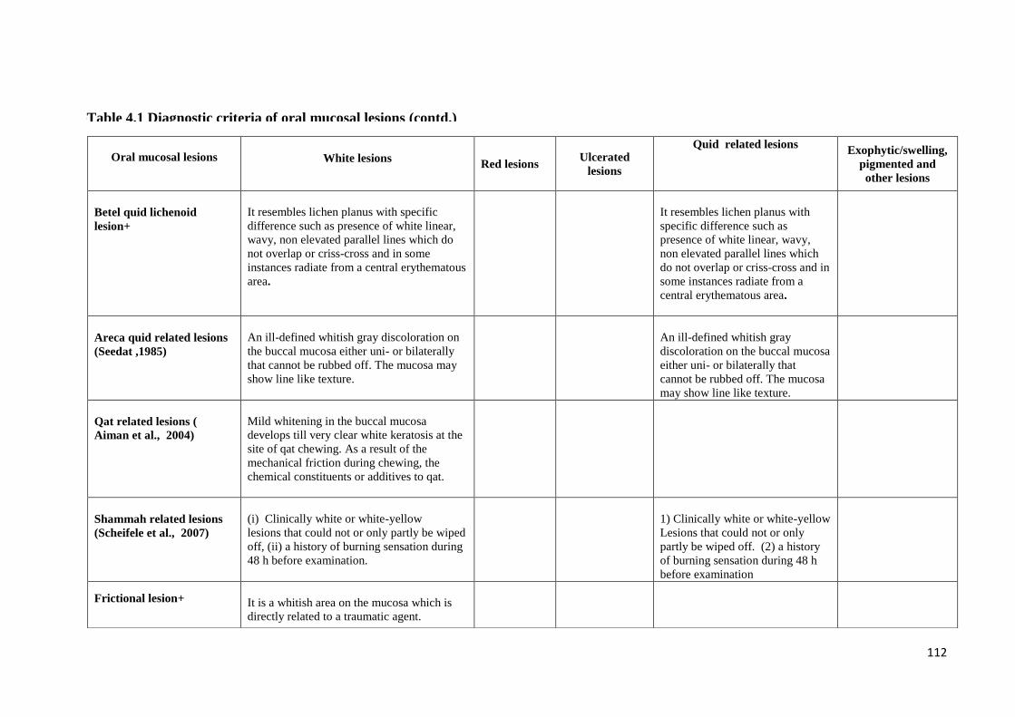
112
Oral mucosal lesions
White lesions Red lesions
Ulcerated
lesions
Quid related lesions Exophytic/swelling,
pigmented and
other lesions
Betel quid lichenoid
lesion+
It resembles lichen planus with specific
difference such as presence of white linear,
wavy, non elevated parallel lines which do
not overlap or criss-cross and in some
instances radiate from a central erythematous
area.
It resembles lichen planus with
specific difference such as
presence of white linear, wavy,
non elevated parallel lines which
do not overlap or criss-cross and in
some instances radiate from a
central erythematous area.
Areca quid related lesions
(Seedat ,1985)
An ill-defined whitish gray discoloration on
the buccal mucosa either uni- or bilaterally
that cannot be rubbed off. The mucosa may
show line like texture.
An ill-defined whitish gray
discoloration on the buccal mucosa
either uni- or bilaterally that
cannot be rubbed off. The mucosa
may show line like texture.
Qat related lesions (
Aiman et al., 2004)
Mild whitening in the buccal mucosa
develops till very clear white keratosis at the
site of qat chewing. As a result of the
mechanical friction during chewing, the
chemical constituents or additives to qat.
Shammah related lesions
(Scheifele et al., 2007)
(i) Clinically white or white-yellow
lesions that could not or only partly be wiped
off, (ii) a history of burning sensation during
48 h before examination.
1) Clinically white or white-yellow
Lesions that could not or only
partly be wiped off. (2) a history
of burning sensation during 48 h
before examination
Frictional lesion+
It is a whitish area on the mucosa which is
directly related to a traumatic agent.
TableTable 4.1 Diagnostic criteria of oral mucosal lesions (contd.)
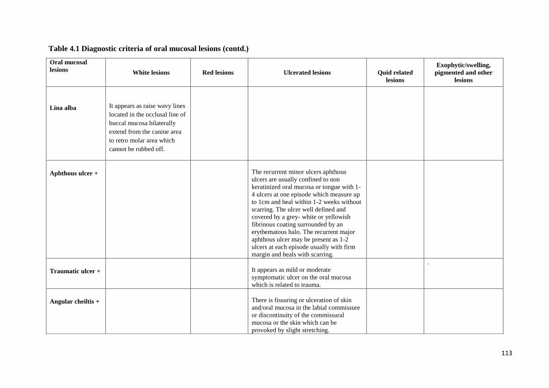
113
Oral mucosal
lesions
White lesions Red lesions Ulcerated lesions
Quid related
lesions
Exophytic/swelling,
pigmented and other
lesions
Lina alba
It appears as raise wavy lines
located in the occlusal line of
buccal mucosa bilaterally
extend from the canine area
to retro molar area which
cannot be rubbed off.
Aphthous ulcer +
The recurrent minor ulcers aphthous
ulcers are usually confined to non
keratinized oral mucosa or tongue with 1-
4 ulcers at one episode which measure up
to 1cm and heal within 1-2 weeks without
scarring. The ulcer well defined and
covered by a grey- white or yellowish
fibrinous coating surrounded by an
erythematous halo. The recurrent major
aphthous ulcer may be present as 1-2
ulcers at each episode usually with firm
margin and heals with scarring.
Traumatic ulcer +
It appears as mild or moderate
symptomatic ulcer on the oral mucosa
which is related to trauma.
.
Angular cheiltis +
There is fissuring or ulceration of skin
and/oral mucosa in the labial commissure
or discontinuity of the commissural
mucosa or the skin which can be
provoked by slight stretching.
Table 4.1 Diagnostic criteria of oral mucosal lesions (contd.)
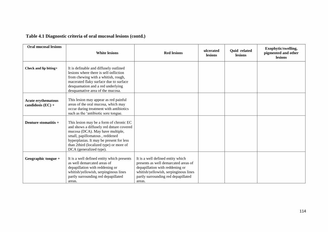
114
Oral mucosal lesions
White lesions Red lesions
ulcerated
lesions
Quid related
lesions
Exophytic/swelling,
pigmented and other
lesions
Check and lip biting+
It is definable and diffusely outlined
lesions where there is self-infliction
from chewing with a whitish, rough,
macerated flaky surface due to surface
desquamation and a red underlying
desquamative area of the mucosa.
Acute erythematous
candidosis (EC) +
This lesion may appear as red painful
areas of the oral mucosa, which may
occur during treatment with antibiotics
such as the ‗antibiotic sore tongue.
Denture stomatitis +
This lesion may be a form of chronic EC
and shows a diffusely red dnture covered
mucosa (DCA). May have multiple,
small, papillomatous , reddened
hyperplasias. It may be present for less
than 2third (localized type) or more of
DCA (generalized type).
Geographic tongue +
It is a well defined entity which presents
as well demarcated areas of
depapillation with reddening or
whitish/yellowish, serpinginous lines
partly surrounding red depapillated
areas.
It is a well defined entity which
presents as well demarcated areas of
depapillation with reddening or
whitish/yellowish, serpinginous lines
partly surrounding red depapillated
areas.
Table 4.1 Diagnostic criteria of oral mucosal lesions (contd.)
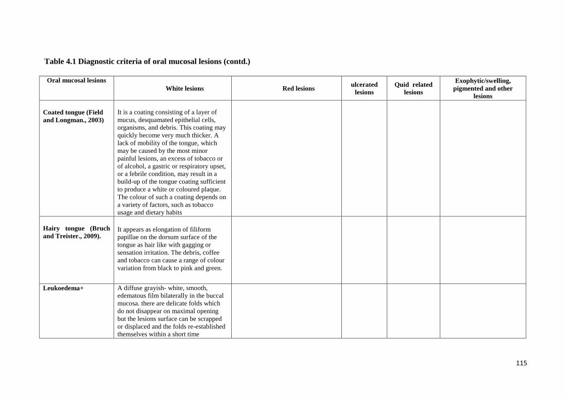
115
Oral mucosal lesions
White lesions Red lesions ulcerated
lesions
Quid related
lesions
Exophytic/swelling,
pigmented and other
lesions
Coated tongue (Field
and Longman., 2003)
It is a coating consisting of a layer of
mucus, desquamated epithelial cells,
organisms, and debris. This coating may
quickly become very much thicker. A
lack of mobility of the tongue, which
may be caused by the most minor
painful lesions, an excess of tobacco or
of alcohol, a gastric or respiratory upset,
or a febrile condition, may result in a
build-up of the tongue coating sufficient
to produce a white or coloured plaque.
The colour of such a coating depends on
a variety of factors, such as tobacco
usage and dietary habits
Hairy tongue (Bruch
and Treister., 2009).
It appears as elongation of filiform
papillae on the dorsum surface of the
tongue as hair like with gagging or
sensation irritation. The debris, coffee
and tobacco can cause a range of colour
variation from black to pink and green.
Leukoedema+ A diffuse grayish- white, smooth,
edematous film bilaterally in the buccal
mucosa. there are delicate folds which
do not disappear on maximal opening
but the lesions surface can be scrapped
or displaced and the folds re-established
themselves within a short time
Table 4.1 Diagnostic criteria of oral mucosal lesions (contd.)
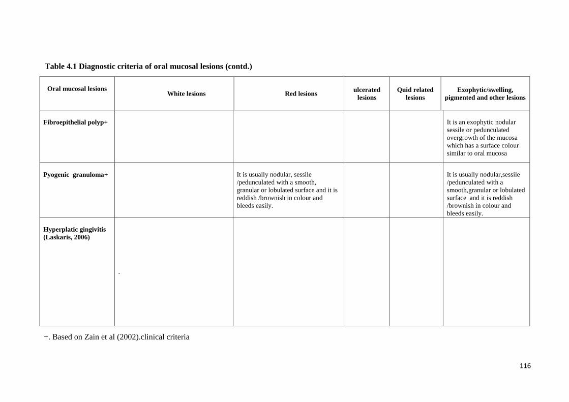
116
Oral mucosal lesions
White lesions Red lesions ulcerated
lesions
Quid related
lesions
Exophytic/swelling,
pigmented and other lesions
Fibroepithelial polyp+
It is an exophytic nodular
sessile or pedunculated
overgrowth of the mucosa
which has a surface colour
similar to oral mucosa
Pyogenic granuloma+
It is usually nodular, sessile
/pedunculated with a smooth,
granular or lobulated surface and it is
reddish /brownish in colour and
bleeds easily.
It is usually nodular,sessile
/pedunculated with a
smooth,granular or lobulated
surface and it is reddish
/brownish in colour and
bleeds easily.
Hyperplatic gingivitis
(Laskaris, 2006)
.
+. Based on Zain et al (2002).clinical criteria
Table 4.1 Diagnostic criteria of oral mucosal lesions (contd.)
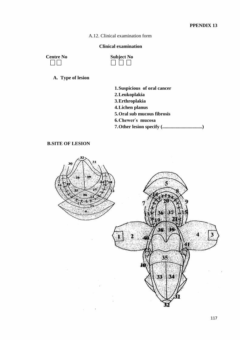
117
PPENDIX 13
A.12. Clinical examination form
Clinical examination
Centre No Subject No
A. Type of lesion
1. Suspicious of oral cancer
2. Leukoplakia
3. Erthroplakia
4. Lichen planus
5. Oral sub mucous fibrosis
6. Chewer s mucosa
7. Other lesion specify (.................................)
B.SITE OF LESION
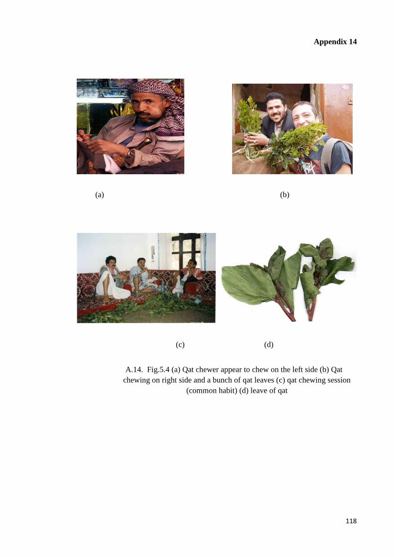
118
Appendix 14
(a) (b)
(c) (d)
A.14. Fig.5.4 (a) Qat chewer appear to chew on the left side (b) Qat
chewing on right side and a bunch of qat leaves (c) qat chewing session
(common habit) (d) leave of qat