Embed Size (px)
Citation preview

1
CHAPTER ONE
LITERATURE REVIEW
1.1 Breast Cancer Epidemiology
1.1.1 The Global burden of breast cancer
For a large number of women newly diagnosed in the world, it has been ascertain
that, breast cancer is a neglected disease in terms of other numerically more frequent
health problems. It has also been described as an orphan disease, in the sense that the
very detailed knowledge about tumor characteristics and the necessary host biology
capable of providing basic care is absent. Current international cancer policy and planning
initiatives are irrelevant to breast cancer, with the exception of nutritional
recommendation. However, progress with declines in mortality in some developed
countries has been reported (Ginsburg et al., 2011).
Breast cancer is the most prevalent cancer in the world (4.4 million survivors up to
5 years following diagnosis) and the second most common cause of cancer related
mortality in women wide world (Parkin et al., 2005). It also accounts for 23% (1.38 million)
of the total new cancer cases and 14% (458,400) of the total cancer deaths in 2008 and
ranks second most common cancer overall (10.9% of all cancers) but ranks fifth as cause
of death (Ferlay et al., 2010). 1.15 million new breast cancer cases were recorded in
2004 and over 500,000 deaths reported around the world and more than half of all cases
occurred in industrialized countries (Parkin and Fernandez, 2006). Breast cancer
incidence rates vary from 19.3 per 100,000 women in Eastern Africa to 89.7 per 100,000
women in Western Europe. They are normally high in developed regions of the world
(except Japan) and low in most of the developing regions. Due to more favorable survival
of breast cancer in developed regions, the range of mortality rates is very much less,
approximately 6-19 per 100,000. Notwithstanding, it is still the most frequent cause of
cancer death in women in both developing (269 000 deaths, 12.7% of total) and
developed regions, where the estimated 189 000 deaths is almost equal to the estimated
number of deaths from lung cancer (188 000 deaths) (Ferlay et al., 2010).
For some time now, there have been some encouraging in both breast cancer
incidence and mortality trends with the incidence of new cases stabilizing as well as death
rates falling in some high income or developed countries. However, this appears to be
vice versa in developing countries (Kanavos, 2006). Notably, breast cancer incidence
rates have leveled off since 1990, with a decrease of 3.5%/year from 2001 to 2004 (Li et
al., 2003). In the same manner, breast cancer mortality rates have also declined by 24%,
2
with the greatest impact among young women and as well as women with estrogen
receptor (ER)-positive disease (Berry et al., 2005). Also, both incidence and mortality
declined in the United States; between 1999 and 2006, incidence rates decreased by
2.0% per year, and mortality decreased by 1.9% annually between 1998 and 2006
(Horner et al., 2006). The decline in breast cancer mortality has been largely attributed to
the combination of early detection with screening programs and the advent of more
efficacious adjuvant systemic therapy.
Breast cancer is common in women both in the developed and the developing
countries, comprising 16% of all female cancers. Although it is thought to be a common
cancer in the developed countries, a majority (69%) of all breast cancer deaths occurs in
developing world. Indeed, increase life expectancy, increase urbanization and adoption of
western lifestyles have increased the incidence of breast cancer in the developing
countries (Kanavos, 2006). Eventhough it is now the most common cancer both in
developed and developing regions with around 690 000 new cases estimated in each
region, much of the burden of incidence, morbidity, and mortality will occur in the
developing world with population ratio of 1:4 (Ferlay et al., 2010). As developing countries
succeed in achieving lifestyles similar to those in advanced economies, they will also
encounter much higher cancer rates, particularly cancers of the breast. This forms part of
a larger epidemiological transition in which the burden of chronic, non-communicable
disease once limited to industrialized nations, is now increasing in less developed
countries (Kanavos, 2006).
A report by Stewart et al (Stewart and Kleigues, 2003), mentioned that many of the
new cancer cases are now occurring among women from low and middle income
countries, where the incidence is increasing by as much as 5% per each year and there
are about three fourths of breast cancer deaths occurring worldwide. Of the 411,000
breast cancer deaths around the world in 2002, 221,000 (54%) occurred in low- and
middle-income countries (LMCs). The incidence of breast cancer rose from 126,227 cases
in 2002 in China (IARC: Cancer Epidemiology Database, GLOBOCAN. 2002) to over
169,000 in 2008 (IARC: Cancer Epidemiology Database, GLOBOCAN. 2008).
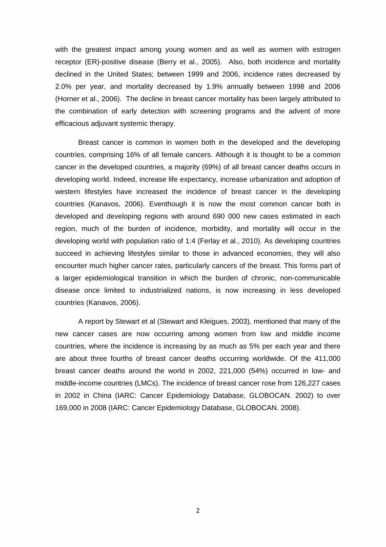
3
Figure 1.1: Chart showing worldwide prevalent Breast Cancer cases (X1000), Adult population.
(GLOBOCAN, 2008)
1.1.2 Differences in Population of Breast Cancer
Breast cancer variation among population, or the regional differences in the types
have been attributed to the following: prevalence of major risk factors, availability and use
of medical practices such as cancer screening, availability and quality of treatment,
completeness of reporting, and age structure. However, geographic areas, and counties
and parishes within countries also determine the frequency of the most commonly
diagnosed cases or deaths (Garcia M et al., 2007). The highly penetrant but rare
susceptibility genes, BRCA1 and BRCA2 (Fackenthal et al., 2007) and more prevalent,
but lower penetrance genes, CHEK2 and FGFR (Easton et al., 2007) have been indicated
to be the key inter-individual and inter-group differences in the distribution of reproductive
risk factors. Countries with massive economic development over the past 50 years, such
Japan, Singapore, and urban areas of China have experience an increase in breast
cancer incidence (Horn-Ross et al., 2000).
Age-standardized incidence rates for breast cancer 1998–2002 were 110 (non-
Hispanic Caucasians, California), 82.3 (Ontario, Canada), 41.3 (Hong Kong) and 14.7
(Jiashan, China) (Curado et al., 2007). Reports on migration studies reveal that the
incidence of breast cancer changes significantly over one to two generations to more
closely reflect the breast cancer risk in the adopted country (Ziegler et al., 1993), which
seems to occur in parallel with dynamics in diet and certain indicators of acculturation
4
(Porter, 2008). Notably, evaluation of differences in risk factors and natural history of all
tumor types, would permit for comparisons based on geographical regions,
socioeconomic status and levels of industrialization (Ginsburg et al, 2011).
Other differences in population of breast cancer are outlined below:
In a study by Li et al (Li et al., 2002), it was showen that the majority of breast tumours
from Asian women are estrogen receptor (ER) negative. Also it has been indicated that
both pre-and postmenopausal Asian women with breast cancer, are likely to have ER
positive tumors as Caucasians (Uy et al., 2007). In addition, greater proportion of ER+
tumors in a Vietnamese cohort, has been found in a studies on ER positivity among
premenopausal breast cancer cases as compared with the comparison group of
Caucasian women in Australia. (Tran and Lawson, 2004).
Considerably, variation in the gene profiles of tumors from populations of different
genetic/ethnic backgrounds have also been reported. About 15% of sporadic breast
cancer, which are BRCA1 origin in Caucasian women appears to have the basal
phenotype. On the other hand, other studies have also suggested that breast cancer in
women of African ancestry may have a higher proportion of basal phenotype (Carey et
al., 2006). In similar manner among Nigerians, a high frequency of basallike tumors was
observed, where 87 of 148 (59%) breast cancer cases were both ER- and HER2-
(Olopade et al., 2004).
5
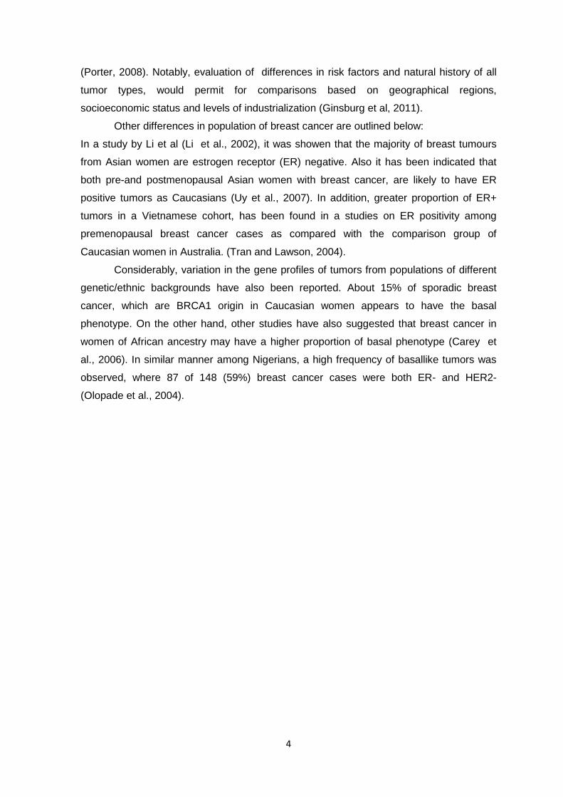
Figure 1.2: Incidence of female breast cancer by age in selected population, 1988-1993.
(Parkin et al., 1997; Tavassoli and Devilee, 2003)
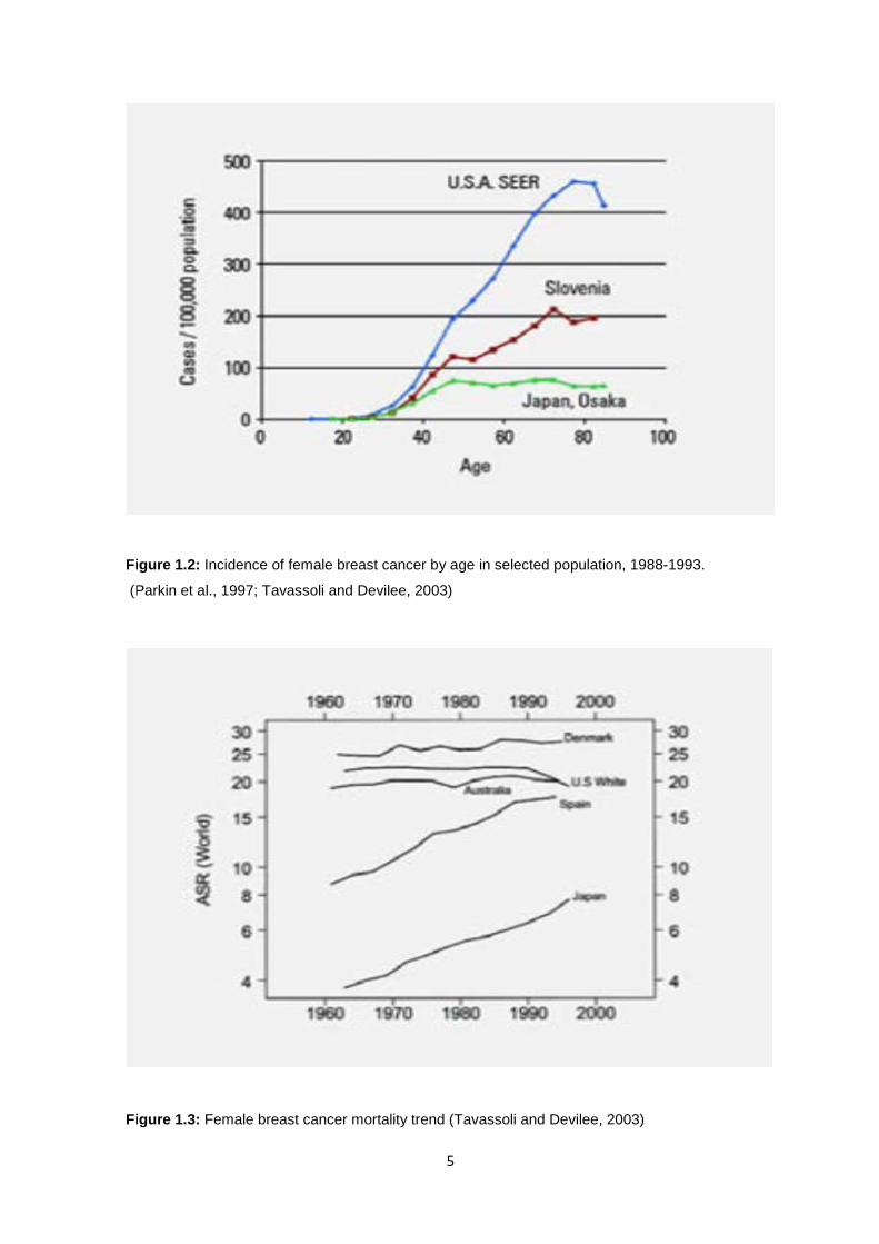
Figure 1.3: Female breast cancer mortality trend (Tavassoli and Devilee, 2003)
6
1.2 General characteristics of Breast Cancer
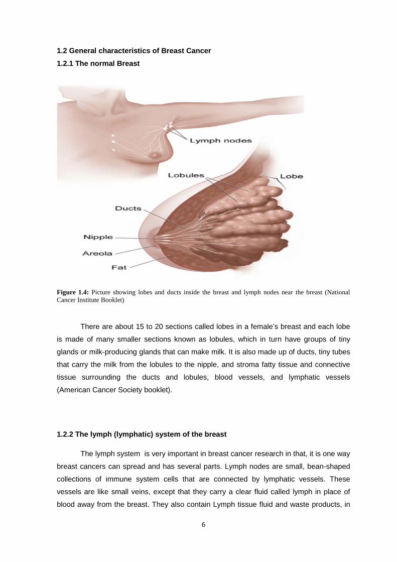
1.2.1 The normal Breast
Figure 1.4: Picture showing lobes and ducts inside the breast and lymph nodes near the breast (National Cancer Institute Booklet)
There are about 15 to 20 sections called lobes in a female’s breast and each lobe
is made of many smaller sections known as lobules, which in turn have groups of tiny
glands or milk-producing glands that can make milk. It is also made up of ducts, tiny tubes
that carry the milk from the lobules to the nipple, and stroma fatty tissue and connective
tissue surrounding the ducts and lobules, blood vessels, and lymphatic vessels
(American Cancer Society booklet).
1.2.2 The lymph (lymphatic) system of the breast
The lymph system is very important in breast cancer research in that, it is one way
breast cancers can spread and has several parts. Lymph nodes are small, bean-shaped
collections of immune system cells that are connected by lymphatic vessels. These
vessels are like small veins, except that they carry a clear fluid called lymph in place of
blood away from the breast. They also contain Lymph tissue fluid and waste products, in

7
addition to immune system cells. Breast cancer cells can enter lymphatic vessels and
begin to grow in lymph nodes. Most lymphatic vessels in the breast connect to lymph
nodes under the arm (axillary nodes). Some lymphatic vessels that connect to lymph
nodes inside the chest are called internal lymph nodes, and those either above or below
the collarbone are called supraclavicular or infraclavicular nodes (American Cancer
Society booklet).
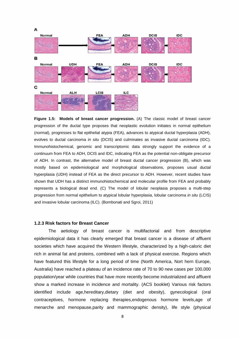
There are several types of breast cancer, but some of them are quite rare.
Currently, majority of all breast cancers worldwide are the ductal and lobular subtypes
.However, the ductal subtype accounting acounts for the majority of the diagnosed cases
,constituting for about 40–75% (Rakha et al., 2006). In addition, several linear models of
breast cancer initiation, transformation and progression, as depicted in Fig. 1.5 have been
formulated. There are two models for the ductal subtype The first ‘ductal’ model, reported
by Lerwill (Lerwill, 2008) are as follow: First of all, it recognizes flat epithelial atypia (FEA),
to atypical ductal hyperplasia (ADH) and then ductal carcinoma in situ (DCIS) as the non-
obligate precursors of the advanced invasive and metastatic ductal carcinoma. In the
second model, usual epithelial ductal hyperplasia (UDH) was proposed as an intermediate
stage of progression between FEA and DCIS (Page et al., 1985). In the case of lobular
subtype, atypical lobular hyperplasia (ALH) and lobular carcinoma in situ (LCIS) was also
proposed as the non-obligate precursor lesions to invasive lobular carcinoma. (Boecker et
al., 2002).
8
Figure 1.5: Models of breast cancer progression. (A) The classic model of breast cancer
progression of the ductal type proposes that neoplastic evolution initiates in normal epithelium
(normal), progresses to flat epithelial atypia (FEA), advances to atypical ductal hyperplasia (ADH),
evolves to ductal carcinoma in situ (DCIS) and culminates as invasive ductal carcinoma (IDC).
Immunohistochemical, genomic and transcriptomic data strongly support the evidence of a
continuum from FEA to ADH, DCIS and IDC, indicating FEA as the potential non-obligate precursor
of ADH. In contrast, the alternative model of breast ductal cancer progression (B), which was
mostly based on epidemiological and morphological observations, proposes usual ductal
hyperplasia (UDH) instead of FEA as the direct precursor to ADH. However, recent studies have
shown that UDH has a distinct immunohistochemical and molecular profile from FEA and probably
represents a biological dead end. (C) The model of lobular neoplasia proposes a multi-step
progression from normal epithelium to atypical lobular hyperplasia, lobular carcinoma in situ (LCIS)
and invasive lobular carcinoma (ILC). (Bombonati and Sgroi, 2011)
1.2.3 Risk factors for Breast Cancer
The aetiology of breast cancer is multifactorial and from descriptive
epidemiological data it has clearly emerged that breast cancer is a disease of affluent
societies which have acquired the Western lifestyle, characterized by a high-caloric diet
rich in animal fat and proteins, combined with a lack of physical exercise. Regions which
have featured this lifestyle for a long period of time (North America, Nort hern Europe,
Australia) have reached a plateau of an incidence rate of 70 to 90 new cases per 100,000
population/year while countries that have more recently become industrialized and affluent
show a marked increase in incidence and mortality. (ACS booklet) Various risk factors
identified include age,hereditary,dietary (diet and obesity), gynecological (oral
contraceptives, hormone replacing therapies,endogenous hormone levels,age of
menarche and menopause,parity and mammographic density), life style (physical
9
activity,smoking and alcohol) ,oxygen reactive species,radiation and environmental
pollutants. (Fig. 1.6).
With age,breast cancer incidence is known to drastically increase up to the age of
50, after which it increases slowly (Mitruen and Hirvonen, 2003). Hereditary factors are
observed in about one fourth of the total cases of breast cancer,which involves two
classes of genes; high and low penetrance genes. High penetrance genes with allelic
variants that are relatively rare,such as BRCA1/2,tumor protein 53 gene (TP53) and ataxia
telangiecttasi mutated gene (ATM). Low penetrance genes such as the genes encoding
for the enzymes involved in estrogen and carcinogen metabolism as well as in the
detoxification of reactive oxygen species,for instance P450 cytochrome and Gluthioone-S-
transferases (GSTs) on the other hand are more common and allelic variants confer low
risk of breast cancer (Mitruen and Hirvonen ,2003).
In the case of dietary, the human diet contains variety of natural carcinogens and
anticarcinogens. Uptake of fruits and vegetables which are rich in antioxidants reduces
the risk (McKeown, 1999) whereas increase in polysaturated fatty acids (Bartsch et al.,
1999) and meat consumption (Zheng et al., 1998) increases the risk of braest cancer.
Obesity has been reported to be associated with an increase in estrogen levels and as
well as a risk in postmenopausal women,who have most of their estrogen derived from the
conversion of androgens,in the adipose tissue,as a result of aromatse enzyme activity
(Hunter et al., 1993). However, in premenopausal women, it has been indicated that,
obesity can have aprotective effect, due to the higher period of frequent ovulation which
reduces estrogen levels (Mannisto et al., 1996). Physical activity is a lifestyle factor which
is considered as a breast cancer risk factor. It is considered protective against braest
cancer because it reduces the regualar ovulatory cycles and increases the level of
catechol-O-methylated estrogens (Henderson et al., 1985).
Cigarette smoke is very rich in carcinogens and reactive oxygen species and may
be considered as one with high risk in breast cancer (Mitruen et al., 2003). Its function is
controversial ,in that it can serve as a protective against cancer.It may contain anti-
estrogenic effect,such as nicotine wich inhibit aromatase. Further more,women who
smoke tend to reach menopause earlier than nonsmokers (baron et al., 1990). Another
fact to consider is alcohol. 15% of alcoholic women have the risk of developing breast
cancer (Kuper et al., 2000). It has been stipulated alcoholic women have higher levels of
estrogen than non-alcoholic (Reichman et al., 1993).
The use of oral contraceptive increases breast cancer risk but disappears after ten years
of cessation, whereas hormone replacement therapy disappears in five years. However,
breast cancer cases in hormone replacement therapy (HRT) tend to be less advanced at
10
the time of diagnosis, and biologically less agressive compared to women who never used
such therapy (Holli et al., 1997).
Relationship between endogenous estrogen levels and breast cancer has been
indicated. High estrogen levels in the serum or urine, and low levels of sex hormone
binding protein (SHBG), resulting in high bioavailability of free estradiol also point for an
important role for endogenous and exogenous estrogens in the risk of breast cancer
(Kristensen et al., 2000).
Another strong marker of breast cance risk is the degree of mammographic
density. It has been indicated that, the risk in women with more dense breast is four to six
times higher than those with less dense breast (Boyd et al., 1995). Evidence suggest
that,the etiology of mammographic density may be due to the exposure to steroid
hormone, since it decreases with age (Boyd et al., 2002) as well as in women on
tamoxifen (Cuzick et al., 2004) and also increases in women who are on hormone
replacement therapy (Rutter et al., 2001).
Environnmental pollutants similar to hormones can also interfere in the control of a
large family of nuclear hormone receptors,which in turn can upregulate various genes
involved in the cell cycle,such as TP53, Retinoblatoma (RB) and the serine.threonine-
protein kinase proto-oncogene RAF by transcriptional activation induced by ligand
(Kristensen et al., 2000).
These polluatnts are designated xeno-estrogens, which include pesticides, dyes, food
preservatives and other polluatnts and can play a role in the etiology of breast
cancer,since they interfere with the activity of endogenous estrogens (Garner et al., 2000).
Ionizing radiation (John and Kelsey, 1993) and history of benign breast cancer (Mitruen et
al., 2003) have also been established to increase breast cancer risk.
11
Figure 1.6: Aetiological factors involved in the development of breast cancer (Tavassoli and
Devilee, 2003)
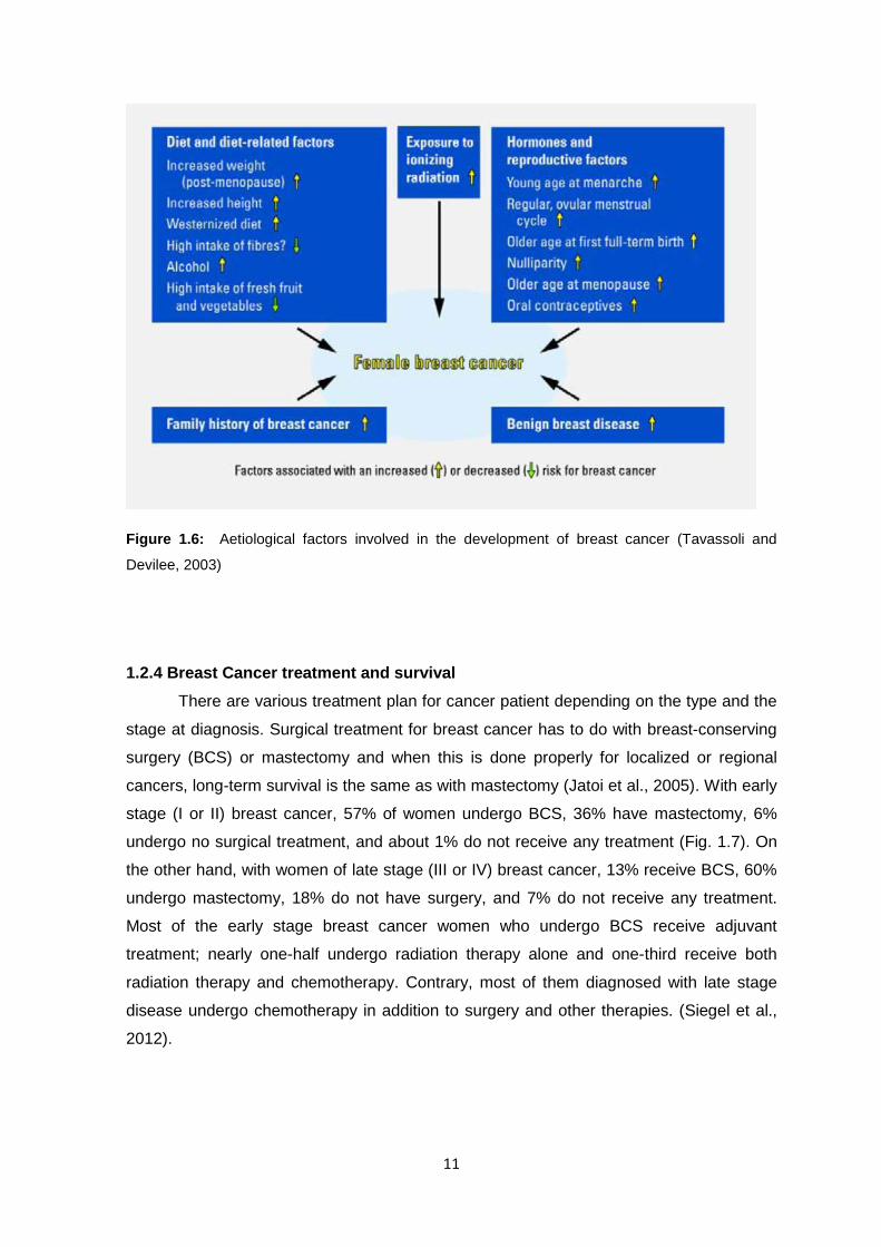
1.2.4 Breast Cancer treatment and survival There are various treatment plan for cancer patient depending on the type and the
stage at diagnosis. Surgical treatment for breast cancer has to do with breast-conserving
surgery (BCS) or mastectomy and when this is done properly for localized or regional
cancers, long-term survival is the same as with mastectomy (Jatoi et al., 2005). With early
stage (I or II) breast cancer, 57% of women undergo BCS, 36% have mastectomy, 6%
undergo no surgical treatment, and about 1% do not receive any treatment (Fig. 1.7). On
the other hand, with women of late stage (III or IV) breast cancer, 13% receive BCS, 60%
undergo mastectomy, 18% do not have surgery, and 7% do not receive any treatment.
Most of the early stage breast cancer women who undergo BCS receive adjuvant
treatment; nearly one-half undergo radiation therapy alone and one-third receive both
radiation therapy and chemotherapy. Contrary, most of them diagnosed with late stage
disease undergo chemotherapy in addition to surgery and other therapies. (Siegel et al.,
2012).
12
Figure 1.7: Female Breast Cancer Treatment Patterns by Stage, 2008. BCS indicates breast-
conserving surgery; RT, radiation therapy; chemo, chemotherapy (may include common targeted
therapies). Percentages do not sum to 100% due to rounding. (Siegel et al., 2012)
Patients can expect to be cured or to experience at least long-term survival of
more than 10 years. There has been an improvement for the overall 5-year relative
survival rate for female breast cancer patients, from 75.1% between 1975 to 1977 to
90.0% for 2001 through 2007. And this is attributed to the fact that there has been an
improvement in chemotherapy and hormone therapy treatment and also due to earlier
diagnosis resulting from the widespread use of mammography (Siegel et al., 2012). In the
case of localized breast cancer, the 5-year relative survival rate is 98.6%; which declined
to 83.8% for regional stage and 23.3% for distant stage. Other factors that influence
survival include tumor grade, hormone receptor status, and human epidermal growth
factor receptor 2 (HER2) status.
There are differences between African American women and white women on the
basis of stage and survival. African American women are less likely than white women to
be diagnosed with local stage breast cancer (51% vs 61%) and have lower survival rates
than white women within each stage of disease. Though difficult to explain this reasons, it
may be explained in large part by a combination of socioeconomic factors, less access to
care among African American women, and biological differences in cancers (Siegel et al.,
2012).
13
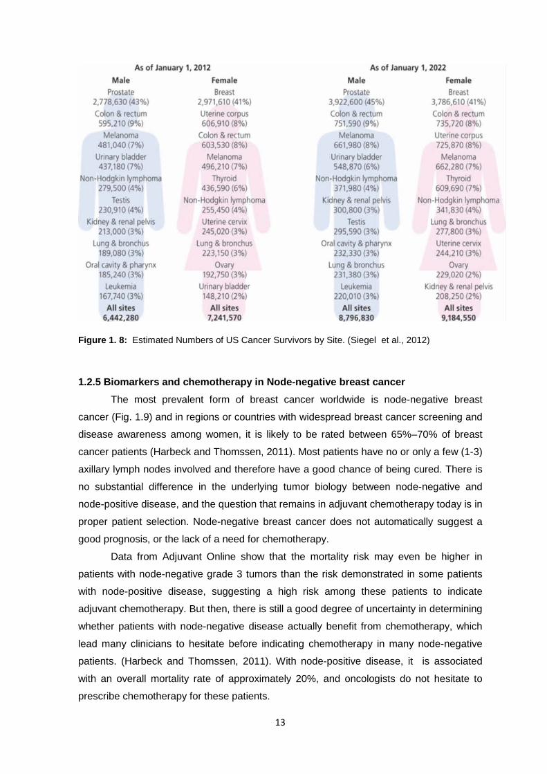
Figure 1. 8: Estimated Numbers of US Cancer Survivors by Site. (Siegel et al., 2012)
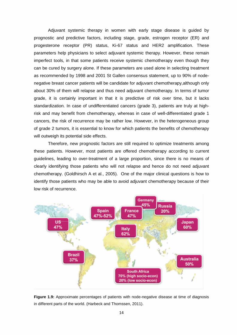
1.2.5 Biomarkers and chemotherapy in Node-negative breast cancer The most prevalent form of breast cancer worldwide is node-negative breast
cancer (Fig. 1.9) and in regions or countries with widespread breast cancer screening and
disease awareness among women, it is likely to be rated between 65%–70% of breast
cancer patients (Harbeck and Thomssen, 2011). Most patients have no or only a few (1-3)
axillary lymph nodes involved and therefore have a good chance of being cured. There is
no substantial difference in the underlying tumor biology between node-negative and
node-positive disease, and the question that remains in adjuvant chemotherapy today is in
proper patient selection. Node-negative breast cancer does not automatically suggest a
good prognosis, or the lack of a need for chemotherapy.
Data from Adjuvant Online show that the mortality risk may even be higher in
patients with node-negative grade 3 tumors than the risk demonstrated in some patients
with node-positive disease, suggesting a high risk among these patients to indicate
adjuvant chemotherapy. But then, there is still a good degree of uncertainty in determining
whether patients with node-negative disease actually benefit from chemotherapy, which
lead many clinicians to hesitate before indicating chemotherapy in many node-negative
patients. (Harbeck and Thomssen, 2011). With node-positive disease, it is associated
with an overall mortality rate of approximately 20%, and oncologists do not hesitate to
prescribe chemotherapy for these patients.
14
Adjuvant systemic therapy in women with early stage disease is guided by
prognostic and predictive factors, including stage, grade, estrogen receptor (ER) and
progesterone receptor (PR) status, Ki-67 status and HER2 amplification. These
parameters help physicians to select adjuvant systemic therapy. However, these remain
imperfect tools, in that some patients receive systemic chemotherapy even though they
can be cured by surgery alone. If these parameters are used alone in selecting treatment
as recommended by 1998 and 2001 St Gallen consensus statement, up to 90% of node-
negative breast cancer patients will be candidate for adjuvant chemotherapy,although only
about 30% of them will relapse and thus need adjuvant chemotherapy. In terms of tumor
grade, it is certainly important in that it is predictive of risk over time, but it lacks
standardization. In case of undifferentiated cancers (grade 3), patients are truly at high-
risk and may benefit from chemotherapy, whereas in case of well-differentiated grade 1
cancers, the risk of recurrence may be rather low. However, in the heterogeneous group
of grade 2 tumors, it is essential to know for which patients the benefits of chemotherapy
will outweigh its potential side effects.
Therefore, new prognostic factors are still required to optimize treatments among
these patients. However, most patients are offered chemotherapy according to current
guidelines, leading to over-treatment of a large proportion, since there is no means of
clearly identifying those patients who will not relapse and hence do not need adjuvant
chemotherapy. (Goldhirsch A et al., 2005). One of the major clinical questions is how to
identify those patients who may be able to avoid adjuvant chemotherapy because of their
low risk of recurrence.
Figure 1.9: Approximate percentages of patients with node-negative disease at time of diagnosis
in different parts of the world. (Harbeck and Thomssen, 2011).
15
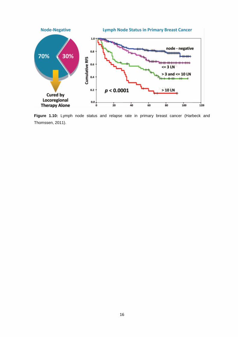
1.2.6 Node status and relapse Rate Another clinical question is how to identify individuals with high risk who may
benefit from adjuvant chemotherapy. One important risk factor for disease relapse is nodal
stays. Most patients with node-negative breast cancer have a fairly good ten-year overall
survival with loco-regional treatment alone, (Fig 1.10) however, about 30% relapse
developing distant metastasis. In fact, approximately 70% of node-negative patients
respond sufficiently to surgery, radiotherapy, and endocrine therapy, and do not require
additional chemotherapy. The problem faced by most clinicians now face is that
approximately 30% of node-negative patients will need chemotherapy because of their
risk for recurrence, but there are limited tools currently available to identify this subset of
patients. Data from Adjuvant! Online have shown that even patients with grade 1,
estrogen receptor– negative, node-negative tumors may have a high relapse rate of
almost 20% over 10 years. Indicating that the relapse rate is even higher in patients with
grade 2 and grade 3 tumors (Harbeck and Thomssen, 2011).
Therefore, markers to predict individual risk who may benefit from adjuvant
chemotherapy and also to identify patients not requiring aggressive adjuvant therapy are
urgently needed, so as to avoid unnecessary exposure of women to the potential toxicity
and side-effects of such treatment, and also to reduce the overall cost of breast cancer
management as well as preventing under-treatment of node-negative breast cancer are
needed.
The main cause of morbidity and mortality in patients with cancer is the formation
of distant metastases, which is a multistep event involving local invasion, degradation of
extracellular matrix, angiogenesis, intravasation, evasion of apoptosis and survival in
circulation, extravasation and growth at secondary site. Key mediators of this process
include certain proteinases such as uPA, PAI-1, MMPs and ADAMs which has caused
increase attention to be drawn on these factors as potential prognostic markers for risk
assessment in node-negative breast cancer. However, unlike MMPs, uPA , and PAI-1,
little work has been done on ADAMs role as a prognostic factor.
16
Figure 1.10: Lymph node status and relapse rate in primary breast cancer (Harbeck and
Thomssen, 2011).
17
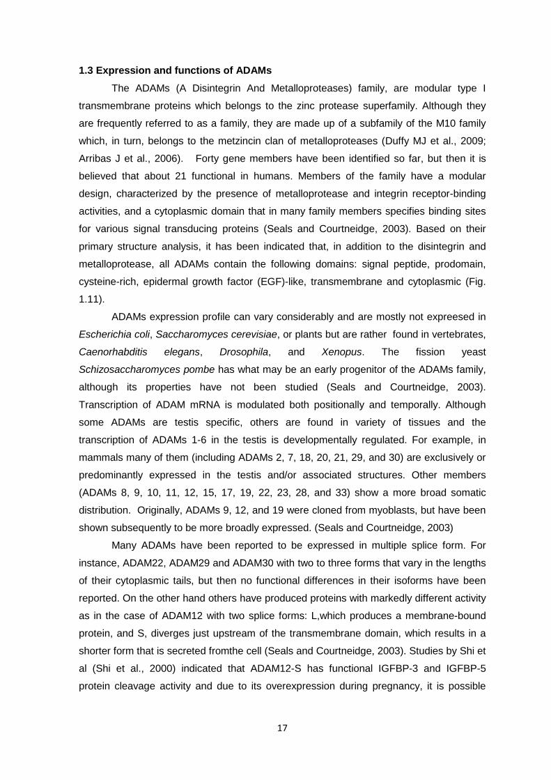
1.3 Expression and functions of ADAMs The ADAMs (A Disintegrin And Metalloproteases) family, are modular type I
transmembrane proteins which belongs to the zinc protease superfamily. Although they
are frequently referred to as a family, they are made up of a subfamily of the M10 family
which, in turn, belongs to the metzincin clan of metalloproteases (Duffy MJ et al., 2009;
Arribas J et al., 2006). Forty gene members have been identified so far, but then it is
believed that about 21 functional in humans. Members of the family have a modular
design, characterized by the presence of metalloprotease and integrin receptor-binding
activities, and a cytoplasmic domain that in many family members specifies binding sites
for various signal transducing proteins (Seals and Courtneidge, 2003). Based on their
primary structure analysis, it has been indicated that, in addition to the disintegrin and
metalloprotease, all ADAMs contain the following domains: signal peptide, prodomain,
cysteine-rich, epidermal growth factor (EGF)-like, transmembrane and cytoplasmic (Fig.
1.11).
ADAMs expression profile can vary considerably and are mostly not expreesed in
Escherichia coli, Saccharomyces cerevisiae, or plants but are rather found in vertebrates,
Caenorhabditis elegans, Drosophila, and Xenopus. The fission yeast
Schizosaccharomyces pombe has what may be an early progenitor of the ADAMs family,
although its properties have not been studied (Seals and Courtneidge, 2003).
Transcription of ADAM mRNA is modulated both positionally and temporally. Although
some ADAMs are testis specific, others are found in variety of tissues and the
transcription of ADAMs 1-6 in the testis is developmentally regulated. For example, in
mammals many of them (including ADAMs 2, 7, 18, 20, 21, 29, and 30) are exclusively or
predominantly expressed in the testis and/or associated structures. Other members
(ADAMs 8, 9, 10, 11, 12, 15, 17, 19, 22, 23, 28, and 33) show a more broad somatic
distribution. Originally, ADAMs 9, 12, and 19 were cloned from myoblasts, but have been
shown subsequently to be more broadly expressed. (Seals and Courtneidge, 2003)
Many ADAMs have been reported to be expressed in multiple splice form. For
instance, ADAM22, ADAM29 and ADAM30 with two to three forms that vary in the lengths
of their cytoplasmic tails, but then no functional differences in their isoforms have been
reported. On the other hand others have produced proteins with markedly different activity
as in the case of ADAM12 with two splice forms: L,which produces a membrane-bound
protein, and S, diverges just upstream of the transmembrane domain, which results in a
shorter form that is secreted fromthe cell (Seals and Courtneidge, 2003). Studies by Shi et
al (Shi et al., 2000) indicated that ADAM12-S has functional IGFBP-3 and IGFBP-5
protein cleavage activity and due to its overexpression during pregnancy, it is possible

18
that ADAM12-S is responsible for increasing the pool of IGF in the bloodstream during
pregnancy through IGFBP proteolysis.
Another protein to consider is ADAM28. It produces isoforms with different
subcellular localization patterns and tissue expression. There are three isoforms in Murine
ADAM28 , two larger ones for encoding membrane-anchored proteins and expressed in
the epididymis and lung, and smaller one predicted to encode a secreted protein with
testis-specific expression. However,in human there are only two forms: the secreted form
which is preferentially expressed in the spleen, and the membrane-bound form with
specificity to lymph node (Seals and Courtneidge, 2003). Other ADAMs with documented
evidence of alternativel splicing are ADAM9, ADAM10, ADAM11 and ADAM3.
The ADAMs have been implicated in processes such as the activation of the
proforms of certain growth factors and cytokines as well as the shedding of the
extracellular domains of growth factor receptors and adhesion proteins, control of
membrane fusion, and cell migration, as well as physiological processes such as muscle
development, fertilization, neurogenesis, adipogenesis, myogenesis and cell fate
determination. Another function is the activation of NOTCH signalling by Notch ligand
Delta shedding from the cell surface by ADAM-10 (Rocks et al., 2008). Accumulating
evidence demonstrates ADAMs as proteins that support both proteolytic activity and cell
adhesion, making them candidates to mediate both the remodelling of the extracellular
matrix (ECM) and the changes in cell adhesion that characterize certain pathological
processes such as tumor development, bacterial infection, cardiac hypertrophy, and
asthma (Murphy, 2008). Of these different diseases, it is in cancer where most research
has been carried out .
Key features of malignant tumours are their abilities to invade surrounding tissues,
to have access to the vascular and lymphatic systems, and to disseminate to distant
organs by metastatic spreading (Butler et al., 2006). Major ADAMs shown to play a role in
cancer include ADAM8, -9, -10, -12, -15, -17, -19, -28 and ADAMTS1, -4 and -5. The
related group of ADAMTS are secreted soluble proteins that contain a variable number of
thrombospondin-like repeats (Fig. 1.11). Consistent with a causative role in cancer,
several ADAMs are emerging as potential cancer biomarkers for aiding cancer diagnosis
and predicting patient outcome. Furthermore, a number of selective ADAM inhibitors,
especially against ADAM10 and ADAM17, have been shown to have anti-cancer effects.
Collectively these results have led to the proposal of these metalloproteases as putative
targets of anti-tumor therapy.
19
Figure 1.11: The topography of the ADAMs and related metalloproteases. Comparison of domain
structures of ADAMs, SVMP P-II, SVMP P-III, SVMP-IV, ADAM-TS and MMPs. ADAM protein
contains an N-terminal signal peptide (S.P.), a pro-peptide domain, a metalloprotease domain, a
disintegrin-like domain, a cysteine-rich region, an EGF-like domain, a transmembrane domain(TM)
and a cytoplasmic domain (Cyt. Tail). (Lu et al., 2007).
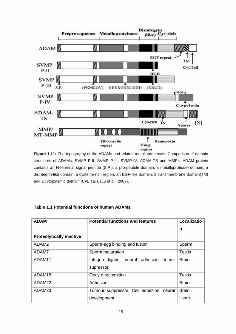
Table 1.1 Potential functions of human ADAMs
ADAM Potential functions and features Localizatio
n
Proteolytically inactive
ADAM2 Sperm-egg binding and fusion Sperm
ADAM7 Sperm maturation Testis
ADAM11 Integrin ligand, neural adhesion, tumor
supressor
Brain
ADAM18 Oocyte recognition Testis
ADAM22 Adhesion Brain
ADAM23 Tumour suppressor, Cell adhesion, neural
development
Brain,
Heart
20
ADAM29 Unknown Testis
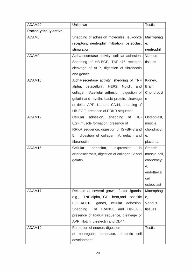
Proteolytically active
ADAM8 Shedding of adhesion molecules, leukocyte
receptors, neutrophil infiltration, osteoclast
stimulation
Macrophag
e,
neutrophil
ADAM9 Alpha-secretase activity, cellular adhesion,
Shedding of HB-EGF, TNF-p75 receptor,
cleavage of APP, digestion of fibronectin
and gelatin,
Various
tissues
ADAM10 Alpha-secretase activity, shedding of TNF
alpha, betacellulin, HER2, Notch, and
collagen IV,cellular adhesion, digestion of
gelatin and myelin, basic protein, cleavage
of delta, APP, L1, and CD44, shedding of
HB-EGF, presence of RRKR sequence.
Kidney,
Brain,
Chondrocyt
e
ADAM12 Cellular adhesion, shedding of HB-
EGF,muscle formation, presence of
RRKR sequence, digestion of IGFBP-3 and
5, digestion of collagen IV, gelatin and
fibronectin
Osteoblast,
muscle,
chondrocyt
e,
placenta
ADAM15 Cellular adhesion, expression in
arteriosclerosis, digestion of collagen IV and
gelatin
Smooth
muscle cell,
chondrocyt
e,
endothelial
cell,
osteoclast
ADAM17 Release of several growth factor ligands,
e.g., TNF-alpha,TGF beta,and specific
EGFR/HER ligands, cellular adhesion,
Shedding of TRANCE and HB-EGF,
presence of RRKR sequence, cleavage of
APP, Notch, L-selectin and CD44
Macrophag
e,
Various
tissues
ADAM19 Formation of neuron, digestion
of neuregulin, sheddase, dendritic cell
development.
Testis
21
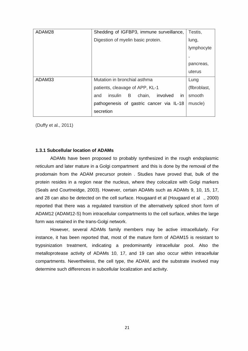
ADAM28 Shedding of IGFBP3, immune surveillance,
Digestion of myelin basic protein.
Testis,
lung,
lymphocyte
,
pancreas,
uterus
ADAM33 Mutation in bronchial asthma
patients, cleavage of APP, KL-1
and insulin B chain, involved in
pathogenesis of gastric cancer via IL-18
secretion
Lung
(flbroblast,
smooth
muscle)
(Duffy et al., 2011)
1.3.1 Subcellular location of ADAMs
ADAMs have been proposed to probably synthesized in the rough endoplasmic
reticulum and later mature in a Golgi compartment and this is done by the removal of the
prodomain from the ADAM precursor protein . Studies have proved that, bulk of the
protein resides in a region near the nucleus, where they colocalize with Golgi markers
(Seals and Courtneidge, 2003). However, certain ADAMs such as ADAMs 9, 10, 15, 17,
and 28 can also be detected on the cell surface. Hougaard et al (Hougaard et al ., 2000)
reported that there was a regulated transition of the alternatively spliced short form of
ADAM12 (ADAM12-S) from intracellular compartments to the cell surface, whiles the large
form was retained in the trans-Golgi network.
However, several ADAMs family members may be active intracellularly. For
instance, it has been reported that, most of the mature form of ADAM15 is resistant to
trypsinization treatment, indicating a predominantly intracellular pool. Also the
metalloprotease activity of ADAMs 10, 17, and 19 can also occur within intracellular
compartments. Nevertheless, the cell type, the ADAM, and the substrate involved may
determine such differences in subcellular localization and activity.

22
Figure 1.12: A schematic overview of ADAMs synthesis, processing, and function.
(Seals and Courtneidge, 2003).
1.3.2 Structure and Domain activity of ADAM Proteins
The generalised structure of an ADAM protein contains 8 distinct domains or
regions. In the typical ADAM protein, these domains are a signal domain, a prodomain, a
metalloproteinase domain, a disintegrin or integrin-binding domain, a cysteine rich
region, an EGF (epidermal growth factor)-like domain, a transmembrane sequence and
an intracellular C-terminal end (McGowan et al., 2007). Like most proteases, the ADAMs
are initially synthesised as enzymatically-inactive precursor proteins.
23
1.3.2.1 The prodomain
At the N terminus of ADAMs recides a signal sequence that directs ADAMs into
the secretory pathway and a prodomain that lead to ADAMs maturation. (Seals and
Courtneidge, 2003). The metalloprotease site of ADAMs are kept inactive, through a
cysteine switch by the prodomain .The conserved cysteine residue located within the
prodomain preferentially coordinates the required active site zinc atom, and thereby
sequesters the metalloprotease domain in an inactive conformation (Edwards et al.,
2008; Becker et al., 1995) . For protease activation, this prodomain is removed by a furin-
like convertase or by autocatalysis, depending on the specific ADAM (Murphy, 2008). The
mechanism employed is the cleavage of the prodomain from the rest of the protein by
proprotein convertases (PCs) at a conserved Rx(R/K)R motif, thereby effectively releasing
the prodomain and switching the zinc coordination to the metalloprotease domain,
enabling it to undertake its catalytic activity. Several studies support this mechanism
(Seals and Courtneidge, 2003).
Aside this mechanisms, there are cases in which ADAMs may undergo
autocatalytic activation. Example is when, activity-blocking mutations in the
metalloprotease domains of ADAM8 and ADAM28, produces only the precursor form of
the protein in transfected cells (Schlomann et al., 2002). Another functional aspect of the
prodomain is to chaperone the proper folding of the metalloprotease domain of ADAMs. It
has been suggested by studies that the removal of the prodomain of ADAM17 generates
a protease-inactive protein (Milla et al., 1999). Similarly, an ADAM10 construct lacking its
prodomain is catalytically inactive in vivo. Hence, it can be concluded from this evidence
that, the prodomain appears to be necessary in assisting in the proper folding of ADAMs,
in the structuring of the catalytic active site, and in the proper transit of ADAMs throughout
the secretory pathway (Seals and Courtneidge, 2003).
1.3.2.2 The metalloprotease domain This domains are well conserved, but then, only 25 out of a known total of 40
members of the family (ADAMs 1, 8-10, 12, 13, 15, 16, 17, 19-21, 24-26, 28, 30, and 33-
40), have the zinc binding catalytic site consensus sequence of HEXXHXXGXXHD (single
letter amino acid code) where X is stands for any other amino acid; the three H residues
bind zinc, the G allows a turn and E constitutes the catalytic residue, comprised of a water
molecule tetrahedrally coordinated to the zinc, and the E residue acting as a catalytic
base (Lu et al., 2007).
24
Metalloprotease domain is located next to prodomain and only about 60% of
ADAMs exhibit nonprotease activity, although they all posses this domain. On the basis of
their structural definition, of the 21 human ADAMs identified, only 13 are proteolytically
active. ADAMs shown to exhibit protease activity include ADAM8, 9, 10, 12, 15, 17, 19, 28
and 33 (Kawaguchi et al., 2002). The mechanism of the proteolytic activity, which is the
best defined function of ADAMs currently, has been more accurately evaluated through
crystallization of the metalloprotease domain. At the active site is zinc and water atoms
necessary for the hydrolytic processing of protein substrates, and which are coordinated
by three conserved histidine residues and a downstream methionine. The methionine lies
in a Met-turn motif that loops around to face the consensus HExxHxxGxxH site. Although
individual proteins among the various metzincins exhibit certain distinguishable structural
features that may impart specificity for substrates and protease inhibitors, there exist
remarkable conservation within this catalytic site (Seals and Courtneidge, 2003).
1.3.2.3 The disintegrin domain The disintegrin-like domain located at the downstream of the metalloprotease
domain, consists of 60 to 90 amino acid long with 6 to 15 Cys residues with sequence
similarity to that of the snake venom disintegrins (Marcinkiewicz C, 2005). This sequence
is found in all ADAMs and they binds to integrins, which are a group of adhesion proteins
implicated in cell adhesion, migration and cell signalling (Stupack, 2007) . Snake venom
disintegrins , which confers the ability of these molecules to interact with integrins in
different cell systems have been characterized as potent inhibitors of the function of
various integrins. The disulfide bridge pattern of RGD-containing disintegrins, which may
be important to their biological activity, especially their potency and selectivity has been
determined by means of chemical methods, NMR spectroscopy and crystallography. Not
all ADAMs have the RGD sequence and in place of that, others contain sequences such
as KGD, MVD, MLD, VGD, ECD, or MDG (single letter amino acid code). (Lu et al., 2007)
ADAMs distegrin derived its name for its presence in the snake venom
metalloproteases (SVMPs), involved in binding of platelet integrin receptors.
Consequently,the binding to this receptors, prevents the association of platelets with their
natural ligands such as fibrinogen, and results in a block in platelet aggregation at the
wound site. This disintegrin-mediated interaction of SVMPs along with the breakdown of
basement membrane components by their metalloprotease activity leads to the severe
hemorrhaging caused by bites from snakes harboring these toxins (Seals and
Courtneidge, 2003). Although, the disintegrin domain has been widely described as being

25
able to interact with integrin molecules and therefore mediating cell-cell and cell-matrix
interactions (Reiss et al., 2006), it has been shown not to be available for protein binding
due to protein folding (Takeda et al., 2006). It may be therefore considered as a structural
feature rather than an integrin ligand.
1.3.2.4 The cysteine-rich and EGF-like domains The Cys-rich and EGF-like domains, though not much is known about them may
play very important role for interactions of ADAMs with other proteins such as chaperones
involved in biosynthesis and or with other partners on the cell surface. Structurally, the two
domain consist of about 160 amino acid with 10 to 14 Cys residues and about 40 amino
acid with 6 Cys residues, respectively. EGF domains have been indicated to be found in
proteins that are either completely secreted or have transmembrane regions that tether
the protein to the cell surface (Lu et al., 2007). On the other hand, the Cys-rich domain is
located at the carboxy terminal end of ADAMs, which has been considered likely to
complements the binding capacity of the disintegrin-like domain and imparts specificity to
disintegrin domain-mediated interactions (Emi et al., 1993). The Cys-rich domain of
TACE/ADAM-17 may play a role in the release of the pro-domain and may be required for
the shedding of interleukin 1 receptor type II as well aid in as well the recruitment of
accessory proteins involved in targeting TACE to some substrates (Reddy et al., 2000)
Several other fuctions have been reported for Cystein-rich domain as follows:
It has been indicated to be involved in cell-cell fusion (Huovila et al., 1996), regulating
protease activity and controlling substrate specificity (Reiss et al., 2006), function as a
ligand for the cell-adhesion molecule syndecan, especially syndecan-4 (Iba et al., 1999,
2000).
1.3.2.5 Transmembrane domain Mostly, ADAMs belong to the type I membrane proteins, anchored through a
transmembrane (TM) domain near the C-terminus and also have an alternatively spliced
form that diverges before the TM domain, leading to the production of a soluble, secreted
form. All of the ADAMTSs lack a TM domain and are therefore termed as secreted
proteases. Examples of ADAMs with transmembarne domain include 11, 12, 17, and 28
(Cerretti et al., 1999; Lu et al., 2007).
The ability to posses both soluble and membrane-anchored forms, allows ADAMs
to regulate events not only on or near the cell surface, but also at a distance from cells.
26
However, very little is known as to whether all the membrane-anchored ADAMs have a
soluble counterpart generated through either alternative splicing or shedding from the cell
surface. (Cerretti et al., 1999; Lu et al., 2007).
1.3.2.6 The cytoplasmic tail
ADAMs have unusual cytoplasm tail, with many rich in proline, serine, glutamic
acid and or lysine. They are highly variable both in length ( between 40 to 250 amino acid)
and in sequence and contains a phosphorylation sites and SH3 binding domains .This
domain contains noticeable and specialized motifs that have been postulated to be
involved in the inside-out regulation of metalloprotease activity, the outside-in regulation of
cell signaling, and or the control of maturation and subcellular localization (Lu et al., 2007;
Seals and Courtneidge, 2003). PxxP binding sites for SH3 domain-containing proteins
are the most notable motifs which can be found in human ADAMs 7, 8, 9, 10, 12, 15, 17,
19, 22, 29, and 33. Several ADAMs also have potential phosphorylation sites for serine-
threonine and/or tyrosine kinases. Not only might this regulate ADAM function directly, but
the resulting phosphotyrosine residues could also provide ligands for SH2 domain-
containing proteins (Seals and Courtneidge, 2003).
In a study, ADAM9 binds to endophilin I and SH3PX1 (Howard et al., 1999). Given
the potential function of endophilin I and SH3PX1 in vesicle sorting, it is speculated that
these interactions may play a part in the regulation of ADAM maturation and or subcellular
localization. Also it was reported that the membrane proximal region of the tail of ADAM9
associates with the catalytic domain of protein kinase C-alpha (PKC-alpha) (Izumi et al.,
1998). The tail of ADAM9 can be phosphorylated by PKC in vitro at one or more serine
and threonine residues. It therefore speculated that PKC-alpha helps to recruit ADAM9 to
specific sites on the plasma membrane and upon phosphorylation or activation of ADAM9,
shedding of HB-EGF occurs (Seals and Courtneidge, 2003).
1.3.3 Mechanisms by which ADAMs play a role in cancer
There are several different mechanisms through which ADAMs promote cancer
formation and progression. Some of these processes includes Cell proliferation,
Angiogenesis and Apoptosis.
27
1.3.3.1 Cell Proliferation Cell proliferation by ADAMs can occur through the following processes:
1.3.3.1.1 Activation of positively-stimulating pathways
Several proteolytically active ADAMs regulate cell proliferation by cleaving growth
factors or cell surface proteins or by activation of positively-stimulating growth factors.
Many of these growth factors are first and foremost synthesised as inactive
transmembrane precursor proteins that require ectodomain shedding for activation in
order to fuction at its exert maximun capacity. Ligands for several growth factor receptors
are processed by ADAM family members and amongst the best-studied growth-
stimulating factors that are activated by ADAMs are the EGFR/HER family of ligands
(EGF receptor ligands (heparin-binding EGF (HB-EGF), amphiregulin, betacellulin,
epiregulin) with primary conversion mediated by either ADAM10 or ADAM17 (Duffy et al.,
2011). However, other ADAMs such as ADAM8, 9, 12, 17 and 19 can also activate one or
more of these ligands (Horiuchi et al., 2007) .
What actually happens is that, the shed form of the ligands binds to one or more of
the EGFR/HER family of receptors. Upon homo- or heterodimerisation, several different
pathways including the mitogenactivated protein kinase (MAPK) pathway, the
phosphatidylinositol 3-kinase (PI3K) pathway and janus kinase/signal transducer and
activator of transcriptional (JAK/STAT) pathway, are activated by downstream signalling
which can results in some of the classical hall markers of malignancy such as enhanced
cell proliferation, increased cell motility and increased cell survival (Peeters et al., 2009;
Duffy et al., 2011).

28
Figure 1.13: Mode of action of ADAMs in the activation of EGFR/HER receptor signalling ADAMs are involved in proteolytic ectodomain shedding of membrane bound ligands. The
released ligands (EGF, HB-EGF, TGFa, heregulins) are free to bind to and activate EGFR, HER3
and HER4. Following receptor dimerisation , downstream signalling through many pathways is
activated, including MAPK, PI3K and JAK/STAT. (Duffy MJ et al., 2011).
In a work reported by Singh et al it was showed that UV irradiation of skin cancer
cells activated ADAM9 and 17 which was followed by amphiregulin shedding, EGFR
transactivation and increased cell proliferation. ADAM-10 also contributes to cell
proliferation by modulating b-catenin signalling through E-cadherin shedding and
increasing gene cyclin D1 levels (Shtutman et al., 1999).
1.3.3.1.2 Inactivation of growth-inhibitory pathways
Inactivation of growth-inhibitory pathways has been indicated in TGFb which
signals via TGFbR1 and TGFbR. It has been proposed that, in normal and early
malignant cells, TGFb inhibits proliferation where as in progressive malignancy, TGFb
promotes proliferation (Ikushima and Miyazono, 2010 ; Duffy et al., 2011). ADAM17 was
reported to mediate shedding of the type 1 TGFb receptor, thereby drcreasing TGFb
signalling which led to decreased growth inhibition and the reduction in growth inhibition
complements the growth stimulation, resulting from increased release of the EGFR/HER
ligands (Liu et al., 2009; Duffy et al., 2011).
29
1.3.3.1.3. Shedding of adhesion proteins One of the means of increased cell proliferation is by means of shedding of
adhesion proteins by ADAMs. For example ADAM10 appears to be the major sheddase
for the release of EGF and betacellulin (Sahin et al., 2004, 2007 ) and also contributes to
E-Cadherin shaddase (Ito et al., 1999) . The sunsequent release of soluble E-cadherin in
the extracellular milieu leads to the abrogation of cell-cell contacts, thereby facilitating cell
migration. ADAM-10 also contributes to cell proliferation by modulating b-catenin
signalling through E-cadherin shedding and increasing gene cyclin D1 levels (Shtutman et
al., 1999) Najy et al also reported that ADAM15-mediated shed form of cadherin E bound
to and activated HER2 in breast cancer cells. The increased proliferation and migration
observed was attributed to the fact, the shed form of cadherin E formed a complex with
HER2 and HER3, resulting in enhanced ERK signalling.
Apart from increased cell proliferation, this shedding might also be expected to
weaken cell:cell interaction and thus allow dissociation of potential invasive and metastatic
cells in the primary cancer which could potentially place a malignant cell or group of cells
on their pathway to metastais. Shedding of other adhesion proteins such as L-selectin,
ICAM-1 or VCAM, on the other hand, might be expected to modulate binding of tumour
cells to the vasculature wall and thus play a role in the intravasation (Duffy et al., 2011)
1.3.3.2 Angiogenesis
Cancer growth and metastasis can be mediated by angiogenesis which consists of
the formation of new blood vessels devoted to vascularise the tumour tissue. This process
is essential for tumours to grow beyond approximately 2 mm in diameter. (Duffy et al.,
2011). Angiogenesis process is under the dependence of a balance of pro- and
antiangiogenic factors (Bajou et al., 2004). Proteinases in general, have been initially
considered as positive regulators of angiogenesis but recent studies have evidenced
complex and sometimes opposite roles of MMPs, ADAMs and ADAMTSs in regulating
tumoral angiogenesis (Handsley and Edwards, 2005). However, several evidences have
also proved that ADAMs may promote cancer growth and metastasis through this
process. Some of these evidences are: Pulmonary hypovascularisation in mice expressed
catalytically inactive ADAM17 (Zhao et al., 2001), and the deletion of ADAM17 resulted in
pathological neovascularisation and reduced growth of injected tumour cells in a mouse
model (Weskamp et al., 2010).
30
Some studies carried out has indicated the recombinant disintegrin domain (RDD)
of ADAM-15 as a potent inhibitor of angiogenesis. ADAM-15 RDD induces a reduction of
MDA-MB-231 tumour growth associated with less tumour vascularization in vivo.
Transgenic B16F10 melanoma cells form less metastasis in mouse lungs after turning on
RDD expression (Trochon-Joseph et al., 2004). It has been proposed that mechanisms
implicating ADAM-15 in the regulation of angiogenesis could be related to the presence of
Arg-Gly-Asp (RGD) sequence in the disintegrin domain which binds integrins. In addition,
certain ADAM proteinases have been indicated to control cell apoptosis. In a mammary
cancer model induced by the expression of polyoma middle T oncoprotein, ADAM-12 has
been shown to increase stromal cell apoptosis and decrease tumour cell apoptosis
(Kveiborg et al, 2005) ADAM-10 knock-out (KO) embryos suffer from cell growth arrest
and apoptosis associated with an overexpression of full-length E-cadherin (Maretzky et
al., 2005).
1.3.4 Evidence of a Role for ADAMs in Cancer and their potential use as Biomarkers
Based on the ability of ADAMs to release ligands which is capable of stimulating
cell proliferation as well as migration , several studies from cell lines grown in culture,
animal models and human malignancies suggest that a number of ADAMs in cancer
formation. The most established ones include ADAM9, ADAM10, ADAM12, ADAM15 and
ADAM17 are involved in cancer formation and/or progression of which the strongest
evidence for a role in malignancy exists for ADAM17 (Duffy et al., 2009) .
Several studies have shown that increased expression of certain ADAMs
enhanced in vitro invasion, proliferation and promoted tumour formation in vivo (McGowan
et al., 2007; Borrell-Pages et al., 2003), while decreased expression reduced these
processes. It has been shown that deficiency of specific ADAMs such as ADAM9, 15 and
17 resulted in decreased growth of heterotopically injected tumour cells in mice models
(Guaiquil et al., 2009). Biomarkers are potentially useful in cancer detection, prognosis
assessment, and predicting therapy outcome or likely resistance to therapy as well as
monitoring ongoing therapy.
31
Figure 1.14: An overview of a disintegrin and metalloproteinases (ADAM) in cancer biology.
Five different pathways may be involved in ADAM mediated cancer cell proliferation and
progression. (1) ProADAMs are activated by furin or matrix metalloproteinases (MMPs). (2)
Sheddase activity of ADAMs is stimulated by external factors (e.g. 12-Otetradecanoylphorbol-13-
acetate [TPA]), leading to shedding of cell surface ligands such as heparinbinding- epidermal
growth factor (HP-EGF) and transforming growth factor (TGF)-α. This process perhaps involves
protein kinase C (PKC) and mitogenactivated protein kinase (MAPK) pathways. Then, soluble
growth factors such as HP-EGF activate epidermal growth factor receptor on the cells in autocrine
and paracrine manners. (3) The interaction of the disintegrin and cysteine-rich domains of ADAM
with integrins or syndecans on the cells may help them to cleave the substrates (e.g. extracellular
matrix [ECM]). (4) ADAMs modulate extracellular matrix–integrin interactions, and thus they can
indirectly promote proliferation signals through integrins. (5) ADAM may process other
undetermined membrane-anchored molecules such as chemokines, cytokines and their receptors,
which are related to cancer cell proliferation and progression. CR, cysteine-rich domain; CT,
cytosolic tail; Dis, disintegrin domain; E, epidermal growth factor-like domain; MP,
metalloproteinase domain; Pro, propeptide domain; TM, transmembrane domain.
( Mochizuki S and Okada Y, 2007)
32
1.3.5 Contribution of ADAMs in different types of cancer as diagnostic marker 1.3.5.1 Lung cancer
It has been proved in several studies that dysregulation of the production of
several ADAMs leads to lung cancer. Some of the have been indicated here. Primary
bronchial epithelial cells and bronchial cell lines exposed to smoke components showed
an increased proliferation rate associated with EGFR phosphorylation with possible
mediation by ADAM-17 which can activate several EGFR ligands (Lemjabbar et al., 2003).
Also ADAM17 was reported to be upregulated in non-small cell lung carcinoma (NSCLC),
with possible heregulin3 (HER3) signalling (Zhou et al., 2006).
Another example is ADAM-8, which was strongly expressed in NSCLC by tissue
microarray analysis, and correlates with clinical stage of the disease (Ishikawa et al.,
2004). Also, ADAM-9 mRNA and protein expression levels are enhanced in EBC-1 lung
cancer cell line displaying a tropism for brain metastasis as compared to parent EBC-1 or
EBC-1 cell line with a tropism for bone tissue (Shintani et al., 2004).
ADAM-12 mRNA and protein levels was reported to increase in NSCLC when
compared to non-cancerous tissues (Rocks et al., 2006) ADAM-15 was reported to be
expressed in both small cell lung carcinoma (SCLC) and NSCLC cell lines with higher
expression in tumoral cells than in normal epithelial cells of pulmonary tumours (Schutz et
al., 2005).
ADAM28 was one of the first ADAMs shown to be elevated in serum from patients with
cancer (Kuroda et al., 2010). ADAM-28, cleaved insulin-like growth factor binding protein-
3 (IGFBP- 3), and was found to be about 16-fold over-expressed in NSCLC (Mochizuki
et al., 2007).
1.3.5.2 Brain tumours
ADAM-22 , restricted to the brain is implicated in cell-cell and cell-matrix
interactions through their binding to integrins and extracellular matrix and may be
involved in neural development (Sagane et al., 1998). Cytoplasmic variants of ADAM-22
have been indicated to have been expressed differently in normal human brain tissue and
gliomas (Harada et al., 2000), which gives evidence of ADAM genes expression in brain
tumours. Brain tumour cell lines cultured under hypoxic conditions demonstrated an
upregulation of ADAM-17 expression levels, and its activity correlated with increased
tumour cell invasion (Zheng et al., 2007). Also, ADAM-8 and ADAM-19 mRNA are
upregulated in primary brain tumours and their expression and activity are correlated with
33
invasiveness of glioma cells (Wildeboer et al., 2006). There was an overexpression of the
membrane- bound ADAM-12 variant in glioblastomas (Kodama et al., 2004). However,
treatment of cultured glioblastoma cells with an ADAM-12 inhibitor decreased the
production of the mature HB-EGF indicating its role in HB-EGF signalling pathway in
those cells.
1.3.5.3 Prostate cancer There is a possiblity of the existence of both androgen-independent and androgen
dependent cases in prostate cancer though early stages involve androgen-dependent
(Bertram et al., 2006). Androgen-dependency has been suggested to interfere with
ADAM-related regulation processes since the mRNA expression of several ADAMs is
regulated by androgens (McCulloch et al., 2000). Androgen or serum starvation enhances
ADAM-9 protein expression in androgen receptor- positive prostate cancer cells
(Shigemura et al., 2007). Peduto et al. reported that well-differentiated prostate cancers
developed in ADAM-9–deficient mice compared with poorly differentiated tumors in control
mice expressing ADAM-9 . They also showed that overexpression of ADAM-9 in mouse
prostate epithelial cells gave rise to epithelial hyperplasia and prostate intraepithelial
neoplasia,a putative precursor lesion for prostate cancer because of its ability to cleave
EGFR ligands and the receptor for fibroblast growth factor (Peduto et al., 2005).
In studies on prostate cancer cell lines in culture,overexpression of ADAM-9 was
found to be associated with the conversion of LNCaP cells to an androgen-independent
and metastatic state (Sung et al., 2008). In prostate carcinoma, ADAM-9 levels were
significantly associated with prostate-specific antigen (PSA)relapse-free survival
(Fritzsche et al., 2008). ADAM-8 protein expression has been demonstrated to be
significantly associated with higher cancer stages including positive nodal status, and
higher Gleason scores (Fritzsche et al., 2006). Najy et al. also found that down-
regulation of ADAM-15 in the prostate cancer cell line PC3 decreased migration and
adhesion to specific extracellular protein matrix proteins, such as fibronectin, vitronectin,
and laminin.
1.3.5.4 Liver cancer In activated hepatic stellate cells, TGF-b1 induces ADAM-12 expression which
might also participate in tumour progression (Le Pabic et al., 2005). This suggest that,
ADAM-12 might contribute to growth inhibitory signalling in normal epithelial cells which is
lost during tumour progression. Several studies have indicated ADAM-17 as a
34
contributing factor to EGFR-ligand release and induction of cell proliferation and invasion
(Rocks et al., 2008). In a study, it was reported that ADAM-17 mRNA levels were higher in
hepatocellular carcinomas than in paired non-cancerous liver tissues suggesting that this
proteinase might be implicated in tumour invasiveness by either activating EGFR by
amphiregulin (Lemjabbar et al., 2003) or TGFalpha (Borrell-Pages et al., 2003). ADAM-9
in one study has been reported to promote invasiveness of liver metastatic carcinoma
cells by degrading basement membrane components such as laminin-1 (Mazzocca et al.,
2005).
1.3.5.5 Breast cancer There has been several reports of ADAMs implications in breast cancer patients.
One of the first ADAMs shown to have diagnostic potential was ADAM12 in breast cancer.
ADAM- 12, an apoptosis-modulating gene accelerated the development of tumour by
delaying tumour cell apoptosis by the overexpression of its soluble form lacking the
cytoplasmic tail (secreted splice variant of ADAM-12) (Kveiborg et al., 2005; Rocks et al.,
2008). In urine, using Western blotting ,ADAM-12 levels were enhanced in breast cancer
patients vis-à-vis a healthy control group which suggest a potentially important non-
invasive biomarker in breast cancer (Roy et al., 2004).
Using logistic regression analysis, the authors calculated that the predictive
probability of the presence of breast cancer was ≥ 80%, when levels of ADAM12
exceeded 40 arbitrary units (Roy et al., 2004). In a follow-up study to above, Pories et al
found that urinary ADAM12 levels were also increased in women with putative
premalignant lesions of invasive breast cancer such as atypical hyperplasia and lobular
carcinoma in situ, compared to levels in healthy controls. This finding, if confirmed,
suggests that measurement of ADAM12 in urine could identify women at increase risk of
developing breast cancer. In the PyMT mouse model, overexpression of ADAM-12 was
found to promote breast cancer progression (Kveiborg et al., 2005). Also, expression of
both ADAM12 isoforms was found to be significantly elevated in human malignant breast
tissue overexpression which resulted in increased tumor take, tumor size, and metastasis
in vivo. Of the two isoforms, only the secreted isoform, ADAM12-S, enhanced the ability of
tumor cells to migrate and invade in vitro and resulted in a higher incidence of local and
distant metastasis in vivo (Roy et al., 2004 ).
In vitro studies have shown that overexpression of ADAM- 17 in breast cancer
cells increases invasion and proliferation (McGowan et al., 2007) and targeting it, reverts
the malignant phenotype by preventing shedding of TGF-a and amphiregulin (Kenny and
Bissell, 2007). Active ADAM-28 was found to be overexpressed in breast cancinoma cells
,contributing to the regulation of cell proliferation through IGFBP-3 cleavage, enhancing
35
the bioavailability of IGF-I (Mitsui et al., 2006). Alternative splicing could also be an
important tool used by cancer cells to acquire an invasive phenotype. For example,
different isoforms of ADAM-9 proteins and ADAM-15 mRNA have been detected in breast
cancer cells (Ortiz et al., 2004) which calls for attention to set up a powerful diagnostic tool
by studying the differential production of ADAM-9 or -15 domains. In breast cancer,
ADAM-9 expression was significantly higher in node-positive than node-negative primary
cancers whereas the active form of ADAM-17 was increased in high-grade versus
lowgrade tumors (McGowan et al., 2007) .
In vivo, loss of ADAM-15 decreased metastasis to bone and using breast cancer
cell lines,it was reported that ADAM-15 cleaved cadherin E after growth factor deprivation
(Najy et al., 2008). In another study,it was shown that the human breast cancer cell lines,
MCF-7 and MDA-MB453, strongly express ADAM 9, 12, and 17, whereas ADAM 10 and
15 were expressed at a lower level, indicating a putative pathophysiological role of these
ADAMs in breast cancer biology (Lendeckel et al., 2005). ADAM17 processed through
major histocompatibility complex (MHC) class I molecules was showned to be expressed
in breast, ovarian and prostate cancer ,making it a potential immunotherapeutic target in
these cancers. (Sinnathamby et al., 2011).
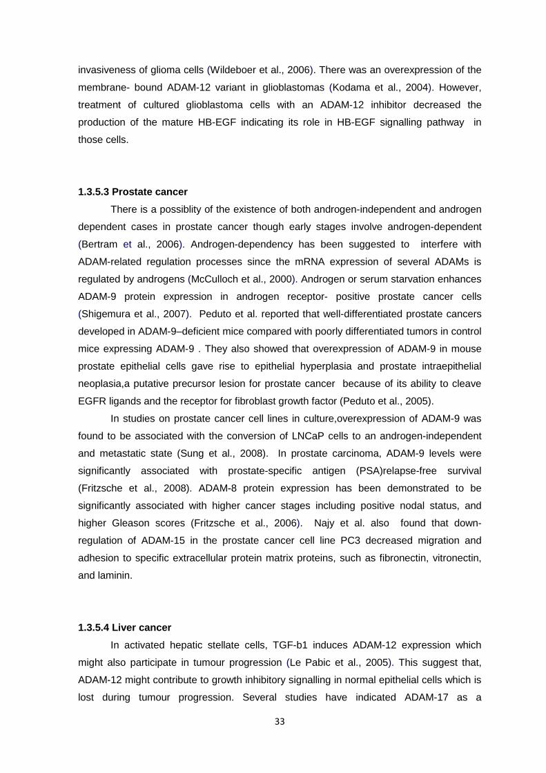
Figure 1.15 Prospective role of ADAMs in breast carcinoma cell proliferation.
ADAM28 is overexpressed as active forms in breast carcinoma cells. ADAM28 cleaves insulin-like
growth factor binding protein-3 (IGFBP-3) and releases insulin-like growth factor-I (IGF-I) through
the IGF-I–IGFBP-3 complex. IGF-I induces cell proliferation through phosphorylation of the IGF
type I receptor (IGF-IR) and extracellular signal-regulated kinase 1/2 (ERK1/2). IGFBP-3 cleavage
can be inhibited by treatment with anti-ADAM28 antibody or an ADAM inhibitor, KB-R7785, as well
as ADAM28 small interfering RNA (siRNA) (Mochizuki S and Okada Y, 2007)
36
1.3.5.6 Gastric and colon carcinoma In vivo, ADAM-10 and -17 are overexpressed in antral mucosa during H. pylori
infection and it has been established that , ADAM-9, -10, -12, -15, and -17 are increased
in gastric tumours (Carl-McGrath et al., 2005). ADAM-10, which is found to be
overexpressed in vitro after gastric cell infection, could establish a link between
Helicobacter pylori-induced inflammation and carcinogenesis in stomach and it acts
through EGFR ligand shedding leading to gastric cell proliferation (Joy et al., 2005). In
colon carcinomas, ADAM-17 is overexpressed independently of tumour stage or grade
and is involved in tumour growth and angiogenesis possibly via an autocrine/paracrine
pathway implicating EGFR (Blanchot-Jossic et al., 2005). ADAM-9 is also overexpressed
in a colon cell line and is co-localized with E-cadherin suggesting a potential role in E-
Cadherin-mediated metastasis. It has been showed that a soluble form of ADAM-9
secreted by hepatic stellate cells promoted colon cancer cell invasion in vitro. (Mazzocca
et al., 2005).
1.3.5.7 Kidney, bladder carcinoma Inhibition of ADAM-17 by a dominant negative ADAM-17 mutant prevents pro-HB-
EGF cleavage, EGFR activation and cell proliferation in kidney carcinoma cells ,indicating
the importance of EGFR signalling in the development of kidney cancer since (Schafer et
al., 2004). ADAM-12 mRNA was found to be overexpressed in bladder cancer and its
levels correlated with disease stage. In another study, the levels of ADAM-12 was also
found to be higher in the urine from patients with bladder cancer compared with healthy
control subjects (Frohlich et al., 2006) and concentrations tended to be higher in those
with the largest invasive tumours. In some cases , urinary ADAM12 levels decreased
following surgical removal of the bladder cancer but increased again with recurrent
disease (Fröhlich et al., 2006) which suggesting the propability of using the
measurement of urinary ADAM12 for monitoring patients with bladder cancer.
1.3.5.8 Pancreatic carcinoma Some of the ADAMs implicated in pancratic cancinoma are ADAM-9, -10 and -17
but are restricted to specific compartments. Analysis of mRNA expression levels in
microdissected cancer samples shows an overexpression of ADAM-9 and -15 proteinases
in pancreatic tumour cells (Rocks et al., 2008). ADAM-17 was once again overexpressed
in all pancreatic ductal adenocarcinoma (PDAC) and pancreatic cancer cell lines.
(Yamada et al., 2007).

37
1.3.5.9 Squamous Cell Carcinoma This G-protein which are known to activate ADAMs can also transactivate
epidermal growth factor receptor (EGFR) (Ohtsu et al., 2006). For example, ultraviolet
(UV) radiation of skin cancer cells activates ADAMs and induces EGFR ligand shedding
and EGFR transactivation (Singh et al., 2009). One mechanism for this process is the
likelihood that, the UV irradiation induced reactive oxygen species (ROS) generation,
which in turn activated ADAM9 and ADAM17, and finally cleaving EGFR ligands,
particularly AR. Skin cancer proliferation can be induced by the binding of the soluble
form of AR to EGFR. Overexpression of protein kinase Cε (PKCε) in mouse epidermis
which resulted in the rapid development of papilloma independent metastatic SCCs via
the two-stage model of carcinogenesis have also been reported (Wheeler et al., 2003).
PKCε transgenic mice have elevated serum TNF-α levels during skin tumor promotion by
12- O-tetradecanoylphorbol-13-acetate (TPA).
Since TNF-α is linked to skin tumor promotion by TPA, this increase may be linked
to the development of metastatic SCC. TPA stimulated shedding of TNF-α could be
completely prevented in PKCε transgenic mice and isolated keratinocytes by an ADAM17
inhibitor, TAPI-1. These results indicate that PKCε signal transduction pathways to TPA-
stimulated TNF-α ectodomain shedding are mediated by ADAM17. Injection of a TNF-α
synthesis inhibitor during skin tumor promotion completely prevented the development of
metastatic sqamous cell carcinoma in PKCε transgenic mice (Wheeler et al., 2003).
1.3.5.10 Basal Cell Carcinoma.
The most common type of skin tumor is Basal Cell Carcinoma which rarely
metastasizes but is locally invasive and highly destructive. ASAMs implicated in this
disease are ADAM10, 12, and 17 . Compared with central areas of basal cell carcinoma
tumor cell nests,these ADAMs increased at the peripheral tumor margin . Expression of
ADAM10 and ADAM12 is increased in the deep margin of invading tumor cell nests. In
contrast, ADAM17 is increased in superficial basal cell carcinoma. All the three ADAMs
showed different expression patterns in basal cell carcinoma histologic subtypes,
indicating their different roles in the pathogenesis of BCC (Oh et al., 2009).
1.3.5.11 Malignant Melanoma
ADAM9, ADAM10, ADAM12, ADAM15, ADAM17, HB-EGF and TGF-α are
overexpressed in more than 1 malignant melanoma (MM) cell lines which can induced
the migration of MM cells. (Singh et al., 2009). In ADAM10, the expression was

38
significantly elevated in melanoma metastases compared with primary melanomas (Lee et
al., 2010). Expression of several components of the Notch pathway, which can be cleaved
by ADAM10, were also upregulated in malignant melanoma compared with common
melanocytic nevi (Massi et al., 2006). Down-regulation of ADAM10 with specific siRNA
resulted in the suppression of cell growth and the reduced migration of MM cells .
ADAM10 has also been implicated in constitutive CD44 cleavage from malignanat
melanoma cells, and its expression can impair tumor cell proliferation (Anderegg et al.,
2009). In malignant melanoma cells, both ADAM10 and ADAM17 are significantly
expressed in histological sections but only ADAM10 is involved in the constitutive
shedding of native CD44 from this cells. (Ahrens et al., 2001), ADAM9 is detected in
malignant melanoma cells and in peritumoral stromal fibroblasts, while it is absent in
fibroblasts distal to the tumor site. In contrast, in nevi, ADAM9 expression is absent both
in nevus cells and in stromal cells close to nevus cell nests (Zigrino et al., 2005).
1.3.6 ADAMs as prognostic markers
With increased ADAMs involvement in cancer proliferation and progression,
several studies have investigated for their potential prognostic impact in patients with
cancer and it has been indicated that ,there exist a correlations between levels of
specific ADAMs and parameters of tumour progresion (eg. tumour size, grade, metastasis
to local lymph nodes and patient outcome) in human cancers (Valkovskaya et al., 2007).
These markers are important in the management of patients with cancer as they help
avoid the overtreatment of indolent disease and undertreatment of aggressive cases,
especially in the cases of breast and prostate cancer. It has been indicated that, none of
the available serum markers for breast cancer are increased in patients with early disease
and are thus of little value in identifying women at increased risk of developing this
malignancy (Duffy, 2006). In breast cancer cases, prognostic markers may help identify
those patients whose prognosis is so good that they are unlikely to benefit from receiving
adjuvant chemotherapy whiles at the same time identifing patients with aggressive
disease that may derive benefit from receiving such therapy.
One of the best validated ADAMs for predicting patient outcome is ADAM17 in
breast cancer with the active form being more associated with breast cancer progression
than the precursor form (McGowan et al., 2007). It has been showed that patients with
breast cancers expressing high levels of ADAM17 protein had significantly shorter overall
survival compared to those with low expression of the protein (McGowan et al., 2008) and
the prognostic impact was independent of tumour size, grade and lymph node status.
39
Also high expression of ADAM17 mRNA was also found to predict adverse outcome in
patients with breast cancer (Kenny et al., 2007). For the first time it was reported that the
increased expression of ADAM17 in gastric cancer is associated significantly with
aggressive progression and poor prognosis which suggested ADAM17 as an an
important molecular marker for predicting the carcinogenesis, progression, and prognosis
of gastric cancer. ( Zhang et al., 2011).
Another prognostic marker for breast cancer is ADAM15 which have four isoforms
or variants (ADAM15-A, ADAM15-B, ADAM15-C and ADAM15-D) with different effects in
vitro (Zhong et al., 2008). In this sudy, ADAM15-A was found to increase cell invasion,
migration and adhesion, while ADAM15-B was shown to decrease adhesion. However,
high expression of both predicted shortened relapse-free survival in lymph node-negative
breast cancer patients. ADAM15-C, on the other hand, correlated with improved relapse-
free survival in lymph node-positive but not in lymph node-negative patients. In another
study, it was suggest that ADAM15 is generally overexpressed in adenocarcinoma and is
highly associated with metastatic progression of prostate and breast cancers. (Kuefer et
al., 2006). Fritzche et al, showed that increased expression of ADAM9 in prostate cancer
was significantly associated with shortened relapse-free survival as measured by
increasing serum PSA levels and the prognostic impact was independent of the
conventionally used factors for this malignancy such as tumour size, Gleason grade and
preoperative PSA level.
This independent prognostic impact of ADAM9 was found in both the total
population of patients investigated as well as in those treated with anti-androgens
(Fritzsche al., 2007). ADAM9 has also been shown to have prognostic value in renal
(Roemer et al., 2004) and pancreatic cancers (Grützmann et al., 2004). Also , an
increased expression of ADAM9 in hepatocellular carcinoma was indicated to be an
independent prognostic marker of overall survival following hepatectomy with a very have
poor outcome (Tao et al., 2010) Furthermore, Using publicly available gene expression
data for patients with lymph node-negative breast tumors who did not receive systemic
treatment, it was shown that ADAM12L is the only ADAM whose expression level is
significantly associated with decreased distant metastasis-free survival times (Hui Li et al.,
2012).
40
1.3.7 ADAMs as therapy predictive markers Some ADAMs could be relevant markers of therapeutic response. Predictive
markers are factors that are associated with upfront response or resistance to a particular
therapy (Duffy et al., 2005).They are important in the management of cancer patients as
tumours of the same histological type or tissue of origin vary widely in their response to
most available systemic therapies. Few studies have been carried out on ADAMs as
therapy predictive biomarkers, and in one of them ,it was reported that high levels of
mRNA for ADAM9 and 11 but not for ADAM10 or ADAM12 were associated with
increased benefit from tamoxifen in patients with recurrent breast cancer (Sieuwerts et al.,
2005).
This finding was especially true for patients whose primary tumour contained large
amounts of stroma. In this same study,ADAM9 but not ADAM11 provided independent
predictive information over estrogen receptors, progesterone receptors, menopausal
status and dominant site of relapse. ADAM-9 and ADAM11mRNA levels in tumours are
indeed associated with better response to tamoxifen therapy and ADAM-9 protein
production is an indicator of poor prognosis (O’Shea et al., 2003; Sieuwerts et al., 2005).
1.3.8 ADAMs inhibitors as Therapeutic Targets for the Treatment of Cancer
In many types of cancers, ADAMs are upregulated and it might be expected that
inhibition of these proteases could be used to treat cancer. Several recent studies have
therefore highlighted the potential of targeting ADAMs family members has therefore been
highlighted as a new approach for antitumor therapy (Kataoka, 2009). Inhibitors of ADAMs
metalloprotease activity fall into four broad classes: those that inhibit by denaturation;
those that inhibit by Zn-chelation; small molecule inhibitors of catalysis; and proteinacious
inhibitors called TIMPs.
The first two categories represent nonselective inhibitors such as reducing agents
or zinc chelating agents. The third class arose from efforts to develop inhibitors of both
MMPs and ADAMs, and comprise hydroxamate- based inhibitors that bind competitively
to the active site. These have proved to be useful tools for studying ADAMs (Moss et al.,
2001) and have resulted in the development of at least four potential approaches exist to
block ADAM protease activity. These include use of low molecular weight synthetic
inhibitors (table 2), purified or synthetic forms of ADAM prodomains ,modified TIMPs and
monoclonal antibodies (Lendeckel et al., 2005; Rapti et al., 2008).
41
Of these potential approaches, only the use of low molecular weight synthetic
inhibitors has been subjected to detailed investigation. Most of the low molecular weight
ADAM inhibitors use hydroxamate as the zinc binding group and were designed to interact
or bind to the prime subsites of the MMP-like catalytic site (Moss et al., 2008). Although
the majority of those described, inhibited a number of MMPs as well as some ADAMs,
some are relatively selective for specific ADAMs especially ADAM10 and or ADAM17
(Zhou et al., 2006). Some of these molecule inhibitors despite their potency, are not
always selective for MMPs, but are equally potent inhibitors of ADAMs (Moss et al., 2001).
For example Batimastat and Ro-31-9790 inhibit ADAM17 better than several MMPs
(Barlaam et al., 1999). TIMP-3 has been reported to also inhibits ADAM17 and ADAM12
where as ADAM10 is inhibited by both TIMP-1 and TIMP-3 (Amour et al, 2000).
Of the ADAMs inhibted compouds indicated ,the most widely investigated for
anticancer activity are INCB3619 and INCB7839 (Incyte Corporation, Wilmington, DE)
(Witters et al., 2008). INCB3619 an orally active low molecular weight compound that
selectively inhibits ADAM-10 and ADAM-17 with half maximal inhibitory concentration
(IC50) values of 14 and 22 nM/L, respectively (Incyte Corporation; Zhou et al., 2006). In
contrast, the IC50 for ADAM-8, ADAM-9, and ADAM-33 were 1,000, >5,000, and 1,036
nM/L. INCB3619, a selective inhibitor of a subset of ADAM proteinases, blocks the
shedding of ErbB ligands, reduces ErbB ligand shedding in vivo and inhibits ErbB
pathway signalling, tumour cell proliferation and survival.
In an early study, using non small cell lung cancer (NSCLC) cells in culture,
INCB3619 was reported to block release of the HER3 ligand, heregulin, rendering these
cells sensitive to the EGFR inhibitor, gefitinib (Zhou et al., 2006). Also, using NSCLC cell,
INCB3619 increased apoptosis and reduced the apoptotic threshold for response to
paclitaxel (Zhou et al., 2006). In another study, the addition of INCB3619 to MCF-7 breast
cancer cells in vitro resulted in minimal growth inhibition. However when combined with
the dual EGFR/HER2 tyrosine kinase inhibitor, GW2974 (Sigma Aldrich), synergistic
growth inhibition was observed (Fridman et al., 2007). The combination of INCB3619 and
GW2974 also gave rise to decreased phosphorylation of ERK and AKT,
Furthermore, using lung cancer, mice bearing non–small cell lung cancer
xenografts were administered gefitinib, INCB3619, or a combination of the two agents.
When used singularly, neither gefitinib nor INCB3619 were effective but the combination
of INCB3619 and gefitinib significantly reduced tumor growth (Zhou et al., 2006). In
preclinical models, INCB3619 has also been shown to syngerize with cisplatin in reducing
the growth of head and neck cancers, and with paclitaxel in inhibiting the growth of breast
cancer in a xenograft model (Zhou et al., 2006).
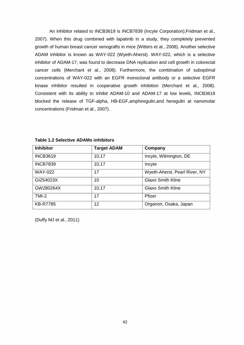
42
An inhibitor related to INCB3619 is INCB7839 (Incyte Corporation);Fridman et al.,
2007). When this drug combined with lapatinib in a study, they completely prevented
growth of human breast cancer xenografts in mice (Witters et al., 2008). Another selective
ADAM inhibitor is known as WAY-022 (Wyeth-Aherst). WAY-022, which is a selective
inhibitor of ADAM-17, was found to decrease DNA replication and cell growth in colorectal
cancer cells (Merchant et al., 2008). Furthermore, the combination of suboptimal
concentrations of WAY-022 with an EGFR monoclonal antibody or a selective EGFR
kinase inhibitor resulted in cooperative growth inhibition (Merchant et al., 2008).
Consistent with its ability to inhibit ADAM-10 and ADAM-17 at low levels, INCB3619
blocked the release of TGF-alpha, HB-EGF,amphiregulin,and heregulin at nanomolar
concentrations (Fridman et al., 2007).
Table 1.2 Selective ADAMs inhibitors
Inhibitor Target ADAM Company
INCB3619 10,17 Incyte, Wilmington, DE
INCB7839 10,17 Incyte
WAY-022 17 Wyeth-Aherst, Pearl River, NY
GI254023X 10 Glaxo Smith Kline
GW280264X 10,17 Glaxo Smith Kline
TMI-2 17 Pfizer
KB-R7785 12 Organon, Osaka, Japan
(Duffy MJ et al., 2011)
43
1.4 Characteristics and specific functions of ADAM17 1.4.1 Strucrure of ADAM17
Like other ADAMs, ADAM17, a modular type I protein, contains in addition to the
signal peptide, four functionally distinct extracellular domains followed by a
transmembrane region and a cytoplasmic tail (Fig. 1.15) (Arribas and Esselens, 2009;
Duffy et al., 2009) as previously described for all ADAMs. Its involvement in major
contemporary pathologies like cancer, inflammatory and vascular diseases seem to be
connected to its cleavage abilities and this is due to the large variety of substrates it is
able to cut. Certain growth factors and receptors can be activated and inactivated
repectively by ADAM17 by shedding their extracellular domain from the cell membrane
and in the same way, it can detach cells by cleaving cell adhesion molecules. These
proteolytic activities normally form part of cleavage cascades reffered to as Regulated
Intramembrane Proteolysis, leading to intracellular signaling.
ADAM17 (also named as tumor necrosis factor-alpha-converting enzyme,TACE) is
a memeber of the ADAM family and a transmembrane metalloprotease and primary
sheddase for multiple EGFR pro-ligands, including transforming growth factor-alpha (TGF-
a), heparin-binding epidermal growth factor (HB-EGF), and amphiregulin and all these
ligands have been implicated in cancer development and progression (Xu P et al., 2010).
Of all ADAM members, the most extensively investigated and the strongest evidence for a
role in malignancy exists for ADAM17, which has been demonstrated to be overexpressed
in the breast, ovary, kidney, colon, and pancreas cancer tissues and regulates important
biological phenomena in these cancers through EGFR/PI3K/ AKT pathway.
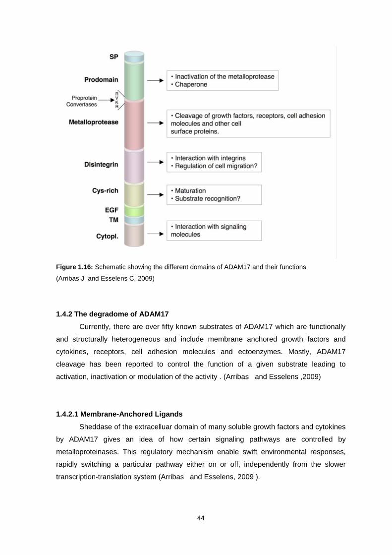
44
Figure 1.16: Schematic showing the different domains of ADAM17 and their functions
(Arribas J and Esselens C, 2009)
1.4.2 The degradome of ADAM17
Currently, there are over fifty known substrates of ADAM17 which are functionally
and structurally heterogeneous and include membrane anchored growth factors and
cytokines, receptors, cell adhesion molecules and ectoenzymes. Mostly, ADAM17
cleavage has been reported to control the function of a given substrate leading to
activation, inactivation or modulation of the activity . (Arribas and Esselens ,2009)
1.4.2.1 Membrane-Anchored Ligands
Sheddase of the extracelluar domain of many soluble growth factors and cytokines
by ADAM17 gives an idea of how certain signaling pathways are controlled by
metalloproteinases. This regulatory mechanism enable swift environmental responses,
rapidly switching a particular pathway either on or off, independently from the slower
transcription-translation system (Arribas and Esselens, 2009 ).
45
This can be seen in the following instances: The release of TGF-alpha, constituted
a straightforward activation process, due to the inactive state of the membrane-bound
proTGF-alpha (Borrell-Pages et al., 2003 ). Also it was indicated that the cleavage of
TNF-alpha by ADAM17 resulted in a transition between two active forms with different
properties (Aggarwal., 2003). In confirmation to the above study, the human carcinoma
cell line Colo205, is resistant to the soluble TNF-alpha, whereas is sensitive to
membrane-associated proTNF-alpha (Grell et al., 1995).
In the same manner, a ligand called neuregulins, were fully active even when
membrane bound and its cleavage by ADAM17 converted from physically restricted
growth factor into a diffusible isoform (Montero et al., 2000).
1.4.2.2 Receptors
The ADAM17 degradome includes many receptors, aside ligands. Receptors
cleavage offers another way to regulate the response of the cell to growth factors and
cytokines. In this case, shed receptor in addition to being unable to transmit signals to the
cell, can also act as a ligand-sequestring scavenger (Arribas and Esselens, 2009). This
is shown in a study by Aggarwal, where the shedding of the receptors for TNF-a by
ADAM17 and the subsequent sequestering of TNF-alpha by the soluble receptors,
prevented the binding of the cytokine to the full-length receptors (Aggarwal., 2003).
ADAM17, can also be an activator of certain receptors. Typical example is a
receptor that determines the developmental fates of mainly neuronal and hematopoietic
cell populations, called Notch receptor. ADAM17 carry this process out by first releasing
the extracellular domain of Notch following ligand binding, while the “stub”,
(transmembrane and cytoplasmic domain), stays behind in the membrane. The Notch
intracellular domain released, then translocate to the nucleus in order to regulate its target
genes expression (Selkoe and Kopan, 2003). ADAM17 has been indicated to mediate the
cleavage of ErbB4, N-cadherin and CD44 in the same manner as indicated above.
(Arribas and Esselens, 2009).
46
1.4.2.3 Cell Adhesion Molecules Shedding of cell adhesion molecules by ADAM17 results in the weakening of cell-
cell interactions (Arribas et al., 2006), which is very impotant in signal transduction. For
instance, the shedding of CD44 by ADAM17 is followed by a Regulated Intramembrane
Proteolysis (RIP) event, that releasing the intracellular domain (ICD) of CD44. The
released ICD in turn is translocated to the nucleus, where it finally regulates the
transcription of target genes (Nagano and Saya, 2004)
1.4.2 ADAM17 Dysfuntion and cancer
Despite the remarkably large number of ADAM17 substrates identified, most of the
proposed pathological roles in cancer of the metalloproteinase are related to just a few,
namely, TNF-a and ligands for HER in general. Upon ligand binding, the receptors
undergo homo- or heterooligomerization and activation. Activated HER receptors
transduce signals through a plethora of intracellular factors that, ultimately, regulate the
expression of groups of genes that control cellular proliferation, migration, adhesion,
differentiation and apoptosis (Yarden et al., 2001).
Different components of the EGFR pathway, such as HER2, are overactivated and play a
causal role in breast cancer progression (Baselga, 2001). ADAM17 is frequently
overexpressed in breast cancers (Borrell-Pages et al., 2003) and its overexpression is
associated with tumor progression and metastasis ( McGowan et al., 2007).
1.5.1 Release/Shedding of TNF-a by ADAM-17.
ADAM-17 was originally identified by its ability to release membrane-bound TNF-a from its
precursor and its purification was based on its ability to hydrolyze both peptide and full-
length TNF-a–based substrates at their physiologic cleavage site (Moss et al., 1997).
TNF-a , is a pluripotent peptide with multiple activities potentially important in tumor
formation and/or progression.
Apart from TNF-a, ADAM-17 has been shown to shed several membrane-bound
proteins, including E-selectin, p75 TNF receptor, transforming growth factor-a,
amphiregulin, heparin-binding EGF, and epiregulin. The shedding of a number of these
molecules is potentially important in cancer progression. ADAM-17 implication in TNF-a
release was documented in vivo where T cells derived from ADAM-17–deficient mice
(TACEDZn/ZnD) lost almost all of their ability to shed TNF-a (Black et al., 1997).

47
TNF-a role in malignancy include its ability to up-regulate MMP expression, induce
angiogenic factors, enhance cell migration, promote the epithelial-to-mesenchy, induce
expression of the transcription factor nuclear factor nB, induce reactive oxygen species
that damage DNA, and induction of matrix degrading proteases and release of cytokines
and chemokines (Pilarsky et al., 2004; Babbar et al., 2006). Suganuma et al. showed that
a deficiency of this cytokine rendered mice resistant to chemically induced skin
carcinogenesis (Suganuma et al., 1999). Because ADAM-17 catalyzes the formation of
active TNF-a, it might also be expected to play a role in malignancy via the generation of
this cytokine.
1.5.2 Shedding of HER ligands by ADAM-17 implicated in breast cancer The HER (also known as ErbB) proteins belong to the subclass I of the superfamily
of receptor kinases. There are four members of the family: EGFR/ErbB1/HER1,
HER2/ErbB2, HER3/ErbB3, and HER4/ErbB4 with an extracellular domain, a
transmembrane domain and an intracellular domain. Two members of this family (EGFR
and HER2) mediate cell growth, cell survival, cell migration, angiogenesis, and invasion
(Olayioye et al., 2000). Because of their key role in these processes, altered expression of
EGFR and HER2 has been implicated in the formation/progression of multiple cancer
types and these two proteins are currently among the best-validated targets for cancer
treatment , especially HER2 in breast cancer (Hynes et al., 2006).
The HER tyrosine kinases are activated by a number of ligands that are initially
synthesized as transmembrane precursors,which are necessary for biological activity.
There are three main groups: (a) EGF,TGF- a, amphiregulin, and epigen, which
specifically bind to EGFR; (b) heparin-binding (HP)-EGF, epiregulin, and betacellulin,
which bind to EGFR or HER4; and (c) neuregulin (also known as heregulin), which binds
to HER3 and HER4 (Olayioye et al., 2000; Hynes et al., 2006). No naturally occurring
ligand has yet been identified that specifically bind to HER2, but then it is preferred
dimerization partner for the other three ligand-activated HER members. It has been
reported that most of the HER ligands can be released from their precursor forms by
specific ADAMs. For example, a knockout studies with the use of mouse embryonic cells
showed that ADAM-17 was the major sheddase for TGF-a, amphiregulin, HB-EGF, and
epiregulin (Sahin et al., 2004).
ADAM-mediated release of HER ligands can be activated by a number of
physiologic and pharmacologic stimuli. Among the best-studied stimuli are agonists for G
protein–coupled receptors. The binding of these agonists to their receptor results in
48
ADAM-mediated release of HER ligands and transactivation of EGFR (Ohtsu et al., 2007).
Thus, in recent years, there has been a growing interest in ADAM17 as a new therapeutic
target in several EGFR-dependent tumour types. Literally, it is so obvious that ADAM17
fulfills a key role in diverse processes and pathologies, making it a prime target for
developing therapies.
Several reports have been documented on the evidence implicating ADAM17-
mediated growth factor ligand release and EGFR. For example ADAM17, via ligand
release and activation of the EGFR-PI3K-AKT pathway, enhanced in vitro breast cancer
cell proliferation and invasion. (Zheng et al., 2009) . Also, amphiregulin released by
ADAM-17 cleavage enhances cell proliferation of cancer cells (Gschwind et al., 2003;
Zhang, et al., 2006). It was also shown by Mendelson et al that treating mouse embryonic
fibroblasts with platelet derived growth factor receptor beta (PDGFRb) led to activation of
ADAM17, release of EGFR ligands and EGFR/ERK signalling which ultimately resulted in
enhanced migration.
1.6 Blocking of ADAM17 The structure of the proteolytic domain of ADAM17 at a resolution of 1.7 Å, has
enable the development of numerous inhibitors (Table 1.3) with various degrees of
specificity. Some compounds block, in addition to ADAM17, closely related ADAMs and
MMPs, while other seem to be highly specific. ADAM17 inhibitors are being assayed in
different preclinical models as in clinical trials, as shown in the table below.
Using a three-dimensional culture model of human breast cancer progression,
Kenny et al (Kenny et al., 2007) reported that either knockdown of ADAM-17 expression
with the use of small interfering RNA or inhibition of protease activity with the use of a
low–molecular weight inhibitor (TNF-alpha protease inhibitor-2) reversed the malignant
phenotype by preventing the release of amphiregulin and TGF-alpha. In renal cancer
cells, silencing of ADAM-17 was found to restore a dependence on exogenous growth
factors, reduce invasion,and block in vivo tumor formation. The role of ADAM-17 in
pancreatic cancer is underscored by experiments showing that inhibition of ADAM-17
gene expression, by using small interfering RNA (siRNA) technique, affects invasiveness
of tumour cells (Ringel et al., 2006). Treatment of breast cancer cell lines with anti-
ADAM17 antibodies leads to a decrease in cell proliferation (Lendeckel et al., 2005).
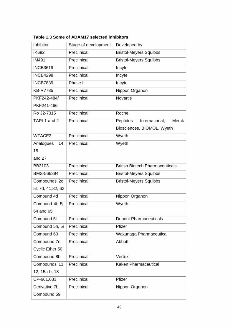
49
Table 1.3 Some of ADAM17 selected inhibitors
Inhibitor Stage of development Developed by
IK682 Preclinical Bristol-Meyers Squibbs
IM491 Preclinical Bristol-Meyers Squibbs
INCB3619 Preclinical Incyte
INCB4298 Preclinical Incyte
INCB7839 Phase II Incyte
KB-R7785 Preclinical Nippon Organon
PKF242-484/
PKF241-466
Preclinical Novartis
Ro 32-7315 Preclinical Roche
TAPI-1 and 2 Preclinical Peptides International, Merck
Biosciences, BIOMOL, Wyeth
WTACE2 Preclinical Wyeth
Analogues 14,
15
and 27
Preclinical Wyeth
BB3103 Preclinical British Biotech Pharmaceuticals
BMS-566394 Preclinical Bristol-Meyers Squibbs
Compounds 2o,
5l, 7d, 41,32, 62
Preclinical Bristol-Meyers Squibbs
Compund 4d Preclinical Nippon Organon
Compund 4t, 5j,
64 and 65
Preclinical Wyeth
Compund 5l Preclinical Dupont Pharmaceuticals
Compund 5h, 5i Preclinical Pfizer
Compund 60 Preclinical Wakunaga Pharmaceutical
Compound 7e,
Cyclic Ether 50
Preclinical Abbott
Compound 8b Preclinical Vertex
Compounds 11,
12, 15a-b, 18
Preclinical Kaken Pharmaceutical
CP-661,631 Preclinical Pfizer
Derivative 7b,
Compound 59
Preclinical Nippon Organon

50
DPC-A38088
DPH-067517
Preclinical Bristol-Myers Squibb
GW280264X Preclinical GlaxoSmithKline
(Arribas J and Esselens C, 2009)
51
1.7 Objectives of study
The general aim of this study was to determine the expression of the impact of ADAM17
in node-negative Breast Carcinoma
Specific aims were as follow:
To evaluate the tissue-based protein expression of ADAM17 and correlate it with
clinical and pathological features of breast cancer.
To compare the expression of ADAM17 in node-negative and node-positive
breast Cancer
To differentiate among the heterogeneous grade 2 patients to aid in grouping them
into high and low risk category
To determine the potential prognostic effect of ADAM17 to aid in differentiating
indolent node-negative Breast Cancer from aggressive node-negative Breast
Cancer
52
CHAPTER TWO
MATERIAL AND METHODS
2.1 Patient population
50 Cases of cancer patients who have undergone radical mastectomy with no prior
treatment, with ages ranging from 24 - 89 were obtained from Saint Anthony General
Hospital, Porto Portugal together with an informed consent form of participation in the
project. Patients involved were pathologically confirmed of the following node-negative
and node-positive breast cancer: Invasive ductal carcinoma, In situ ductal carcinoma and
Invasive lobular carcinoma with histological grades 1 to 3. There were 40 node-negative
cases and 10 node-positive cases with tumor size ranging from 0.6 cm to 4.7 cm. In
addition to ADAM17, HER2, Ki67, Progesterone and Estrogen receptors were also
determined. The ADAM17 protein expression in breast cancer tissue samples were
confirmed by immunohistochemistry analysis, which was performed on formalin-fixed,
paraffin-embedded, 3 micro meters thick tissue sections using the avidin-biotin-peroxidase
complex method.
2.2 Sample Processing
2.2.1 Microscopic examination
The tissue samples were collected as quickly as possible after the removal of the
organ from the subjects (patients) and immediately fixed in 10% formalin. This was to
preserve the cells and cellular constituents in a state as close as possible to the living
cells and also maintains the antigenicity to allow them to be processed without change.
Each sample was stratified according to the following: Age, size, weight, anatomical
location, tumor relation to surgical margin and nodal status. They were then sectioned and
placed on embedding cassette for tissue processing.
2.2.3 Tissue processing
The main steps in tissue processing are dehydration and clarification. In order to
remove the fixative and water from the tissues, they were sent through graded alcohols of
increasing concentrations (70% - 95% - 100%). The alcohol was then replaced by a
53
‘’clearing’’ solution, Xylene to subsequently aid in carrying out the impregnation with
paraffin wax and was left overnight, since xylene is miscible with paraffin wax.
2.2.4 Impregnation and sectioning
The processed tissues in cassettes were properly aligned and oriented in paraffin
at 58-60 degree Celsius. They were then allowed to cool to form paraffin blocks. With the
help of a microtome, the tissue in the paraffin block were first trimmed to exposed the
tissue of interest after which it was cut into thin sections of about 3 micro meters. The cut
sectioned were placed in cold water to remove any creases that may exist to enable it
stretch. They were then picked with a coated glass slide and placed again in warm water
about 60 degrees Celsius for adhesion and extension onto the slides. The slides with the
tissues were then put in a refrigerator under room temperature for 30 minutes.
2.2.5 Immunohistochemical staining for all the antibodies using avidin-biotin peroxidase complex protocol
2.2.5.1 Dewaxing and Hydration
Slides with tissues were de-waxed (removal of wax) by placing them in xylene
twice for 5 minutes each. They were then hydrated in series of alcohol in decreasing
concentration (100% - 90% - 75%) for at least 1 minute each and finally washed under
running water.
2.2.5.2 Antigen retrieval
To increase the antigenicity of the tissues, 50ml of 10% Dako target retrieval
solution was prepared by adding 45ml of water to 5ml of Dako target retrieval solution.
50ml solution is enough for 15 slides. The solution was then poured in plastic container
after which the slides were added and waited for 5 minutes for the solution to be clear.
The container with the slides were then put in water bath under 96-100 degrees Celsius
for 20 minutes. The water bath was put off and the slides and container were removed
and placed in cold water till it reached room temperature and finally placed in humid
container to prevent drying up of the slides.
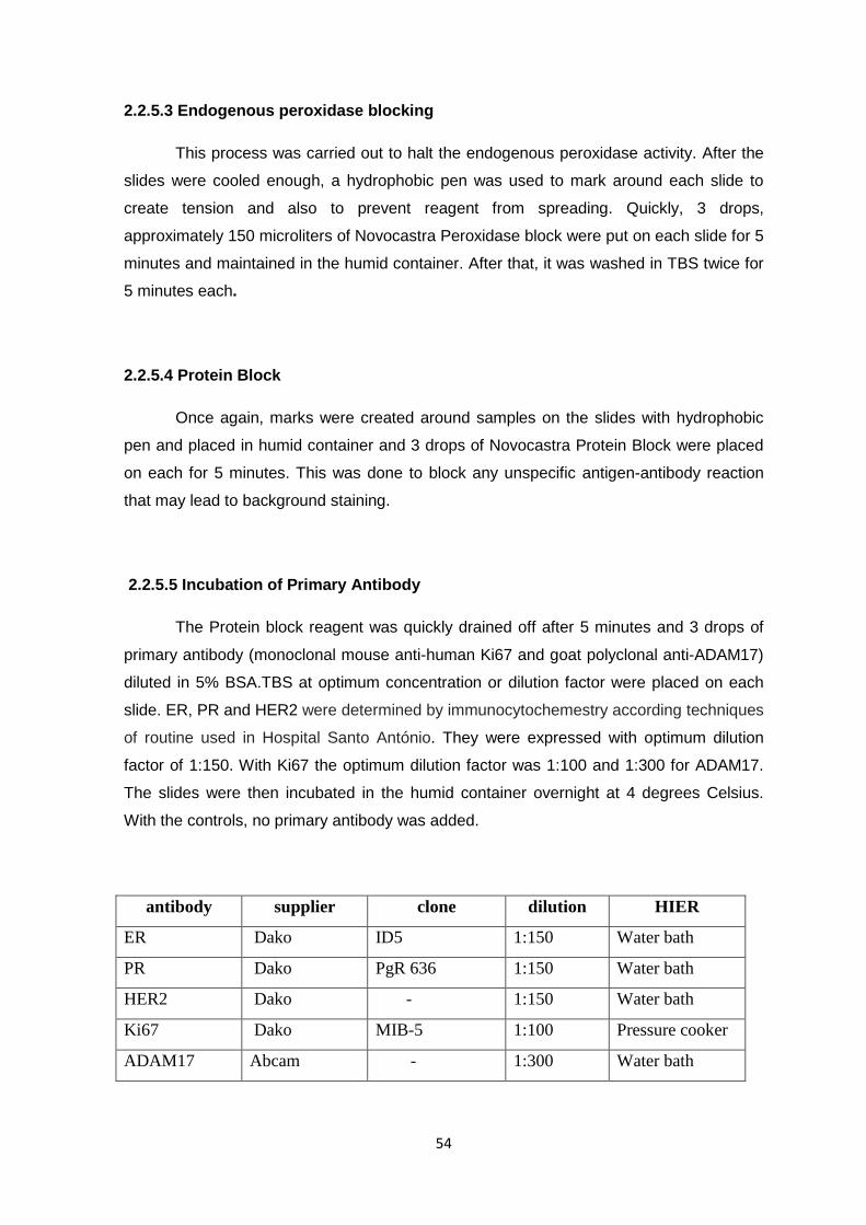
54
2.2.5.3 Endogenous peroxidase blocking
This process was carried out to halt the endogenous peroxidase activity. After the
slides were cooled enough, a hydrophobic pen was used to mark around each slide to
create tension and also to prevent reagent from spreading. Quickly, 3 drops,
approximately 150 microliters of Novocastra Peroxidase block were put on each slide for 5
minutes and maintained in the humid container. After that, it was washed in TBS twice for
5 minutes each.
2.2.5.4 Protein Block
Once again, marks were created around samples on the slides with hydrophobic
pen and placed in humid container and 3 drops of Novocastra Protein Block were placed
on each for 5 minutes. This was done to block any unspecific antigen-antibody reaction
that may lead to background staining.
2.2.5.5 Incubation of Primary Antibody
The Protein block reagent was quickly drained off after 5 minutes and 3 drops of
primary antibody (monoclonal mouse anti-human Ki67 and goat polyclonal anti-ADAM17)
diluted in 5% BSA.TBS at optimum concentration or dilution factor were placed on each
slide. ER, PR and HER2 were determined by immunocytochemestry according techniques
of routine used in Hospital Santo António. They were expressed with optimum dilution
factor of 1:150. With Ki67 the optimum dilution factor was 1:100 and 1:300 for ADAM17.
The slides were then incubated in the humid container overnight at 4 degrees Celsius.
With the controls, no primary antibody was added.
antibody supplier clone dilution HIER
ER Dako ID5 1:150 Water bath
PR Dako PgR 636 1:150 Water bath
HER2 Dako - 1:150 Water bath
Ki67 Dako MIB-5 1:100 Pressure cooker
ADAM17 Abcam - 1:300 Water bath
55
2.2.5.6 Post/Secondary Antibody
In avidin-biotin-peroxidase complex protocol, secondary antibodies are needed to
conjugate to biotin and function as a link between tissue-bound primary antibody and
avidin-biotin-peroxidase complex. In other words, the secondary antibody is needed in
detecting the primary anti-body. The slides were washed twice in TBS for 5 minutes each.
150 microliter of Novocastra post primary antibody were placed on each slide for 30
minutes and spread carefully for even distribution. They were washed in TBS twice for 5
minutes each.
2.2.5.7 Polymerization (Avidin-Biotin)
Again, 150 microliters of Novolink Polymer was put on the slides and incubated in
a humid container for 30 minutes. They were then washed with TBS twice for 5 minutes
each. Polymerization is necessary in that it enable the polymer or the enzyme to first react
with DAB to aid in optimum visualization.
2.2.5.8 Chromogen reaction
Chromogens are electron donors which upon being oxidized becomes insoluble
colored product. An example is 3,3-diaminobenzidinetrahydrochloride (DAB). 50
microliters of DAB for each 1000 microliters of substrate buffer were prepared in 15ml
falcon tube. In aiding to visualize the results, 150ml of DAB solution were placed on each
slides for few minutes or till reaction occurs. Slides were quickly placed in container with
distilled water and placed under running water for 15 minutes. DAB, upon being oxidized
produced brown end colour which aid in the visualization of tissue on slides.
2.2.5.9 Hematoxylin staining
For counterstain, slides were inserted in Hematoxylin for several times (6-8 times)
depending on the concentration of the Hematoxylin and how frequently it has been used.
They were again placed under running water for 15 minutes.
56
2.2.5.10 Dehydration and clearing
Slides were placed in 100% alcohol twice and moved up and down for several
times in each case, to remove the water after which they were placed in xylene twice and
also moved several times in each case.
2.2.5.11 Mounting of slides
Slides were removed from the xylene one after the other and were mounted with
entellan. Each slide was pressed carefully after placing the cover slip to get rid of any
bubbles present. Quickly, they were passed through xylene and cleaned after which they
were allowed to dry for microscopic examination. In each case, the non-cancerous breast
cancer tissues were used as control tissues and the omission of the primary antibody
served as negative control.
The HER-2/neu was considered negative when scores of 0 and +1, and positive
when scores of +2 and +3 were recorded. To be considered as +2, +3, the cellular
membrane should be completely stained in more than 10% of the tumor cells. In the cases
where a score 2 was found by IHC, it was further studied by FISH and classified as
positive or negative according FISH results. Cells without staining, or with weak staining in
part of the cell membrane and in less than 10% of the tumor cells were considered
negative. In the case of ER and PR they were graded by considering 1% of tumor cells
stained in the nucleus as negative and more than 10% of stained tumor cells as positive.
With Ki67, scores were obtained by counting at least 100 cells (excluding mitotic cells)
and taken the percentage of stained cells in the nucleus. The cells were counted at places
where there were high proliferation of cells.
ADAM17 levels of expression were determined by using staining intensity. The
staining intensity was evaluated using four-tier grading system (0- negative; 1-weak; 2-
moderate and 3-strong). To delineate between low and high levels of ADAM17
expression, the tumors with strong ADAM17 expression were grouped against those with
none to moderate ADAM17 expression.

57
2.2.6 Statistical Analysis of Results
Statistical significance was calculated using Fisher’s exact test to assess the
relationships between ADAM17 protein expression and different clinical parameters
through IBM SPSS Statists 20.0 software. This is due to the smaller sample size and the
fact that some of the cells had values less than five. Significance was accepted at p<0.05.
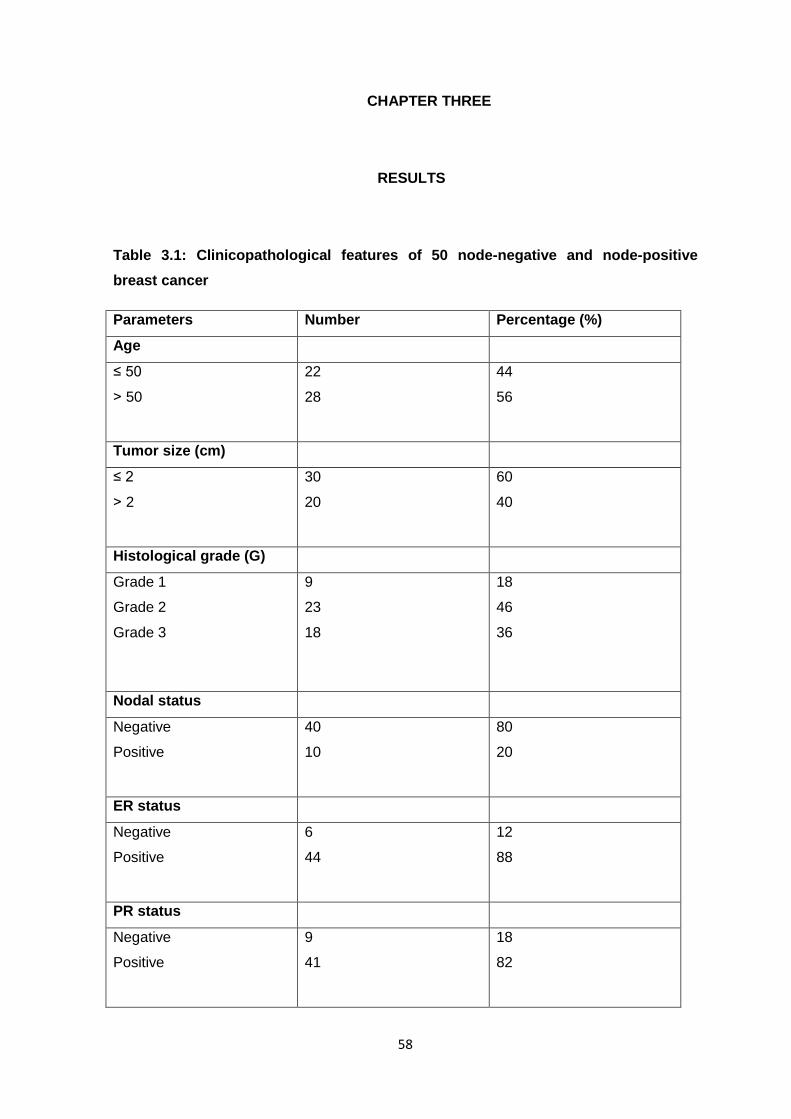
58
CHAPTER THREE
RESULTS
Table 3.1: Clinicopathological features of 50 node-negative and node-positive breast cancer
Parameters Number Percentage (%)
Age
≤ 50
˃ 50 22
28
44
56
Tumor size (cm)
≤ 2
˃ 2
30
20
60
40
Histological grade (G)
Grade 1
Grade 2
Grade 3
9
23
18
18
46
36
Nodal status
Negative
Positive
40
10
80
20
ER status
Negative
Positive
6
44
12
88
PR status
Negative
Positive
9
41
18
82
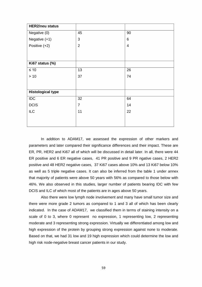
59
HER2/neu status
Negative (0)
Negative (+1)
Positive (+2)
45
3
2
90
6
4
Ki67 status (%)
≤ 10
˃ 10
13
37
26
74
Histological type
IDC
DCIS
ILC
32
7
11
64
14
22
In addition to ADAM17, we assessed the expression of other markers and
parameters and later compared their significance differences and their impact. These are
ER, PR, HER2 and Ki67 all of which will be discussed in detail later. In all, there were 44
ER positive and 6 ER negative cases, 41 PR positive and 9 PR ngative cases, 2 HER2
positive and 48 HER2 negative cases, 37 Ki67 cases above 10% and 13 Ki67 below 10%
as well as 5 triple negative cases. It can also be inferred from the table 1 under annex
that majority of patients were above 50 years with 56% as compared to those below with
46%. We also observed in this studies, larger number of patients bearing IDC with few
DCIS and ILC of which most of the patients are in ages above 50 years.
Also there were low lymph node involvement and many have small tumor size and
there were more grade 2 tumors as compared to 1 and 3 all of which has been clearly
indicated. In the case of ADAM17, we classified them in terms of staining intensity on a
scale of 0 to 3, where 0 represent no expression, 1 representing low, 2 representing
moderate and 3 representing strong expression. Virtually we differentiated among low and
high expression of the protein by grouping strong expression against none to moderate.
Based on that, we had 31 low and 19 high expression which could determine the low and
high risk node-negative breast cancer patients in our study.
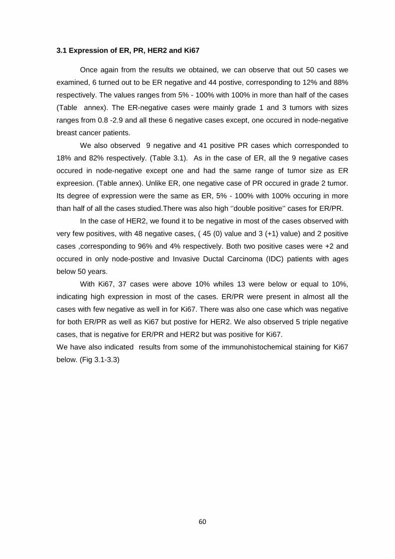
60
3.1 Expression of ER, PR, HER2 and Ki67
Once again from the results we obtained, we can observe that out 50 cases we
examined, 6 turned out to be ER negative and 44 postive, corresponding to 12% and 88%
respectively. The values ranges from 5% - 100% with 100% in more than half of the cases
(Table annex). The ER-negative cases were mainly grade 1 and 3 tumors with sizes
ranges from 0.8 -2.9 and all these 6 negative cases except, one occured in node-negative
breast cancer patients.
We also observed 9 negative and 41 positive PR cases which corresponded to
18% and 82% respectively. (Table 3.1). As in the case of ER, all the 9 negative cases
occured in node-negative except one and had the same range of tumor size as ER
expreesion. (Table annex). Unlike ER, one negative case of PR occured in grade 2 tumor.
Its degree of expression were the same as ER, 5% - 100% with 100% occuring in more
than half of all the cases studied.There was also high ‘’double positive’’ cases for ER/PR.
In the case of HER2, we found it to be negative in most of the cases observed with
very few positives, with 48 negative cases, ( 45 (0) value and 3 (+1) value) and 2 positive
cases ,corresponding to 96% and 4% respectively. Both two positive cases were +2 and
occured in only node-postive and Invasive Ductal Carcinoma (IDC) patients with ages
below 50 years.
With Ki67, 37 cases were above 10% whiles 13 were below or equal to 10%,
indicating high expression in most of the cases. ER/PR were present in almost all the
cases with few negative as well in for Ki67. There was also one case which was negative
for both ER/PR as well as Ki67 but postive for HER2. We also observed 5 triple negative
cases, that is negative for ER/PR and HER2 but was positive for Ki67.
We have also indicated results from some of the immunohistochemical staining for Ki67
below. (Fig 3.1-3.3)
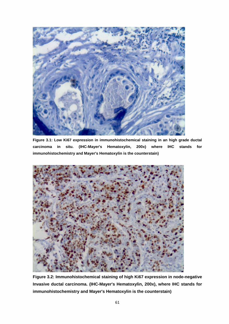
61
Figure 3.1: Low Ki67 expression in immunohistochemical staining in an high grade ductal carcinoma in situ. (IHC-Mayer's Hematoxylin, 200x) where IHC stands for immunohistochemistry and Mayer's Hematoxylin is the counterstain)
Figure 3.2: Immunohistochemical staining of high Ki67 expression in node-negative Invasive ductal carcinoma. (IHC-Mayer's Hematoxylin, 200x), where IHC stands for immunohistochemistry and Mayer's Hematoxylin is the counterstain)
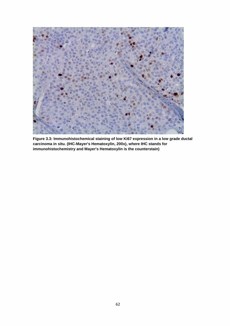
62
Figure 3.3: Immunohistochemical staining of low Ki67 expression in a low grade ductal carcinoma in situ. (IHC-Mayer's Hematoxylin, 200x), where IHC stands for immunohistochemistry and Mayer's Hematoxylin is the counterstain)
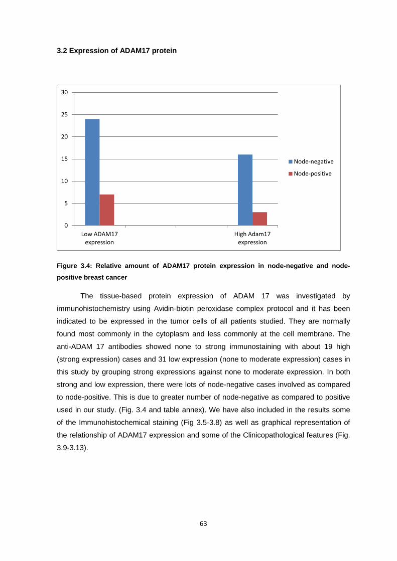
63
3.2 Expression of ADAM17 protein
Figure 3.4: Relative amount of ADAM17 protein expression in node-negative and node-
positive breast cancer
The tissue-based protein expression of ADAM 17 was investigated by
immunohistochemistry using Avidin-biotin peroxidase complex protocol and it has been
indicated to be expressed in the tumor cells of all patients studied. They are normally
found most commonly in the cytoplasm and less commonly at the cell membrane. The
anti-ADAM 17 antibodies showed none to strong immunostaining with about 19 high
(strong expression) cases and 31 low expression (none to moderate expression) cases in
this study by grouping strong expressions against none to moderate expression. In both
strong and low expression, there were lots of node-negative cases involved as compared
to node-positive. This is due to greater number of node-negative as compared to positive
used in our study. (Fig. 3.4 and table annex). We have also included in the results some
of the Immunohistochemical staining (Fig 3.5-3.8) as well as graphical representation of
the relationship of ADAM17 expression and some of the Clinicopathological features (Fig.
3.9-3.13).
0
5
10
15
20
25
30
Low ADAM17 expression
High Adam17 expression
Node-negative
Node-positive
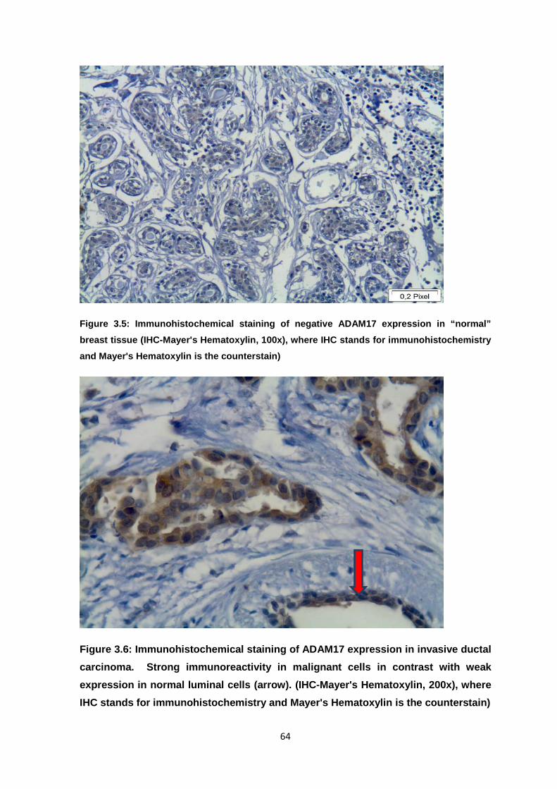
64
Figure 3.5: Immunohistochemical staining of negative ADAM17 expression in “normal” breast tissue (IHC-Mayer's Hematoxylin, 100x), where IHC stands for immunohistochemistry and Mayer's Hematoxylin is the counterstain)
Figure 3.6: Immunohistochemical staining of ADAM17 expression in invasive ductal carcinoma. Strong immunoreactivity in malignant cells in contrast with weak expression in normal luminal cells (arrow). (IHC-Mayer's Hematoxylin, 200x), where IHC stands for immunohistochemistry and Mayer's Hematoxylin is the counterstain)
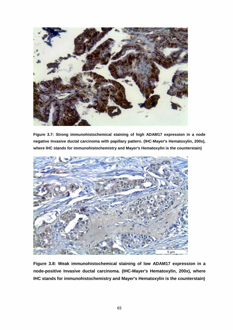
65
Figure 3.7: Strong immunohistochemical staining of high ADAM17 expression in a node negative Invasive ductal carcinoma with papillary pattern. (IHC-Mayer's Hematoxylin, 200x),
where IHC stands for immunohistochemistry and Mayer's Hematoxylin is the counterstain)
Figure 3.8: Weak immunohistochemical staining of low ADAM17 expression in a node-positive Invasive ductal carcinoma. (IHC-Mayer's Hematoxylin, 200x), where IHC stands for immunohistochemistry and Mayer's Hematoxylin is the counterstain)
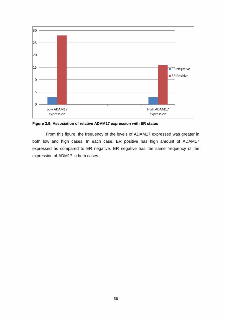
66
Figure 3.9: Association of relative ADAM17 expression with ER status
From this figure, the frequency of the levels of ADAM17 expressed was greater in
both low and high cases. In each case, ER positive has high amount of ADAM17
expressed as compared to ER negative. ER negative has the same frequency of the
expression of ADM17 in both cases.
0
5
10
15
20
25
30
Low ADAM17 expression
High ADAM17 expression
ER Negative
ER Positive
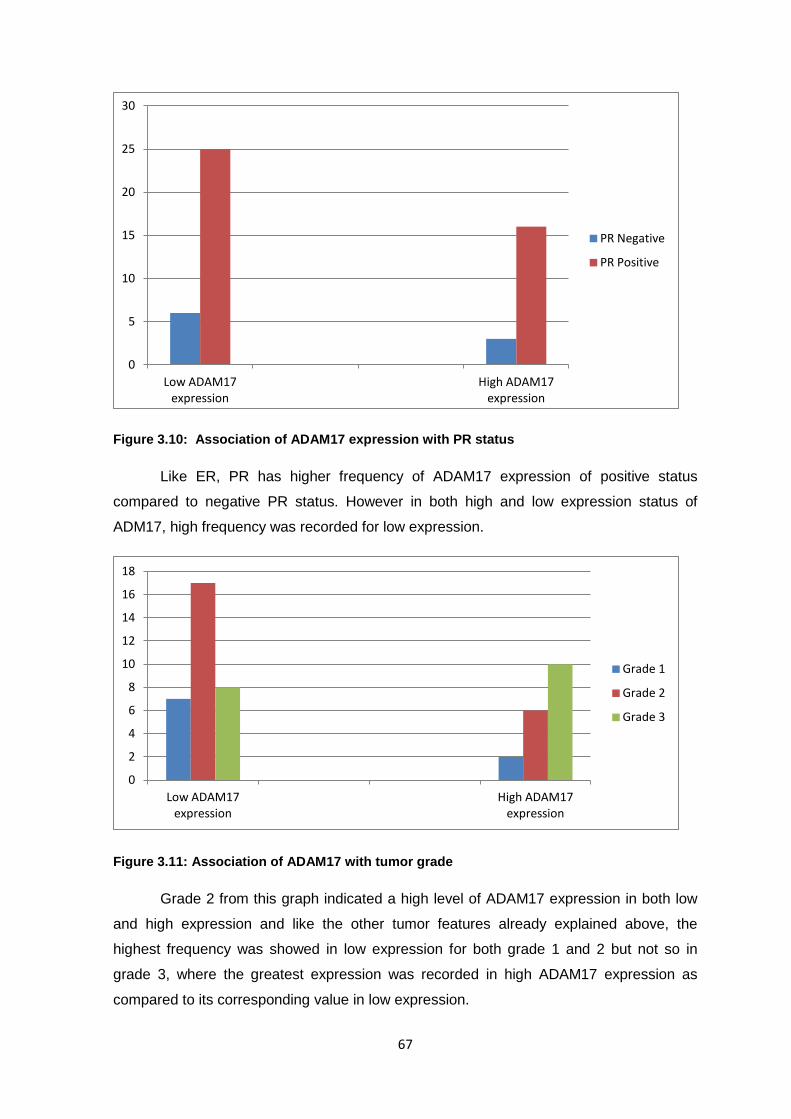
67
Figure 3.10: Association of ADAM17 expression with PR status
Like ER, PR has higher frequency of ADAM17 expression of positive status
compared to negative PR status. However in both high and low expression status of
ADM17, high frequency was recorded for low expression.
Figure 3.11: Association of ADAM17 with tumor grade
Grade 2 from this graph indicated a high level of ADAM17 expression in both low
and high expression and like the other tumor features already explained above, the
highest frequency was showed in low expression for both grade 1 and 2 but not so in
grade 3, where the greatest expression was recorded in high ADAM17 expression as
compared to its corresponding value in low expression.
0
5
10
15
20
25
30
Low ADAM17 expression
High ADAM17 expression
PR Negative
PR Positive
0
2
4
6
8
10
12
14
16
18
Low ADAM17 expression
High ADAM17 expression
Grade 1
Grade 2
Grade 3
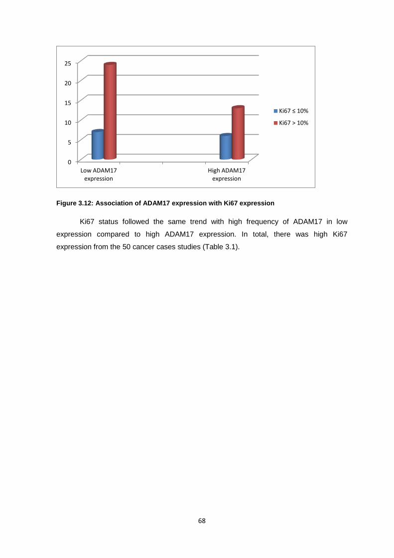
68
Figure 3.12: Association of ADAM17 expression with Ki67 expression
Ki67 status followed the same trend with high frequency of ADAM17 in low
expression compared to high ADAM17 expression. In total, there was high Ki67
expression from the 50 cancer cases studies (Table 3.1).
0
5
10
15
20
25
Low ADAM17 expression
High ADAM17 expression
Ki67 ≤ 10%
Ki67 ˃ 10%
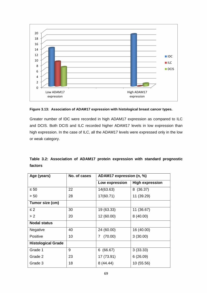
69
Figure 3.13: Association of ADAM17 expression with histological breast cancer types.
Greater number of IDC were recorded in high ADAM17 expression as compared to ILC
and DCIS. Both DCIS and ILC recorded higher ADAM17 levels in low expression than
high expression. In the case of ILC, all the ADAM17 levels were expressed only in the low
or weak category.
Table 3.2: Association of ADAM17 protein expression with standard prognostic factors
Age (years) No. of cases ADAM17 expression (n, %)
Low expression High expression
≤ 50
˃ 50
22
28
14(63.63)
17(60.71)
8 (36.37)
11 (39.29)
Tumor size (cm)
≤ 2
˃ 2
30
20
19 (63.33)
12 (60.00)
11 (36.67)
8 (40.00)
Nodal status
Negative
Positive
40
10
24 (60.00)
7 (70.00)
16 (40.00)
3 (30.00)
Histological Grade
Grade 1
Grade 2
Grade 3
9
23
18
6 (66.67)
17 (73.91)
8 (44.44)
3 (33.33)
6 (26.09)
10 (55.56)
0 2 4 6 8
10 12 14 16 18 20
Low ADAM17 expression
High ADAM17 expression
IDC
ILC
DCIS
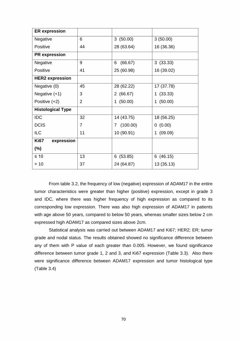
70
ER expression
Negative
Positive
6
44
3 (50.00)
28 (63.64)
3 (50.00)
16 (36.36)
PR expression
Negative
Positive
9
41
6 (66.67)
25 (60.98)
3 (33.33)
16 (39.02)
HER2 expression
Negative (0)
Negative (+1)
Positive (+2)
45
3
2
28 (62.22)
2 (66.67)
1 (50.00)
17 (37.78)
1 (33.33)
1 (50.00)
Histological Type
IDC
DCIS
ILC
32
7
11
14 (43.75)
7 (100.00)
10 (90.91)
18 (56.25)
0 (0.00)
1 (09.09)
Ki67 expression (%)
≤ 10
˃ 10
13
37
6 (53.85)
24 (64.87)
6 (46.15)
13 (35.13)
From table 3.2, the frequency of low (negative) expression of ADAM17 in the entire
tumor characteristics were greater than higher (positive) expression, except in grade 3
and IDC, where there was higher frequency of high expression as compared to its
corresponding low expression. There was also high expression of ADAM17 in patients
with age above 50 years, compared to below 50 years, whereas smaller sizes below 2 cm
expressed high ADAM17 as compared sizes above 2cm.
Statistical analysis was carried out between ADAM17 and Ki67; HER2; ER; tumor
grade and nodal status. The results obtained showed no significance difference between
any of them with P value of each greater than 0.005. However, we found significance
difference between tumor grade 1, 2 and 3, and Ki67 expression (Table 3.3). Also there
were significance difference between ADAM17 expression and tumor histological type
(Table 3.4)
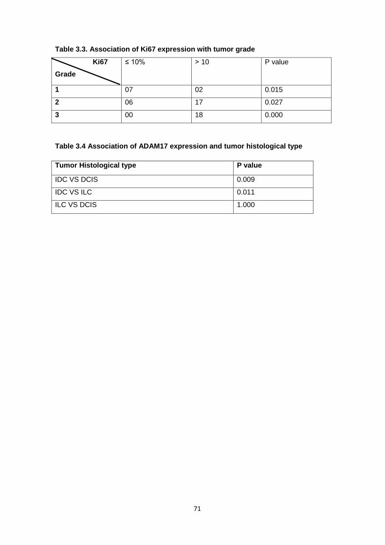
71
Table 3.3. Association of Ki67 expression with tumor grade
Ki67 Grade
≤ 10% ˃ 10 P value
1 07 02 0.015
2 06 17 0.027
3 00 18 0.000
Table 3.4 Association of ADAM17 expression and tumor histological type
Tumor Histological type P value
IDC VS DCIS 0.009
IDC VS ILC 0.011
ILC VS DCIS 1.000
72
CHAPTER FOUR
DISCUSSION AND CONCLUSION
4.1 Expression of ER, PR, HER2 and Ki67 Progress in the molecular understanding of breast cancer and the development of
sophisticated diagnostic tools has led to the availability of several prognostic tools as well
that aid in the determination of the outcome or the progression of individual tumors.
Breast cancer prognostic factors are essential to identify patients at risk of distant
metastasis development and to decide whether adjuvant treatments are needed or not.
The most validated biological marker in non-metastatic breast cancer are tumor size,
histological grade, mitotic index, Ki67 rate, axillary lymph node involvement, Estrogen
Receptor (ER), Progesterone Receptor (PR) and HER2 status. However, several review
articles have addressed their potential uses and as well as some drawbacks. Locoregional
treatment is mostly used in treating patients with lymph node-negative breast cancers, of
which about 70% respond positively, however 30% relapse.
Aside these markers, many other molecules have also been assessed, including
proliferation antigen Ki-67. Since its identification as a nuclear antigen associated with cell
proliferation in 1983 (Gerdes et al., 1983), it has been studied with considerable enthusiasm.
In breast cancer, ER/PR, HER2, and Ki-67 are important biological markers for predicting
prognosis and making effective treatment decisions. Treatment decisions for breast cancer
are commonly made based on the information derived from the immunohistochemistry (IHC)
of these biological markers. For example, the application of chemotherapy is commonly used
for patients with high Ki-67 values, endocrine therapy for ER/PR positive, and anti-HER2
(trastuzumab) therapy for HER2 positive. However, chemotherapy is once again
recommended for patients with high Ki67, ER-positive and HER2 negative by St. Gallen
consensus recommendation. (1999 and 2011)
73
4.1.1 ER/PR expression
Estrogen receptors (ER) and/or progesterone receptors (PR) presence is currently
a component of routine evaluation of breast cancer specimens. ER was the first analyzed
in breast cancer in the late 1950s, and was the first molecular marker evaluated for
prognosis and therapy response for breast cancer. The status of ER has been shown to
have significant predictive value on tumor response to hormone therapy in metastatic
disease as well as for adjuvant therapy after local excision (Dowsett M et al., 2008). On
the other hand, the role of PR status in predicting tumor response to therapy is still
unclear, although it has shown promise. From our current study, most of the cases were
postive for both ER and PR, corresponding to 88% and 82% respectively which have
been confirmed with other studies.
This finding can indicate a better response to hormonal therapy in those patients
with ‘double-positive’ ER/PR and clinicians will not heistate in subjecting these patients to
hormonal therapy for better reponse. This has been confirmed by another studies where it
was reported that a ‘double-positive’ ER+/PR+ tumor responded better to hormonal
therapy than ‘single-positive’ ER+/PR− or ER−/PR+ tumors (Dowsett M et al., 2008). In
addition to their predictive value, as to whether they also have prognostic value has been
extensively studied, but remains under debate.
Estrogens are generated by women to boost the development and maintenance of
female attributes and they are necessary for the sexual organs, really important during
pregnancy. Among the many parts of the women body that are the targets of the
estrogens, are breasts and uterus and they bind to inner parts of the cells called estrogen
receptors. In most of the cases this binding process leads to the proliferation state of the
cell which is normal physiological state but can lead to breast or cancer of the uterus in
most cases. We noticed that more than half of the cases were 100% ER positive even in
early stage node-negative grade 1 tumors. This can be attributed to the large amount of
estrogen already or naturally present in breast which binds to their receptors for normal
physiological process.
74
4.1.2 HER2 expression
Another major prognostic marker that is currently recommended for the evaluation
of primary invasive breast cancer is the human epidermal growth factor receptor 2, also
known as HER2. HER2 is an oncogene belonging to the EGF receptor (EGFR) family.
Gene amplification of HER2 has been shown to occur in 10–40% of primary tumors and
HER2 protein overexpression is found in almost 25% of breast cancers conferring a more
aggressive biology and have been shown to be associated with worse overall survival and
twice the mortality rate compared with women with no HER2 expression. (Paikn et al.,
1990; Van de Vijver et al., 1980). Our results obtained was not in consistent with many
studies on HER2, which records higher percentage of its presence. In our study, we
recorded more negative than positive expression of HER2 (98% negative and 2%
positive). However, all the positive expression occured in only node-positive which could
mean that they are mostly or normally expressed in advanced breast cancer cases or
lymph node metastatic stages. Most of the studies carried on HER2 have been seen on
advanced or distant metastasis. Also one contributing factor to higher negative result
could be the small sample size of our studies.
The HER tyrosine kinases are activated by a number of ligands that are initially
synthesized as transmembrane precursors and these ligands can be released from their
precursor forms by specific ADAMs. Knockout studies with the use of mouse embryonic
cells showed that ADAM-17 was the major sheddase for TGF-a, amphiregulin, HB-EGF,
and epiregulin (Sahin et al., 2004; Borrell-Pages et al., 2003), which are all ligands for
HER2. Once again, this report seem not to be in consitent with our results. It is expected
that at least there would be more postive HER2 cases where there was positive or high
expression of ADAM17, but we recorded only one such case.
Instead most of the high ADAM17 expression corresponded to negative HER2
expression. This could suggest a possible alternative pathway by which ADAM17 causes
breast cancer initiation, progression and invasion in addtion to the already or commonly
known release of ligands for EGFR/HER2. This can be confirmed by other studies
reporting ADAM17 to have act on a large group of cell adhesion molecules, resulting in
weakening of cell-cell interactions as well as cleavage and activation of certain receptors
(Arribas et al., 2006).
75
4.1.3 Ki67 expression Ki–67 was first discovered by J. Gerdes et al in 1983 suggesting its use for
marking proliferating cells and it is expressed in the nucleus during cell cycle. Cancer cells
are well known because of their multiplying behaviour that characterizes the disease. For
this reason putting the Ki–67 antibody to work produces a reaction, obtaining the desired
nuclear staining. This method is used by oncologists for diagnosing breast cancer, but it is
not enough to conclude that cancer is present or not. Parallel tests have to be carried out
for hormone receptors, metastasis and HER2-neu to prescribe the suitable treatment. Ki–
67 is present in normal breast tissue but in a very low percentage, a figure that is notably
higher when cancer is present. Furthermore, high percentage of Ki–67 is synonym of poor
prognosis, which has been proven to have good response to chemotherapy treatment. For
example several authors measured Ki67 in tamoxifen-treated breast cancer patients, all
reporting a decrease in Ki67 (Johnston SR et al., 1994; Makris A et al., 1998).
In confirmation to this assertion, our study indicated a high expression of Ki67 in
74% of the cases which is in accordance with several research work of Ki67 expression in
breast cancer. The expression was even high in most of ADAM17 negative expression.
According to St Gallen consensus, patients with high Ki67 expression and ER positive are
recommended to be given chemotherapy, which means if the parameters used in their
recommendation is to be followed, about 74% of the patients in this study may be given
chemotherapy, which may not be necessary and they can be spared form the toxic effect
of adjuvant chemotherapy. We also recorded only one negative case which corresponded
to high ADAM17 expression and this can be a good candidate with the treatment of
ADAM17 inhibitor or chemotherapy since it was also negative for both ER and PR.
There was significance difference between Ki67 and tumor grade 1, 2 and 3 with P
values 0.015, 0.027 and 0.000 respectively, showing a strong correlation between tumor
grade and Ki67. Triple negative cancers are very aggressive and difficult to treat since
they do not respond to HER2, ER or PR target treatment. In our study all the triple
negative cases has a corresponding higher expression of Ki67 which are good candidate
for chemotherapy according to St Gallen consensus recommendation.It also
corresponded to both high and low ADAM17 expression which could also means that
inhibitors of ADAM17 can be given for treatment,especially those who expressed high
levels, aside chemotherapy.
76
4.2 ADAM17 protein expression ADAM family has been demonstrated to be involved in the process of proteolytic
‘shedding’ of membrane associated proteins and hence the rapid modulation of key cell
signaling pathways in the tumor microenvironment (Arribas J et al., 2006; Duffy MJ et al.,
2009). Through the cytoplasmic domain, they influence downstream signalling cascade,
especially EGFR through processing of EGFR-ligands. And this is of particular interest
since EGFR is a well established drug target for breast cancer (Ohtsu H et al., 2006). It
was established before that ADAM17, an important member of the ADAMs family, is the
most extensively studied ADAM molecule. It sheds a variety of important cell surface
molecules, including cytokines, growth factors, and adhesion molecules.
Among those genes that are up-regulated in breast cancer, the epidermal growth
factor receptor genes have achieved considerable attention. While gene amplification
rarely exceeds eight-fold, mRNA and protein expression can be up to 120-fold higher than
normal levels. They play key roles in tumor angiogenesis, invasion, and metastatic
potential as well as resistance to anti-endocrine, chemo- and radiotherapy. ADAM17 is
involved in cell proliferation and invasion by releasing several ligands and overexpressed
in cancers of the breast, ovary, kidney, colon, and prostate. Because of its ability to
release biologically important ligands such as TNF-a, transforming growth factor-a,
amphiregulin, heparin-binding EGF, and epiregulin, for these receptors, it might be
expected to play an important role in cancer progression.
In our study, there was expression of ADAM17 protein in both node-negative and
positive breast cancer,(Figure 3.4) which was verified by immunohistochemistry analysis.
The expression was clearly upregulated or high in some of the node-negative as well as
node-positive breast cancer cases and as well showed low expression in both.The
evidence of low expression in some of the node-negative cases through to high
expression in some of the node-positive cases may suggest that, ADAM17 expression in
breast cancer may play an important role in the process of tumor initiation and
dissemination of tumor cells to lymph nodes and distant organs. In general, there was
higher number of low expression as compared to higher expression of ADAM17 and also
higher expression occured in more node-negative than node-positive. The high frequency
of low expression in general and also in node-positive contradict with other studies where
almost all the breast cancer cases under study show significant upregulation. (McGowan
Pm et al., 2008). This could be due to the fact that, our study was primarily aimed at node-
negative or early breast cancer cases and not advanced, and we included lot of node-
negative as compared to node-positive, just for comparing their differential expression.
77
Most of the reports we have read on ADAM17 expression in breast cancer has to do with
metastatic stage or advanced stage.
The number of low expression was more than its corresponding high expression in
each of the clinicopathological features examined, except in the case of IDC and grade 3
tumors where it was otherwise. More than half of the cases with high expression ocurred
in grade 3 tumors. The low and high expression observed in all the clinico-pathological
features, which could indicate high and low risk individual gave an indication that, those
parameters alone will not help in selecting individuals for adjuvant chemotherapy. We also
observed high or upregulated expression in tumor cells than their adjacent non-cancerous
cells which is in consistent with several work carried on ADAM17 in breast and other
cancers. It was established before that ADAM family memebers could be differentially
expressed between mammal and pathological mammary gland, but their pattern of
expression and intimate mechanism of action are not yet precisely establised (McGowan
et al., 2008; Roy R et al., 2004).
For instance in a study performed by Lendeckel U et al., (Lendeckel U et al., 2005)
on 24 breast cancer specimen and corresponding non-cancerous tissue, mRNA
expression of ADAMs 9,12 and 17 were increased in cancerous tissue compared to
normal adjacent tissues. Again, it was reported that, in a study on ADAM expression in
breast cancer tissue, there was an elevated expression ADAM 17 (Borrell-Pages et al.
2003) in breast carcinomas compared with non-neoplastic or adjacent healthy tissue,
which could be confirmed by this study. Once again, another study on ADAM17
expression pattern, observed in gastric normal mucosa and cancers, reported significantly
higher expression in cancer than in normal tissue (Yoshimura T et al., 2002).
The high expression we observed are also in accordance with many other
published reports, (McGowan PM et al., 2008; Lendeckel U et al., 2005). In human
prostate cancer cells, ectopic overexpression of ADAM17 resulted in increased cell
proliferation, and ADAM17 promoted G1- to S-phase transition concomitantly with
upregulation of cyclin E, CDK2, and downregulation of p21 and p27 proteins (Lin P et al.,
2011). It has been shown that ADAM17-mediated EGFR ligand cleavage enhances the
proliferation and survival of squamous cell carcinoma cells as well as lung cancer cells
(Baumgart A et al., 2010). In addition, ADAM17 promotes the breast cancer cell’s
malignant phenotype by increased proliferation, invasion, and angiogenesis. It contributes
to breast cancer progression through the activation of the EGFRPI3K-AKT signal pathway
(McGowan PM et al., 2007).
78
We found significantly higher concentration of ADAM17 expressed in grade 3 than
1 and 2 as well as higher concentration in ductal compared to lobular. (figure 3.11 and
3.13). In the case of high ADAM17 expression, the grades were expressed in increasing
order, with grade 1 having the lowest, followed by grade 2 and grade 3. This wasnt so in
the low expression of ADAM17,where grade 2 had the highest expression, followed by 1
and the 3. This confirms ADAM17 expression having correlation with tumor grade. It could
also mean that, though a its expression occurs at the earliest state f a disease, normally
expressed at the later or high stage. In More expression in ductal than lobular may
suggest that ADAM17 may have a different role in the formation of two most frequently
observed histological type of breast concer, with its expression more associated with the
most commen breast cancer type. This finding was in accordance with published study by
McGwan PM et al (McGwan PM et al., 2008).The high expression of ADAM17 in Invasive
ductal carcinoma and in grade 3 tumors, may indicate that, it is normally expressed in
andvance breast cancer cases and also indicate or confirm its involvement in tumor
initiation and progression.
There was also high expression in ER, PR, positive and Ki67 high expression
cases as compared to their corresponding negative and positive cases respectively, but
was higher in negative HER2 expressio.The high expression of ADAM17 in the positive
cases in these markers could confirm the aggressiveness of patients diagnosed positive
or having high expression of these markers. (Figure 3.9, 3.10 and 3.12). Also in figure
3.12, we observed that, some of the high Ki67 expression corresponded to low levels of
ADAM17. Since Ki67 is well known in its determination of cell proliferation, it could be due
to the fact that, ADAM17 uses other alternative pathway in tumor initiation and
progression as explained in the case of HER2 apart from cell proliferation.
Notwithstanding, there was high frequency of high Ki67 expression that corresponded to
high ADAM17 expression, confirming the ki67 as a proliferative marker. But then,
according to St Gallen consensus, chemotherapy is recommended for ER/PR postive and
high Ki67 expression. If we are to follow this recommendation, more than half of the our
cases will have to be given adjuvant chemotherapy. From our data almost all of the
patients that showed these features had low ADAM17 expression also, which could mean
that not all of them may necessarily need chemotherapy since they may be classifed as
low risk due to their low expression of ADAM17. On the other hand, some of the patients
with low Ki67 and negative ER/PR actually expressed high protein of ADAM17, these
people may also be classified as high risk due to their high expression of ADAM17 and
they may benefit from adjuvant chemotherapy.
79
In analyzing the relationship between ADAM17 expression and clinicopathological
parameters of breast cancer, our results obtained in aggreement to study published by
McGowan et al (McGowan et al., 2008) showed that expression level was independent of
all the conventional prognostic factors, that is age, tumor size, ER, PR, Ki67, HER2 and
grade nodal status. However there was a significance difference between ADAM17 and
histological type. This suggest ADAM17 may be an independent prognostic factor.
A number of studies have indicated that high expression of ADAM17 was
associated with poor outcome. For example using both ELISA and western blotting,
McGowan et al, Kenny and Bissell, showed that elevated levels of ADAM-17 predict poor
outcome in patients with breast cancer. Also patients with high expression had a
significantly shorter survival compared with low expression. Evidence of ADAM17 as a
potential prognostic marker as well as predictive of therapy, confirming the results of our
studies and its importance have been shown in chapter one in various cancers.
As well as predicting patient outcome, ADAM-17 is also a potential marker of
therapy outcome and a target for anticancer therapy. As a therapy predictive marker,
ADAM-17 may have a role in identifying patients likely to be resistant to therapies directed
against EGFR and HER-2, (tyrosine kinase inhibitors and trastuzmab respectively, due to
excess formation of ligands that activate EGFR and promote heterodimerization with
HER-2. Thus, in a recent study in an animal model, trastuzumab-resistant human breast
cancer cells were found to express a number of EGRF ligands such as TGF-a, HB-EGF
and EGF with specific reference to TGF-a (Hynes NE and Lane HA, 2005). Similarly,
excess production of TGF-a as well as amphiregulin, has been shown to confer resistance
to gefitinib in lung cancer (Kakiuchi S et al., 2004,2005).
ADAM-17 may also be a target for anticancer treatment. Recently, a number of
relatively selective inhibitors have been described for this enzyme (Moss Ml and Bartsch
JW, 2004). One of these, known as INCB3619 (Incyte, Wilmington, MA), blocked tumor
cell proliferation and was synergistic with existing drugs in inhibiting tumor growth in
model system. An ADAM inhibitor related to INCB3619, (INCB7839) is currently in clinical
trials against a number of cancers, including breast cancer. In this context, it is worth
noting that etanercept, a recombinant human soluble p75 TNF receptor that inhibits TNF-a
biological activity, has been evaluated in clinical trials for the treatment of advanced breast
and ovarian cancer (Madhusudan S et al., 2004;2005).
ADAM17 levels were also found to be significantly associated with those of uPA.
uPA is a serine protease causally involved in invasion and metastasis and is one of the
most potent biological prognostic factors thus far described for breast cancer (Duffy MJ,
80
2002). Recently, its prognostic effect in lymph node–negative breast cancer patients was
validated in both a prospective randomized trial and a pooled analysis. The correlation
between ADAM-17 levels and uPA in the breast cancers could result from ADAM-17–
mediated shedding of ligands that induce expression of uPA such as amphiregulin and
TNF-a (Yang WL et al., 2004).This could confirm ADAM17 as a potential prognostic
marker.
4.3 OVERALL DISCUSSION
In this study, our main focus was on the expression of ADAM17 in node-negative
breast cancer and its ability to identify individuals who may be classified as high or low
risk of breast cancer.We correlated its impact on other clinicopathological features of
breast cancer and in that essence we engaged other conventional prognostic markers
such ER, PR, HER2 and Ki67 expression.
From our study population, most of the patients have smaller size of tumor with
ages above 50 years. Grade 2 patients appeared to outnumber both grade 1 and 3, which
call for proper attention on those patients. For instance, using grade as a marker, it is very
easy to classify grade 1 as low stage of the disease and grade 3 as high and clinicians
will find it quiet easy selecting patients for treatment. The most difficult population will be
grade 2 since they are heterogenous which need other markers to differenctiate this
population into low and high risk, which was one of the motivation behind this study.
In dealing with grade 2 cancer patients for instance, other markers that are able to
differentiate them are urgently needed and some of these markers that has been validated
include Ki67 rate, axillary lymph node involvement, Estrogen Receptor (ER),
Progesterone Receptor (PR) and HER2 status. These markers have also been employed
based on the St Gallen consensus in delivering adjuvant chemotherapy in node-negative
patients. However, about only 30% of node-negative patients relapse after loco-regional
treatment and may need adjuvant chemotherapy. Several review articles have addressed
the potential uses of these markers as well as their drawbacks. This assertion forms the
bases of our present study. We were able to differentiate among grade 2 patients into high
and low ADAM17 expression which could help in allocating them into low and high risk.
81
We recorded 6 high cases and 17 low expressions in grade 2 patients. Also from
the results we obtained, high and positive expression of ER and PR were seen in most of
the cases. These patients can be said to respond to hormone target therapy whiles the
high cases of HER2 negative may mean those patients will not benefit from EGFR target
therapy.
Considering the main focus of our study, impact of ADAM17 as a potential
prognostic factor, all the results obtained was confirmed with other established studies.
We recorded higher number of low expression than high expression which could be
attributed to the fact that most of the cases we involved were node-negative breast cancer
which were in the early stage of the disease process. Both low and high expressions were
found in node-negative and node-positive and moreover a great number of established
studies have also indicated upregulation or overexpression of ADAM17 in breast cancer in
advanced cases. This established result confirms that ADAM17 is involved or contributes
in the initiation, progression and invasion in breast cancer. We also recorded high
expression in grade 3 than 2 and 1 and also in IDC as compared to ILC and DCIS,
indicating its expression at later stage of breast cancer and commonly expressed in the
most common histological type.
From our results, grouping those with strong expression levels of ADAM17 against
those with none to moderate (low expression), we obtained two groups of breast cancer
patients, low and high ADAM17 patients. We had 19 high expression and 31 low
expression, which could lead to high and low risk due to ADAM17 involvement in breast
cancer initiation,progression and distant metastasis. We saw that almost 80% of the cases
will be reccommended for adjuvant chemotherapy based on St Gallen recommendation
on treatment selection. A large number of patients with ER/PR positive and high Ki67
expression corresponded to low expression of ADAM17, which means they may be
spared from the toxic effect of adjuvant chemotherapy. On the other hand, some patients
with negative ER/PR and low Ki67 also expressed high levels of ADAM17, and these
patients could be among the 30% of the people that may actually benefit from
chemotherapy.
Some of the positive expression of ADAM17 corresponded to negative HER2
status, which could be that, those tumors were not EGFR signalling. This is because,
since ADAM17 has been reported of shedding ligands for HER2, we were actually
expecting to see positive expression of ADAM17 corresponding to positive HER2 status.It
could use other alternative way such as activation of certain receptors and shedding
adhesion molecules.

82
Statistical analysis carried on ADAM17 with conventional prognostic markers did
not correlate, except in the case of histological type, suggeting that ADAM17 may be an
independent prognostic marker. ADAM17 has been found highly expressed in several
cancers such gastric cancer, prostate cancer, ovarian cancer, breast cancer indicating
that its expression may be a common event in human malignant. With its involvement in
signalling cascades, ADAM17, its a good candidate in designing target therapy against
cancers involving EGFR/HER2 signallings. Selective inhibition of ADAM-17 could prevent
the release of multiple ligands potentially important in promoting tumor growth. In recent
years, a number of selective inhibitors of ADAM-17 have been described some of which
have been shown to reduce tumor growth in model systems.
From our study, ADAM17 prognostic effect was determined by the low and high
expression levels using the staining intensity obtained by immunohistochemical
staining.This enables us to suggest those with low expression as low risk individuals and
high expression as high risk individual, considering the results of other studies giving
evidence of ADAM17 upregulations in breast cancer and its high expression
corresponding to poor disease outcome as well as poor less patient survival and vise
versa.
4.4 CONCLUSION AND FUTURE DIRECTIONS
In summary, our study provides further evidence that ADAM17 protein is involved
or upregulated in human breast cancer initiation, progression and distant metastasis,
indicating a putative pathophysiological role in breast cancer biology. It may directly
influence downstream signalling cascades of the EGFR through processing of EGFR-
ligands. This is of particular interest, as the EGFR is a well-established drug target for
breast cancer. Though we were not able to carry our further investigation from our current
study to proove ADAM17 prognostic impact on individual patients, relating our results with
a study where its levels of expression were significantly associated with uPA, and also
with other various published works with its upregulation in other tumors, as well as with
poor prognosis in high expression cases, it could be confirmed that, ADAM17 may be a
risk assessment marker, capable of differenciating aggressive node-negative breast
cancer patients from indolent ones. Thus we suggested that those with high expression
may have high risk of the disease and vise versa.
83
Based on that assertation and from our results, we were able to obtain two groups
of patients based on their levels of expression. That is individuals with high levels of
ADAM17 who may be at high risk and may also benefit from adjuvant chemotherapy as
well as those at low risk who may be spared from the toxic effect of adjuvant
chemotherapy, and as well as differentiated among grade 2, (a heterogenous group)
patient. As a result, we had 31 cases with low expression of which we classified them as
low risk and 19 high expression as high risk patuients. If our results are confirmed further
using in vivo experiments with humans, ADAM-17 could be a new target for the treatment
of breast cancer and as well a very potent prognostic marker capable of differentiating
aggressive node-negative from indolent ones. Finally, our results provide an impetus for
further work on ADAM-17 inhibitors for the treatment of cancer.
Like some reports on prognostic markers in cancer, this study has a number of
limitations. These include its retrospective nature and the fact that the samples
investigated were very small. Other limitations in our study include our inability to
expressed ADAM17 quantitatively amd also in its two forms (soluble and secreted forms).
Therefore in future, it would be necessary to increase sample size to enable stronger
associations to be observed. This will also help in carrying multivariate analysis instead of
doing only univariate, as we did. Due to the retrospective nature of our study, we could not
carry out survival analysis and it is our aim to carry on an prospective studies where we
can actually assess the impact of ADAM17 on patients survival after treatment. Also in
future studies, we aim at expressing all ADAMs involved in breast cancer and correlate
their impact and also assess ADAM17 expression as a potential biomaker for predictive
therapy outcome. Once again, we also seek to set a reference point of ADAM17
expression by which we can confidently group patients into high risk or low risk by
expressing it quantitatively.
Nevertheless, this is one of the few works already established to assess the
prognostic impact of ADAM17 expression, specifically in node-negative breast cancer and
also able to differentiate among the heterogenous group grade 2 patients as well. Our
results further confirm the importance of this kind of studies and the need for more of such
studies to help in treatment selection especially among node-negative breast cancer
patients.
84
BIBLIOGRAPHY
Aggarwal BB. (2003): Signalling pathways of the TNF superfamily: a double-edged
sword. Nat Rev Immunol. 3, 745-756.
Ahrens T, Assmann V, Fieber C et al. (2001): CD44 is the principal mediator of
hyaluronic-acid-induced melanoma cell proliferation. Journal of Investigative
Dermatology. 116, 93–101.
America Cancer Society Booklet. Page 2-13. (www.cancer.org).
Amour A, Knight CG, Webster A, Slocombe PM, Stephens PE, Knauper V,
Docherty J and Murphy G. (2000): The in vitro activity of ADAM-10 is inhibited by
TIMP-1 and TIMP-3. FEBS Lett. 473, 275–279.
Anderegg U, Eichenberg T, Parthaune T et al. (2009): ADAM10 is the constitutive
functional sheddase of CD44 in human melanoma cells. Journal of Investigative.
Dermatology. 129, 1471–1482.
Arribas J and Esselens C. (2009): ADAM17 as a Therapeutic Target in Multiple
Diseases. Current Pharmaceutical Design. 15, 2319-2335.
Arribas J, Bech-Serra JJ, Santiago-Josefat B. (2006): ADAMs, cell migration and
cancer. Cancer Met Rev. 25, 57–68.
Babbar N, Casero RA, Jr. Tumor necrosis factor-a increases reactive oxygen species
byinducing spermine oxidase in human lung epithelial cells: a potential mechanism
for inflammation-induced carcinogenesis.Cancer Res.66,11125-11130.
Bajou K, Maillard C, Jost M, et al. (2004): Host-derived plasminogen activator
inhibitor-1concentration is critical for in vivo tumoral angiogenesis and growth.
Oncogene. 2004. 23, 6986-6990.
Balkwill F. (2002): Tumour necrosis factor or tumour promoting factor? Cytokine
Growth Factor Rev. 13: 135 - 141.
Barlaam B, Bird TG, Lambert-Van Der Brempt C, Campbell D, Foster SJ and
Maciewicz R. (1999): New alpha-substituted succinate-based hydroxamic acids as
TNF-alpha convertase inhibitors. J. Med. Chem. 42: 4890–4908.
Barrett AJ, Rawling ND, Woessner JF (2000): Handbook of proteolytic enzymes, 3rd
Edition, pages 4-10.
Baron J, Vecchia CL, Levi F. (1990): The anti-estrogenic effect of cigarette smoking in
women. American Journal of Obsterics and Gynecology. 162, 502-514.
Bartsch H, Nair J, Owen R. (1999): Dietary polyunsaturated fatty acids and cancers of
the breast and colorectum:emerging evidence for their role as risk modifiers.
Carcinigenesis. 20, 2209-2218.
85
Baselga J. (2001)Targeting the epidermal growth factor receptor: a clinical reality. J Clin
Oncol. 19, 41S-44S.
Baumgart A, Seidl S, Vlachou P, et al. (2010): ADAM17 regulates epidermal growth
factor receptor expression through the activation of Notch1 in non-small cell lung
cancer. Cancer Res. 70:5368–5378.
Berry DA, Cronin KA, Plevritis SK, et al. (2005): Effect of screening and adjuvant
therapy on mortality from breast cancer. N Engl J Med. 353, 1784-1792.
Bertram J, Peacock JW, Fazli L et al. (2006): Loss of PTEN is associated with
progression to androgen independence. Prostate. 66, 895-902.
Becker JW, Marcy AI, Rokosz LL, Axel MG, Burbaum JJ, Fitzgerald PM, Cameron
PM, Esser CK, Hagmann WK, Hermes JD. et al. (1995): Stromelysin-1: Three-
dimensional structure of the inhibited catalytic domain and of the C-truncated
proenzyme. Protein Sci. 4, 1966–1976.
Black RA, Rauch CT, Kozlosky JJ, et al. (1997): A metalloproteinase disintegrin that
releases tumour necrosis factor-alpha. Nature. 385:729-733.
Blanchot-Jossic F, Jarry A, Masson D et al. (2005): Up-regulated expression of
ADAM17 in human colon carcinoma: co-expression with EGFR in neoplastic and
endothelial cells. J. Pathol. 207,156-163.
Boecker W, Moll R, Dervan P, et al. (2002): Usual ductal hyperplasia of the breast is a
committed stem (progenitor) cell lesion distinct from atypical ductal hyperplasia
and ductal carcinoma in situ. J Pathol. 198, 458–467.
Bombonati A and Sgroi DC. (2011): The Molecular pathology of Breast Cancer.
J Pathol. 223, 307-317.
Borrell-Pages M, Rojo F, Albanell J, Baselga J, Arribas J. (2003): TACE is required for
the activation of the EGFR by TGF-alpha in tumors. EMBO J. 22, 1114-1124.
Boyd N et al. (1995): Quantitative classification of mammographic Densities and breast
cancer risk: results from Canadian National Breast Screening Study. Journal of the
National Cancer Institute,1995. 87, 670-675.
Boyd N et al. (2002): A longitudinal study of the effects of menopause on
mammographic features. Cancer Epidemiology Biomarkers and Prevention. 11,
1048-1053.
ButlerCA, Darragh KM, Currie GP and Anderson WJ. (2006): Variation in lung
cancer survival rates between countries: do differences in data reporting
contribute? Respir. Med. 100,1642-1646.
Carey LA, Perou CM, Livasy CA, Dressler LG, Cowan D, et al. (2006): Race, breast
cancer subtypes and survival in the Carolina Breast Cancer Study. JAMA. 295,
2492–2502.
86
Carl-McGrath S, Lendeckel U, Ebert M, Roessner A, Rocken C. (2005): The
disintegrin-metalloproteinases ADAM9, ADAM12, and ADAM15 are upregulated in
gastric cancer. Int. J. Oncol. 26, 17-24.
Cerretti DP, DuBose RF, Black RA, and Nelson N. (1999): Isolation of two novel
metalloproteinase-disintegrin (ADAM) cDNAs that show testis-specific gene
expression. Biochem. Biophys. Res. Commun . 263, 810–815.
Curado MP, Edwards B, Shin HR, Storm H, Ferlay J, Heanue M, Boyle P. (Eds).
(2007): Cancer incidence in Five Continents. Lyon: Vol. IX IARC Scientific
Publications, No. 160.
Cuzick J et al. (2004): Tamoxifen and breast density in women at increased risk of
breast cancer. Journal of the National Cancer Institute. 96, 621-628.
Dowsett M, Allred C, Knox J, et al. (2008): Relationship between quantitative estrogen
and progesterone receptor expression and human epidermal growth factor
receptor 2 (HER-2) status with recurrence in the Arimidex, Tamoxifen, Alone or in
Combination trial. J Clin. Oncol. 26, 1059–1065.
Duffy MJ. (2005): Predictive markers in breast and other cancers: a review.
Clin Chem. 51, 494-503.
Duffy MJ. (2006): Serum tumor markers in breast cancer: are they of clinical value?
Clin Chem. 52, 345-51.
Duffy MJ, McKiernan E, O’Donovan N, McGowan P. (2007): Role of ADAMs in
cancer formation and progression. Clin Cancer Res. 13, 2335-2343.
Duffy MJ, McKiernan E, O’Donovan N, et al. (2009): The role of ADAMs in disease
pathophysiology. Clin Chim Acta. 403, 31–36.
Duffy MJ, Mullooly M, O'Donovan N, Sukor S, Crown J, Pierce A, McGowan PM.
(2011): The ADAMs family of proteases: new biomarkers and therapeutic targets
for cancer? Clin Proteomics. 8, 8-9.
Easton DF, Pooley KA, Dunning AM, pharoah PDP, Thompson D, Ballinger D, et al.
(2007): Genomewide asociated study identifies novel breast cancer susceptibility
loci. Nature. 447, 1087-1094.
Edwards DR, Handsley MH and Pennington CJ. (2008): The ADAM
metalloproteinases. Mol Aspects Med. 29, 258-89.
Emi M, Katagiri T, Harada Y, Saito H, Inazawa J, Ito I, Kasumi F, Nakamura
Y. (19993): A novel metalloprotease/disintegrin-like gene at 17q21.3 is somatically
rearranged in two primary breast cancers. Nat. Genet. 5, 151-157.
Fackenthal JD and Olopade O. (2007): Breast cancer risk associates with BRCA1 and
BRCA2 mutations in diverse populations. Nat Rev cancer. 7, 937-947.
87
Ferlay J, Shin HR, Bray F, Forman D, Mathers C, Parkin DM. (2010): Estimates of
worldwide burden of cancer in 2008: GLOBOCAN 2008. Int. J. Cancer. 127, 2893–
2917.
Fridman JS, Caulder E, Hansbury M et al. (2007): Selective inhibition of ADAM
metalloproteases as a novel approach for modulating ErbB pathways in cancer.
Clin Cancer Res. 13, 1892–1902.
Fritzsche FF, Jung M, Xu C et al. (2006): ADAM8 expression in prostate cancer is
associated with parameters of unfavorable prognosis. Virchows Arch. 449, 628-
636.
Fritzsche FR, JungM and TolleA. (2008): ADAM9 expression is a significant and
independent prognostic marker of PSA relapse in prostate cancer. Eur Urol. 54,
1097-10108.
Fritzsche FR, Jung M, Tolle A, et al. (2007): ADAM9 Expression is a Significant and
Independent Prognostic Marker of PSA Relapse in Prostate Cancer. Eur Urol, 54,
1097-10106.
Frohlich C, Albrechtsen R, Dyrskjot L, Rudkjaer L, Orntoft TF, Wewer UM. (2006):
Molecular profiling of ADAM12 in human bladder cancer.
Clin. Cancer Res. 12, 7359-7368.
Garcia M, Jemal A, Ward EM, Center MM, Hao Y, Siegel RL, Thun MJ. (Eds). (2007):
Global Cancer Facts and Figures 2007. Atlanta, GA: American Cancer Society.
Page 1-3.
Garner C, Mathews H, Burka L. (2000): Phenolphthalein metabolite inhibits catechol-
O-methyltransferase-mediated etabolism of catechol estrogens: a possible
mechanism for carcinogenecity. Toxicology and Applied Pharmacology, 2000. 162,
124-131.
Ginsburg OM and Love RR. (2011): Breast Cancer: a neglected disease for the
majority of affected women worldwide. Breast J. 17, 289-295.
Goldhirsch A, Glick JH, Gelber RD, Coates AS, Thurlimann B, Senn HJ. (2005):
Meeting highlights: international expert consensus on the primary therapy of early
breast cancer. Ann Oncol. 16, 1569-1583.
Grell M, Douni E, Wajant H, Lohden M, Clauss M, Maxeiner B, et al. (1995): The
transmembrane form of tumor necrosis factor is the prime activating ligand of the
80 kDa tumor necrosis factor receptor. Cell. 8), 793-802.
Grützmann R, Lüttges J, Sipos B, Ammerpohl O, Dobrowolski F, Alldinger I, Kersting
S, Ockert D, Koch R, Kalthoff H, Schackert HK, Saeger HD, Klöppel G, Pilarsky C.
(2004): ADAM9 expression in pancreatic cancer is associated with tumour type
and is a prognostic factor in ductal adenocarcinoma. Br J Cancer. 90, 1053-8.
88
Guaiquil V, Swendeman S, Yoshida T, Chavala S, Campochiaro PA, Blobel CP.(2009):
ADAM9 is involved in pathological retinal neovascularization.
Mol Cell Biol. 29, 2694-703.
Handsley MM, Edwards DR. (2005): Metalloproteinases and their inhibitors in tumor
angiogenesis. Int. J. Cancer. 115, 849-860.
Harada T, Nishie A, Torigoe K et al. (2000): The specific expression of three novel
splice variant forms of human metalloprotease-like disintegrinlike cysteine-rich
protein 2 gene in brain tissues and gliomas. Jpn. J. Cancer Res. 91, 1001-1006.
Harbeck N and Thomssen C. (2011) A new look at node-negative breast cancer.
The Oncologist. 16, 51-60.
Henderson B et al. (1985): Do regular ovulatory cycless increase breast cancer risk?.
Cancer. 56, 1206-1208.
Holli K, Isola J, Cuzick J. (1997): Hormone replacement therapy and biological
aggressiveness of breast cancer. Lancet. 350, 1704-1705.
Horiuchi K, Le Gall S, Schulte M, et al. (2007): Substrate selectivity of epidermal
growth factor-receptor ligand sheddases and their regulation by phorbol esters and
calcium influx. Mol Biol Cell. 18, 176-88.
Horn-Ross PL, Lee M, John EM, Koo J. (2000): Sources of phytoestrogen exposure
among non-Asian women in California.Cancer Causes and Control.11, 299-301.
Horner MJ, Ries LAG, Krapcho M, et al. (eds). (2006): SEER Cancer Statistics Review,
1975-2006, National Cancer Institute. Bethesda, MD.
Hougaard S, Loechel F, Xu X, Tajima R, Albrechtsen R and Wewer. (2000):
UMTrafficking of human ADAM 12-L: Retention in the trans-Golgi network.
Biochem. Biophys. Res. Commun. 275, 261–267.
Howard L, Nelson KK, Maciewicz RA and Blobel CP. (1999): Interaction of the
metalloprotease disintegrins MDC9 and MDC15 with two SH3 domain-containing
proteins, endophilin I and SH3PX1. J. Biol. Chem. 274, 31693–31699.
Hui Li, Sara Duhachek-Muggy, Yue Qi, Yan Hong, Fariba Behbod, Anna Zolkiewska.
(2012): An essential role of metalloprotease-disintegrin ADAM12 in triple-negative
breast cancer. Breast Cancer Res Treat. 135, 759–769.
Hunter D and Willet W. (1993): Diet, body size and breast cancer. Epidemiology
Reviews. 15, 110-132.
Huovila AP, Almeida EA, White JM. (1996): ADAMs and cell fusion. Curr Opin Cell Biol.
8, 692-699.
Hynes NE and SchlangeT. (2006): Targeting ADAMs and ErbBs in lung cancer.
Cancer Cell, 2006. 10, 7-11.
IARC: Cancer Epidemiology Database, GLOBOCAN. 2008. (http://www-dep.iarc.fr)
89
IARC: Cancer Epidemiology Database, GLOBOCAN. 2002. (http://www-dep.iarc.fr)
Iba K, Albrechtsen R, Gilpin BJ, Loechel F and Wewer UM. (1999): Cysteine-rich
domain of human ADAM 12 (meltrin -alpha) supports tumor cell adhesion.
Am. J. Pathol. 154, 489–1501.
Iba K, Albrechtsen R, Gilpin B, Frohlich C, Loechel F, Zolkiewska A, Ishiguro
K, Kojima T, Liu W, Langford JK. et al. (2000): The cysteine-rich domain of
human ADAM 12 supports cell adhesion through syndecans and triggers signaling
events that lead to alpha-1 integrin-dependent cell spreading.
J. Cell. Biol. 149, 1143–1156.
Ikushima H and Miyazono K. (2010): TGFbeta signalling: a complex web in cancer
progression. Nat Rev Cancer. 2010. 10, 415-24.
Ito K, Okamoto I, Araki N, et al., (1999): Calcium influx triggers the sequential
proteolysis of extracellular and cytoplasmic domains of E-cadherin, leading to loss
of beta-catenin from cell-cell contacts. Oncogene. 18, 7080-7090.
Izumi Y, Hirata M, Hasuwa H, Iwamoto R, Umata T, Miyado K, Tamai Y,
Kurisaki T, Sehara-Fujisawa A, Ohno S. et al. (1998): A metalloprotease-
disintegrin, MDC9/meltrin-beta/ADAM9 and PKC-gamma are involved in TPA-
induced ectodomain shedding of membrane-anchored heparin-binding EGF-like
growth factor. EMBO J. 17, 7260–7272.
Jatoi I, Proschan MA. (2005): Randomized trials of breast-conserving therapy versus
mastectomy for primary breast cancer: a pooled analysis of updated results. Am J
Clin Oncol. 28,289-294.
Joh T, Kataoka H, Tanida S et al. (2005): Helicobacter pylori-stimulated interleukin-8
(IL-8) promotes cell proliferation through transactivation of epidermal growth factor
receptor (EGFR) by disintegrin andmetalloproteinase (ADAM) activation. Dig. Dis.
Sci. 50, 2081-2089.
John E and Kelsey J. (1993): Radiation and other environmental exposures and breast
cancer. Epidemiology Reviews, 1993. 15,157-162.
Johnston SR, MacLennan KA, Sacks NP, Salter J, Smith IE and Dowsett M. (1994):
Modulation of Bcl-2 and Ki-67 expression in oestrogen receptor-positive human
breast cancer by tamoxifen. Eur J Cancer. 30, 1663–1669.
Kakiuchi S, Daigo Y, Ishikawa N et al. (2004): Prediction of sensitivity of advanced ´
nonsmall cell lung cancer to gefitinib. Human Mol Genet. 2004. 13, 3029–3043.
Kanavos P. (2006): The rising burden of cancer in the developing world. Ann Oncol.
17, 15-23.
90
Kataoka H. (2009): EGFR ligands and their signaling scissors, ADAMs, as new
molecular targets for anticancer treatments. Journal of Dermatological Science.
56, 148–153.
Kawaguchi N, Xu X, Tajima R. et al. (2002): ADAM 12 protease induces adipogenesis
in transgenic mice. Am. J. Pathol, 2002. 160, 1895-1903.
Kenny PA and Bissell MJ. (2007): Targeting TACE-dependent EGFR ligand shedding
in breast cancer. J Clin Invest. 117, 337–45.
Kodama , Ikeda E, Okada A et al. (2004): ADAM12 is selectively overexpressed in
human nglioblastomas and is associated with glioblastoma cell proliferation and
shedding of heparin-binding epidermal growth factor. Am. J. Pathol. 165, 1743-
1753.
Kristensen VN et al. (2000): Genetic variants of CYP19 (aromatase) and breast cancer
risk. Oncogene. 19,1329-1333.
Kuefer R, Day KC, Kleer CG et al. (2006): ADAM15 disintegrin is associated with
aggressive prostate and breast cancer disease. Neoplasia. 8:319-329.
Kuper H et al. (2000): Alcohol and breast cancer risk: the alcoholism paradox.
British Journal of Cancer. 83, 949-951.
Kveiborg M, Fröhlich C, Albrechtsen R, Tischler V, Dietrich N, Holck P, Kronqvist P,
Rank F, Mercurio AM, Wewer UM. (2005): A role for ADAM12 in breast tumor
progression and stromal cell apoptosis. Cancer Res. 65, 4754-4761.
Kuroda H, Mochizuki S, Shimoda M, Chijiiwa M, Kamiya K, Izumi Y, Watanabe M,
Horinouchi H, Kawamura M, Kobayashi K, Okada Y. (2010): ADAM28 is a
serological and histochemical marker for non-small-cell lung cancers.
Int J Cancer. 127, 1844-56.
Le Pabic H, L’Helgoualc’h A, Coutant A et al. (2005): Involvement of the
serine/threonine p70S6 kinase inTGF-beta1-inducedADAM12 expression in
cultured human hepatic stellate cells. J. Hepatol. 43,1038-1044.
Lee SB, Schramme A, Doberstein K et al. (2010): ADAM10 is upregulated in
melanoma metastasis compared with primary melanoma. Journal of Investigative
Dermatology. 130, 763–773.
Lemjabbar H, Li D, Gallup M, Sidhu S, Drori E, Basbaum C. (2003): Tobacco smoke-
induced lung cell proliferation mediated by tumor necrosis factor alpha-converting
enzyme and amphiregulin. J Biol Chem. 278, 26202-26207.
Lendeckel U, Kohl J, Arndt M, et al. (2005): Increased expression of ADAM family
members in human breast cancer and breast cancer cell lines.
J Clin Res Clin Oncol. 131, 41-48.
Lerwill MF. (2008): Flat epithelial atypia of the breast. Arch Pathol Lab Med.132, 615–621.
91
Li CL, Malone KE and Daling JR (2002): Differences in breast cancer hormone
receptor status and histology by race and ethnicity among women 50 years of age
and older. Cancer Epidemiol Biomarkers Prev. 11, 601-607.
Li CL, Anderson BO, Daling JR, et al. (2003): Trends in incidence rates of invasive
lobular and ductal breast carcinoma. J Am Med Assoc. 289, 1421-1424.
Lin P, Sun X, Feng T, et al. (2011): ADAM17 regulates prostate cancer cell proliferation
through mediating cell cycle progression by EGFR/PI3K/AKT pathway.
Mol Cell Biochem. 359, 235-43.
Liu C, Xu P, Lamouille S, Xu J, and Derynck R. (2009): TACE-mediated ectodomain
shedding of the type I TGF-beta receptor downregulates TGF-beta signaling. Mol
Cell. 35, 26-36.
Lu X, Lu D, Scully MF and Kakkar VV. (2007): Structure-Activity Relationship Studies on
ADAM Protein-Integrin Interactions. Cardiovascular & Hematological Agents in Medicinal
Chemistry. 5, 29-42.
Madhusudan S, Foster M, Muthuramalingam SR, et al. (2004): A phase II study of
etanercept (Enbrel), a tumour necrosis factor alpha inhibitor in patients with
metastatic breast cancer. Clin Cancer Res. 10, 6528-6534.
Madhusudan S, Muthuramalingam SR, Braybrooke JP, et al. (2005): Study of
etanercept, a tumour necrosis factor alpha inhibitor, in recurrent ovarian cancer. J
Clin Oncol, 2005. 23, 5950-5959.
Mannisto S et al. (1996): Body size indicators na drisk of breast cancer according to
menopause an destrogen receptor status. International Journal of Cancer. 68,
8-13.
Marcinkiewicz C. (2005): Functional characteristic of snake venom disintegrins:
potential therapeutic implication. Curr. Pharm. Des. 11, 815-827.
Maretzky T, Reiss K, Ludwig A et al. (2005): ADAM10 mediates E-cadherin shedding
and regulates epithelial cell-cell adhesion,migration,and β-catenin translocation.
Proc Natl Sci. 102, 9182-9187.
Makris A, Powles TJ, Allred DC, Ashley S, Ormerod MG, Titley JC and Dowsett M.
(1998): Changes in hormone receptors and proliferation markers in tamoxifen
treated breast cancer patients and the relationship with response.
Breast Cancer Res Treat. 48 ,11–20.
Massi D, Tarantini F, Franchi A et al. (2006): Evidence for differential expression of
Notch receptors and their ligands in melanocytic nevi and cutaneous malignant
melanoma. Modern Pathology. 19, 246–254.
92
Mazzocca A, Coppari R, De Franco R et al. (2005): A secreted form of ADAM9
promotes carcinoma invasion through tumor-stromal interactions.
Cancer Res. 65, 4728-4738.
McCulloch DR, Harvey M, Herington AC. (2001): The expression of the ADAMs
proteases in prostate cancer cell lines and their regulation by dihydrotestosterone.
Mol. Cell Endocrinol. 167, 11-21.
McGowan PM, Ryan BM, Hill AD, McDermott E, O’Higgins N, Duffy MJ. (2007):
ADAM-17 expression in breast cancer correlates with variables of tumor
progression. Clin Cancer Res. 13, 2335–2343.
McGowan P, Ryan B, Hill A et al. (2008): ADAM17 predicts adverse outcome in
patients with breast cancer. Ann Oncol. 19:1075-1081.
McKeown N. (1999): Antioxidants and breast cancer. Nutrition Reviews. 57,321-324.
Merchant NB, Voskresensky I, Rogers CM, et al. (2008): TACE/ADAM-17, a
component of epidermal growth factor receptor axis and a promising therapeutic
target in colorectal cancer. Clin Cancer Res. 14, 1182-191.
Milla ME, Leesnitzer MA, Moss ML, Clay WC, Carter HL, Miller AB, Su JL, Lambert
MH, Willard DH, Sheeley DM. et al. (1999): Specific sequence elements are
required for the expression of functional tumor necrosis factor-alpha-converting
enzyme (TACE). J. Biol. Chem. 274, 30563–30570.
Mitruen K, and Hirvonen A. (2003): Molecular epidemiology of sporadic breast cacer:
The role of polymorphic genes involved estrogen biosynthesis and metabolism.
Mutation .544, 9-41.
Mitsui Y, Mochizuki S, Kodama K et al. (2004): ADAM28 is overexpressed in human
breast carcinomas: implications for carcinoma cell proliferation through cleavage
of insulin-like growth factor binding protein-3. Cancer Res. 66, 9913-9920.
Mochizuki S and Okada Y. (2007): ADAMs in cancer cell proliferation and
progression. Cancer Sci. 98, 621-628.
Montero JC, Yuste L, Diaz-Rodriguez E, Esparis-Ogando A, Pandiella A. (2000):
Differential shedding of transmembrane neuregulin isoforms by the tumor necrosis
factor-alpha-converting enzyme. Mol Cell Neurosci.16, 631-48.
Moss ML, Jin SL, Milla ME, et al. (1997): Cloning of a disintegrin metalloproteinase
that processes precursor tumour- necrosis factor alpha. Nature. 385, 733-736.
Moss ML, White JM, Lambert MH and Andrews RC. (2001): TACE and other
ADAM proteases as targets for drug discovery. Drug Discov. Today. 6,
417–426.
93
Moss ML, Sklair-Tavron L, Nudelman R. (2008): Drug insight. tumor necrosis factor-
converting enzyme as a pharmaceutical target for rheumatoid arthritis.
Nat Clin Pract Rheumatol. 4, 300-900.
Moss ML and Bartsch JW. (2004): Therapeutic benefits from targeting of
ADAM family members. Biochemistry. 43, 7227–7235.
Murphy G. (2008): The ADAMs: Signaling scissors in the tumour microenvironment.
Nat Rev Cancer. 8, 929-941.
Nagano O and Saya H. (2004)Mechanism and biological significance of CD44
cleavage. Cancer Sci. 95, 930-935.
National Cancer Institue booklet. Pages 1-2. www.cancer.gov
Najy AJ, Day KC, Day ML. (2008): The ectodomain shedding of E-cadherin by
ADAM15 supports ErbB receptor activation. J Biol Chem. 2008. 283,
18393-18401.
Ohtsu H, Dempsey PJ and Eguchi S. (2006): ADAMs as mediators of EGF receptor
transactivation by G protein-coupled receptors. American Journal of Physiology.
291, 1–10.
Oh ST, Schramme A, Stark A, Tilgen W, Gutwein P and Reichrath J. (2009): The
disintegrin-metalloproteinases ADAM 10, 12 and 17 are upregulated in invading
peripheral tumor cells of basal cell carcinomas. Journal of Cutaneous Pathology.
2009, 36:395–401.
Olayioye MA, Neve RM, Lane HA, Hynes NE. (2000): The erbB signalling network:
heterodimerization in development and cancer. EMBO J. 19, 3159-3167.
Olopade OI, Ikpatt OF, Dignam JJ, Khramstov Z, Tetriakova M, Grushko T, et al.
(2004):“Intrinsic Gene Expression” subtypes correlated with grade and
morphometric parameters reveal a high proportion of aggressive basal-like tumors
among black women of African ancestry. Proc Am Soc Clin Oncol. 23, 9509.
(abstract)
Ortiz RM, Karkkainen I and Huovila AP. (2004): Aberrant alternative exon use and
increased copy number of human metalloprotease-disintegrin ADAM15 gene in
breast cancer cells. Genes Chromosomes Cancer. 41, 366-378.
O’Shea C, McKie N, Buggy Y et al. (2003): Expression of ADAM-9 Mrna and
protein in human breast cancer. Int. J. Cancer. 105, 754-761.
Page DL, Dupont WD, Rogers LW, et al . (1985): Atypical hyperplastic lesions of the
female breast: A long-term follow-up study. Cancer. 55, 2698–2708.
Paik S, Hazan R, Fisher ER, et al. (1990): Pathologic findings from the National Surgical Adjuvant Breast and Bowel Project: prognostic significance of erbB-2 protein overexpression in primary breast cancer. J. Clin. Oncol. 8, 103–112.

94
Parkin DM, Bray F, Ferlay J, Pisani P. (2005): Global cancer statistics, 2002.
CA Cancer J. Clin. 55, 74-108.
Parkin DM and Fernandez LM. (2006): Use of statistics to assess the global burden of
breast cancer.Breast J. 12, S70-S80.
Peduto L, Reuter VE, Shaffer DR et al. (2005): Critical function for ADAM9 inmouse
prostate cancer. Cancer Res. 65, 9312-9319.
Parkin DM, Whelan SI, Ferlay J, Raymond L, Young J. (1997). Cancer incidence in
FiveContinents Lyon : IARC Scientific Publications. 7, 13-14.
Peeters M, Price T, Van Laethem JL. (2009): Anti-epidermal growth factor receptor
monotherapy in the treatment of metastatic colorectal cancer: where are we today?
Oncologist. 14, 29-39.
Pilarsky E, Porat RM, Stein I, et al. (2006): NF-nB functions as a tumor promoter in
inflammation-associated cancer. Nature. 431:461-466.
Porter P. (2008): Westernizing women’s risks? Bresat cancer in low income countries.
New Eng J med. 358, 213-216.
Rapti M, Atkinson SJ, Lee MH, Trim A, Moss M, Murphy G. (2008): The isolated N-
terminal domains of TIMP-1 and TIMP-3 are insufficient for ADAM10 inhibition.
Biochem J. 411, 433-439.
Reddy P, Slack JL, Davis R, Cerretti DP, Kozlosky CJ, Blanton RA, Shows D,
Peschon JJ, Black RA. (2000): Functional analysis of the domain structure of
tumor necrosis factor-alpha converting enzyme. J. Biol. Chem. 275,
14608-14614.
Reichman M et al. (1993): Effects of alcohol consumption on plasma and urinary
hormone concentrations in premenopausal women. Journal of the National Cancer
Institute. 85, 722-727.
Reiss K, Ludwig A, Saftig P. (2006): Breaking up the tie: disintegrin-like
metalloproteinases as regulators of cell migration in inflammation and invasion.
Pharmacol Ther. 111, 985-1006.
Rakha EA, Putti TC, Abd El-Rehim Dm, et al. (2006): Morphological and
immunophenotypic analysis of breast carcinoma with basal and myoepithelial
differentiation. J Pathol. 208, 495-506.
Rocks N, Paulissen G, El Hour M, Quesada F, Crahay C, Gueders M, Foidart JM, Noel
A, Cataldo D. (2008): Emerging roles of ADAM and ADAMTS metalloproteinases
in cancer. Biochemie. 90, 369-379
Roy R, Wewer UM, Zurakowski D, Pories SE, Moses MA. 92004): ADAM12
cleaves extracellular matrix proteins and correlates with cancer status and
stage.J. Biol. Chem. 279, 51323-51330.
95
Rutter C et al. (2001): Changes in breast density associated with initiation,
discontinuation and continuing use of hormone replacement therapy. Journal of the
American Medical Association, 2001. 285, 171-176.
novel drug targets with therapeutic potential? Eur J Cell Biol, 2011. 90:527–35.
Sagane K, Ohya Y, Hasegawa Y, and Tanaka I. (1998): Metalloproteinase- like,
disintegrin-like, cysteine-rich proteins MDC2 and MDC3: Novel human cellular
disintegrins highly expressed in the brain. Biochem. J.334, 93–98.
Sahin U, Weskamp G, Kelly K, et al. (2004): Distinct roles for ADAM10 and ADAM17
in ectodomain shedding of six EGFR ligands. J Cell Biol. 164, 769-779.
Sahin U and Blobel CP. (2007): Ectodomain shedding of the EGF-receptor ligand
epigen is mediated by ADAM17. FEBS Lett. 581, 41-44.
Seals DF and Courtneidge . (2003): The ADAMs family of proteases: Multidomain
proteins with multi functions. Genes Dev. 17, 7-30.
Schafer B, Marg B, Gschwind A, Ullrich A. (2004): Distinct ADAM
metalloproteinases regulate G protein-coupled receptor-induced cell proliferation
and survival. J. Biol. Chem. 279, 47929-47938.
Schlomann U, Wildeboer D, Webster A, Antropova O, Zeuschner D, Knight CG,
Docherty AJ, Lambert M,SkeltonL,Jockusch H.et al. (2002): The metalloprotease
disintegrin ADAM8: Processing by autocatalysis is required for proteolytic activity
and cell adhesion. J. Biol. Chem. 277, 48210-482148219.
Schutz A, Hartig W, Wobus M, Grosche J, Wittekind C, Aust G. (2005): Expression
of ADAM15 in lung carcinomas. Virchows Arch. 446, 421-429.
Selkoe D, Kopan R. (2003) Notch and Presenilin: regulated intramembrane proteolysis
links development and degeneration. Annu Rev Neurosci. 26, 565-597.
Shigemura K, Sung SY, Kubo H et al. Reactive oxygen species mediate androgen
receptor- and serum starvation-elicited downstream signaling of ADAM9
expression in human prostate cancer cells. Prostate. 67, 722-731.
Shintani Y, Higashiyama S, Ohta M, Hirabayashi H, Yamamoto S, Yoshimasu T,
Matsuda H, Matsuura. (2004): Overexpression of ADAM9 in non-small cell lung
cancer correlates with brain metastasis. Cancer Res. 64, 4190-4196.
Shi Z, Xu W, Loechel F, Wewer UM, and Murphy LJ. (2000): ADAM 12, a disintegrin
metalloprotease, interacts with insulin-like growth factor-binding protein-3.
J. Biol. Chem. 275, 18574–18580.
Siegel R, DeSantis C, Virgo K, Stein K, Mariotto A et al. (2012): Cancer Treatment
and Survivorship Statistics. CA Cancer J Clin . 6, 220-241.
96
Sieuwerts AM, Meijer-van Gelder ME, Timmermans M, Trapman AM, Garcia RR,
Arnold M, Goedheer AJ, Portengen H, Klijn JG, Foekens JA. (2005): How ADAM-
9 and ADAM-11 differentially from estrogen receptor predict response to tamoxifen
treatment in patients with recurrent breast cancer: a retrospective study. Clin
Cancer Res. 11, 7311-21.
Singh B, Schneider M, Knyazev P and Ullrich A. (2009): Uvinduced EGFR signal
transactivation is dependent on proligand shedding by activated metalloproteases
in skin cancer cell lines. International Journal of Cancer. 124, 531–539.
Sinnathamby G, Zerfass J, Hafner J, et al. (2011): ADAM metallopeptidase domain 17
(ADAM17) is naturally processed through major histocompatibility complex (MHC)
class I molecules and is a potential immunotherapeutic target in breast, ovarian
and prostate cancers. Clin Exp Immunol. 163, 324–32.
Shtutman M, Zhurinsky J, Simcha I et al. (1999): The cyclin D1 gene is a target of the
beta-catenin/LEF-1 pathway. Proc. Natl. Acad. Sci. 96, 5522-5527.
Stewart BW, Kleigues P (Eds). (2003): World Cancer Report. IARC Press; Lyon
France.
Stupack DG. (2007): The Biology of integrins. Oncology. 21, 6-12.
Suganuma O, Okabe S, Marino MW, et al. (1999): Essential role of tumour necrosis
factor a (TNF-a) in tumour promotion as revealed byTNF-a deficient mice. Cancer
Res. 59, 4516- 8.
Sung S-Y, Kubo H, Shigemura K et al. (2006): Oxidative stress induces ADAM9
protein expression in human prostate cancer cells. Cancer Res. 66, 9519-9526.
Takeda S, Igarashi T, Mori H, Araki S. (2006): Crystal structures of VAP1 reveal
ADAMs’ MDC domain architecture and its unique C-shaped scaffold.
EMBO J. 25, 2388-2396.
Tavassoli FA, Devilee P. (Eds.) (2003). World Health Organization Classification of
Tumours: Pathology and Genetics of Tumours of the Breast and Female
Genital Organs. IARC Press: Lyon 5:14-15.
Tran D and Lawson J. (2004): Rates of estrogen receptor-alpha expression are no
different in low-risk (Vietnam) and high-risk (Austarlian) breast cancer. Appl
Immunohistochem Mol Morphol, 2004. 12:139-141.
Trochon-Joseph V, Martel-Renoir D, Mir LM, et al. (2004): Evidence of antiangiogenic
and antimetastatic activities of the recombinant disintegrin domain of metargidin.
Cancer Res. 64, 2062-2069.
Uy GB, Laudico AV, Fernandez AM, Lim FG, et al. (2007): Immunohistochemical
assay of hormone receptors in breast cancer at the Philippine General Hospital:
Importance of early fixation of specimen. Philipp Jsurg Spec. 62,123-127.
97
Valkovskaya N, Kayed H, Felix K et al. (2007): ADAM8 expression is associated with
increased invasiveness and reduced patient survival in pancreatic cancer.
J Cell Mol Med. 11, 1162-1174.
Van de Vijver MJ, Mooi WJ, Wisman P, Peterse JL, Nusse R. (1980):
Immunohistochemical detection of the neu protein in tissue sections of human
breast tumors with amplified neu DNA. Oncogene .2, 175–178.
Weskamp G, Mendelson K, Swendeman S, Le Gall S, Ma Y, Lyman S, Hinoki A,
Eguchi S, Guaiquil V, Horiuchi K, Blobel CP. (2010): Pathological
neovascularization is reduced by inactivation of ADAM17 in endothelial cells but
not in pericytes. Circ Res. 106, 932-940.
Wheeler DL, Ness KJ, Oberley TD and Verma AK. (2003): Protein kinase Cε is linked
to 12-O-tetradecanoylphorbol-13-acetate-induced tumor necrosis factor-α
ectodomain shedding and the development of metastatic squamous cell carcinoma
in protein kinase Cε transgenic mice. Cancer Research, 2003. 63, 6547–6555.
Wildeboer D, Naus S, Amy Sang QX, Bartsch JW, Pagenstecher A. (2006):
Metalloproteinase disintegrins ADAM8 and ADAM19 are highly regulated in human
primary brain tumors and their expression levels and activities are associated with
invasiveness. J. Neuropathol. Exp. Neurol. 65, 516-527.
Witters L, Scherle P, Friedman S, et al. (2008): Synergistic Inhibition with a dual
epidermal growth factor receptor/HER-2/neu tyrosine kinase inhibitor and a
disintegrin and metalloprotease inhibitor. Cancer Res. 68, 7083-7089.
Xu P and Derynck R. )2010): Direct activation of TACE-mediated ectodomain
shedding by p38 MAP kinase regulates EGF receptor-dependent cell proliferation.
Mol Cell. 37, 551-566.
Yamada D, Ohuchida K, Mizumoto K et al. (2007): Increased expression of ADAM 9
and ADAM 15 mRNA in pancreatic cancer. Anticancer Res. 27, 793-799.
Yang WL, Godwin AK, Xu XX. (2004): Tumour necrosisalpha-induced matrix
proteolytic enzyme production and basement membrane remodelling by human
ovarian surface epithelial cells: molecular basis linking ovulation and cancer risk.
Cancer Res. 64, 1534^40.
Yarden Y and Sliwkowski MX. )2001=Ç Untangling the ErbB signalling network. Nat
Rev Mol Cell Biol. 2, 127-137.
Yoshimura T, Tomita T, Dixon MF, et al. (2002): ADAMs (a disintegrin and
metalloproteinase) messenger RNA expression in Helicobacter pylori-infected,
normal, and neoplastic gastric mucosa. J Infect Dis. 185,332–40.
Zhang TC, Zhu WG, Huang MD. (2011): Prognostic value of ADAM17 in human
gastric cancer. Med Oncol. 29, 2684-2690.
98
Zhang Q, Thomas SM, Lui VW, et al. (2006): Phosphorylation of TNF-alpha
converting enzyme by gastrin-releasing peptide induces amphiregulin release and
EGF receptor activation. Proc. Natl. Acad. Sci. 103, 6901-6906.
Zhao J, Chen H, Peschon JJ, et al. (2001): Pulmonary hypoplasia in mice lacking
tumor necrosis factor-alpha converting enzyme indicates an indispensable role for cell
surface protein shedding during embryonic lung branching morphogenesis. Dev Biol.
232, 204-218.
Zheng W et al. (1998): Well-done meat intake and the risk of breast cancer. Journal of
the National Cancer Institute. 90, 1724-1729.
Zheng X, Jiang F, Katakowski M et al. (2007): Inhibition of ADAM17 reduces
hypoxia-induced brain tumor cell invasiveness. Cancer Sci. 98, 674-684.
Zheng X, Jiang F, Katakowski M, et al. (2009): ADAM17 promotes breast cancer cell
malignant phenotype through EGFRPI3K-AKT activation. Cancer Biol Ther. 8,
1045-1054.
Zhou B-BS, Petyon M, He B et al. (2006): Targeting ADAM-mediated ligand cleavage
to inhibit HER2 and EGFR pathways in non-small cell lung cancer.
Cancer Cell. 10, 39–50.
Ziegler R, Hoover RN, Pike MC, Hildesheim A, Nomura A, et al. (1993): Migration
Patters and Breast Cancer Risks in Asian-American Women. JNCI. 85, 1819-
1827.
Zigrino P, Mauch C, Fox JW and Nischt R. (2005): ADAM-9 expression and
regulation in human skin melanoma and melanoma cell lines. International Journal
of Cancer. 116, 853–859.
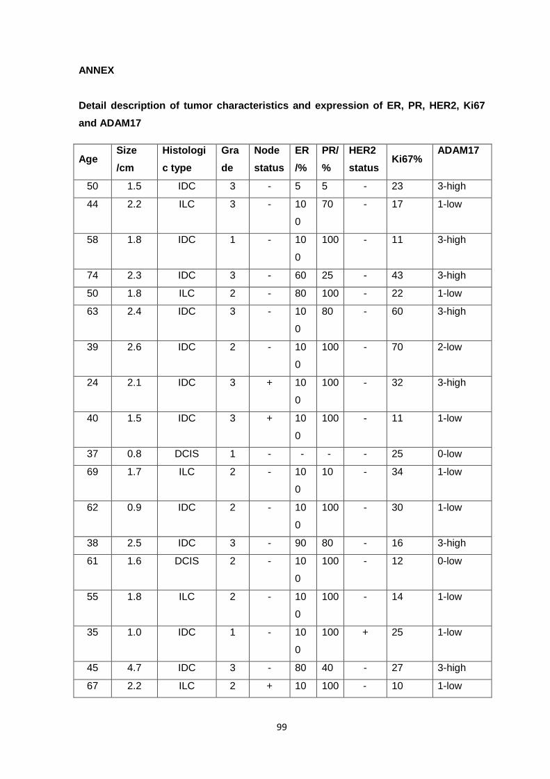
99
ANNEX
Detail description of tumor characteristics and expression of ER, PR, HER2, Ki67 and ADAM17
Age Size /cm
Histologic type
Grade
Node status
ER/%
PR/%
HER2 status
Ki67% ADAM17
50 1.5 IDC 3 - 5 5 - 23 3-high
44 2.2 ILC 3 - 10
0
70 - 17 1-low
58 1.8 IDC 1 - 10
0
100 - 11 3-high
74 2.3 IDC 3 - 60 25 - 43 3-high
50 1.8 ILC 2 - 80 100 - 22 1-low
63 2.4 IDC 3 - 10
0
80 - 60 3-high
39 2.6 IDC 2 - 10
0
100 - 70 2-low
24 2.1 IDC 3 + 10
0
100 - 32 3-high
40 1.5 IDC 3 + 10
0
100 - 11 1-low
37 0.8 DCIS 1 - - - - 25 0-low
69 1.7 ILC 2 - 10
0
10 - 34 1-low
62 0.9 IDC 2 - 10
0
100 - 30 1-low
38 2.5 IDC 3 - 90 80 - 16 3-high
61 1.6 DCIS 2 - 10
0
100 - 12 0-low
55 1.8 ILC 2 - 10
0
100 - 14 1-low
35 1.0 IDC 1 - 10
0
100 + 25 1-low
45 4.7 IDC 3 - 80 40 - 27 3-high
67 2.2 ILC 2 + 10 100 - 10 1-low

100
0
54 0.6 ILC 1 - 10
0
100 - 13 1-low
52 1.1 IDC 2 - 10
0
100 - 17 1-low
52 1.8 IDC 3 - - - - 61 3-high
40 1.8 IDC 3 +
50
- ++ 60 2-low
49 3.6 IDC 2 +
80
60 - 40 1-low
53 2.8 ILC 2 - 10
0
100 - 23 1-low
67 2.8 IDC 2 - 10
0
100 - 10 3-high
55 1.2 IDC 2 - 10
0
100 + 36 3-high
45 1.2 IDC 3 - 80 80 - 8 1-low
56 0.9 ILC 2 - 80 80 - 9 1-low
45 2.0 IDC 3 + 10
0
75 - 11 1-low
45 1.0 IDC 1 - 10
0
100 - 10 3-high
52 2.1 DCIS 1 - - - - 9 1-low
39 1.4 ILC 2 - 10
0
100 - 9 1-low
60 2.0 IDC 3 + 10
0
100 - 21 1-low
38 1.6 DCIS 2 - 10
0
100 + 27 1-low
72 2.1 DCIS 3 - - - - 2 1-low
50 2.8 IDC 3 + 10
0
100 - 51 1-low
39 3.1 ILC 2 - 10
0
100 - 3 3-high
39 1.6 IDC 3 - 60 30 - 28 3-high
41 2.9 IDC 3 - - - - 46 3-high
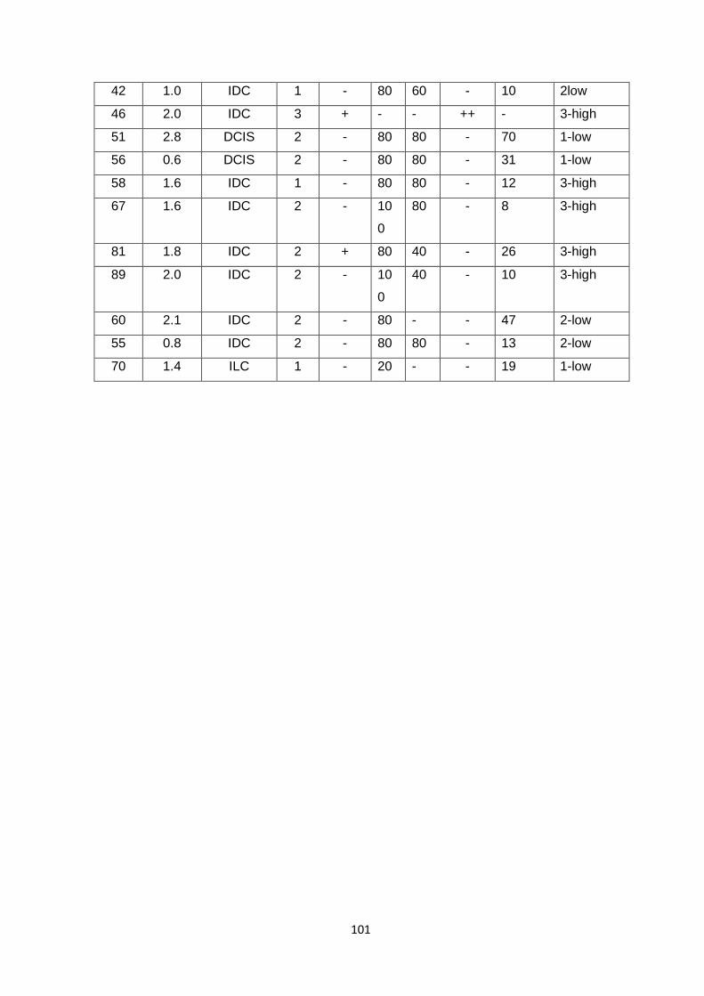
101
42 1.0 IDC 1 - 80 60 - 10 2low
46 2.0 IDC 3 + - - ++ - 3-high
51 2.8 DCIS 2 - 80 80 - 70 1-low
56 0.6 DCIS 2 - 80 80 - 31 1-low
58 1.6 IDC 1 - 80 80 - 12 3-high
67 1.6 IDC 2 - 10
0
80 - 8 3-high
81 1.8 IDC 2 + 80 40 - 26 3-high
89 2.0 IDC 2 - 10
0
40 - 10 3-high
60 2.1 IDC 2 - 80 - - 47 2-low
55 0.8 IDC 2 - 80 80 - 13 2-low
70 1.4 ILC 1 - 20 - - 19 1-low