Embed Size (px)
DESCRIPTION
Human Anatomy and Physiology Respiratory System
Citation preview
CHAPTER TENRESPIRATORY SYSTEM
Chapter Objectives
At the end of the chapter, the student should be able to:- Describe the purpose of the respiratory system- Differentiate between external and internal respiration- Name all of the structures of the respiratory system- Explain how food and foreign materials are kept out of the respiratory tract- Explain the mechanism for the pulmonary ventilation- List and define five breathing volumes- Describe in which respiration is regulated
General Function
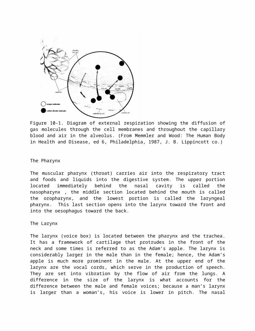
A primary requirement for all body cell activities and growth is oxygen, which is needed to obtain energy from food. The fundamental purpose of the respiratory system is to supply oxygen to the individual tissue cells and to remove their gaseous waste product, carbon dioxide. Breathing, or ventilation, refers to the inhalation and exhalation of air. Air is a mixture of oxygen, nitrogen, carbondioxide and other gases; the pressure of these gases varies, depending on the elevation above sea level. The first, called external expiration, takes place only in the lungs, where oxygen from the outside air enters the blood and carbondioxide leaves the blood to be breathed into the outside air (Figure 10-1). In the second, called internal respiration, gas exchanges take place between the blood and the body cells, with oxygen leaving the blood and entering the cells at the same time that carbon dioxide leaves the cells and enters the blood.
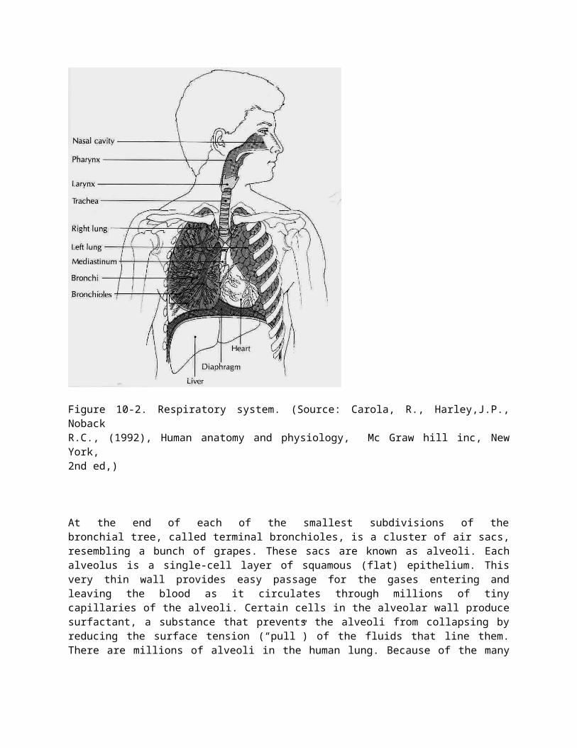
The respiratory system is an intricate arrangement of spaces and passageways that conduct air into the lungs. These spaces include the nasal cavities; the pharynx, which is common to the digestive and respiratory systems; the voice box, or larynx; the windpipe, or trachea; and the lungs themselves, with their conducting tubes and air sacs. The entire system might be thought of as a pathway for airbetween the atmosphere and the blood (Figure 10-2).
Structure and Function of Respiratory Pathways
The Nasal Cavities
Air makes its initial entrance into the body through the openings in the nose called the nostrils. Immediately inside the nostrils, located between the roof of the mouth and the cranium, are the two spaces known as the nasal cavities. These two spaces are separated from each other by a partition, the nasal septum. The septum and the walls of the nasal cavities are constructed of bone covered with mucous membrane. From the lateral (side) walls of each nasal cavity are three projections called the conchae. The conchae greatly increase the surface over winch air must travel on its way through the nasal cavities.
The lining of the nasal cavities is a mucous membrane, which contains many blood vessels that bring heat and moisture to it. The cells of this membrane secrete a large amount of fluid. It is better to breath through the nose than through the mouth because of changes produced in the air as it comes in contact with the lining of the nose:1. Foreign bodies, such as dust particles and pathogens, are filtered out by the hairs of the nostrils or caught in the surface mucus.2. Air is warned by the blood in the vascular membrane.3. Air is moistened by the liquid secretion
The sinuses are small cavities lined with mucous membrane in the bones of the skull. The sinuses communicate with the nasal cavities, and they are highly susceptible to infection.
Figure 10-1. Diagram of external respiration showing the diffusion of gas molecules through the cell membranes and throughout the capillary blood and air in the alveolus. (From Memmler and Wood: The Human Body in Health and Disease, ed 6, Philadelphia, 1987, J. B. Lippincott co.)
The Pharynx
The muscular pharynx (throat) carries air into the respiratory tract and foods and liquids into the digestive system. The upper portion located immediately behind the nasal cavity is called the nasopharynx , the middle section located behind the mouth is called the oropharynx, and the lowest portion is called the laryngeal pharynx. This last section opens into the larynx toward the front and into the oesophagus toward the back.
The Larynx
The larynx (voice box) is located between the pharynx and the trachea. It has a framework of cartilage that protrudes in the front of the neck and some times is referred to as the Adam’s apple. The larynx is considerably larger in the male than in the female; hence, the Adam’s apple is much more prominent in the male. At the upper end of the larynx are the vocal cords, which serve in the production of speech. They are set into vibration by the flow of air from the lungs. A difference in the size of the larynx is what accounts for the difference between the male and female voices; because a man’s larynx is larger than a
woman’s, his voice is lower in pitch. The nasal cavities, the sinuses, and the pharynx all serve as resonating chambers for speech, just as the cabinet does for a stereo speaker.
The space between these two vocal cords is called the glottis, and the little leaf-shaped cartilage that covers the larynx during swallowing is called the epiglottis. The epiglottis helps keep food out of the remainder of the respiratory tract. As the larynx moves upward and forward during swallowing, theepiglottis moves downward, covering the opening into the larynx. You can feel the larynx move upward toward the epiglottis during this process by placing the flat ends of your fingers on your larynx as you swallow.
The larynx is lined with ciliated mucous membrane. The cilia trap dust and other particles, moving them upward to the pharynx to be expelled by coughing, sneezing, or blowing the nose.
The Trachea (Windpipe)
The trachea is a tube that extends from the lower edge of the larynx to the upper part of the chest above the heart. It has a framework of cartilages to keep it open. These cartilages, shaped somewhat like a tiny horseshoe or the letter C, are found along the entire length of the trachea. All the open sections of these cartilages are at the back so that the esophagus can bulge into this section during swallowing. The purpose of the trachea is to conduct air between the larynx and the lungs.
The Bronchi and Bronchioles
The trachea divides into two bronchi which enter the lungs. The right bronchus is considerably larger in diameter than the left and extends downward in a more vertical direction. Therefore, if a foreign body is inhaled, it is likely to enter the right lung. Each bronchus enters the lung at a notch or depression called the hilus or hilum. The blood vessels and nerves also connect with the lung in this region.
The Lungs
The lungs are the organs in which external respiration takes place through the extremely thin and delicate lung tissues. The two lungs, set side by side in the thoracic cavity, are constructed in the following manner:
Each bronchus enters the lung at the hilus and immediately subdivides. Because the subdivision of the bronchi resembles the branches of a tree, they have been given the common name bronchial tree. The bronchi subdivide again and again, forming progressively smaller divisions, the smallest of which are called bronchioles. The bronchi contain small bits of cartilage, which give firmness to the walls and serve to hold the passageways open so that air can pass in and out easily. However, as the bronchi become smaller, the cartilage decreases in amount. In the bronchioles there is no cartilage at all; what remains is mostly smoothly muscle, which is under the control of the autonomic nervous system.
Figure 10-2. Respiratory system. (Source: Carola, R., Harley,J.P., NobackR.C., (1992), Human anatomy and physiology, Mc Graw hill inc, New York,2nd ed,)
At the end of each of the smallest subdivisions of the bronchial tree, called terminal bronchioles, is a cluster of air sacs, resembling a bunch of grapes. These sacs are known as alveoli. Each alveolus is a single-cell layer of squamous (flat) epithelium. This very thin wall provides easy passage for the gases entering and leaving the blood as it circulates through millions of tiny capillaries of the alveoli. Certain cells in the alveolar wall produce surfactant, a substance that prevents the alveoli from collapsing by reducing the surface tension (“pull”) of the fluids that line them. There are millions of alveoli in the human lung. Because of the many air spaces, the lung is light in weight; normally a piece of lung tissue dropped into a glass of water will float.
As mentioned the pulmonary circuit brings blood to and from the lungs. In the lungs bl9od passes through the capillaries around the alveoli, where the gas exchange takes place.
The Lung Cavities
The lungs occupy a considerable portion of the thorax cavity, which is separated from the abdominal cavity by the muscular partition known as the diaphragm. Each lung is enveloped in a double sac of serous membrane called the pleura. The portion of the pleura that is attached to the chest wall is called parietal pleura, while the portion that is reflected onto the surface of the lung is called visceral pleura.
The pleural cavity around the lungs is an air-tight space with a partial vacuum, which causes the pressure in this space to be less than atmospheric pressure. Because the pressure inside the lungs is higher than that in the surrounding pleural cavity, the lungs tend to remain inflated. The entire thoracic cavity is flexible, capable of expanding and contracting along with the lungs. The region between the lungs, the mediastinum, contains the heart, great blood vessels, esophagus, trachea, and lymph nodes.
Physiology of Respiration
Pulmonary Ventilation
Ventilation is the movement of air into and out of the lungs, as in breathing. There are two phases of ventilation (Figure10-3):1. Inhalation is the drawing of air into the lungs.2. Exhalation is the expulsion of air from the lungs.
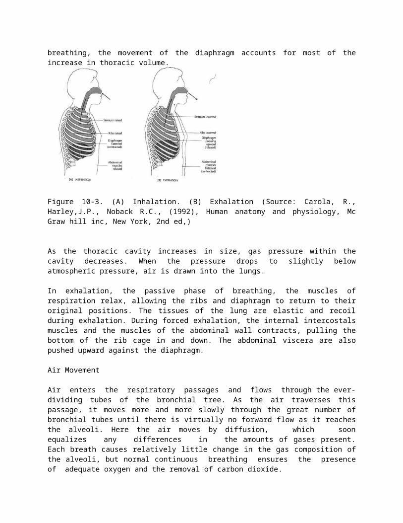
In inhalation, the active phase of breathing, the respiratory muscles contract to enlarge the thoracic cavity. The diaphragm is a strong dome-shaped muscle attached around the base of the rib cage. The contraction and relaxation of the diaphragm cause a piston-like downward motion that result in an increase in the vertical dimension of the chest. The rib cage is also moved upward and outward by contraction of the external intercostals muscles and, during exertion, by contraction of other muscles of the neck and chest. During quiet breathing, the movement of the diaphragm accounts for most of the increase in thoracic volume.
Figure 10-3. (A) Inhalation. (B) Exhalation (Source: Carola, R., Harley,J.P., Noback R.C., (1992), Human anatomy and physiology, Mc Graw hill inc, New York, 2nd ed,)
As the thoracic cavity increases in size, gas pressure within the cavity decreases. When the pressure drops to slightly below atmospheric pressure, air is drawn into the lungs.
In exhalation, the passive phase of breathing, the muscles of respiration relax, allowing the ribs and diaphragm to return to their original positions. The tissues of the lung are elastic and recoil during exhalation. During forced exhalation, the internal intercostals muscles and the muscles of the abdominal wall contracts, pulling the bottom of the rib cage in and down. The abdominal viscera are also pushed upward against the diaphragm.
Air Movement
Air enters the respiratory passages and flows through the ever-dividing tubes of the bronchial tree. As the air traverses this passage, it moves more and more slowly through the great number of bronchial tubes until there is virtually no forward flow as it reaches the alveoli. Here the air moves by diffusion, which soon equalizes any differences in the amounts of gases present. Each breath causes relatively little change in the gas composition of the alveoli, but normal continuous breathing ensures the presence of adequate oxygen and the removal of carbon dioxide.
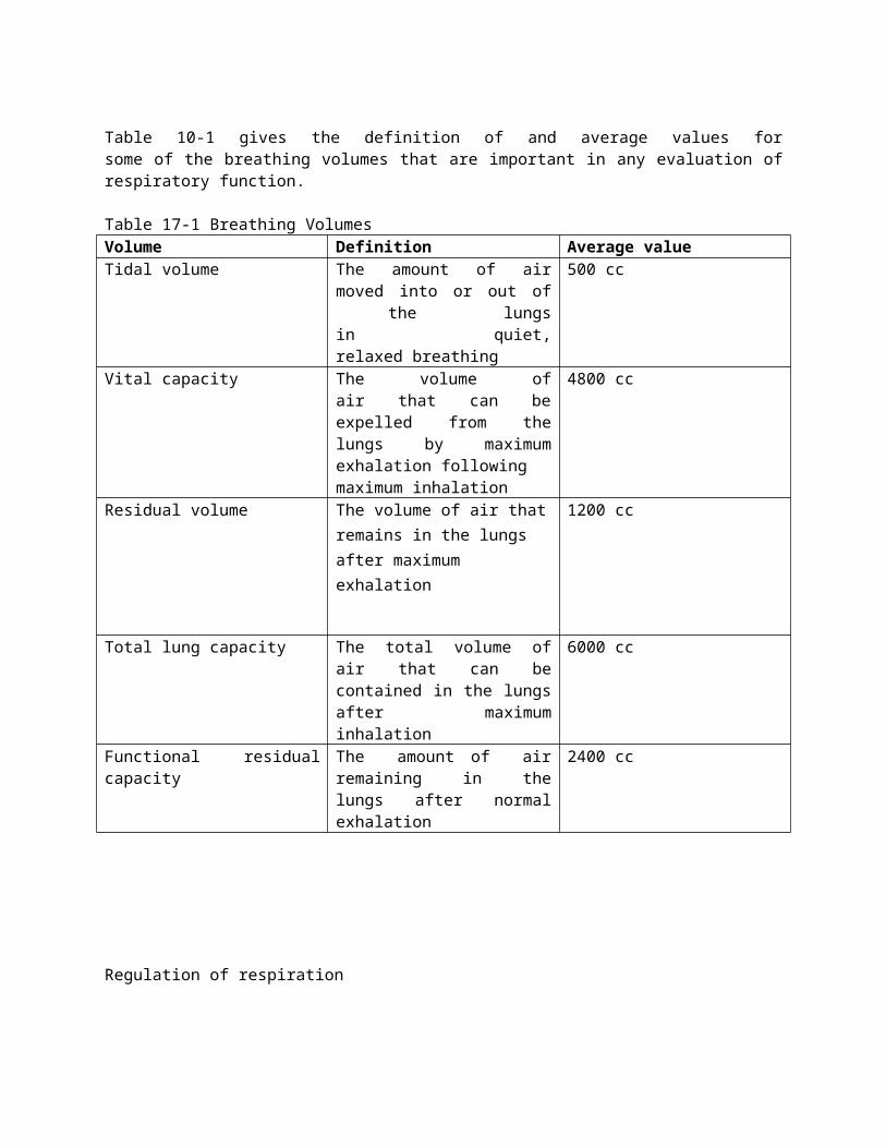
Table 10-1 gives the definition of and average values for some of the breathing volumes that are important in any evaluation of respiratory function.
Table 17-1 Breathing VolumesVolume Definition Average valueTidal volume The amount of air moved into or
out of the lungs in quiet, relaxed breathing
500 cc
Vital capacity The volume of air that can be expelled from the lungs by maximum exhalation followingmaximum inhalation
4800 cc
Residual volume The volume of air that remains in the lungs after maximum exhalation
1200 cc
Total lung capacity The total volume of air that can be contained in the lungs after maximum inhalation
6000 cc
Functional residual capacity The amount of air remaining in the lungs after normal exhalation
2400 cc
Regulation of respiration
Regulation of respiration is a complex process that must keep pace with moment-to-moment changes in cellular oxygen requirements and carbon dioxide production. Regulation depends primarily on the respiratory control centers located in the medulla and pons of the brain stem. Nerve impulses from the medulla are modified by the centers in the pons. Respiration is regulated so that the levels of oxygen, carbon dioxide, and acid are kept within certain limits. The control centers regulate the rate, depth, and rhythm of respiration.
From the respiratory center in the medulla, motor nerve fibers extend into the spinal cord. From the cervical (neck) part of the cord, these nerve fibers continue through the phrenic nerve to the diaphragm. The diaphragm and the other muscles of respiration are voluntary in the sense that they can be regulated by messages from the higher brain centers, notably the cortex. It is possible for a person to deliberately breath more rapidly or more slowly or to hold his breath and not breath at all for a time. Usually we breath without thinking about it, while the respiratory centers in the medulla and pons do the controlling.
Of vital importance in the control of respiration are the chemoreceptors. These receptors are found in structures called the carotid and aortic bodies, as well as out side the medulla of the brain stem. The carotid bodies are located near the bifurcation of the common carotid arteries, while the aortic bodies are located in the aortic arch. These bodies contain many small blood vessels and sensory neurons, which are sensitive to decreases in oxygen supply as well as to increases in carbon dioxide and acidity (H+). Impulses are sent to the brain from the receptors in the carotid and aortic bodies. The receptor cells outside the medulla are affected by the concentration of hydrogen ion in cerebrospinal fluid (CSF) as determined by the concentrations of carbon dioxide in the blood.
Review Questions
1. What is the purpose of respiration and what are its two components?2. Trace the pathway of air from the outside into the blood.3. What are the advantages of breathing through the nose?4. Describe the lung cavities.5. What muscles are used for inhalation? Forceful expiration?6. What are chemoreceptors and how do they function to regulate breathing?7. Define four volumes used to measure breathing.
CHAPTER ELEVEN
DIGESTIVE SYSTEM
Chapter Objectives
At the end of the chapter, the student should be able to:- Name the two main functions of the digestive system- Describe the four layers of the digestive tract wall- Describe the peritoneum- Name and describe the organs of the digestive tract- Name and describe the accessory organs of digestion and biliary apparatus- List the functions of each organ involved in digestion- Explain the role of enzymes in digestion and give examples of enzymes- Name the end products of fats, proteins, and carbohydrates digestion- Define absorption- Define villi and state how villi function in absorption- Describe how bile functions in digestion- List the main functions of the liver- Explain the use of feedback in regulating digestion and give several examples
General Function
Every body cell needs a constant supply of nutrients to proviide energy and building blocks for the manufacture of body substances. Food as we take it in, however, is too large to enter the cells. It must first be broken down into particles small enough to pass through the cell membrane. This process is known as digestion. After digestion, food must be carried to the cells in every part of the body by the circulation. The transfer of food into the circulation is called absorption. Digestion and absorption are the two chief functions of the digestive system.
Structure and Function of Organs of Digestion and
Accessory Organs
For our purpose the digestive system may be divided into two groups of organs:
1. The digestive tract, a continuous passageway beginning at the mouth, where food is taken in, and terminating at the anus, where the solid waste products of digestion are expelled from the body2. The accessory organ, which are necessary for the digestive process but are not a direct part of the digestive tract. They release substances into the digestive tract through ducts.
The Walls of the Digestive Tract
Although specified for specific tasks in different organs, the wall of the digestive tract, from the esophagus to the anus, is similar in structure throughout. Follow the diagram of the small intestine in Figure 11-1 as we describe the layers of this wall from the innermost to the outermost surface. First is the mucous membrane, so called because its epithelial layer contains many mucus-secreting cells. The type of epithelium is simple columnar. The layer of connective tissue beneath this, the submucosa, contains blood vessels and some of the nerves that help regulate digestive activity. Next are two
layers of smooth muscle. The inner layer has circular fibers, and the outer layer has longitudinal fibers. The alternate contractions of these muscles create the wavelike movement that propels food through the digestive tract and mixes it with digestive juices. This movement is called peristalsis. The outermost layer of the wall consists of fibrous connective tissue. Most of the abdominal organs have an additional layer of serous membrane that is part of the peritoneum.
The Peritoneum
The abdominal cavity is lined with a thin, shiny serous membrane that also covers most of the abdominal organs (Figure 11-2). The portion of this membrane that lines the abdomen is called the parietal peritoneum; that covering the organ is called the visceral peritoneum. In addition to these single layered portions of the peritoneum there are a number of double-layered structures that carry blood vessels, lymph vessels, and nerves, and sometimes act as ligaments supporting the organs. The mesentery is a double-layered portion of the peritoneum shaped somewhat like a fan. The handle portion is attached to the back wan, and the expanded long edge is attached to the small intestine. Between the two layers of membrane that fill the mesentery are the blood vessels, lymphatic vessels, and nerves that supply the intestine. The section of the peritoneum that extends from the colon to the back wall is the mesocolon.
A large double layer of the peritoneum containing much fat hangs like an apron over the front of the intestine. This greater omentum extends from the lower border of the stomach into the pelvic part of the abdomen and then loops back up to the transverse colon. There is also a smaller membrane, called the lesser omentum that extends between the stomach and the liver.
Figure 11-1. Diagram of the wall of the small intestine showing the numerous villi. (From Memmler, Ruth Lundeen et al: The human body in Health and disease,ed. 8, New York, 1996,Lippincott.)
Figure 11-2. Diagram of the abdominal cavity showing the peritoneum(Source: Carola, R., Harley,J.P., Noback R.C., (1992), Human anatomy and physiology, Mc Graw hill inc, New York, 2nd ed,)
The Digestive Tract
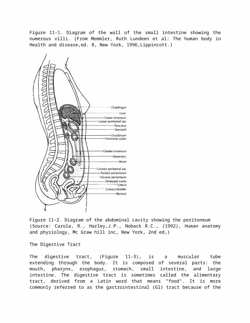
The digestive tract, (Figure 11-3), is a muscular tube extending through the body. It is composed of several parts: the mouth, pharynx, esophagus, stomach, small intestine, and large intestine. The digestive tract is sometimes called the alimentary tract, derived from a Latin word that means "food". It is more commonly referred to as the gastrointestinal (Gl) tract because of the major importance of the stomach and intestine in the process of digestion.
Figure 11-3. Digestive system (Source: Carola, R., Harley,J.P., NobackR.C., (1992), Human anatomy and physiology, Mc Graw hill inc, New York,2nd ed,)
The Mouth
The mouth, also called the oral cavity, is where a substance begins its travels through the digestive tract (Figure 11-4). The mouth has three digestive functions:1. To receive food, a process called ingestion2. To prepare food for digestion3. To begin the digestion of starch.
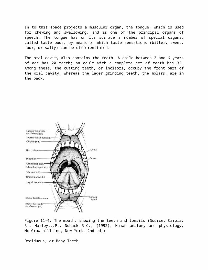
In to this space projects a muscular organ, the tongue, which is used for chewing and swallowing, and is one of the principal organs of speech. The tongue has on its surface a number of special organs, called taste buds, by means of which taste sensations (bitter, sweet, sour, or salty) can be differentiated.
The oral cavity also contains the teeth. A child between 2 and 6 years of age has 20 teeth; an adult with a complete set of teeth has 32. Among these, the cutting teeth, or incisors, occupy the front part of the oral cavity, whereas the lager grinding teeth, the molars, are in the back.
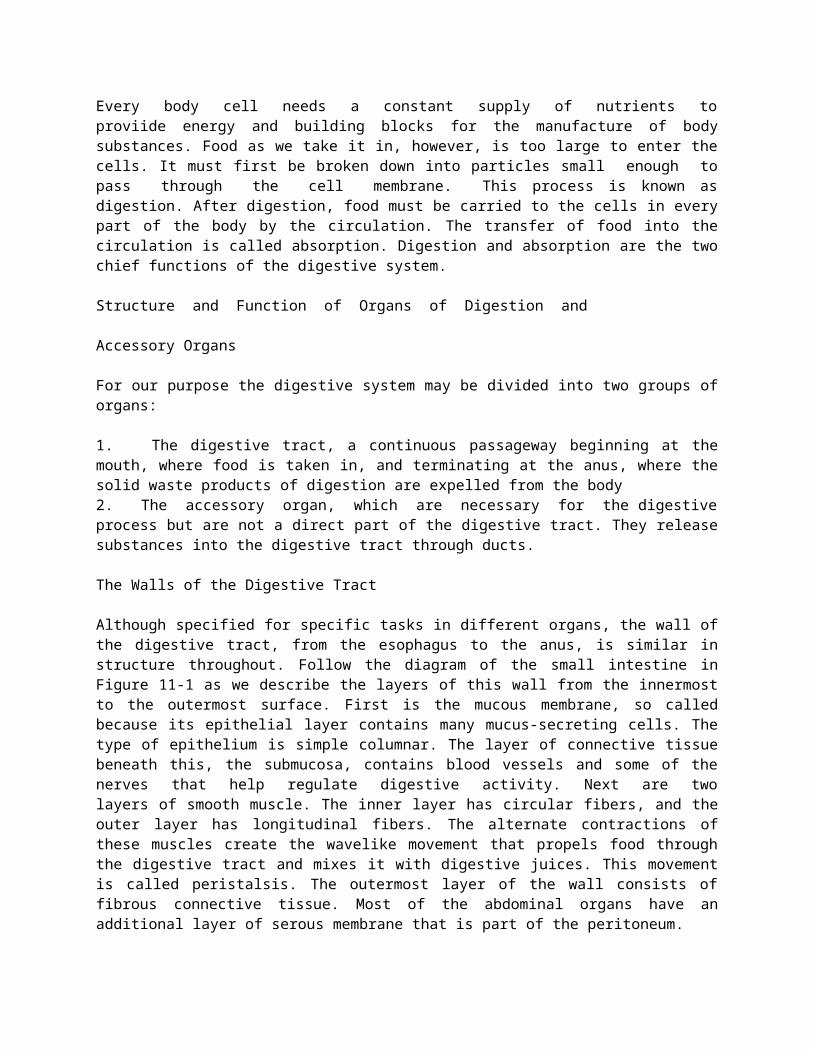
Figure 11-4. The mouth, showing the teeth and tonsils (Source: Carola, R., Harley,J.P., Noback R.C., (1992), Human anatomy and physiology, Mc Graw hill inc, New York, 2nd ed,)
Deciduous, or Baby Teeth
The first eight deciduous teeth to appear through the gums are the incisors. Later the canines (eye teeth) and molar appear. Usually, the 20 baby teeth have all appeared by the time a child has reached the age of 2 or 21/2 years. During the first 2 years the permanent teeth develop within the jawbonesfrom buds that are present at birth. The first permanent tooth to appear is the important 6-year molar. This permanent tooth comes in before the baby incisors are lost. Because decay and infection of adjacent deciduous molars may spread to and involve new, permanent teeth, deciduous teeth need proper care.
Permanent Teeth
As a child grows, the jawbones grow, making space for additional teeth. After the 6-year molars have appeared, the baby incisors loosen and are replaced by permanent incisors. Next, the baby canines (cuspids) are replaced by permanent canines, and finally, the baby molars are replaced by the bicuspids (premolars) of the permanent teeth.
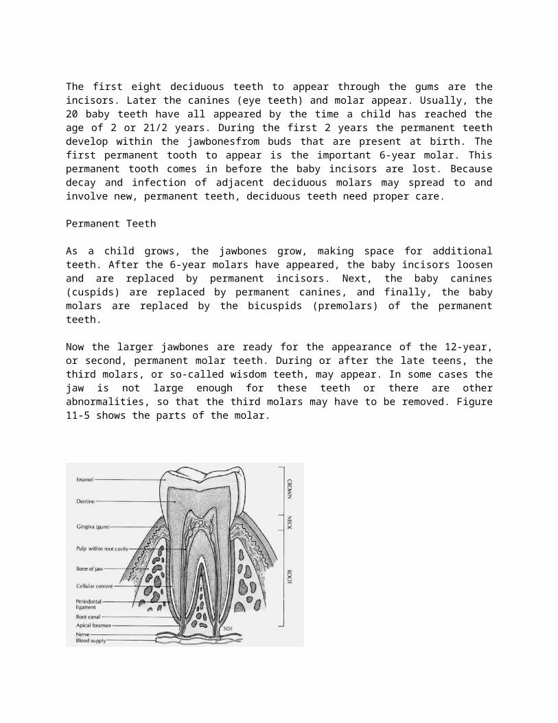
Now the larger jawbones are ready for the appearance of the 12-year, or second, permanent molar teeth. During or after the late teens, the third molars, or so-called wisdom teeth, may appear. In some cases the jaw is not large enough for these teeth or there are other abnormalities, so that the third molars may have to be removed. Figure 11-5 shows the parts of the molar.
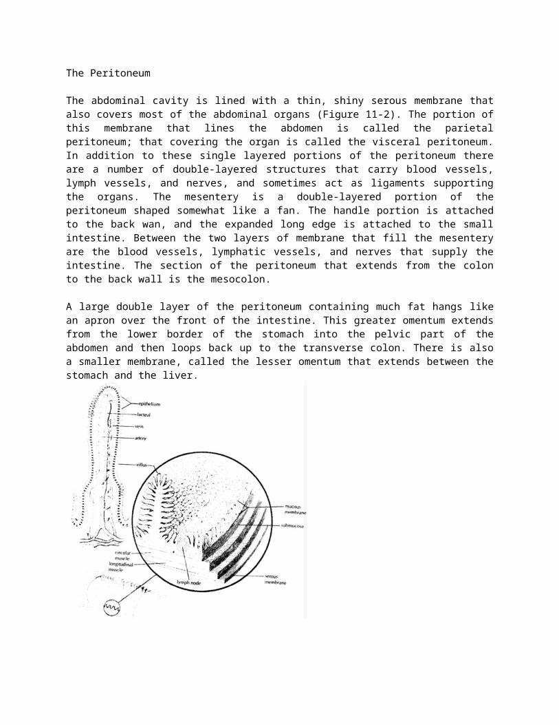
Figure 11-5. A molar tooth (Source: Carola, R., Harley,J.P., Noback R.C., (1992), Human anatomy and physiology, Mc Graw hill inc, New York, 2nd ed,)
The Salivary Glands
While food in the mouth, it is mixed with saliva, one purpose of which is to moisten the food and facilitate the processes of chewing, or mastication, and swallowing, or deglutition. Saliva also helps keep the teeth and mouth clean and reduce bacterial growth.
This watery mixture contains mucus and an enzyme called salivary amylase, which begins the digestive process by converting starch to sugar. It is manufactured mainly by three pairs of glands that function as accessory organs:1. The parotid glands, the largest of the group, are located below and in front of the ear.2. The submandibular, or sumaxillary, glands are located near the body of the lower jaw3. The sublingual glands are under the tongue.
All these glands empty by means of ducts into the oral cavity.
The Pharynx and Esophagus
The pharynx is commonly referred to as the throat. The oral part of the pharynx is visible when you look into an open mouth and depress the tongue. The palatine tonsils may be seen at either side. The pharynx also extends upward to the nasal cavity and downward to the level of the larynx. The soft palate is tissue that forms the back of the roof of the oral cavity. From it hangs a soft, fleshy, V -shaped mass called the uvula.
In swallowing, a small portion of chewed food mixed with saliva, called a bolus, is pushed by the tongue into the pharynx, swallowing occurs rapidly by an involuntary reflex action. At the same time, the soft palate and uvula are raised to prevent food and liquid from entering the nasal cavity, and the tongue is raised to seal the back of the oral cavity. The entrance of the trachea is guarded during swallowing by a leaf-shaped cartilage, the epiglottis, which covers the opening
of the larynx. The swallowed food is then moved by peristalsis into esophagus, a muscular tube about 25cm (10 inches) long that carries food into the stomach. No additional digestion occurs in the esophagus.
Before joining the stomach, the esophagus must pass through the diaphragm. It passes through a space in the diaphragm called the esophageal hiatus. If there is a weakness in the diaphragm at this point, a portion of the stomach or other abdominal organ may protrude through the space, a condition called hiatal hernia.
The Stomach
The stomach is an expanded J-shaped organ in the upper left region of the abdominal cavity (Figure 11-6). In addition to the two muscle layers already described, it has a third, inner oblique (angled) layer that aids in grinding food and mixing it with digestive juices. The left-facing arch of the stomach is the greater curvature, whereas the right surface forms the lesser curvature. Each end of the stomach is guarded by a muscular ring, or sphincter, that permits the passage of substances in only one direction.
Between the esophagus and the stomach is the lower esophageal sphincter (LES). This valve has also been called cardiac sphincter because it separates the esophagus from the region of the stomach that is close to the heart. We are sometimes aware of the existence of this sphincter; sometimes it does not relax as it should, produce a feeling of being unable to swallow past that point. Between the distal, or far, end of the stomach and the small intestine is the pyloric sphincter. The region of the stomach leading into this sphincter, the pylorus, is important in regulating how rapidly food moves into the small intestine.
The stomach serves as a storage pouch, digestive organ, and churn. When the stomach is empty, the lining forms many folds called rugae. These folds disappear as the stomach expands. (It may be stretched to hold one half of a gallon of food and liquid.) Special cells in the lining of the stomach secrete substances that mix together to form gastric juice, the two main components of which are:1. Hydrochloric acid (HCL), a s1rong acid that softens the connective tissue in meat and destroys foreign organisms2. Pepsin, a protein-digesting enzyme. This enzyme is produced in an inactive form and is activated only when food enters the stomach and HCL is produced.The semi-liquid mixture of gastric juice and food that leaves the stomach to enter the small intestine is called chyme.
The Small Intestine
The small intestine is the longest part of the digestive tract. It is known as the small intestine because, although it is longer than the large intestine, it is smaller in diameter, with an average width of about 2.5 cm (1 inch). When relaxed to its full length, the small intestine is about 6 m (20 feet) long. The first 25 cm (10 inches) or so of the small intestine make up the duodenum. Beyond the duodenum are two more divisions: the jejunum, which forms the next two fifths of the small intestine, and the ileum, which constitutes the remaining portion.
The wall of the duodenum contains glands that secrete large amounts or mucus to protect the small intestine from the strongly acid chyme entering from the stomach. Cells of the small intestine also secrete enzymes that digest proteins and carbohydrates. In addition, digestive juices from the liver and
pancreas enter the small intestine through a small opening in the duodenum. Most of the digestive process takes place in the small intestine under the effects of these juices.
Most absorption of digested food also occurs through the walls of the small intestine. To increase the surface area of the organ for this purpose, the mucosa is formed into millions of tiny, finger-like projections, called villi (see Figure 11-1), Which give the inner surface a velvety appearance. In addition, each epithelial cell has small projecting folds of the cell membrane known as microvilli. These create a remarkable increase in the total surface area available in the small intestine for the absorption of nutrients.
The Large Intestine
Any material that cannot be digested as it passes through the digestive tract must be eliminated from the body. In addition, most of the water secreted into the digestive tract for proper digestion must be reabsorbed into the body to prevent dehydration. The storage and elimination of undigested waste and the reabsorption of water are the functions of the large intestine.
The large intestine is about 6.5 cm (2.5 inches) in diameter and about 1.5 m (5 feet) long. The outer longitudinal muscle fibers form three separate bands on the surface. These bands draw up the wall of the organ to give it its distinctive puckered appearance.
The large intestine begins in the lower right region of the abdomen. The first part is a small pouch called the cecum. Between the ileum of the small intestine and the cecum is a sphincter, the ileocecal valve that prevents food from traveling backward into the small intestine. Attached to the cecum is a small, blind tube containing lymphoid tissue; it is called the vernriform appendix (vermiform means "wotmlike"). Inflammation of this tissue as a result of infection or obstruction is appendicitis.
The second portion, the ascending colon, extends upward along the right side of the abdomen toward the liver. The large intestine then bends across the abdomen, forming the transverse colon. At this point it bends sharply and extends downward on the left side of the abdomen into the pelvis, forming the descending colon. The lower part of the colon bends posteriorly in an S shape and continues downward as the sigmoid colon. The sigmoid colon empties into the rectum, which serves as a temporary storage area for indigestible or unabsorbable food residue (see Figure 11-3). Enlargement of the veins in this area constitutes haemorrhoids. A narrow portion of the distal large intestine is called the anal canal. This leads to the outside of the body through an opening called the anus.
Large quantities of mucus, but no enzymes, are secreted by the large intestine. At intervals, usually after meals, the involuntary muscles within the walls of the large intestine propel solid waste material, called feces or stool, toward the rectum. This material is then eliminated from the body by both voluntary an involuntary muscle actions, a process called defecation.
While the food residue is stored in the large intestine, bacteria that nomla11y live in the colon act on it to produce vitamin K and some of the B-complex vitamins. As mentioned systemic antibiotic therapy may destroy these bacteria and others living In the large intestine, causing undesirable side effects.
The Accessory Structures
The Liver
The liver, often referred to by the word root hepat, is the largest glandular organ of the body (Figure 11-7). It is located in the upper right portion of the abdominal cavity under the dome of the diaphragm. The lower edge of a normal-sized liver is level with the lower margin of the ribs. The human liver is the same reddish brown color as the animal liver seen in the supermarket. It has a large right lobe and a smaller left lobe; the right lobe includes two inferior smaller lobes. The liver is supplied with blood through two vessels: the portal vein and the hepatic artery. These vessels deliver about 11/2 quarts of blood to the liver every minute. The hepatic artery carries oxygenated blood, whereas the portal system of veins carries blood that is rich in the end products of digestion. This most remarkable organ has so many functions that only some of its major activities can list here:
1. The storage of glucose (simple sugar) in the form of glycogen, in animal starch. When the blood sugar level falls below normal, liver cells convert glycogen to glucose and release it into the bloodstream; this serves to restore the normal concentration of blood sugar.2. The formation of blood plasma proteins, such as albumin, globulins, and clotting factors3. The synthesis of urea, a waste product of protein metabolism. Urea is released into the blood and transported to the kidneys for elimination.4. The modification of fats, so cells all over the body can use them more efficiently5. The manufacture of bile6. The destruction of old red blood cells. The pigment released from these cells in both the liver and the spleen is eliminated in the bile. This pigment (bilirubin) gives the stool its characteristic dark color.7. The detoxification (removal of the poisonous properties) of harmful substances such as alcohol and certain drugs8. The storage of some vitamins and iron
The main digestive function of the liver is the production of bile. The salts contained in bile act like a detergent to emulsify fat, that is, to break up fat into small droplets that can be acted on more effectively by digestive enzymes. Bile also aids in the absorption of fat from the small intestine. Bile leaves the lobes of the liver by two ducts that merge to form the common hepatic duct. After collecting bile from the gallbladder, this duct, now called common bile duct, delivers bile into the duodenum.
The Gallbladder
The gallbladder is a muscular sac on the inferior surface of the liver that serves as a storage pouch for bile. Although the liver may manufacture bile continuously, the body is likely to need it only a few times a day. Consequently, bile from the liver flows into the hepatic ducts and then up through the cystic duct connected with the gallbladder. When chyme enters the duodenum, the gallbladder contracts, squeezing bile through the cystic duct and into the common bile duct leading to the duodenum.
The Pancreas
The pancreas is a long gland that extends from the duodenum to the spleen. The pancreas produces enzymes that digest fats, proteins, carbohydrates, and nucleic acids. The protein digesting enzymes are produced in inactive forms, which must be converted to active forms in the small intestine by other
enzymes. The pancreas also produces large amounts of alkaline fluid, which neutralizes the chyme in the small intestine, thus protecting the lining of the digestive tract. These juices collect in a main duct that joins the common bile duct or empties into the duodenum near the common bile duct. Most persons also have an additional smaller duct that opens into the duodenum.
Because they arte usually confined to proper channels, pancreatic enzymes do not damage body tissues. However, if the bile ducts become blocked, pancreatic enzymes back up into the pancreas. Also, in some cases of gallbladder disease, disease, infection may extend to the pancreas and cause abnormal activation of the pancreatic enzymes. In either circumstance, the pancreas suffers destruction by its own juice, and the outcome can be fatal; this condition is known as acute pancreatitis.
The pancreas also functions as an endocrine gland, producing the hormones insulin and glucagons that regulate sugar metabolism. These secretions of the islets cells are released directly into the blood.
Digestion and Absorption of Carbohydrates, Fats, and Proteins
Digestion
Digestion, a complex process that occurs in the alimentary canal, consists of physical and chemical changes that prepare food for absorption. Mechanical digestion breaks food into tiny particles, mixes them with digestive juices, moves them along the alimentary canal, and finally eliminates the digestive wastes from the body. Chewing or mastication, swallowing or deglutition, peristalsis, and defecation are the main processes of mechanical digestion. Chemical digestion breaks down large, nonabsorbable food molecules−molecules that are able to pass through the intestinal mucosa into blood and lymph. Chemical digestion consists of numerous chemical reactions catalyzed by enzymes in saliva, gastric juice, pancreatic juice, and intestinal juice.
Carbohydrate Digestion
Very little digestion of carbohydrates (starches and sugars) occurs before food reaches the small intestine. Salivary amylase usually has little time to do its work because so many of us swallow our food so fast. Gastric juice contains no carbohydrate-digesting enzymes. But after the food reaches the small intestine, pancreatic and intestinal juice enzymes digest the starches and sugars. A pancreatic enzyme (amylase) starts the process by changing starches into a double sugar, namely, maltose. Three intestinal enzymes−rnaltase, sucrase, and lactase−digest double sugars by changing them into simple sugars, chiefly glucose (dextrose). Maltase digests maltose (malt sugar), sucrase digests sucrose (ordinary cane sugar), and lactase digests lactose (milk sugar). The end product of carbohydrate digestion is the so-called simple sugar; the most abundant is glucose.
Protein Digestion
Protein digestion starts in the stomach. Two enzymes (renin and pepsin) in the gastric juice cause the giant protein molecules to break up into somewhat simpler compounds. Pepsinogen, a component of gastric juice, is converted into active pepsin enzyme by hydrochloric acid (also in gastric juice). In the intestine, other enzymes (trypsin in the pancreatic juice and peptidases in the intestinal juice)
finish the job of protein digestion. Every protein molecule is made up of many amino acids joined together. When enzymes have split up the large protein molecule into its separate amino acids, protein digestion is completed. Hence the end product of protein digestion is amino acids. For obvious reasons, the amino acids are also referred to as protein building blocks.
Fat Digestion
Very little carbohydrate and fat digestion occurs before food reaches the small intestine. Most fats are undigested until after emulsification by bile in the duodenum (that is, fat droplets are broken into very small droplets). After this takes place, pancreatic lipase splits up the fat molecules into fatty acids and glycerol (glycerine). The end products of fat digestion, then, are fatty acids and glycerol.
Table 11-1 summarizes the main facts about chemical digestion. Enzyme names indicate the type of food digested by the enzyme. For example, the name amylase indicates that the enzyme digests carbohydrates (starches and sugars), protease indicates a protein- digesting enzyme, and lipase means a fat-digesting enzyme. When carbohydrate digestion has been completed, starches (polysaccharides) and double sugars (disaccharides) have been changed mainly to glucose, a simple sugar (monosaccharide). The end products of protein digestion, on the other hand, are amino acids. Fatty acids and glycerol are the end products of fat digestion.
Absorption
After food is digested, it is absorbed; that is, it moves through the mucous membrane lining of the small intestine into the blood and lymph. In other words, food absorption is the process by which molecules of amino acids, glucose, fatty acids, and glycerol goes from the inside of the intestines into the circulating fluids of the body. Absorption of foods is just as essential as digestion of foods. The reason is fairly obvious. As long as food stays in the intestines, it cannot nourish the millions of cells that compose all other parts of the body. Their lives depend on the absorption of digested food and its transportation to them by the circulating blood.
Structural adaptations of the digestive tube, including folds in the lining mucosa, villi, and micro villi, increase the absorptive surface and the efficiency and speed of transfer of materials from the intestinal lumen to body fluids. Many salts such as sodium are actively transported through the intestinal mucosa. Water follows by osmosis. Other nutrients are also actively transported into the blood of capillaries in the intestinal villi. Fats enter the lymphatic vessels or lacteals found in intestinal villi.
Table 11-1 Chemical DigestionDigestive juices and enzymes Substance Digested (or
hydrolyzed)Resulting Products*
SalivaAmylase
Starch (Polysaccharide) Maltose (disaccharide)
Gastric JuiceProtease (Pepsin)Hydrochloric acid
Proteins Partially digested proteins
Pancreatic JuiceProtease (trypsin)LipaseAmylase
Proteins (intact of partially digested)Fats emulsified by bileStarch
Fatty acids, amino acids and GlycerolMaltose
Intestinal JuicePeptidasesSucraseLactase Maltase
PeptidesSucrose (cane sugar)Lactose (milk sugar)Maltose (malt sugar)
Amino AcidsGlucose and fructose (simple sugars)Glucose and galactose(simple sugars Glucose)
*Substances underlined are end products of digestion (that is, completely digested foods ready for absorption)
Review Questions1. What organs form the gastrointestinal tract?2. Trace the jejunum, cecum, colon, duodenum, and ileum.3. If you inserted 9 inches of an enema tube through the anus, the tip of the tube would probably be in what structure?4. What is the peritoneum? Name the two layers and describe their locations. Name four double-layered peritoneal structures.5. Differentiate between deciduous and permanent teeth with respect to kinds and numbers.6. What is peristalsis? Name some structures in which it occurs.7. Name two purpose of the acid in gastric juice.8. Name the principal digestive enzymes. Where is each formed? What does each do?9. Where does absorption occur, and what structures are needed for absorption?10. What types of digested materials are absorbed into the blood?11. What types of digested materials are absorbed into the lymph?12. Name the accessory organs of digestion and the functions of each.13. Name five non-digestive functions of the liver.14. Which digestive juice emulsifies fats?15. What juices digest carbohydrates? Proteins? Fats?16. Where are simple sugars and amino acids absorbed into blood capillaries? Where are lipids absorbed into lacteals?17. Where is most of the water absorbed from the lumen of the digestive tract?