Embed Size (px)
Citation preview

C H A P T E R 3 The Skeletal System
1Around 206—some people develop varying numbers of miscel-laneous bones, either sesamoid bones, which form withinsome tendons as a response to stress (such as the patella) orsutural bones, which develop within the sutures of the skull.
C H E C K P O I N T :
Overview: Skeletal System
1. The average human adult has ___________bones in their body, while the average new-born has ___________ bones in theirs.
2. Explain the difference.
49
C H A P T E R 3
The Skeletal System
Overview: Skeletal System
We are born with 270 bones in our bodies, andeven more bones form during childhood. Bythe time we reach adulthood though, severalseparate bones have fused together so that thenumber of our bones has decreased to around2061, which make up the adult skeletal system.An example of this reduction occurs in eachhalf of our pelvis, where three separate bones—the ilium, the ischium, and the pubis—fuse intoone single bone called the os coxa.
The skeletal system is further divided into theaxial skeleton, consisting of the bones of theskull, vertebral column, and the thoracic cage;and the appendicular skeleton, which con-sists of the bones of the upper and lower extremi-ties along with their associated girdles (Table 3.1).
Naming Bony Processes and Other LandmarksBony landmarks are various ridges, spines, depres-sions, pores, bumps, grooves, and articulating struc-tures on the surface of bones. These structures allowfor the passage of blood vessels and nerves; for jointsbetween bones; and for the attachment of liga-ments, muscles, and tendons. Therefore, a workingknowledge of the names of these structures will be avaluable asset when considering the structure andfunction of these surface features.
Some of the most commonly encountered bonylandmarks are listed in Table 3.2.
Table 3.1 Summary of the Bones of the Adult Skeletal System
Axial Skeleton—80 Bones Appendicular Skeleton—126 Bones
Skull and Hyoid 23 bones Pectoral Girdle 4 bonesInner Ear Ossicles 6 bones Upper Extremities 60 bonesVertebral Column 26 bones Pelvic Girdle 2 bonesSternum and Ribs 25 bones Lower Extremities 60 bones
Total – 206 Bones
Bonus Question: Using this information as a reference, how would this tableappear if it was a list of the bones for a newborn?
bro78143_03_49-144.QXD 9/17/07 2:11 PM Page 49

C H A P T E R 3 The Skeletal System
50
Table 3.2 Common Bony Landmarks
FEATURE DESCRIPTION EXAMPLE
Landmarks of Articulation:
Condyle: Smooth, rounded knob occipital condyle of skull
Facet: Smooth, flat, slightly concave or convex articular facets of vertebraearticular surface
Head: Prominent expanded end of a bone, head of the femursometimes rounded
Elevated Landmarks:
Process: Any bony prominence mastoid process of the skull
Spine: Sharp, slender, or narrow process spine of the scapula
Crest: Narrow ridge iliac crest of the pelvis
Line: Slightly raised, elongated ridge nuchal lines of the skull
Tuberosity: Rough surface tibial tuberosity
Tubercle: Small, rounded process greater tubercle of the humerus
Trochanter: Massive processes unique to the femur greater trochanter of the femur
Epicondyle: Projection superior to a condyle medial epicondyle of the femur
Depressions or Flat Surfaces:
Alveolus: Pit or socket tooth socket
Fossa: Shallow; broad, or elongated basin mandibular fossa
Fovea: Small pit fovea capitis of the femur
Sulcus: Groove for a tendon, nerve, or blood vessel intertubercular sulcus of the Humerus
Spaces or openings:
Foramen: Hole through a bone, usually round foramen magnum of the skull
Fissure: Slit through a bone orbital fissure behind the eye
Meatus or canal: Tubular passage or tunnel through a bone auditory meatus of the ear
Sinus: Space or cavity within a bone frontal sinus of the skull
Bonus Question: Alveolus is a common term in human anatomy. How many other examples of alveoli canyou find in Anatomy & Physiology Revealed (APR)?
C H E C K P O I N T :
Naming Bone Processes and Other Landmarks
1. Explain the difference between a crest and a line.
2. Explain the difference between a condyle andan epicondyle.
3. Explain the difference between a foramenand a fissure.
Lines
ProcessCondyleSpine Foramen
Alveolus
Foramen
Sinuses
Crest
Canal(meatus)
EXERCISE 3.1:
Coloring Exercise
Color in the structures with colored pens orpencils.
bro78143_03_49-144.QXD 9/17/07 2:12 PM Page 50

C H A P T E R 3 The Skeletal System
51
A Few Notes About Naming ProcessesA process is any bony prominence—that is, a pieceof bone that sticks out from the rest of the bone.When it comes to naming these processes, there area few rules that need to be followed to minimizeconfusion. For example, let’s consider when aprocess articulates with another bone, such as thezygomatic process of the temporal bone. Thisprocess is a structure on the temporal bone thatarticulates with the zygomatic bone. The formulafor naming one of these processes is:
the x process of the y bone
where x � the name of bone articulated withand y � the name of the bone it is part of
So, with the ZYGOMATIC process of the TEMPO-RAL bone, x � ZYGOMATIC (the bone articulatedwith) and y � TEMPORAL (the bone it is part of).Now, how does the zygomatic process of the temporalbone compare to the temporal process of the zygo-matic bone? If you are not sure, don’t worry, we willcover these structures shortly.
Let’s look at the styloid process of the temporalbone. This styloid process does not articulate with anyother bone, but it shares its name with the styloidprocesses of both the ulna and radius bones of theforearm. Therefore, it must be named in reference tothe bone that it is part of to prevent confusion—hence the name the styloid process of the temporalbone. What problems would you predict could occurif this distinction is not made in an emergency roomscenario?
Some processes, the mastoid process for exam-ple, are unique in name and do not articulate withany other bones. These require no further clarifica-tion when naming them.
ProcessFossaeSpine
Fovea
Head
CrestTrochanters
Line
Eplcondyles
Condyles
Head
Tubercle
Tuberosity
FossaeEpicondyles
Condyles
C H E C K P O I N T :
A Few Notes About Naming Processes
1. Consider the temporal process of thezygomatic bonea) This process articulates with which bone?b) This process is part of which bone?
2. What is the correct way to say the two styloidprocesses of the forearm?
3. Why is it correct to refer to the mastoidprocess as the mastoid process and not themastoid process of the temporal bone?
bro78143_03_49-144.QXD 9/17/07 2:12 PM Page 51

C H A P T E R 3 The Skeletal System
52
• From the Home screen, clickthe drop-down box on the Selectsystem menu.
• From the systems listed, click onSkeletal, and you will see theimage to the right:
Animation: Joint MovementsWe cannot discuss the bones of the skeleton withoutfirst understanding their movements in reference toeach other and with the rest of the body. The loca-tion of this movement or articulation betweenbones is the joint. In the ANIMATIONS section ofthe Skeletal System, several of these joint move-ments are listed for you. By selecting a specific jointmovement, a definition and short animated exam-ple will explain each one.
EXERCISE 3.2:
Joint Movements
• From the opening screen for the Skeletal Sys-tem click the ANIMATIONS button at the topof the screen.
• From the Select animation menu, select eachof the movements listed under the Joint move-ments heading.
• Click the Play button, and view the animation.
• After viewing the animation and reading the defini-tion, define the following joint movements. Click theSelect animation menu in the ANIMATIONLIST window to select the next term:
Flexion:
Extension:
Abduction:
Adduction:
Pronation:
Supination:
Elevation:
Depression:
Circumduction:
• This is the opening screen forthe Skeletal System.
bro78143_03_49-144.QXD 9/17/07 2:12 PM Page 52
C H A P T E R 3 The Skeletal System
53
Rotation:
Dorsiflexion:
Plantar flexion:
Eversion:
Inversion:
Protraction:
Retraction:
1. Define appositional bone growth.
2. Which cells produce bone material?
3. How is a tunnel formed around a blood vessel?
4. How is the tunnel filled in to produce a newosteon?
5. What is the name for the concentric rings thatform the osteon?
• When you are finished with the animation, clickon the Dissection button at the top of the screento begin the following exercises, or click on theExit button at the top right of the screen to exitAnatomy & Physiology | Revealed®.
C H E C K P O I N T :
Joint Movements
1. A movement that raises a body part is . . . .2. A movement of a body part away from the
main axis of the body or structure is . . . .3. A movement at the ankle so that the dorsum
of the foot is elevated is . . . .4. A movement that decreases the angle
between two bones at a joint is . . . .5. A movement that increases the angle
between two bones at a joint is . . . .
Animation: Appositional Bone Growth
• Return to the ANIMATION LIST, and selectAppositional bone growth.
• Click the Play button, and view the animation.
• After viewing the animation, answer the followingquestions:
H E A D S U P !
Anatomy & Physiology | Revealed® has several
animations available to aid your study of different
systems. Watch for the Animation button at the
bottom of the screen to be highlighted green,
which indicates that an animation is available for
the specific structure(s) you are viewing.
Skeleton (Regions)
EXERCISE 3.3:
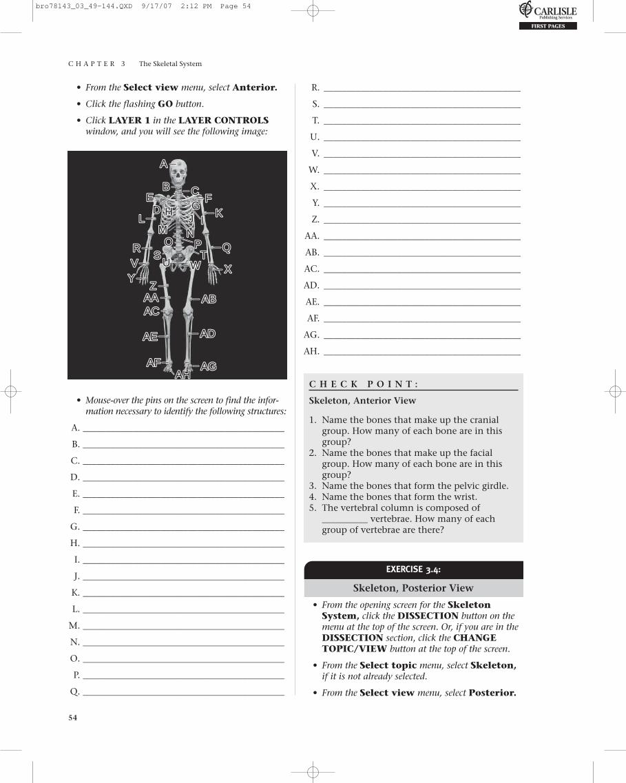
Skeleton, Anterior View
• From the opening screen for the SkeletalSystem, or from the Skeletal SystemAnimations Screen, click the DISSECTIONbutton on the menu at the top of the screen.
• From the Select topic menu, select Skeletalunder the Skeletal (Regions) heading.
bro78143_03_49-144.QXD 9/17/07 2:12 PM Page 53
C H A P T E R 3 The Skeletal System
54
• From the Select view menu, select Anterior.
• Click the flashing GO button.
• Click LAYER 1 in the LAYER CONTROLSwindow, and you will see the following image:
R. ___________________________________________
S. ___________________________________________
T. ___________________________________________
U. ___________________________________________
V. ___________________________________________
W. ___________________________________________
X. ___________________________________________
Y. ___________________________________________
Z. ___________________________________________
AA. ___________________________________________
AB. ___________________________________________
AC. ___________________________________________
AD. ___________________________________________
AE. ___________________________________________
AF. ___________________________________________
AG. ___________________________________________
AH. ___________________________________________
C H E C K P O I N T :
Skeleton, Anterior View
1. Name the bones that make up the cranialgroup. How many of each bone are in thisgroup?
2. Name the bones that make up the facialgroup. How many of each bone are in thisgroup?
3. Name the bones that form the pelvic girdle.4. Name the bones that form the wrist.5. The vertebral column is composed of
__________ vertebrae. How many of eachgroup of vertebrae are there?
AA
BB CC
DDEE FF
GGHHIIJJ KKLL
UU
MM
SSOO
TT
NNPP QQ
XX
ABAB
ADAD
AGAGAHAH
WW
RRVVYY
ZZAAAAACAC
AEAE
AFAF
• Mouse-over the pins on the screen to find the infor-mation necessary to identify the following structures:
A. ____________________________________________
B. ____________________________________________
C. ____________________________________________
D. ____________________________________________
E. ____________________________________________
F. ____________________________________________
G. ____________________________________________
H. ____________________________________________
I. ____________________________________________
J. ____________________________________________
K. ____________________________________________
L. ____________________________________________
M. ____________________________________________
N. ____________________________________________
O. ____________________________________________
P. ____________________________________________
Q. ____________________________________________
EXERCISE 3.4:
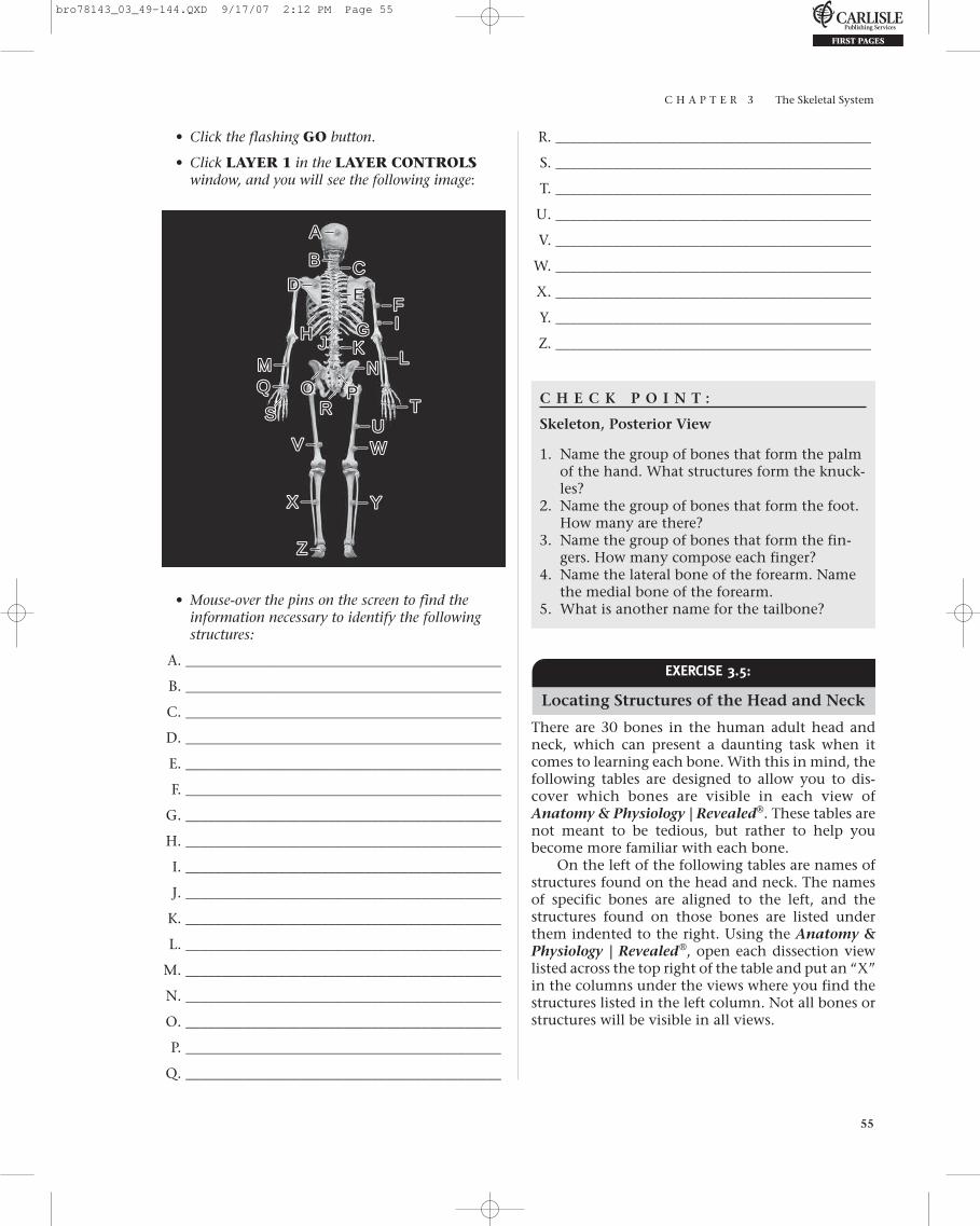
Skeleton, Posterior View
• From the opening screen for the SkeletonSystem, click the DISSECTION button on themenu at the top of the screen. Or, if you are in theDISSECTION section, click the CHANGETOPIC/VIEW button at the top of the screen.
• From the Select topic menu, select Skeleton,if it is not already selected.
• From the Select view menu, select Posterior.
bro78143_03_49-144.QXD 9/17/07 2:12 PM Page 54
C H A P T E R 3 The Skeletal System
55
• Click the flashing GO button.
• Click LAYER 1 in the LAYER CONTROLSwindow, and you will see the following image:
R. ____________________________________________
S. ____________________________________________
T. ____________________________________________
U. ____________________________________________
V. ____________________________________________
W. ____________________________________________
X. ____________________________________________
Y. ____________________________________________
Z. ____________________________________________
AA
BB CCDD EE
FFGGHH
IIJJ
QQRR
KKLL
SS TT
VV
XX
ZZ
MM NNPPOO
UUWW
YY
• Mouse-over the pins on the screen to find theinformation necessary to identify the followingstructures:
A. ____________________________________________
B. ____________________________________________
C. ____________________________________________
D. ____________________________________________
E. ____________________________________________
F. ____________________________________________
G. ____________________________________________
H. ____________________________________________
I. ____________________________________________
J. ____________________________________________
K. ____________________________________________
L. ____________________________________________
M. ____________________________________________
N. ____________________________________________
O. ____________________________________________
P. ____________________________________________
Q. ____________________________________________
C H E C K P O I N T :
Skeleton, Posterior View
1. Name the group of bones that form the palmof the hand. What structures form the knuck-les?
2. Name the group of bones that form the foot.How many are there?
3. Name the group of bones that form the fin-gers. How many compose each finger?
4. Name the lateral bone of the forearm. Namethe medial bone of the forearm.
5. What is another name for the tailbone?
EEXERCISE 3.5:
Locating Structures of the Head and Neck
There are 30 bones in the human adult head andneck, which can present a daunting task when itcomes to learning each bone. With this in mind, thefollowing tables are designed to allow you to dis-cover which bones are visible in each view ofAnatomy & Physiology | Revealed®. These tables arenot meant to be tedious, but rather to help youbecome more familiar with each bone.
On the left of the following tables are names ofstructures found on the head and neck. The namesof specific bones are aligned to the left, and thestructures found on those bones are listed underthem indented to the right. Using the Anatomy &Physiology | Revealed®, open each dissection viewlisted across the top right of the table and put an “X”in the columns under the views where you find thestructures listed in the left column. Not all bones orstructures will be visible in all views.
bro78143_03_49-144.QXD 9/17/07 2:12 PM Page 55
C H A P T E R 3 The Skeletal System
56
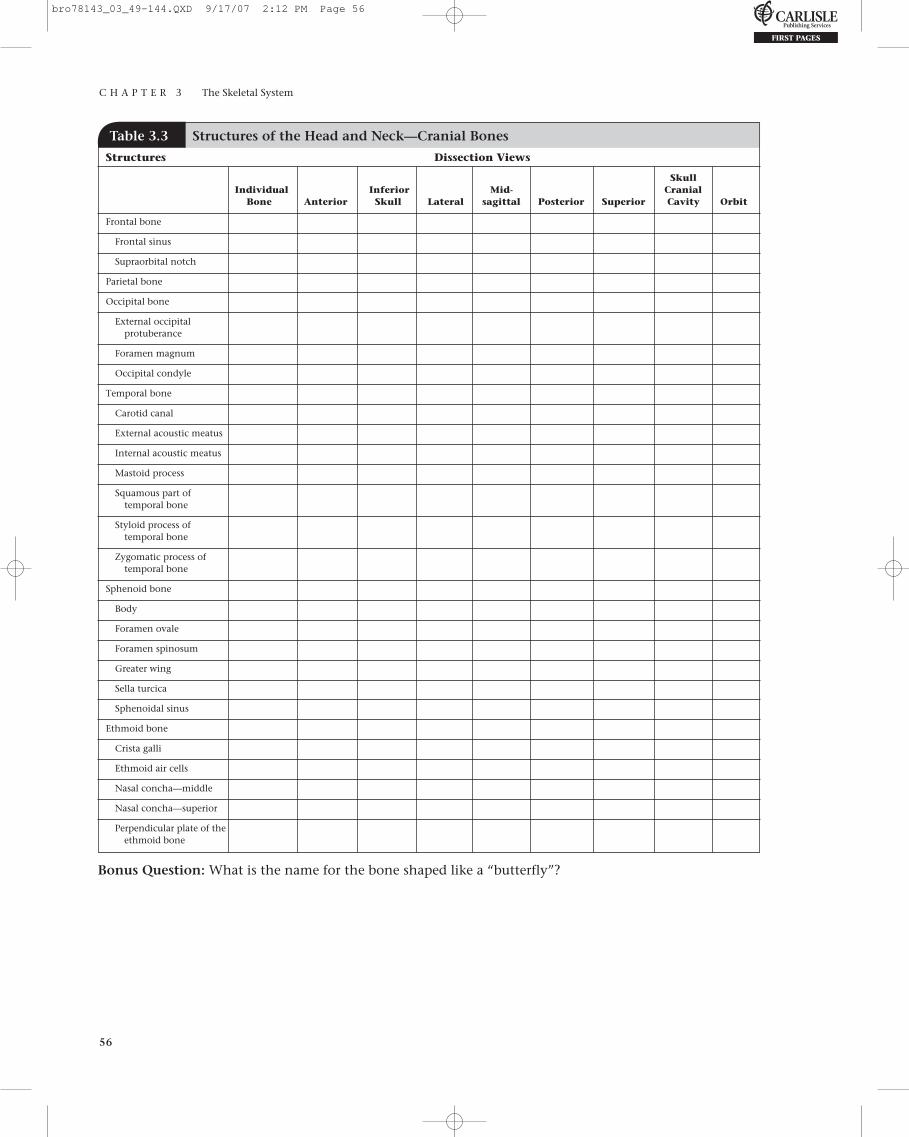
Table 3.3 Structures of the Head and Neck—Cranial Bones
Structures Dissection Views
Skull Individual Inferior Mid- Cranial
Bone Anterior Skull Lateral sagittal Posterior Superior Cavity Orbit
Frontal bone
Frontal sinus
Supraorbital notch
Parietal bone
Occipital bone
External occipital protuberance
Foramen magnum
Occipital condyle
Temporal bone
Carotid canal
External acoustic meatus
Internal acoustic meatus
Mastoid process
Squamous part of temporal bone
Styloid process of temporal bone
Zygomatic process of temporal bone
Sphenoid bone
Body
Foramen ovale
Foramen spinosum
Greater wing
Sella turcica
Sphenoidal sinus
Ethmoid bone
Crista galli
Ethmoid air cells
Nasal concha—middle
Nasal concha—superior
Perpendicular plate of the ethmoid bone
Bonus Question: What is the name for the bone shaped like a “butterfly”?
bro78143_03_49-144.QXD 9/17/07 2:12 PM Page 56

Table 3.4 Structures of the Head and Neck—Facial Bones
Structures Dissection Views
Skull Individual Inferior Mid- Cranial
Bone Anterior Skull Lateral sagittal Posterior Superior Cavity Orbit
Maxilla
Alveolar process of maxilla
Infraorbital foramen
Palatine bone
Zygomatic bone
Temporal process of zygomatic bone
Nasal bone
Vomer bone
Mandible
Alveolar process of mandible
Angle of mandible
Body of mandible
Condylar process of mandible
Coronoid process of mandible
Mandibular foramen
Mental foramen
Ramus of mandible
Bonus Question: What is the anatomical term that refers to the chin area? How do you suppose it receivedthis name?
Table 3.5 Structures of the Head and Neck—Other Skull Structures
Structures Dissection Views
Skull Individual Inferior Mid- Cranial
Bone Anterior Skull Lateral sagittal Posterior Superior Cavity Orbit
Coronal suture
Cranial fossa—anterior
Cranial fossa—middle
Cranial fossa—posterior
Foramen lacerum
Hard palate
Hyoid bone
Jugular foramen
Lambda
Lambdoid suture
Nasal concha (inferior)
Pterion
Sagittal suture
Septal cartilage
Temporomandibular joint (TMJ)
Articular disk of the TMJ
Zygomatic arch
Bonus Question: What is the name for the “cheekbone”?
bro78143_03_49-144.QXD 9/17/07 2:12 PM Page 57

C H A P T E R 3 The Skeletal System
58
Bonus Question: Which bones are characterized by the presence of “transverse foramina”?
EXERCISE 3.6:
Head and Neck, Anterior View
• From the opening screen for the SkeletonSystem, click the DISSECTION button on themenu at the top of the screen. Or, if you are in theDISSECTION section, click the CHANGETOPIC/VIEW button at the top of the screen.
• From the Select topic menu, select Head andneck.
• From the Select view menu, select Anterior.
• Click the flashing GO button.
• Click LAYER 1 in the LAYER CONTROLSwindow, and you will see the following image:
AA
BB CCDDEE FF
GG HH IIJJ
KK
LLMM
RR
NN
PP
SS
QQOO
• Mouse-over the pins on the screen to find theinformation necessary to identify the followingstructures:
A. ____________________________________________
B. ____________________________________________
C. ____________________________________________
D. ____________________________________________
E. ____________________________________________
F. ____________________________________________
G. ____________________________________________
H. ____________________________________________
I. ____________________________________________
J. ____________________________________________
K. ____________________________________________
L. ____________________________________________
M. ____________________________________________
N. ____________________________________________
O. ____________________________________________
P. ____________________________________________
Q. ____________________________________________
R. ____________________________________________
S. ____________________________________________
Table 3.6 Structures of the Head and Neck—Cervical Vertebrae
Structures Dissection Views
Skull Individual Inferior Mid- Cranial
Bone Anterior Skull Lateral sagittal Posterior Superior Cavity Orbit
Atlas (C1 vertebra)
Axis (C2 vertebra)
Cervical vertebrae
Spinous process (cervical)
Transverse process (cervical)
Vertebral body (cervical)
Intervertebral disk
bro78143_03_49-144.QXD 9/17/07 2:12 PM Page 58

C H A P T E R 3 The Skeletal System
59
C H E C K P O I N T :
Head and Neck, Anterior View
1. Name the bones contributing to the orbit.2. Name the bone known as the “collar bone.”3. What structures are formed by the frontal
bone?4. Which is the shortest rib?5. Name the superior part of the sternum.
A. ___________________________________________
B. ___________________________________________
C. ___________________________________________
D. ___________________________________________
E. ___________________________________________
F. ___________________________________________
G. ___________________________________________
H. ___________________________________________
I. ___________________________________________
J. ___________________________________________
K. ___________________________________________
L. ___________________________________________
M. ___________________________________________
N. ___________________________________________
O. ___________________________________________
P. ___________________________________________
Q. ___________________________________________
R. ___________________________________________
S. ___________________________________________
T. ___________________________________________
U. ___________________________________________
V. ___________________________________________
W. ___________________________________________
X. ___________________________________________
Y. ___________________________________________
Z. ___________________________________________
AA. ___________________________________________
AB. ___________________________________________
AC. ___________________________________________
AD. ___________________________________________
AE. ___________________________________________
AF. ___________________________________________
AG. ___________________________________________
AH. ___________________________________________
AI. ___________________________________________
EXERCISE 3.7:
Head and Neck, Lateral View
• From the opening screen for the SkeletonSystem, click the DISSECTION button on themenu at the top of the screen. Or, if you are in theDISSECTION section, click the CHANGETOPIC/VIEW button at the top of the screen.
• From the Select topic menu, select Head andneck, if it is not already selected.
• From the Select view menu, select Lateral.
• Click the flashing GO button.
• Click LAYER 1 in the LAYER CONTROLSwindow, and you will see the following image:
AABB
CC
DDEE
VV
FF
RR
AEAE
AHAHAIAI
GGHH II KK
AFAF AGAG
ZZ
JJLL
PP
WWXX YYABAB
ADADACAC
SSAAAA
UU TT
MMNNOO
• Mouse-over the pins on the screen to find theinformation necessary to identify the followingstructures:
bro78143_03_49-144.QXD 9/17/07 2:12 PM Page 59
C H A P T E R 3 The Skeletal System
60
C H E C K P O I N T :
Head and Neck, Lateral View
1. Name the only bone in the body that doesnot articulate with any other bone.
2. Name the two bone processes that make upthe zygomatic arch.
3. Name the “sockets” for the teeth.4. Describe the pterion.5. What structures of which bones form the
temporomandibular joint?
• Mouse-over the pins on the screen to find the infor-mation necessary to identify the following structures:
A. ____________________________________________
B. ____________________________________________
C. ____________________________________________
D. ____________________________________________
E. ____________________________________________
F. ____________________________________________
G. ____________________________________________
H. ____________________________________________
I. ____________________________________________
J. ____________________________________________
K. ____________________________________________
L. ____________________________________________
M. ____________________________________________
N. ____________________________________________
O. ____________________________________________
P. ____________________________________________
Q. ____________________________________________
EXERCISE 3.8:
Thorax, Anterior View
• From the opening screen for the SkeletonSystem, click the DISSECTION button on themenu at the top of the screen. Or, if you are in theDISSECTION section, click the CHANGETOPIC/VIEW button at the top of the screen.
• From the Select topic menu, select Thorax.
• Anterior will be visible in the Select viewmenu.
• Click the flashing GO button.
• Click LAYER 1 in the LAYER CONTROLSwindow, and you will see the following image:
AABBCCDD EE FFHH
II
KKLLMM
NN
PPOO
JJ
GG
C H E C K P O I N T :
Thorax, Anterior View
1. Name the three parts of the sternum.2. Name a landmark for intramuscular injections.3. Name the structures that attach the ribs to
the sternum.4. Name the two bones that form the gleno-
humeral joint.5. Name the structure found between the bodies
of all but two vertebrae. This structure is lack-ing between which two vertebrae?
EXERCISE 3.9:
Abdomen, Anterior View
• From the opening screen for the SkeletonSystem, click the DISSECTION button on themenu at the top of the screen. Or, if you are in theDISSECTION section, click the CHANGETOPIC/VIEW button at the top of the screen.
• From the Select topic menu, select Abdomen.
• Anterior will be visible in the Select view menu.
bro78143_03_49-144.QXD 9/17/07 2:12 PM Page 60

C H A P T E R 3 The Skeletal System
61
• Click the flashing GO button.
• Click LAYER 1 in the LAYER CONTROLSwindow, and you will see the following image:
N. ____________________________________________
O. ____________________________________________
P. ____________________________________________
Q. ____________________________________________
R. ____________________________________________
S. ____________________________________________
T. ____________________________________________
U. ____________________________________________
V. ____________________________________________
W. ____________________________________________
X. ____________________________________________
Y. ____________________________________________
C H E C K P O I N T :
Abdomen, Anterior View
1. What process occurs to the pubic symphysisduring late pregnancy?
2. What Latin term means “wing”? Where is a structure with this name located?
3. Name a landmark for intramuscular injections.4. What term refers to the hip joint socket?
Which bones contribute to this structure?5. Name the structure formed by five fused
vertebrae.
AABB
CCEE DD
FF
GGHH
LLII
JJKK
MMNN
UUXXWW
PP
SSTT QQRRVV
YY
OO
• Mouse-over the pins on the screen to find theinformation necessary to identify the followingstructures:
A. ____________________________________________
B. ____________________________________________
C. ____________________________________________
D. ____________________________________________
E. ____________________________________________
F. ____________________________________________
G. ____________________________________________
H. ____________________________________________
I. ____________________________________________
J. ____________________________________________
K. ____________________________________________
L. ____________________________________________
M. ____________________________________________
EXERCISE 3.10:
Back, Posterior View
• From the opening screen for the SkeletonSystem, click the DISSECTION button on themenu at the top of the screen. Or, if you are in theDISSECTION section, click the CHANGETOPIC/VIEW button at the top of the screen.
• From the Select topic menu, select Back.
• Posterior will be visible in the Select viewmenu.
• Click the flashing GO button.
bro78143_03_49-144.QXD 9/17/07 2:12 PM Page 61
C H A P T E R 3 The Skeletal System
62
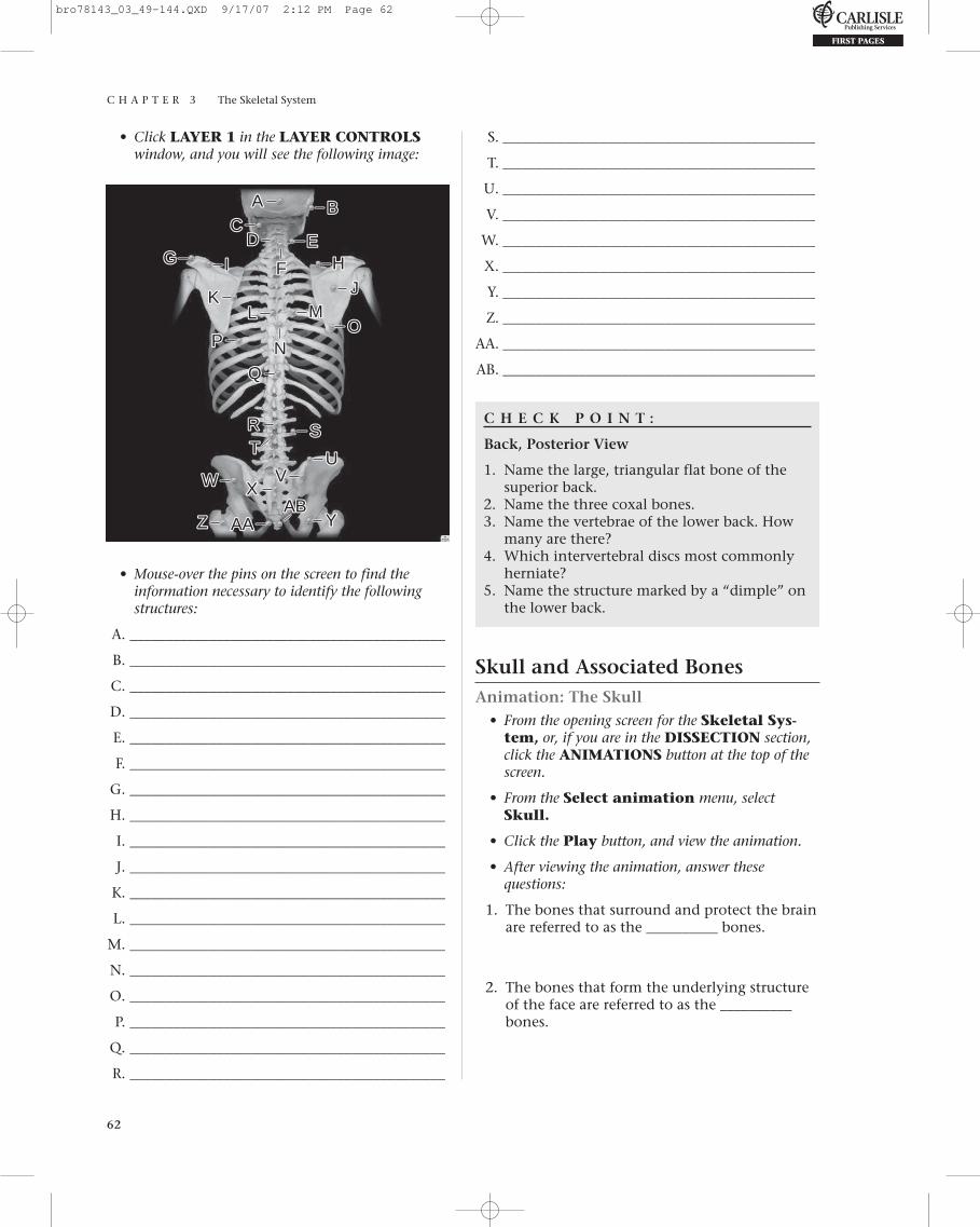
• Click LAYER 1 in the LAYER CONTROLSwindow, and you will see the following image:
S.____________________________________________
T.____________________________________________
U.____________________________________________
V.____________________________________________
W.____________________________________________
X.____________________________________________
Y.____________________________________________
Z.____________________________________________
AA.____________________________________________
AB.____________________________________________
C H E C K P O I N T :
Back, Posterior View
1. Name the large, triangular flat bone of thesuperior back.
2. Name the three coxal bones.3. Name the vertebrae of the lower back. How
many are there?4. Which intervertebral discs most commonly
herniate?5. Name the structure marked by a “dimple” on
the lower back.
Skull and Associated BonesAnimation: The Skull
• From the opening screen for the Skeletal Sys-tem, or, if you are in the DISSECTION section,click the ANIMATIONS button at the top of thescreen.
• From the Select animation menu, selectSkull.
• Click the Play button, and view the animation.
• After viewing the animation, answer thesequestions:
1. The bones that surround and protect the brainare referred to as the __________ bones.
2. The bones that form the underlying structureof the face are referred to as the __________bones.
AA BBCC
DD EE
FF HHII
KKLL MM
NNPP
RR
WW
ZZ
XX
AAAAABAB
VV
TT
OO
SS
UU
YY
JJ
GG
• Mouse-over the pins on the screen to find theinformation necessary to identify the followingstructures:
A. ____________________________________________
B. ____________________________________________
C. ____________________________________________
D. ____________________________________________
E. ____________________________________________
F. ____________________________________________
G. ____________________________________________
H. ____________________________________________
I. ____________________________________________
J. ____________________________________________
K. ____________________________________________
L. ____________________________________________
M. ____________________________________________
N. ____________________________________________
O. ____________________________________________
P. ____________________________________________
Q. ____________________________________________
R. ____________________________________________
bro78143_03_49-144.QXD 9/17/07 2:12 PM Page 62
C H A P T E R 3 The Skeletal System
63
3. With one exception, the bones of the skullarticulate with each other through jointsknown as __________. The exception is the__________.
4. There are numerous holes in the skull called__________.
5. What are the functions of these holes?
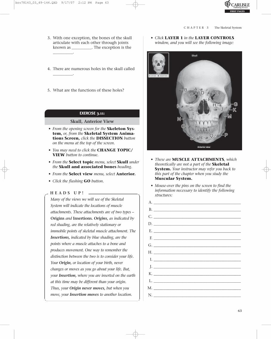
• Click LAYER 1 in the LAYER CONTROLSwindow, and you will see the following image:
EXERCISE 3.11:
Skull, Anterior View
• From the opening screen for the Skeleton Sys-tem, or, from the Skeletal System Anima-tions Screen, click the DISSECTION buttonon the menu at the top of the screen.
• You may need to click the CHANGE TOPIC/VIEW button to continue.
• From the Select topic menu, select Skull underthe Skull and associated bones heading.
• From the Select view menu, select Anterior.
• Click the flashing GO button.
H E A D S U P !
Many of the views we will see of the Skeletal
System will indicate the locations of muscle
attachments. These attachments are of two types –
Origins and Insertions. Origins, as indicated by
red shading, are the relatively stationary or
immobile points of skeletal muscle attachment. The
Insertions, indicated by blue shading, are the
points where a muscle attaches to a bone and
produces movement. One way to remember the
distinction between the two is to consider your life.
Your Origin, or location of your birth, never
changes or moves as you go about your life. But,
your Insertion, where you are inserted on the earth
at this time may be different than your origin.
Thus, your Origin never moves, but when you
move, your Insertion moves to another location.
AABBCC DD EE
FFHHII
KKLL
MMNN
PP
OO
SS
JJGG
RR
• These are MUSCLE ATTACHMENTS, whichtheoretically are not a part of the SkeletalSystem. Your instructor may refer you back tothis part of the chapter when you study theMuscular System.
• Mouse-over the pins on the screen to find theinformation necessary to identify the followingstructures:
A. ____________________________________________
B. ____________________________________________
C. ____________________________________________
D. ____________________________________________
E. ____________________________________________
F. ____________________________________________
G. ____________________________________________
H. ____________________________________________
I. ____________________________________________
J. ____________________________________________
K. ____________________________________________
L. ____________________________________________
M. ____________________________________________
N. ____________________________________________
bro78143_03_49-144.QXD 9/17/07 2:12 PM Page 63

C H A P T E R 3 The Skeletal System
64
O. ____________________________________________
P. ____________________________________________
Q. ____________________________________________
R. ____________________________________________
S. ____________________________________________
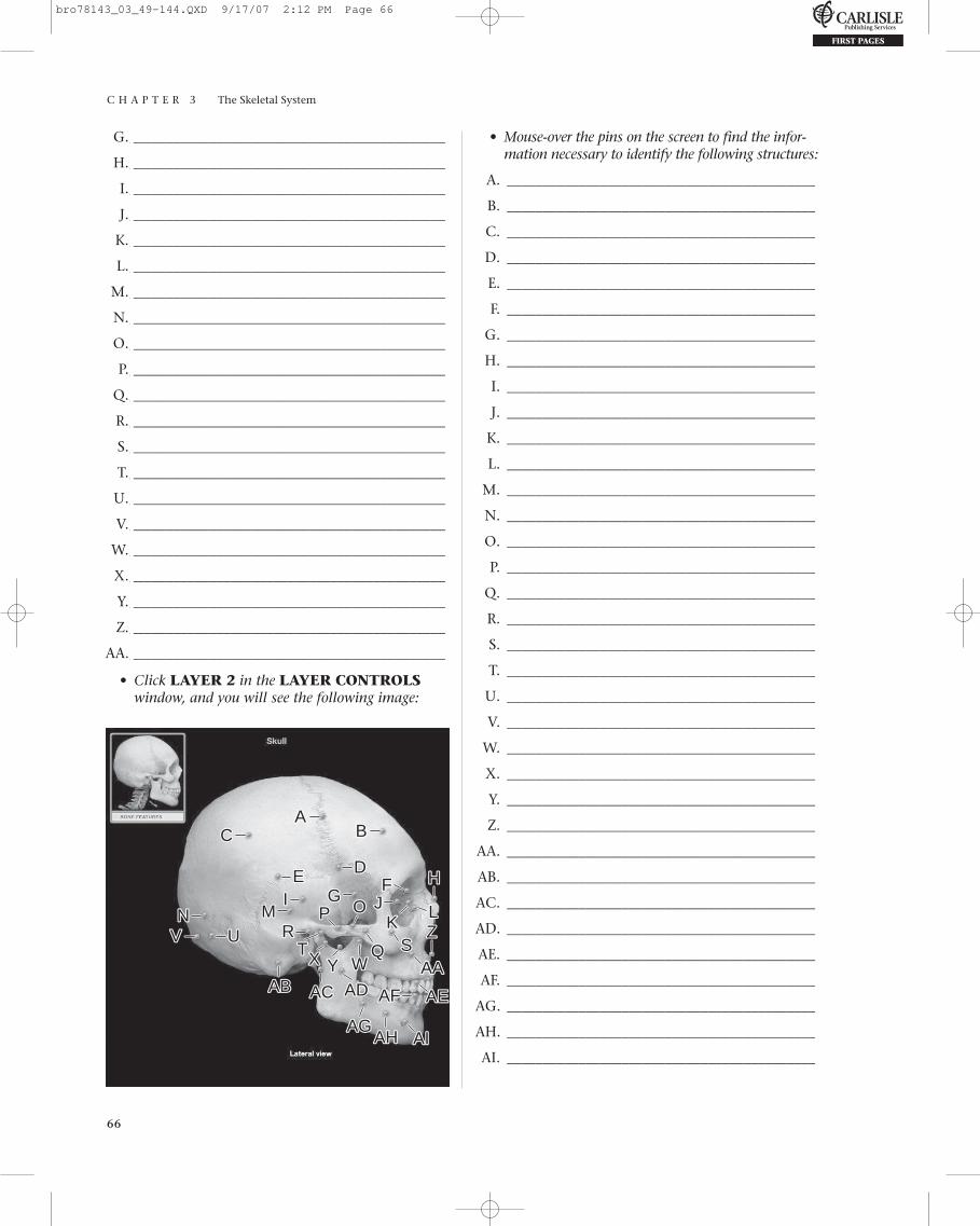
• Click LAYER 2 in the LAYER CONTROLSwindow, and you will see the following image:
K. ____________________________________________
L. ____________________________________________
M. ____________________________________________
N. ____________________________________________
O. ____________________________________________
P. ____________________________________________
Q. ____________________________________________
R. ____________________________________________
S. ____________________________________________
T. ____________________________________________
U. ____________________________________________
V. ____________________________________________
C H E C K P O I N T :
Skull, Anterior View
1. Name the structures transmitted through themental foramen.
2. Name a feature of the skull that can be eithera notch or a foramen.
3. What two bones contain teeth?4. What is the name for the sockets of the
teeth? (You may have to revisit this chapterto answer this one)
5. The nasal septum consists of what specificbones or structures of bones?
AABB
CCDD
EEFF HHII
KK LLMMNN
PP
RR
TT
OO
UUVV
SS
JJGG
• Mouse-over the pins on the screen to find the infor-mation necessary to identify the following structures:
A. ____________________________________________
B. ____________________________________________
C. ____________________________________________
D. ____________________________________________
E. ____________________________________________
F. ____________________________________________
G. ____________________________________________
H. ____________________________________________
I. ____________________________________________
J. ____________________________________________
EXERCISE 3.12:
Skull, Superior View
• From the opening screen for the Skeleton Sys-tem, click the DISSECTION button on themenu at the top of the screen. Or, if you are in theDISSECTION section, click the CHANGETOPIC/VIEW button at the top of the screen.
• From the Select topic menu, select Skull, if itis not already selected.
• From the Select view menu, select Superior.
• Click the flashing GO button.
bro78143_03_49-144.QXD 9/17/07 2:12 PM Page 64

C H A P T E R 3 The Skeletal System
65
• Click LAYER 1 in the LAYER CONTROLSwindow, and you will see the following image:
AABB
CCDD
EEFFGG
• Mouse-over the pins on the screen to find the infor-mation necessary to identify the following structures:
A. ____________________________________________
B. ____________________________________________
C. ____________________________________________
D. ____________________________________________
E. ____________________________________________
F. ____________________________________________
G. ____________________________________________
C H E C K P O I N T :
Skull, Superior View
1. Name the joint between the parietal bones.2. Name the joint between the frontal and the
parietal bones.3. Name the bone type found in or near the
sutures of the skull.4. Where are these bones most often found?5. Name the skull bone that articulates with the
vertebral column.
EXERCISE 3.13:
Skull, Lateral View
• From the opening screen for the SkeletonSystem, click the DISSECTION button on themenu at the top of the screen. Or, if you are in theDISSECTION section, click the CHANGETOPIC/VIEW button at the top of the screen.
• From the Select topic menu, select Skull, if itis not already selected.
• From the Select view menu, select Lateral.
• Click the flashing GO button.
• Click LAYER 1 in the LAYER CONTROLSwindow, and you will see the following image:
AACCBB
DDEEFF
AAAA
UU
GGHH
II
XX
JJ
WW
YY
ZZ
TT KKLL
MMRR
NNPP SS
QQOO
VV
• Mouse-over the pins on the screen to find the infor-mation necessary to identify the following structures:
A. ____________________________________________
B. ____________________________________________
C. ____________________________________________
D. ____________________________________________
E. ____________________________________________
F. ____________________________________________
bro78143_03_49-144.QXD 9/17/07 2:12 PM Page 65
C H A P T E R 3 The Skeletal System
66
G.____________________________________________
H.____________________________________________
I.____________________________________________
J.____________________________________________
K.____________________________________________
L.____________________________________________
M.____________________________________________
N.____________________________________________
O.____________________________________________
P.____________________________________________
Q.____________________________________________
R.____________________________________________
S.____________________________________________
T.____________________________________________
U.____________________________________________
V.____________________________________________
W.____________________________________________
X.____________________________________________
Y.____________________________________________
Z.____________________________________________
AA.____________________________________________
• Click LAYER 2 in the LAYER CONTROLSwindow, and you will see the following image:
• Mouse-over the pins on the screen to find the infor-mation necessary to identify the following structures:
A. ___________________________________________
B. ___________________________________________
C. ___________________________________________
D. ___________________________________________
E. ___________________________________________
F. ___________________________________________
G. ___________________________________________
H. ___________________________________________
I. ___________________________________________
J. ___________________________________________
K. ___________________________________________
L. ___________________________________________
M. ___________________________________________
N. ___________________________________________
O. ___________________________________________
P. ___________________________________________
Q. ___________________________________________
R. ___________________________________________
S. ___________________________________________
T. ___________________________________________
U. ___________________________________________
V. ___________________________________________
W. ___________________________________________
X. ___________________________________________
Y. ___________________________________________
Z. ___________________________________________
AA. ___________________________________________
AB. ___________________________________________
AC. ___________________________________________
AD. ___________________________________________
AE. ___________________________________________
AF. ___________________________________________
AG. ___________________________________________
AH. ___________________________________________
AI. ___________________________________________
AACC BB
DDEE FF
AAAA
UU
GGHH
II
XX
JJ
WWYY
ZZ
ABAB ACAC ADAD AFAF
AGAGAHAH AIAI
AEAE
TT
KKLLMM
RRNN PP
SSQQ
OO
VV
bro78143_03_49-144.QXD 9/17/07 2:12 PM Page 66

C H A P T E R 3 The Skeletal System
67
• Mouse-over the pins on the screen to find the infor-mation necessary to identify the following structures:
A. ____________________________________________
B. ____________________________________________
C. ____________________________________________
D. ____________________________________________
E. ____________________________________________
F. ____________________________________________
• Click LAYER 2 in the LAYER CONTROLSwindow, and you will see the following image:
C H E C K P O I N T :
Skull, Lateral View
1. Name the specific bony structures that formthe temporomandibular joint.
2. What two bones make up most of the lateralskull (one on each side)?
3. What suture is their point of articulation?
EXERCISE 3.14:
Skull, Posterior View
• From the opening screen for the SkeletonSystem, click the DISSECTION button on themenu at the top of the screen. Or, if you are in theDISSECTION section, click the CHANGETOPIC/VIEW button at the top of the screen.
• From the Select topic menu, select Skull, if itis not already selected.
• From the Select view menu, select Posterior.
• Click the flashing GO button.
• Click LAYER 1 in the LAYER CONTROLSwindow, and you will see the following image:
AA
CCBB
DD EEFF
AA
CC
GG
EE
FF
II
DD
HH
BB
• Mouse-over the pins on the screen to find the infor-mation necessary to identify the following structures:
A. ____________________________________________
B. ____________________________________________
C. ____________________________________________
D. ____________________________________________
E. ____________________________________________
F. ____________________________________________
G. ____________________________________________
H. ____________________________________________
I. ____________________________________________
bro78143_03_49-144.QXD 9/17/07 2:12 PM Page 67

C H A P T E R 3 The Skeletal System
68
C H E C K P O I N T :
Skull, Posterior View
1. What suture forms the joint between theparietal and occipital bones?
2. What bone forms most of the posterior skull?3. What is the attachment site for the nuchal
ligament on the skull?
• Mouse-over the pins on the screen to find the information necessary to identify the followingstructures:
A. ____________________________________________
B. ____________________________________________
C. ____________________________________________
D. ____________________________________________
E. ____________________________________________
F. ____________________________________________
G. ____________________________________________
H. ____________________________________________
I. ____________________________________________
J. ____________________________________________
K. ____________________________________________
L. ____________________________________________
• Click LAYER 2 in the LAYER CONTROLSwindow, and you will see the following image:
EXERCISE 3.15:
Skull, Mid-sagittal View
• From the opening screen for the Skeleton Sys-tem, click the DISSECTION button on themenu at the top of the screen. Or, if you are in theDISSECTION section, click the CHANGETOPIC/VIEW button at the top of the screen.
• From the Select topic menu, select Skull, if itis not already selected.
• From the Select view menu, selectMid-sagittal.
• Click the flashing GO button.
• Click LAYER 1 in the LAYER CONTROLSwindow, and you will see the following image:
AACC
BBDDEE FF
GGHH
IIJJ
KKLL
AACC
EE
BB
DDFF
ABABACAC
AAAA
ADAD
UU
GGHHII
XX
JJ
WW
AEAEAFAF
AGAGAIAIAJAJ AKAK
ANANALALAMAM
YYZZ
AHAH
AOAO
TT
KKLL
MMRRNN
PP
SSQQ
OOVV
• Mouse-over the pins on the screen to find the infor-mation necessary to identify the following structures:
A. ____________________________________________
B. ____________________________________________
C. ____________________________________________
D. ____________________________________________
bro78143_03_49-144.QXD 9/17/07 2:12 PM Page 68
C H A P T E R 3 The Skeletal System
69
E. ___________________________________________
F. ___________________________________________
G. ___________________________________________
H. ___________________________________________
I. ___________________________________________
J. ___________________________________________
K. ___________________________________________
L. ___________________________________________
M. ___________________________________________
N. ___________________________________________
O. ___________________________________________
P. ___________________________________________
Q. ___________________________________________
R. ___________________________________________
S. ___________________________________________
T. ___________________________________________
U. ___________________________________________
V. ___________________________________________
W. ___________________________________________
X. ___________________________________________
Y. ___________________________________________
Z. ___________________________________________
AA. ___________________________________________
AB. ___________________________________________
AC. ___________________________________________
AD. ___________________________________________
AE. ___________________________________________
AF. ___________________________________________
AG. ___________________________________________
AH. ___________________________________________
AI. ___________________________________________
AJ. ___________________________________________
AK. ___________________________________________
AL. ___________________________________________
AM. ___________________________________________
AN. ___________________________________________
AO. ___________________________________________
C H E C K P O I N T :
Skull, Mid-sagittal View
1. When your dentist wants to numb yourlower jaw by anesthetizing the nerves thatserve the teeth and skin, what “hole” in whatbone would be used to access those nerves?
2. Name the paranasal sinuses.3. Name the structure that increases the surface
area of the nasal cavity and plays an impor-tant role in warming inhaled air.
4. Name the structure that contains the sublin-gual salivary gland.
5. Name the muscles that attach to the styloidprocess of the temporal bone.
EXERCISE 3.16:
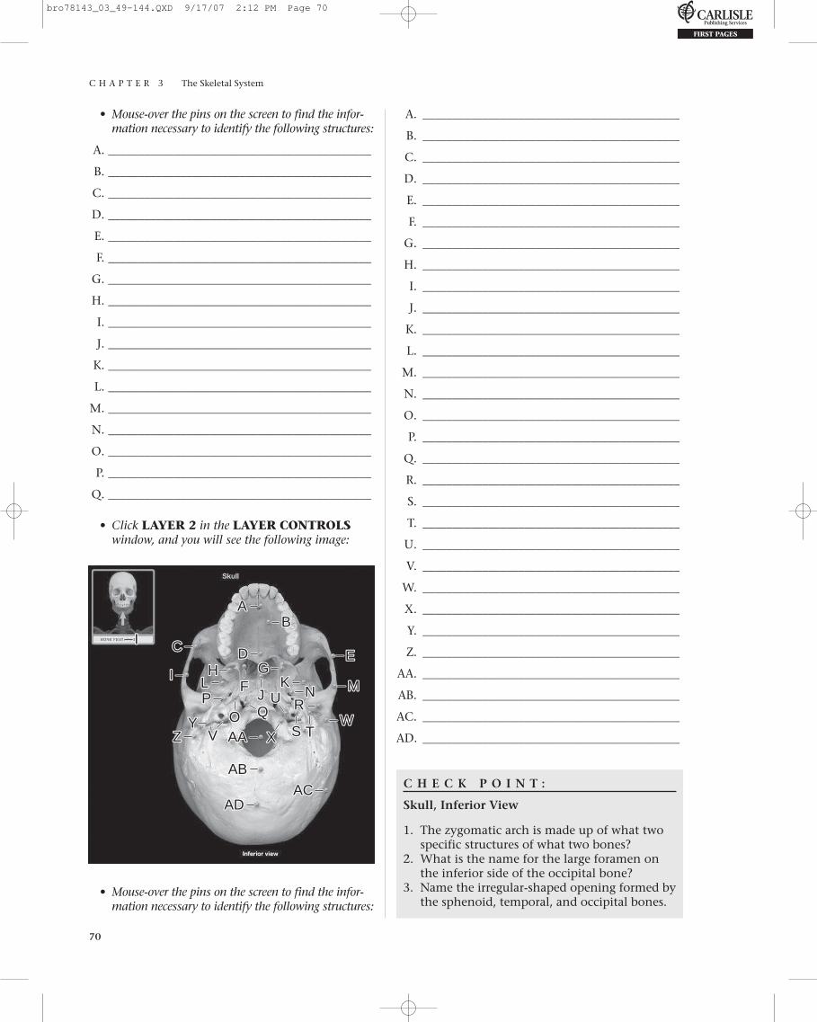
Skull, Inferior View
• From the opening screen for the SkeletonSystem, click the DISSECTION button on themenu at the top of the screen. Or, if you are in theDISSECTION section, click the CHANGETOPIC/VIEW button at the top of the screen.
• From the Select topic menu, select Skull, if itis not already selected.
• From the Select view menu, select Inferior.
• Click the flashing GO button.
• Click LAYER 1 in the LAYER CONTROLSwindow, and you will see the following image:
AABB
CCDD
MM
EEFF
JJ
HH II
LL
OO PPQQ
GG
KK
NN
bro78143_03_49-144.QXD 9/17/07 2:12 PM Page 69
C H A P T E R 3 The Skeletal System
70
• Mouse-over the pins on the screen to find the infor-mation necessary to identify the following structures:
A. ____________________________________________
B. ____________________________________________
C. ____________________________________________
D. ____________________________________________
E. ____________________________________________
F. ____________________________________________
G. ____________________________________________
H. ____________________________________________
I. ____________________________________________
J. ____________________________________________
K. ____________________________________________
L. ____________________________________________
M. ____________________________________________
N. ____________________________________________
O. ____________________________________________
P. ____________________________________________
Q. ____________________________________________
• Click LAYER 2 in the LAYER CONTROLSwindow, and you will see the following image:
A. ___________________________________________
B. ___________________________________________
C. ___________________________________________
D. ___________________________________________
E. ___________________________________________
F. ___________________________________________
G. ___________________________________________
H. ___________________________________________
I. ___________________________________________
J. ___________________________________________
K. ___________________________________________
L. ___________________________________________
M. ___________________________________________
N. ___________________________________________
O. ___________________________________________
P. ___________________________________________
Q. ___________________________________________
R. ___________________________________________
S. ___________________________________________
T. ___________________________________________
U. ___________________________________________
V. ___________________________________________
W. ___________________________________________
X. ___________________________________________
Y. ___________________________________________
Z. ___________________________________________
AA. ___________________________________________
AB. ___________________________________________
AC. ___________________________________________
AD. ___________________________________________
C H E C K P O I N T :
Skull, Inferior View
1. The zygomatic arch is made up of what twospecific structures of what two bones?
2. What is the name for the large foramen onthe inferior side of the occipital bone?
3. Name the irregular-shaped opening formed bythe sphenoid, temporal, and occipital bones.
AABB
CC
II
DD EE
FFHH
II
KKLL MMNNPPQQ RR
WWZZ TTXXAAAA
ABAB
ACACADAD
UUOO
VV SSYY
JJ
GG
• Mouse-over the pins on the screen to find the infor-mation necessary to identify the following structures:
bro78143_03_49-144.QXD 9/17/07 2:12 PM Page 70

C H A P T E R 3 The Skeletal System
71
EXERCISE 3.17:
Skull—Cranial Cavity, Superior View
• From the opening screen for the SkeletonSystem, click the DISSECTION button on themenu at the top of the screen. Or, if you are in theDISSECTION section, click the CHANGETOPIC/VIEW button at the top of the screen.
• From the Select topic menu, selectSkull-cranial cavity under the Skull andassociated bones heading.
• Superior will be visible in the Select view menu.
• Click the flashing GO button.
• Click LAYER 1 in the LAYER CONTROLSwindow, and you will see the following image:
E. ___________________________________________
F. ___________________________________________
G. ___________________________________________
H. ___________________________________________
I. ___________________________________________
J. ___________________________________________
K. ___________________________________________
L. ___________________________________________
M. ___________________________________________
N. ___________________________________________
O. ___________________________________________
P. ___________________________________________
Q. ___________________________________________
R. ___________________________________________
S. ___________________________________________
T. ___________________________________________
U. ___________________________________________
V. ___________________________________________
W. ___________________________________________
X. ___________________________________________
Y. ___________________________________________
Z. ___________________________________________
AA. ___________________________________________
AB. ___________________________________________
AC. ___________________________________________
AD. ___________________________________________
AE. ___________________________________________
AF. ___________________________________________
AG. ___________________________________________
AH. ___________________________________________
AI. ___________________________________________
AJ. ___________________________________________
AK. ___________________________________________
AL. ___________________________________________
AM. ___________________________________________
BBAA
CCDD EE
HHFF GG
IIKK
LLMMNN
PPQQ
RRWW
ALAL
ZZ
AJAJAKAK
AMAM
AEAE AAAA
ABAB
XXADAD AFAF
VVTT
OO
SS
UU
YYAGAG
ACAC
AHAHAIAI
JJ
• Mouse-over the pins on the screen to find theinformation necessary to identify the followingstructures:
A. ____________________________________________
B. ____________________________________________
C. ____________________________________________
D. ____________________________________________
bro78143_03_49-144.QXD 9/17/07 2:12 PM Page 71
C H A P T E R 3 The Skeletal System
72
C H E C K P O I N T :
Skull—Cranial Cavity, Superior View
1. Describe the cribriform plate.2. Name the structures that traverse through the
jugular foramen.3. Name the structure that passes through the
foramen ovale.4. Name the structure contained in the
hypophyseal fossa.5. What structure is transmitted by the foramen
rotundum?
• Mouse-over the pins on the screen to find theinformation necessary to identify the followingstructures:
A. ____________________________________________
B. ____________________________________________
C. ____________________________________________
D. ____________________________________________
E. ____________________________________________
F. ____________________________________________
G. ____________________________________________
H. ____________________________________________
• Click LAYER 2 in the LAYER CONTROLSwindow, and you will see the following image:
EXERCISE 3.18:
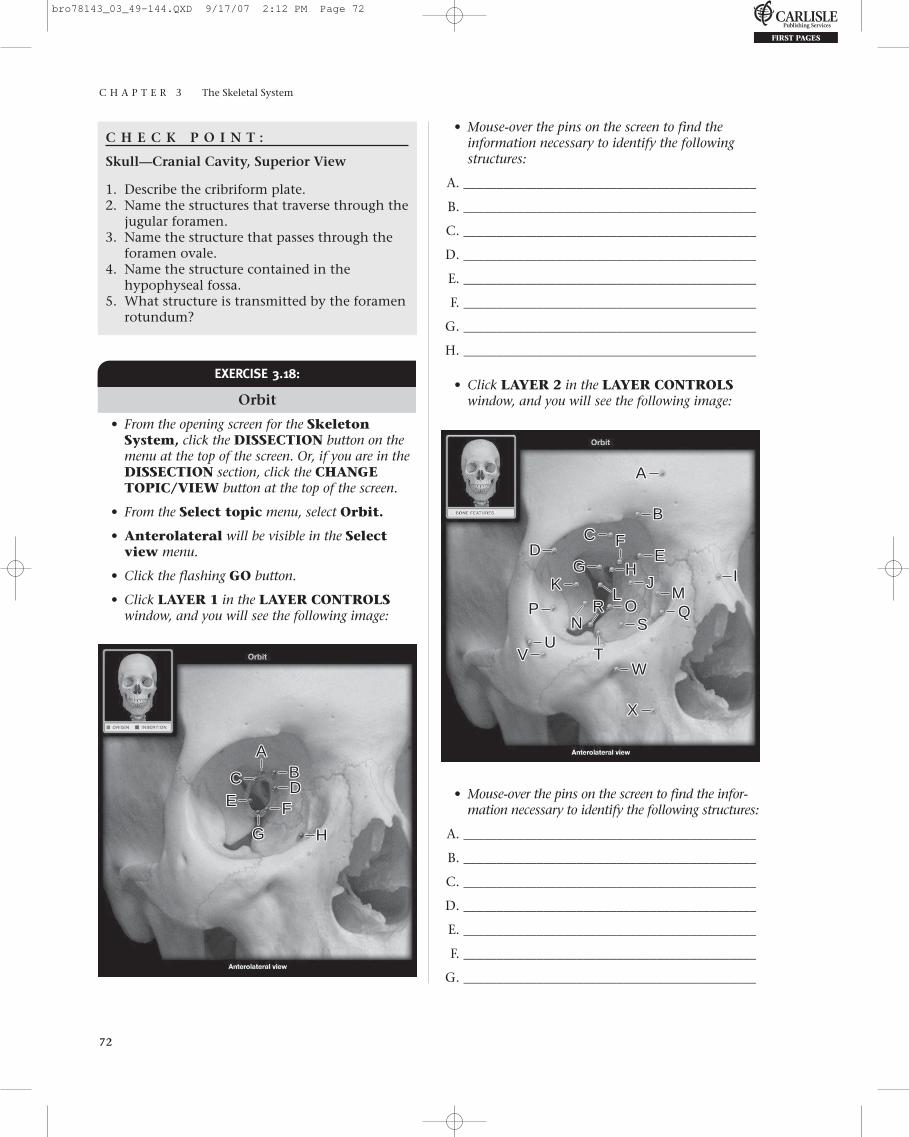
Orbit
• From the opening screen for the SkeletonSystem, click the DISSECTION button on themenu at the top of the screen. Or, if you are in theDISSECTION section, click the CHANGETOPIC/VIEW button at the top of the screen.
• From the Select topic menu, select Orbit.
• Anterolateral will be visible in the Selectview menu.
• Click the flashing GO button.
• Click LAYER 1 in the LAYER CONTROLSwindow, and you will see the following image:
AABBCCDD
FFEE
HHGG
AA
BBCC
DD EEFF
HH IIKKLL MM
NNPP QQRR
WW
XX
VV TT
OOSS
UU
JJGG
• Mouse-over the pins on the screen to find the infor-mation necessary to identify the following structures:
A. ____________________________________________
B. ____________________________________________
C. ____________________________________________
D. ____________________________________________
E. ____________________________________________
F. ____________________________________________
G. ____________________________________________
bro78143_03_49-144.QXD 9/17/07 2:12 PM Page 72
C H A P T E R 3 The Skeletal System
73
H. ____________________________________________
I. ____________________________________________
J. ____________________________________________
K. ____________________________________________
L. ____________________________________________
M. ____________________________________________
N. ____________________________________________
O. ____________________________________________
P. ____________________________________________
Q. ____________________________________________
R. ____________________________________________
S. ____________________________________________
T. ____________________________________________
U. ____________________________________________
V. ____________________________________________
W. ____________________________________________
X. ____________________________________________
C H E C K P O I N T :
Orbit
1. Name the seven bones that constitute theorbit.
2. Name the structure that transmits the opticnerve (CN II) and the ophthalmic artery.
3. Name the nerves and blood vessels transmit-ted by the superior orbital fissure.
4. Name the nerves and blood vessels transmit-ted by the inferior orbital fissure.
5. Name the nerves and blood vessels transmit-ted by the anterior ethmoidal foramen.
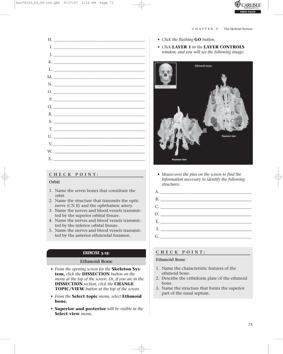
• Click the flashing GO button.
• Click LAYER 1 in the LAYER CONTROLSwindow, and you will see the following image:
EXERCISE 3.19:
Ethmoid Bone
• From the opening screen for the Skeleton Sys-tem, click the DISSECTION button on themenu at the top of the screen. Or, if you are in theDISSECTION section, click the CHANGETOPIC/VIEW button at the top of the screen.
• From the Select topic menu, select Ethmoidbone.
• Superior and posterior will be visible in theSelect view menu.
AA
BB
CCDD
FF
EE
GG
• Mouse-over the pins on the screen to find theinformation necessary to identify the followingstructures:
A. ____________________________________________
B. ____________________________________________
C. ____________________________________________
D. ____________________________________________
E. ____________________________________________
F. ____________________________________________
G. ____________________________________________
C H E C K P O I N T :
Ethmoid Bone
1. Name the characteristic features of theethmoid bone.
2. Describe the cribriform plate of the ethmoidbone.
3. Name the structure that forms the superiorpart of the nasal septum.
bro78143_03_49-144.QXD 9/17/07 2:12 PM Page 73

EXERCISE 3.20:
Frontal Bone
• From the opening screen for the SkeletonSystem, click the DISSECTION button on themenu at the top of the screen. Or, if you are in theDISSECTION section, click the CHANGETOPIC/VIEW button at the top of the screen.
• From the Select topic menu, select Frontalbone.
• Anterior will be visible in the Select viewmenu.
• Click the flashing GO button.
• Click LAYER 1 in the LAYER CONTROLSwindow, and you will see the following image:
C H A P T E R 3 The Skeletal System
74
C H E C K P O I N T :
Frontal Bone
1. Name the structures that traverse the supraor-bital notch.
2. Describe the supraorbital margin.3. Name the sutures associated with the frontal
bone. What bones does the frontal bonearticulate with for each suture?
AA
BBCCDD
• Mouse-over the pins on the screen to find theinformation necessary to identify the followingstructures:
A. ____________________________________________
B. ____________________________________________
C. ____________________________________________
D. ____________________________________________
EXERCISE 3.21:
Mandible
• From the opening screen for the SkeletonSystem, click the DISSECTION button on themenu at the top of the screen. Or, if you are in theDISSECTION section, click the CHANGETOPIC/VIEW button at the top of the screen.
• From the Select topic menu, select Mandible.
• Lateral will be visible in the Select view menu.
• Click the flashing GO button.
• Click LAYER 1 in the LAYER CONTROLSwindow, and you will see the following image:
AA
FF
BBCCDD EE
HHII JJKK
GG
bro78143_03_49-144.QXD 9/17/07 2:12 PM Page 74
C H A P T E R 3 The Skeletal System
75
• Mouse-over the pins on the screen to find the information necessary to identify the following structures:
A. ____________________________________________
B. ____________________________________________
C. ____________________________________________
D. ____________________________________________
E. ____________________________________________
F. ____________________________________________
G. ____________________________________________
H. ____________________________________________
I. ____________________________________________
J. ____________________________________________
K. ____________________________________________
C H E C K P O I N T :
Mandible
1. Name the bone structures that constitute thetemporomandibular joint.
2. What structure is also known as themandibular incisure?
• Click LAYER 1 in the LAYER CONTROLSwindow, and you will see the following image:
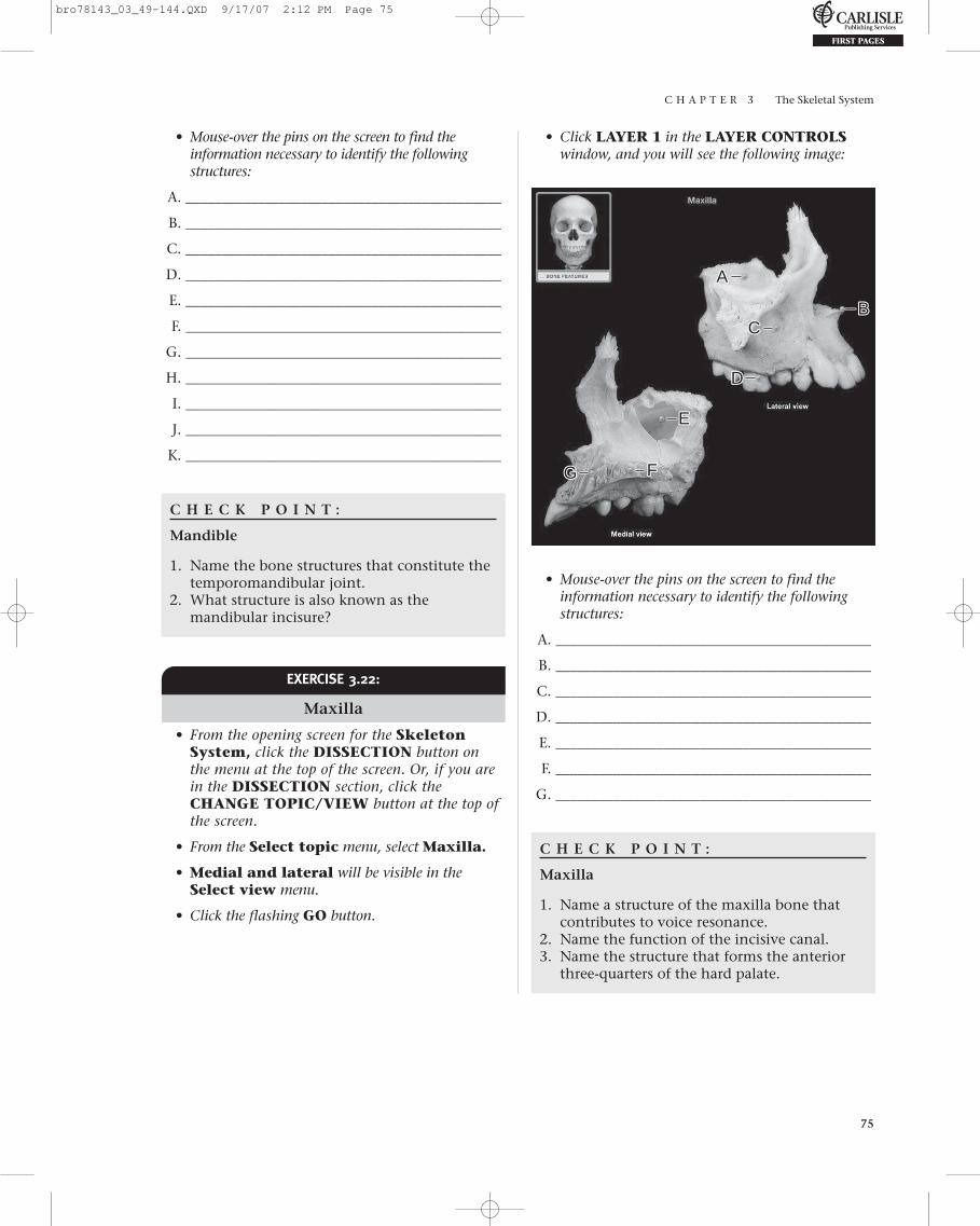
EXERCISE 3.22:
Maxilla
• From the opening screen for the SkeletonSystem, click the DISSECTION button on the menu at the top of the screen. Or, if you arein the DISSECTION section, click theCHANGE TOPIC/VIEW button at the top ofthe screen.
• From the Select topic menu, select Maxilla.
• Medial and lateral will be visible in theSelect view menu.
• Click the flashing GO button.
AA
BBCC
DD
EE
FFGG
• Mouse-over the pins on the screen to find theinformation necessary to identify the followingstructures:
A. ____________________________________________
B. ____________________________________________
C. ____________________________________________
D. ____________________________________________
E. ____________________________________________
F. ____________________________________________
G. ____________________________________________
C H E C K P O I N T :
Maxilla
1. Name a structure of the maxilla bone thatcontributes to voice resonance.
2. Name the function of the incisive canal.3. Name the structure that forms the anterior
three-quarters of the hard palate.
bro78143_03_49-144.QXD 9/17/07 2:12 PM Page 75

C H A P T E R 3 The Skeletal System
76
EXERCISE 3.23:
Occipital Bone
• From the opening screen for the Skeleton Sys-tem, click the DISSECTION button on themenu at the top of the screen. Or, if you are in theDISSECTION section, click the CHANGETOPIC/VIEW button at the top of the screen.
• From the Select topic menu, select Occipitalbone.
• Superior and inferior will be visible in theSelect view menu.
• Click the flashing GO button.
• Click LAYER 1 in the LAYER CONTROLSwindow, and you will see the following image:
H. ____________________________________________
I. ____________________________________________
J. ____________________________________________
C H E C K P O I N T :
Occipital Bone
1. Name the structures that pass through theforamen magnum.
2. What is the function of the occipital condyles?3. What structures are contained within the
cerebellar fossa?
AA
BB
CC
DD
EE
HHII
JJ
GGFF
• Mouse-over the pins on the screen to find theinformation necessary to identify the followingstructures:
A. ____________________________________________
B. ____________________________________________
C. ____________________________________________
D. ____________________________________________
E. ____________________________________________
F. ____________________________________________
G. ____________________________________________
EXERCISE 3.24:
Parietal Bone
• From the opening screen for the SkeletonSystem, click the DISSECTION button on themenu at the top of the screen. Or, if you are in theDISSECTION section, click the CHANGETOPIC/VIEW button at the top of the screen.
• From the Select topic menu, select Parietalbone.
• Lateral will be visible in the Select view menu.
• Click the flashing GO button.
• There are no TAGS for this bone, but be sure toread the information in the STRUCTUREINFORMATION window.
bro78143_03_49-144.QXD 9/17/07 2:12 PM Page 76

C H A P T E R 3 The Skeletal System
77
C. ____________________________________________
D. ____________________________________________
E. ____________________________________________
F. ____________________________________________
G. ____________________________________________
H. ____________________________________________
I. ____________________________________________
J. ____________________________________________
K. ____________________________________________
L. ____________________________________________
M. ____________________________________________
N. ____________________________________________
O. ____________________________________________
P. ____________________________________________
Q. ____________________________________________
C H E C K P O I N T :
Sphenoid Bone
1. What structure is contained in the sella turcica?
2. Name the two structures transmitted throughthe optic canal.
3. Name the paired lateral projections of thesphenoid bone.
C H E C K P O I N T :
Parietal Bone
1. Name the bones that articulate with the pari-etal bone.
2. Name the sutures involved with each articu-lation listed in question 1.
EXERCISE 3.25:
Sphenoid Bone
• From the opening screen for the SkeletonSystem, click the DISSECTION button on themenu at the top of the screen. Or, if you are in theDISSECTION section, click the CHANGETOPIC/VIEW button at the top of the screen.
• From the Select topic menu, select Sphenoidbone.
• Superior and posterior will be visible in theSelect view menu.
• Click the flashing GO button.
• Click LAYER 1 in the LAYER CONTROLSwindow, and you will see the following image:
AA BBCCDDEE
FFHHII
KKLLMM
PP
JJ
NNQQ
OO
GG
• Mouse-over the pins on the screen to find the infor-mation necessary to identify the following structures:
A. ____________________________________________
B. ____________________________________________
EXERCISE 3.26:
Temporal Bone
• From the opening screen for the Skeleton System, click the DISSECTION button on the menu at the top of the screen. Or, if you are in the DISSECTION section, click theCHANGE TOPIC/VIEW button at the top of the screen.
• From the Select topic menu, select Temporalbone.
• Medial and lateral will be visible in theSelect view menu.
• Click the flashing GO button.
bro78143_03_49-144.QXD 9/17/07 2:12 PM Page 77
C H A P T E R 3 The Skeletal System
78
• Click LAYER 1 in the LAYER CONTROLSwindow, and you will see the following image:
AA
BBCCEE DD
GG
HH
JJ
KK
II
FF
• Mouse-over the pins on the screen to find the infor-mation necessary to identify the following structures:
A. ____________________________________________
B. ____________________________________________
C. ____________________________________________
D. ____________________________________________
E. ____________________________________________
F. ____________________________________________
G. ____________________________________________
H. ____________________________________________
I. ____________________________________________
J. ____________________________________________
K. ____________________________________________
C H E C K P O I N T :
Temporal Bone
1. Name the bony canal that ends at thetympanic membrane.
2. Name the bony canal traversed by the facialand vestibulocochlear nerves.
3. Name the bony structure containing theorgans of hearing and equilibrium.
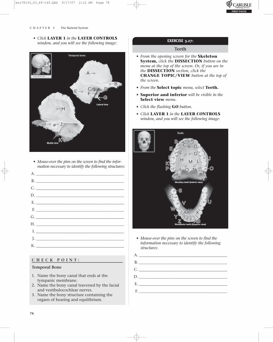
EXERCISE 3.27:
Teeth
• From the opening screen for the SkeletonSystem, click the DISSECTION button on themenu at the top of the screen. Or, if you are inthe DISSECTION section, click theCHANGE TOPIC/VIEW button at the top ofthe screen.
• From the Select topic menu, select Teeth.
• Superior and inferior will be visible in theSelect view menu.
• Click the flashing GO button.
• Click LAYER 1 in the LAYER CONTROLSwindow, and you will see the following image:
AA
SS
BB
TT
EE
WW
CC
UU
FF
XX
DD
VV
GG
YY
HH
ZZ
II
AAAA
KKJJ
ABAB
LLMM
NNPP OORR
• Mouse-over the pins on the screen to find theinformation necessary to identify the followingstructures:
A. ____________________________________________
B. ____________________________________________
C. ____________________________________________
D. ____________________________________________
E. ____________________________________________
F. ____________________________________________
bro78143_03_49-144.QXD 9/17/07 2:12 PM Page 78

C H A P T E R 3 The Skeletal System
79
G.____________________________________________
H.____________________________________________
I.____________________________________________
J.____________________________________________
K.____________________________________________
L.____________________________________________
M.____________________________________________
N.____________________________________________
O.____________________________________________
P.____________________________________________
Q.____________________________________________
R.____________________________________________
S.____________________________________________
T.____________________________________________
U.____________________________________________
V.____________________________________________
W.____________________________________________
X.____________________________________________
Y.____________________________________________
Z.____________________________________________
AA.____________________________________________
AB.____________________________________________
C H E C K P O I N T :
Teeth
1. What four teeth are also known as “wisdomteeth”?
2. What structures are transmitted through thelesser palatine foramen?
3. What structures are transmitted through thegreater palatine foramen?
• From the Select topic menu, select Hyoidbone.
• Anterior and lateral will be visible in theSelect view menu.
• Click the flashing GO button.
• Click LAYER 1 in the LAYER CONTROLSwindow, and you will see the following image:
EXERCISE 3.28:
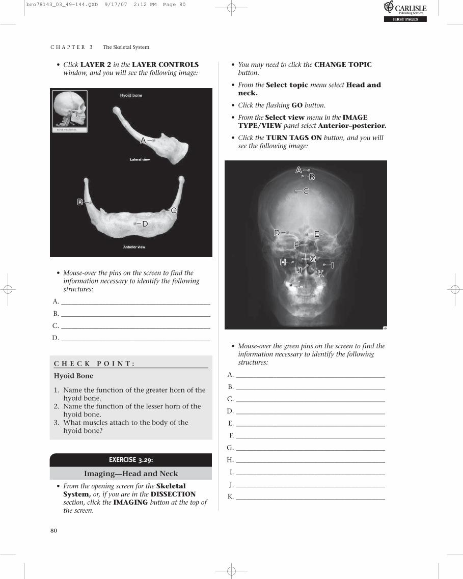
Hyoid Bone
• From the opening screen for the SkeletonSystem, click the DISSECTION button on themenu at the top of the screen. Or, if you are in theDISSECTION section, click the CHANGETOPIC/VIEW button at the top of the screen.
AA
BBCCDD
GGEEFF
HH
• Mouse-over the pins on the screen to find theinformation necessary to identify the followingstructures:
A. ____________________________________________
B. ____________________________________________
C. ____________________________________________
D. ____________________________________________
E. ____________________________________________
F. ____________________________________________
G. ____________________________________________
H. ____________________________________________
bro78143_03_49-144.QXD 9/17/07 2:12 PM Page 79
C H A P T E R 3 The Skeletal System
80
• Click LAYER 2 in the LAYER CONTROLSwindow, and you will see the following image:
• You may need to click the CHANGE TOPICbutton.
• From the Select topic menu select Head andneck.
• Click the flashing GO button.
• From the Select view menu in the IMAGETYPE/VIEW panel select Anterior–posterior.
• Click the TURN TAGS ON button, and you willsee the following image:
AA
BBCC
DD
• Mouse-over the pins on the screen to find theinformation necessary to identify the followingstructures:
A. ____________________________________________
B. ____________________________________________
C. ____________________________________________
D. ____________________________________________
C H E C K P O I N T :
Hyoid Bone
1. Name the function of the greater horn of thehyoid bone.
2. Name the function of the lesser horn of thehyoid bone.
3. What muscles attach to the body of thehyoid bone?
EXERCISE 3.29:
Imaging—Head and Neck
• From the opening screen for the SkeletalSystem, or, if you are in the DISSECTIONsection, click the IMAGING button at the top ofthe screen.
AABB
CC
EEDD
FF
GG
LL
IIKK
HHJJ
• Mouse-over the green pins on the screen to find theinformation necessary to identify the followingstructures:
A. ____________________________________________
B. ____________________________________________
C. ____________________________________________
D. ____________________________________________
E. ____________________________________________
F. ____________________________________________
G. ____________________________________________
H. ____________________________________________
I. ____________________________________________
J. ____________________________________________
K. ____________________________________________
bro78143_03_49-144.QXD 9/17/07 2:12 PM Page 80

C H A P T E R 3 The Skeletal System
81
Non-Skeletal System Structure (blue pin)
L. ____________________________________________
• Click the Select view menu drop-down box inthe IMAGE TYPE/VIEW panel.
• From the Select view menu, select Lateral.
• Click the TURN TAGS ON button and you willsee the following image:
K. ____________________________________________
L. ____________________________________________
M. ____________________________________________
Self TestTake this opportunity to quiz yourself by taking theSELF TEST.
• From the opening screen for the Skeletal Sys-tem, or, if you are in the DISSECTION section,click the SELF TEST button at the top of thescreen.
• From the Select test topic menu, selectSkeleton.
• From the Select region menu, select Skulland associated bones.
• From the Select test type menu, select Clickto identify.
• From the Select number of questions menu,select ALL.
• After completing the SELF TEST and saving yourresults, click the Select test type menu, selectMultiple choice.
• From the Select number of questions menu,select ALL.
• Return to the Select test topic menu, and selectAnimation.
• Click OK on the pop-up box.
• From the Select animation menu, selectSkull.
• Multiple choice is visible in the Select testtype menu.
• ALL is visible in the Select number of ques-tions menu.
• Complete the SELF TEST and save your results.
AA
BB
CCEE
DD
FF GG
JJHH
LL
II
KKMM
• Mouse-over the pins on the screen to find theinformation necessary to identify the followingstructures:
A. ____________________________________________
B. ____________________________________________
C. ____________________________________________
D. ____________________________________________
E. ____________________________________________
F. ____________________________________________
G. ____________________________________________
H. ____________________________________________
I. ____________________________________________
J. ____________________________________________
bro78143_03_49-144.QXD 9/17/07 2:12 PM Page 81
C H A P T E R 3 The Skeletal System
82
S 1
1 1
22
4 4
55
599
7
12
13 13
7
5
I
R L
15
16
17
28
29
30
31
32
33
18
19
20
21
22
23
25
24
26
27
S
2
345
1
13
9
7
12
I
A P
8 8
6
11 1114
EXERCISE 3.30:
Coloring Exercise
Look up the bones of the skull and then colorthem in with colored pens or pencils. Write the
names of important bones or bone features in theblank label lines.
bro78143_03_49-144.QXD 9/17/07 2:12 PM Page 82

C H A P T E R 3 The Skeletal System
83
34
35
36
37
50
26
A
P
R L
L
RHorizontal
section
P A 4
3
5
5
4
2
5
5
1
1
6
2
4
4
2
5
514
10 10
13
7713
25
77
2
2
3
39
40
41
42
43
44
45
46
38
49
48
47
19
5
bro78143_03_49-144.QXD 9/17/07 2:12 PM Page 83

C H A P T E R 3 The Skeletal System
84
❑ 1.__________________________________________
❑ 2.__________________________________________
❑ 3.__________________________________________
❑ 4.__________________________________________
❑ 5.__________________________________________
❑ 6.__________________________________________
❑ 7.__________________________________________
❑ 8.__________________________________________
❑ 9.__________________________________________
❑ 10.__________________________________________
❑ 11.__________________________________________
❑ 12.__________________________________________
❑ 13.__________________________________________
❑ 14.__________________________________________
❑ 15.__________________________________________
❑ 16.__________________________________________
❑ 17.__________________________________________
❑ 18.__________________________________________
❑ 19.__________________________________________
❑ 20.__________________________________________
❑ 21.__________________________________________
❑ 22.__________________________________________
❑ 23.__________________________________________
❑ 24.__________________________________________
❑ 25.__________________________________________
❑ 26.__________________________________________
❑ 27.__________________________________________
❑ 28.__________________________________________
❑ 29.__________________________________________
❑ 30.__________________________________________
❑ 31.__________________________________________
❑ 32.__________________________________________
❑ 33.__________________________________________
❑ 34.__________________________________________
❑ 35.__________________________________________
❑ 36.__________________________________________
❑ 37.__________________________________________
❑ 38.__________________________________________
❑ 39.__________________________________________
❑ 40.__________________________________________
❑ 41.__________________________________________
❑ 42.__________________________________________
❑ 43.__________________________________________
❑ 44.__________________________________________
❑ 45.__________________________________________
❑ 46.__________________________________________
❑ 47.__________________________________________
❑ 48.__________________________________________
❑ 49.__________________________________________
❑ 50.__________________________________________
What Have I Learned?
The following questions cover the material that youjust read, the introduction to the skeleton and theskeletal structures of the head and neck. Apply
what you have learned in answering these questions:
1. The hard palate consists of which bones?
2. What is the name for the large foramen on theinferior side of the occipital bone?
3. What passes through this foramen?
4. Name the two bones that form the bridge of thenose.
5. At the junction of the temporal and occipitalbones, the internal jugular vein passes throughwhich foramen?
I N R E V I E W
Continued
bro78143_03_49-144.QXD 9/17/07 2:12 PM Page 84

C H A P T E R 3 The Skeletal System
85
Thoracic Cage and VertebralColumn
EXERCISE 3.31:
Thoracic Cage
• From the opening screen for the SkeletonSystem, click the DISSECTION button on themenu at the top of the screen. Or, if you are in theIMAGING section, click the DISSECTIONbutton at the top of the screen.
• You may need to click the CHANGE TOPIC/VIEW button to continue.
• From the Select topic menu, select Thoraciccage under the Thoracic cage and verte-bral column heading.
• Anterior will be visible in the Select viewmenu.
• Click the flashing GO button.
• Click LAYER 1 in the LAYER CONTROLSwindow, and you will see the following image:
6. The greater and lesser wings are parts of whatbone?
7. Name the bony canal of the external ear. Whatbone is it a part of?
8. What is the attachment site for the sternocleido-mastoid muscle on the skull?
9. What bony landmark is defined as “any bonyprominence”?
10. What bony landmark is defined as “a pit orsocket”?
11. What bony landmark is defined as “a holethrough a bone, usually round”?
12. What bony landmark is defined as “a smooth,rounded knob”?
AABBCC
EE DDFF
GG HH
II
JJKK
• Mouse-over the pins on the screen to find theinformation necessary to identify the followingstructures:
A. ____________________________________________
B. ____________________________________________
C. ____________________________________________
bro78143_03_49-144.QXD 9/17/07 2:12 PM Page 85
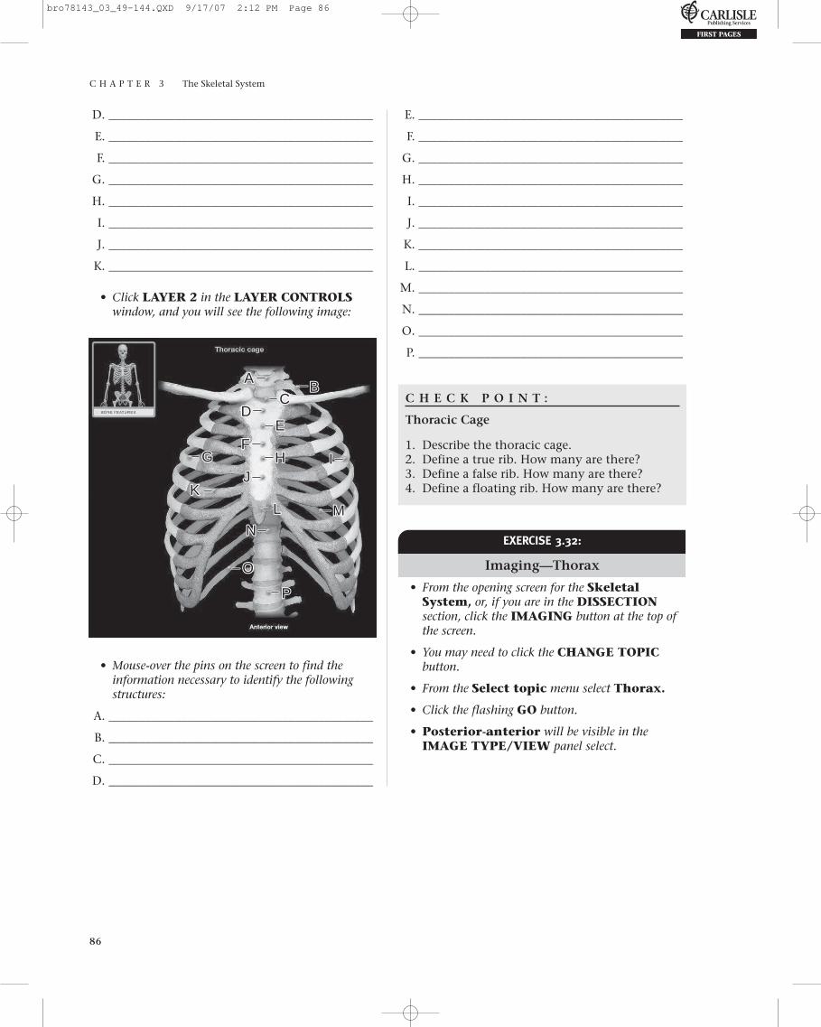
C H A P T E R 3 The Skeletal System
86
D. ____________________________________________
E. ____________________________________________
F. ____________________________________________
G. ____________________________________________
H. ____________________________________________
I. ____________________________________________
J. ____________________________________________
K. ____________________________________________
• Click LAYER 2 in the LAYER CONTROLSwindow, and you will see the following image:
E. ____________________________________________
F. ____________________________________________
G. ____________________________________________
H. ____________________________________________
I. ____________________________________________
J. ____________________________________________
K. ____________________________________________
L. ____________________________________________
M. ____________________________________________
N. ____________________________________________
O. ____________________________________________
P. ____________________________________________
C H E C K P O I N T :
Thoracic Cage
1. Describe the thoracic cage.2. Define a true rib. How many are there?3. Define a false rib. How many are there?4. Define a floating rib. How many are there?
AABB
CC
EEDD
FFGG HH
LL
IIJJ
KKMM
OO
PP
NN
• Mouse-over the pins on the screen to find theinformation necessary to identify the followingstructures:
A. ____________________________________________
B. ____________________________________________
C. ____________________________________________
D. ____________________________________________
EXERCISE 3.32:
Imaging—Thorax
• From the opening screen for the SkeletalSystem, or, if you are in the DISSECTIONsection, click the IMAGING button at the top ofthe screen.
• You may need to click the CHANGE TOPICbutton.
• From the Select topic menu select Thorax.
• Click the flashing GO button.
• Posterior-anterior will be visible in theIMAGE TYPE/VIEW panel select.
bro78143_03_49-144.QXD 9/17/07 2:12 PM Page 86

C H A P T E R 3 The Skeletal System
87
• Click the TURN TAGS ON button, and you willsee the following image:
• Click the flashing GO button.
• Click LAYER 1 in the LAYER CONTROLSwindow, and you will see the following image:
AABB
CC
DD
• Mouse-over the green pins on the screen to find theinformation necessary to identify the followingstructures:
A. ____________________________________________
B. ____________________________________________
C. ____________________________________________
Non-Skeletal System Structure (blue pin)
D. ____________________________________________
EXERCISE 3.33:
Vertebral Column
• From the opening screen for the SkeletonSystem, click the DISSECTION button on themenu at the top of the screen. Or, if you are in theIMAGING section, click the DISSECTIONbutton at the top of the screen.
• You may need to click the CHANGE TOPIC/VIEW button to continue.
• From the Select topic menu, select Vertebralcolumn.
• Anterior, posterior, and lateral will bevisible in the Select view menu.
AABB
CCEEDD
FF GGHH
LL
IIJJ
KK
MM
• Mouse-over the pins on the screen to find theinformation necessary to identify the followingstructures:
A. ____________________________________________
B. ____________________________________________
C. ____________________________________________
D. ____________________________________________
E. ____________________________________________
F. ____________________________________________
G. ____________________________________________
H. ____________________________________________
I. ____________________________________________
J. ____________________________________________
K. ____________________________________________
L. ____________________________________________
M. ____________________________________________
bro78143_03_49-144.QXD 9/17/07 2:12 PM Page 87

C H A P T E R 3 The Skeletal System
88
• Click LAYER 2 in the LAYER CONTROLSwindow, and you will see the following image: C H E C K P O I N T :
Vertebral Column
1. Name the different groups of vertebrae. Howmany of each in a typical skeleton?
2. What structure passes through each interver-tebral foramen?
3. Which vertebra has a dens?4. Which vertebra has no vertebral body?
AA
BB
CCEE
DDFF
GG
HH
LL
IIJJ
KK
MM NN
PP SSQQRR
OO
• Mouse-over the pins on the screen to find the infor-mation necessary to identify the following structures:
A. ____________________________________________
B. ____________________________________________
C. ____________________________________________
D. ____________________________________________
E. ____________________________________________
F. ____________________________________________
G. ____________________________________________
H. ____________________________________________
I. ____________________________________________
J. ____________________________________________
K. ____________________________________________
L. ____________________________________________
M. ____________________________________________
N. ____________________________________________
O. ____________________________________________
P. ____________________________________________
Q. ____________________________________________
R. ____________________________________________
S. ____________________________________________
EXERCISE 3.34:
Cervical Vertebra
• From the opening screen for the SkeletonSystem, click the DISSECTION button on themenu at the top of the screen. Or, if you are in theDISSECTION section, click the CHANGETOPIC/VIEW button at the top of the screen.
• From the Select topic menu, select Cervicalvertebra.
• Superior and lateral will be visible in theSelect view menu.
• Click the flashing GO button.
• Click LAYER 1 in the LAYER CONTROLSwindow, and you will see the following image:
AA
BBCC
EE DD
FF GGHHII
JJ
KK
bro78143_03_49-144.QXD 9/17/07 2:12 PM Page 88

C H A P T E R 3 The Skeletal System
89
• Mouse-over the pins on the screen to find theinformation necessary to identify the followingstructures:
A. ____________________________________________
B. ____________________________________________
C. ____________________________________________
D. ____________________________________________
E. ____________________________________________
F. ____________________________________________
G. ____________________________________________
H. ____________________________________________
I. ____________________________________________
J. ____________________________________________
K. ____________________________________________
C H E C K P O I N T :
Cervical Vertebra
1. Name a characteristic unique to the trans-verse processes of the cervical vertebrae.
2. What structure passes through this openingin C1-C6?
3. What is unique about the spinous process ofmost cervical vertebrae?
4. Which vertebra does not have a spinousprocess?
• Click LAYER 1 in the LAYER CONTROLSwindow, and you will see the following image:
EXERCISE 3.35:
Atlas (C1 Vertebra)
• From the opening screen for the Skeleton System, click the DISSECTION button on the menu at the top of the screen. Or, if you are in the DISSECTION section, click theCHANGE TOPIC/VIEW button at the top of the screen.
• From the Select topic menu, select Atlas (C1 vertebra).
• Superior will be visible in the Select viewmenu.
• Click the flashing GO button.
AABB
CCEEDDFFGG
HH IIJJ
• Mouse-over the pins on the screen to find theinformation necessary to identify the followingstructures:
A. ____________________________________________
B. ____________________________________________
C. ____________________________________________
D. ____________________________________________
E. ____________________________________________
F. ____________________________________________
G. ____________________________________________
H. ____________________________________________
I. ____________________________________________
J. ____________________________________________
C H E C K P O I N T :
Atlas (C1 Vertebra)
1. What is the most posterior structure of theAtlas (C1 vertebra)?
2. What is its function?3. What structure is lacking on the atlas and
coccygeal vertebrae that is present on allother vertebrae?
bro78143_03_49-144.QXD 9/17/07 2:12 PM Page 89

C H A P T E R 3 The Skeletal System
90
H. ____________________________________________
I. ____________________________________________
J. ____________________________________________
K. ____________________________________________
C H E C K P O I N T :
Axis (C2 Vertebra)
1. Name a structure unique to the axis.2. What is another name for this structure?3. What does this structure represent?
EXERCISE 3.36:
Axis (C2 Vertebra)
• From the opening screen for the SkeletonSystem, click the DISSECTION button on themenu at the top of the screen. Or, if you are in theDISSECTION section, click the CHANGETOPIC/VIEW button at the top of the screen.
• From the Select topic menu, select Axis (C2 vertebra).
• Posterosuperior will be visible in the Selectview menu.
• Click the flashing GO button.
• Click LAYER 1 in the LAYER CONTROLSwindow, and you will see the following image:
AA
BB
DDCC
EEFF
GG
KK
HHJJII
• Mouse-over the pins on the screen to find theinformation necessary to identify the followingstructures:
A. ____________________________________________
B. ____________________________________________
C. ____________________________________________
D. ____________________________________________
E. ____________________________________________
F. ____________________________________________
G. ____________________________________________
EXERCISE 3.37:
Thoracic Vertebra
• From the opening screen for the SkeletonSystem, click the DISSECTION button on themenu at the top of the screen. Or, if you are in theDISSECTION section, click the CHANGETOPIC/VIEW button at the top of the screen.
• From the Select topic menu, select Thoracicvertebra.
• Superior and lateral will be visible in theSelect view menu.
• Click the flashing GO button.
• Click LAYER 1 in the LAYER CONTROLSwindow, and you will see the following image:
AA
BB CC
EEDD FFGG
HH
LL
IIJJ KK
bro78143_03_49-144.QXD 9/17/07 2:12 PM Page 90

C H A P T E R 3 The Skeletal System
91
• Mouse-over the pins on the screen to find the information necessary to identify the followingstructures:
A. ____________________________________________
B. ____________________________________________
C. ____________________________________________
D. ____________________________________________
E. ____________________________________________
F. ____________________________________________
G. ____________________________________________
H. ____________________________________________
I. ____________________________________________
J. ____________________________________________
K. ____________________________________________
L. ____________________________________________
C H E C K P O I N T :
Thoracic Vertebra
1. List the characteristic features of thoracicvertebrae.
2. How many thoracic vertebrae do you have?3. Where are they located?
• Click LAYER 1 in the LAYER CONTROLSwindow, and you will see the following image:
EXERCISE 3.38:
Lumbar Vertebra
• From the opening screen for the Skeleton System, click the DISSECTION button on the menu at the top of the screen. Or, if you are in the DISSECTION section, click theCHANGE TOPIC/VIEW button at the top of the screen.
• From the Select topic menu, select Lumbarvertebra.
• Superior and lateral will be visible in theSelect view menu.
• Click the flashing GO button.
AA
BBCCEE
DDFF
GG
HH
II
• Mouse-over the pins on the screen to find the information necessary to identify the followingstructures:
A. ____________________________________________
B. ____________________________________________
C. ____________________________________________
D. ____________________________________________
E. ____________________________________________
F. ____________________________________________
G. ____________________________________________
H. ____________________________________________
I. ____________________________________________
C H E C K P O I N T :
Lumbar Vertebra
1. List the characteristic features of lumbarvertebrae.
2. How many lumbar vertebrae do you have?3. Where are they located?
bro78143_03_49-144.QXD 9/17/07 2:12 PM Page 91
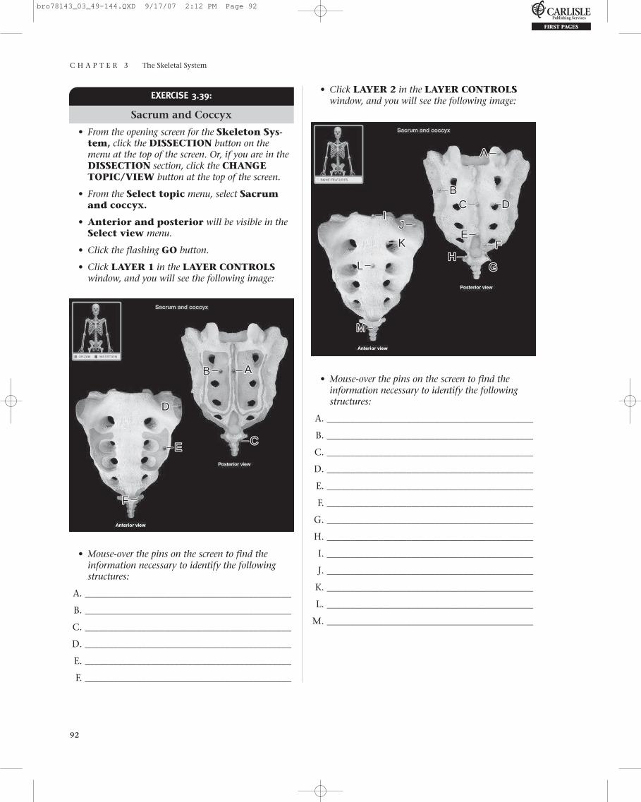
C H A P T E R 3 The Skeletal System
92
EXERCISE 3.39:
Sacrum and Coccyx
• From the opening screen for the Skeleton Sys-tem, click the DISSECTION button on themenu at the top of the screen. Or, if you are in theDISSECTION section, click the CHANGETOPIC/VIEW button at the top of the screen.
• From the Select topic menu, select Sacrumand coccyx.
• Anterior and posterior will be visible in theSelect view menu.
• Click the flashing GO button.
• Click LAYER 1 in the LAYER CONTROLSwindow, and you will see the following image:
• Click LAYER 2 in the LAYER CONTROLSwindow, and you will see the following image:
AABB
CCEE
DD
FF
• Mouse-over the pins on the screen to find the information necessary to identify the followingstructures:
A. ____________________________________________
B. ____________________________________________
C. ____________________________________________
D. ____________________________________________
E. ____________________________________________
F. ____________________________________________
AA
CCBB
EE
DD
FF
GGHH
LL
JJII
KK
MM
• Mouse-over the pins on the screen to find theinformation necessary to identify the followingstructures:
A. ____________________________________________
B. ____________________________________________
C. ____________________________________________
D. ____________________________________________
E. ____________________________________________
F. ____________________________________________
G. ____________________________________________
H. ____________________________________________
I. ____________________________________________
J. ____________________________________________
K. ____________________________________________
L. ____________________________________________
M. ____________________________________________
bro78143_03_49-144.QXD 9/17/07 2:12 PM Page 92

C H A P T E R 3 The Skeletal System
93
C H E C K P O I N T :
Sacrum and Coccyx
1. Name the landmark for establishing femalepelvic dimensions.
2. Name the route for injection in caudal epi-dermal anesthesia.
3. Describe the coccyx.
• From the Select region menu, select Thoraciccage.
• From the Select test type menu, select Clickto identify.
• From the Select number of questions menu,select ALL.
• After completing the SELF TEST and saving yourresults, click the Select test type menu, selectMultiple choice.
• From the Select number of questions menu,select ALL.
• Return to the Select region menu, and take theSELF TEST for Vertebral column andAxial skeleton.
• Complete each SELF TEST and save your results.
Self TestTake this opportunity to quiz yourself by taking theSELF TEST.
• From the opening screen for the Skeletal System,or, if you are in the DISSECTION section, click theSELF TEST button at the top of the screen.
• From the Select test topic menu, selectSkeleton.
What Have I Learned?
The following questions cover the material thatyou just read, the thoracic cage and vertebral col-umn. Apply what you have learned in answering
these questions:
1. Describe the thoracic cage.
2. Define a true rib. How many are there?
3. Define a false rib. How many are there?
4. Define a floating rib. How many are there?
5. Name the different groups of vertebrae. Howmany of each in a typical skeleton?
6. What structure passes through each interverte-bral foramen?
7. Which vertebra has a dens?
8. Which vertebra has no vertebral body?
9. Name a characteristic unique to the transverseprocesses of the cervical vertebrae.
10. What structure passes through this opening inC1-C6?
11. What is unique about the spinous process ofmost cervical vertebrae?
12. Which vertebra does not have a spinous process?
I N R E V I E W
Continued
bro78143_03_49-144.QXD 9/17/07 2:12 PM Page 93

C H A P T E R 3 The Skeletal System
94
13. What is the most posterior structure of the Atlas(C1 vertebra)?
14. What is its function?
15. What structure is lacking on the atlas and coccygeal vertebrae that is present on all othervertebrae?
16. Name a structure unique to the axis.
17. What is another name for this structure?
18. What does this structure represent?
19. List the characteristic features of thoracic vertebrae.
20. How many thoracic vertebrae do you have?
21. Where are they located?
22. List the characteristic features of lumbar vertebrae.
23. How many lumbar vertebrae do you have?
24. Where are they located?
25. Name the landmark for establishing femalepelvic dimensions.
26. Name the route for injection in caudal epidermalanesthesia.
27. Describe the coccyx.
• From the Select topic menu, select Shoulderand arm under the Pectoral girdle andupper limb heading.
• From the Select view menu select Anterior.
• Click the flashing GO button.
Pectoral Girdle and Upper Limb
EXERCISE 3.40:
Shoulder and Arm, Anterior View
• From the opening screen for the Skeleton System, click the DISSECTION button on the menu at the top of the screen. Or, if you are in the DISSECTION section, click theCHANGE TOPIC/VIEW button at the top of the screen.
Continued
bro78143_03_49-144.QXD 9/17/07 2:12 PM Page 94
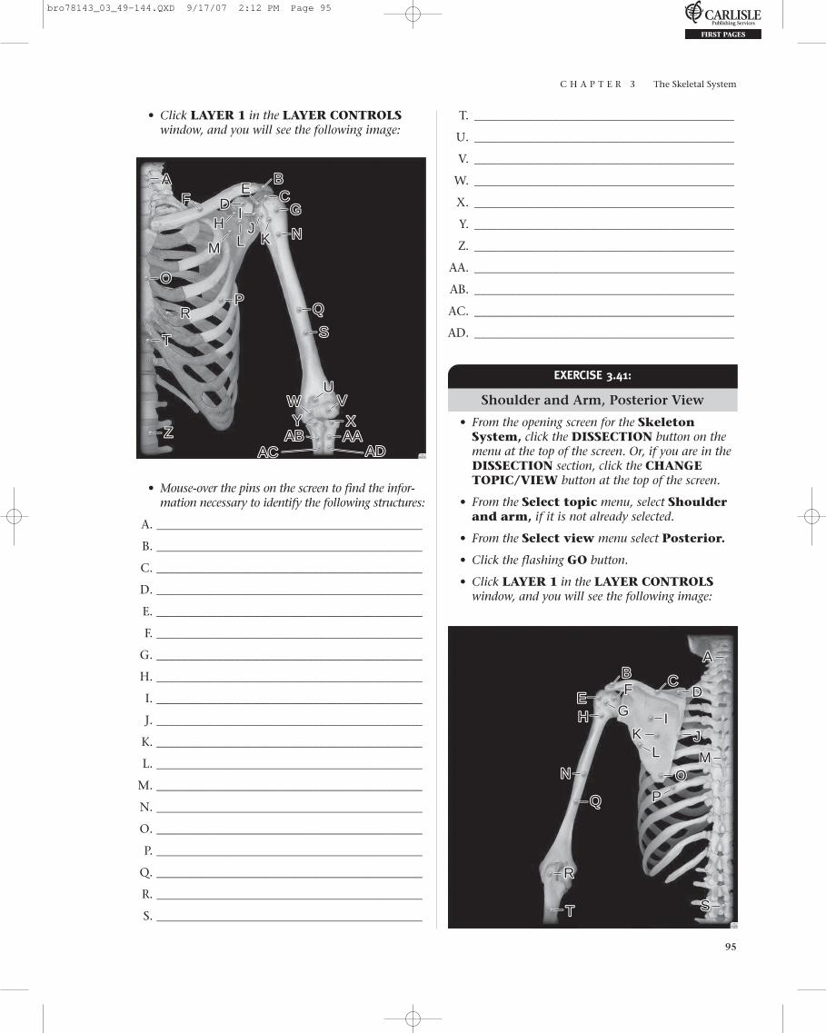
C H A P T E R 3 The Skeletal System
95
• Click LAYER 1 in the LAYER CONTROLSwindow, and you will see the following image:
T. ___________________________________________
U. ___________________________________________
V. ___________________________________________
W. ___________________________________________
X. ___________________________________________
Y. ___________________________________________
Z. ___________________________________________
AA. ___________________________________________
AB. ___________________________________________
AC. ___________________________________________
AD. ___________________________________________
AA BBCCEE
DDFFGG
HHLL
IIJJ
KKMM
NN
OO
UU
XXAAAA
ACAC ADAD
WW
ZZ ABAB
PP
TT
SSRR
VVYY
• Mouse-over the pins on the screen to find the infor-mation necessary to identify the following structures:
A. ____________________________________________
B. ____________________________________________
C. ____________________________________________
D. ____________________________________________
E. ____________________________________________
F. ____________________________________________
G. ____________________________________________
H. ____________________________________________
I. ____________________________________________
J. ____________________________________________
K. ____________________________________________
L. ____________________________________________
M. ____________________________________________
N. ____________________________________________
O. ____________________________________________
P. ____________________________________________
Q. ____________________________________________
R. ____________________________________________
S. ____________________________________________
EXERCISE 3.41:
Shoulder and Arm, Posterior View
• From the opening screen for the SkeletonSystem, click the DISSECTION button on themenu at the top of the screen. Or, if you are in theDISSECTION section, click the CHANGETOPIC/VIEW button at the top of the screen.
• From the Select topic menu, select Shoulderand arm, if it is not already selected.
• From the Select view menu select Posterior.
• Click the flashing GO button.
• Click LAYER 1 in the LAYER CONTROLSwindow, and you will see the following image:
AABB CC
EE DDFF
GGHH
LL
IIJJKK
MMNN
PP
SSTT
RR
OO
bro78143_03_49-144.QXD 9/17/07 2:12 PM Page 95

C H A P T E R 3 The Skeletal System
96
• Mouse-over the pins on the screen to find theinformation necessary to identify the followingstructures:
A. ____________________________________________
B. ____________________________________________
C. ____________________________________________
D. ____________________________________________
E. ____________________________________________
F. ____________________________________________
G. ____________________________________________
H. ____________________________________________
I. ____________________________________________
J. ____________________________________________
K. ____________________________________________
L. ____________________________________________
M. ____________________________________________
N. ____________________________________________
O. ____________________________________________
P. ____________________________________________
Q. ____________________________________________
R. ____________________________________________
S. ____________________________________________
T. ____________________________________________
C H E C K P O I N T :
Shoulder and Arm
1. Which bony structures form the gleno-humeral joint?
2. How many ribs do you have?3. Both true and false ribs articulate with ______.4. Name the characteristic features of the
scapula.
• From the Select topic menu select Shoulderand arm.
• Click the flashing GO button.
• Anterior-posterior will be visible in theIMAGE TYPE/VIEW panel select.
• Click the TURN TAGS ON button, and you willsee the following image:
EXERCISE 3.42:
Imaging—Shoulder and Arm
• From the opening screen for the Skeletal Sys-tem, or, if you are in the DISSECTION section,click the IMAGING button at the top of the screen.
• You may need to click the CHANGE TOPICbutton.
AA
BBCCEE DD FF
GG
• Mouse-over the pins on the screen to find theinformation necessary to identify the followingstructures:
A. ____________________________________________
B. ____________________________________________
C. ____________________________________________
D. ____________________________________________
E. ____________________________________________
F. ____________________________________________
G. ____________________________________________
EXERCISE 3.43:
Clavicle
• From the opening screen for the SkeletonSystem, click the DISSECTION button on themenu at the top of the screen. Or, if you are in theIMAGING section, click the DISSECTIONbutton at the top of the screen.
bro78143_03_49-144.QXD 9/17/07 2:12 PM Page 96

C H A P T E R 3 The Skeletal System
97
• You may need to click the CHANGE TOPIC/VIEW button to continue.
• From the Select topic menu, select Clavicle.
• Superior and inferior will be visible in theSelect view menu.
• Click the flashing GO button.
• Click LAYER 1 in the LAYER CONTROLSwindow, and you will see the following image:
AA
BBCC
EE
DD
FF
• Mouse-over the pins on the screen to find theinformation necessary to identify the followingstructures:
A. ____________________________________________
B. ____________________________________________
C. ____________________________________________
D. ____________________________________________
E. ____________________________________________
F. ____________________________________________
AABB
DD
CC
• Mouse-over the pins on the screen to find theinformation necessary to identify the followingstructures:
A. ____________________________________________
B. ____________________________________________
C. ____________________________________________
D. ____________________________________________
C H E C K P O I N T :
Clavicle
1. The medial end of the clavicle articulates . . . .2. The lateral end of the clavicle articulates . . . .3. Another name for the clavicle is . . . .
EXERCISE 3.44:
Scapula
• From the opening screen for the SkeletonSystem, click the DISSECTION button on themenu at the top of the screen. Or, if you are in theDISSECTION section, click the CHANGETOPIC/VIEW button at the top of the screen.
• Click LAYER 2 in the LAYER CONTROLSwindow, and you will see the following image:
bro78143_03_49-144.QXD 9/17/07 2:12 PM Page 97

C H A P T E R 3 The Skeletal System
98
• From the Select topic menu, select Scapula.
• Anterior, posterior, and lateral will be vis-ible in the Select view menu.
• Click the flashing GO button.
• Click LAYER 1 in the LAYER CONTROLSwindow, and you will see the following image:
P. ____________________________________________
Q. ____________________________________________
R. ____________________________________________
• Click LAYER 2 in the LAYER CONTROLSwindow, and you will see the following image:
AA BBCC
EE
DD
FF
GGHH
LLII
JJ
KK
MM
NN
PPQQ
RR
OO
• Mouse-over the pins on the screen to find the infor-mation necessary to identify the following structures:
A. ____________________________________________
B. ____________________________________________
C. ____________________________________________
D. ____________________________________________
E. ____________________________________________
F. ____________________________________________
G. ____________________________________________
H. ____________________________________________
I. ____________________________________________
J. ____________________________________________
K. ____________________________________________
L. ____________________________________________
M. ____________________________________________
N. ____________________________________________
O. ____________________________________________
AA BBCC
EEDD
FFGG
HH
LL
IIJJ KK
MM
NN
PP
OO
• Mouse-over the pins on the screen to find the infor-mation necessary to identify the following structures:
A. ____________________________________________
B. ____________________________________________
C. ____________________________________________
D. ____________________________________________
E. ____________________________________________
F. ____________________________________________
G. ____________________________________________
H. ____________________________________________
I. ____________________________________________
J. ____________________________________________
K. ____________________________________________
L. ____________________________________________
M. ____________________________________________
N. ____________________________________________
O. ____________________________________________
P. ____________________________________________
bro78143_03_49-144.QXD 9/17/07 2:12 PM Page 98

C H A P T E R 3 The Skeletal System
99
C H E C K P O I N T :
Scapula
1. Name a landmark for intramuscular injections.2. Name another visible subcutaneous landmark.
E. ____________________________________________
F. ____________________________________________
G. ____________________________________________
H. ____________________________________________
I. ____________________________________________
J. ____________________________________________
K. ____________________________________________
L. ____________________________________________
M. ____________________________________________
N. ____________________________________________
O. ____________________________________________
P. ____________________________________________
Q. ____________________________________________
R. ____________________________________________
• Click LAYER 2 in the LAYER CONTROLSwindow, and you will see the following image:
EXERCISE 3.45:
Humerus
• From the opening screen for the SkeletonSystem, click the DISSECTION button on themenu at the top of the screen. Or, if you are in theDISSECTION section, click the CHANGETOPIC/VIEW button at the top of the screen.
• From the Select topic menu, select Humerus.
• Anterior and posterior will be visible in theSelect view menu.
• Click the flashing GO button.
• Click LAYER 1 in the LAYER CONTROLSwindow, and you will see the following image:
AA BB
CCDDEE
FF
GGHH
IIJJ
KK LL
MMNN
PP
OO
RR
AABBCC DD
EE
FF
GG
HHII JJ
KKLL
MMNN
PP
OO
QQ RR
• Mouse-over the pins on the screen to find the infor-mation necessary to identify the following structures:
A. ____________________________________________
B. ____________________________________________
C. ____________________________________________
D. ____________________________________________
bro78143_03_49-144.QXD 9/17/07 2:12 PM Page 99

C H A P T E R 3 The Skeletal System
100
E. ____________________________________________
F. ____________________________________________
G. ____________________________________________
H. ____________________________________________
I. ____________________________________________
J. ____________________________________________
K. ____________________________________________
L. ____________________________________________
M. ____________________________________________
N. ____________________________________________
O. ____________________________________________
P. ____________________________________________
Q. ____________________________________________
R. ____________________________________________
C H E C K P O I N T :
Humerus
1. How many necks are on the proximal end ofthe humerus? Where are they located?
2. Which neck is a common site for fractures?3. Why is your elbow referred to as your “funny
bone”?
EXERCISE 3.46:
Forearm and Hand, Anterior View
• From the opening screen for the Skeleton Sys-tem, click the DISSECTION button on themenu at the top of the screen. Or, if you are in theDISSECTION section, click the CHANGETOPIC/VIEW button at the top of the screen.
• From the Select topic menu, select Forearmand hand.
• From the Select view menu, select Anterior.
• Click the flashing GO button.
• Click LAYER 1 in the LAYER CONTROLSwindow, and you will see the following image:
AA
BB
AA AAAA BB
CC
AACC
DD
AADD
EE
AA EE
FF
AAFF
GGHHII
JJ KKLL MM
NNPP
OO QQRU
WX
RSS
TT
UVVW
X
ZZ
YY
• Mouse-over the pins on the screen to find the infor-mation necessary to identify the following structures:
A. ____________________________________________
B. ____________________________________________
C. ____________________________________________
D. ____________________________________________
E. ____________________________________________
F. ____________________________________________
G. ____________________________________________
H. ____________________________________________
I. ____________________________________________
J. ____________________________________________
K. ____________________________________________
L. ____________________________________________
M. ____________________________________________
N. ____________________________________________
O. ____________________________________________
P. ____________________________________________
Q. ____________________________________________
R. ____________________________________________
S. ____________________________________________
T. ____________________________________________
U. ____________________________________________
bro78143_03_49-144.QXD 9/17/07 2:12 PM Page 100
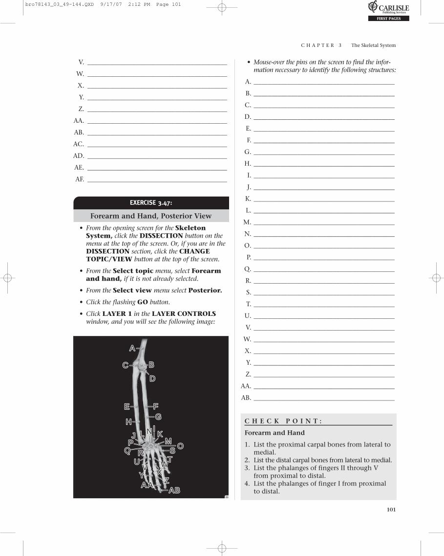
C H A P T E R 3 The Skeletal System
101
V. ___________________________________________
W. ___________________________________________
X. ___________________________________________
Y. ___________________________________________
Z. ___________________________________________
AA. ___________________________________________
AB. ___________________________________________
AC. ___________________________________________
AD. ___________________________________________
AE. ___________________________________________
AF. ___________________________________________
• Mouse-over the pins on the screen to find the infor-mation necessary to identify the following structures:
A.____________________________________________
B.____________________________________________
C.____________________________________________
D.____________________________________________
E.____________________________________________
F.____________________________________________
G.____________________________________________
H.____________________________________________
I.____________________________________________
J.____________________________________________
K.____________________________________________
L.____________________________________________
M.____________________________________________
N.____________________________________________
O.____________________________________________
P.____________________________________________
Q.____________________________________________
R.____________________________________________
S.____________________________________________
T.____________________________________________
U.____________________________________________
V.____________________________________________
W.____________________________________________
X.____________________________________________
Y.____________________________________________
Z.____________________________________________
AA.____________________________________________
AB.____________________________________________
C H E C K P O I N T :
Forearm and Hand
1. List the proximal carpal bones from lateral tomedial.
2. List the distal carpal bones from lateral to medial.3. List the phalanges of fingers II through V
from proximal to distal.4. List the phalanges of finger I from proximal
to distal.
EXERCISE 3.47:
Forearm and Hand, Posterior View
• From the opening screen for the SkeletonSystem, click the DISSECTION button on themenu at the top of the screen. Or, if you are in theDISSECTION section, click the CHANGETOPIC/VIEW button at the top of the screen.
• From the Select topic menu, select Forearmand hand, if it is not already selected.
• From the Select view menu select Posterior.
• Click the flashing GO button.
• Click LAYER 1 in the LAYER CONTROLSwindow, and you will see the following image:
AA
BB
AA AAAABB
CC
DD
EE FFGG
HHII
JJ KKLLMM
NN
PP OOQQ RU W
X
R SSTTU
VV WX
ZZYY
bro78143_03_49-144.QXD 9/17/07 2:12 PM Page 101
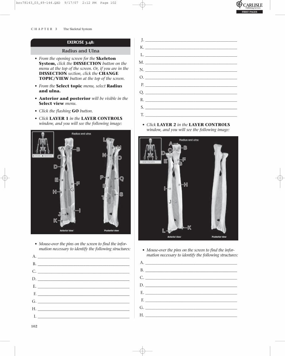
C H A P T E R 3 The Skeletal System
102
EXERCISE 3.48:
Radius and Ulna
• From the opening screen for the SkeletonSystem, click the DISSECTION button on themenu at the top of the screen. Or, if you are in theDISSECTION section, click the CHANGETOPIC/VIEW button at the top of the screen.
• From the Select topic menu, select Radiusand ulna.
• Anterior and posterior will be visible in theSelect view menu.
• Click the flashing GO button.
• Click LAYER 1 in the LAYER CONTROLSwindow, and you will see the following image:
J. ____________________________________________
K. ____________________________________________
L. ____________________________________________
M. ____________________________________________
N. ____________________________________________
O. ____________________________________________
P. ____________________________________________
Q. ____________________________________________
R. ____________________________________________
S. ____________________________________________
T. ____________________________________________
• Click LAYER 2 in the LAYER CONTROLSwindow, and you will see the following image:
AA BB
CC
DD
EEFF
GGHH
IIJJ
KK
LL
MM
NN
PP
OO
RR
SSTT
• Mouse-over the pins on the screen to find the infor-mation necessary to identify the following structures:
A. ____________________________________________
B. ____________________________________________
C. ____________________________________________
D. ____________________________________________
E. ____________________________________________
F. ____________________________________________
G. ____________________________________________
H. ____________________________________________
I. ____________________________________________
AABBCC
DD
EE FFGG
HHII
JJ
KKLL
• Mouse-over the pins on the screen to find the infor-mation necessary to identify the following structures:
A. ____________________________________________
B. ____________________________________________
C. ____________________________________________
D. ____________________________________________
E. ____________________________________________
F. ____________________________________________
G. ____________________________________________
H. ____________________________________________
bro78143_03_49-144.QXD 9/17/07 2:12 PM Page 102
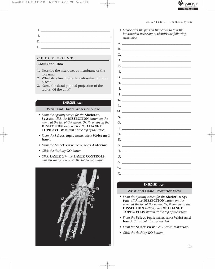
C H A P T E R 3 The Skeletal System
103
I. ____________________________________________
J. ____________________________________________
K. ____________________________________________
L. ____________________________________________
C H E C K P O I N T :
Radius and Ulna
1. Describe the interosseous membrane of theforearm.
2. What structure holds the radio-ulnar joint inplace?
3. Name the distal pointed projection of theradius. Of the ulna?
• Mouse-over the pins on the screen to find theinformation necessary to identify the followingstructures:
A. ____________________________________________
B. ____________________________________________
C. ____________________________________________
D. ____________________________________________
E. ____________________________________________
F. ____________________________________________
G. ____________________________________________
H. ____________________________________________
I. ____________________________________________
J. ____________________________________________
K. ____________________________________________
L. ____________________________________________
M. ____________________________________________
N. ____________________________________________
O. ____________________________________________
P. ____________________________________________
Q. ____________________________________________
R. ____________________________________________
S. ____________________________________________
T. ____________________________________________
U. ____________________________________________
V. ____________________________________________
W. ____________________________________________
X. ____________________________________________
EXERCISE 3.49:
Wrist and Hand, Anterior View
• From the opening screen for the SkeletonSystem, click the DISSECTION button on themenu at the top of the screen. Or, if you are in theDISSECTION section, click the CHANGETOPIC/VIEW button at the top of the screen.
• From the Select topic menu, select Wrist andhand
• From the Select view menu, select Anterior.
• Click the flashing GO button.
• Click LAYER 1 in the LAYER CONTROLSwindow and you will see the following image:
AA BB
CCDDEE
FF
GGHH
II
JJKKLL MM
NNPP
OO
R
U
W
X
RSSTT
U VV
W
X
EXERCISE 3.50:
Wrist and Hand, Posterior View
• From the opening screen for the Skeleton Sys-tem, click the DISSECTION button on themenu at the top of the screen. Or, if you are in theDISSECTION section, click the CHANGETOPIC/VIEW button at the top of the screen.
• From the Select topic menu, select Wrist andhand, if it is not already selected.
• From the Select view menu select Posterior.
• Click the flashing GO button.
bro78143_03_49-144.QXD 9/17/07 2:12 PM Page 103

C H A P T E R 3 The Skeletal System
104
• Click LAYER 1 in the LAYER CONTROLSwindow, and you will see the following image:
S. ____________________________________________
T. ____________________________________________
U. ____________________________________________
AABB
CC DDEE FF
GGHHIIJJ KK LL
MMNN
PP OO QQ
R
U
RSS
TT
U
• Mouse-over the pins on the screen to find theinformation necessary to identify the followingstructures:
A. ____________________________________________
B. ____________________________________________
C. ____________________________________________
D. ____________________________________________
E. ____________________________________________
F. ____________________________________________
G. ____________________________________________
H. ____________________________________________
I. ____________________________________________
J. ____________________________________________
K. ____________________________________________
L. ____________________________________________
M. ____________________________________________
N. ____________________________________________
O. ____________________________________________
P. ____________________________________________
Q. ____________________________________________
R. ____________________________________________
AA BB
AA AAAABB
AA AA
AA
AA
CC
CC
DD
DD
EE
EE
FF
FF
GGHH
IIJJKK LL
MM
NN
PP
OO
R
U
WX
R
SS
TT UVV
WX
ZZYY
EXERCISE 3.51:
Wrist and Hand, Anterior and Posterior View
• From the opening screen for the SkeletonSystem, click the DISSECTION button on themenu at the top of the screen. Or, if you are in theDISSECTION section, click the CHANGETOPIC/VIEW button at the top of the screen.
• From the Select topic menu, select Wrist andhand, if it is not already selected.
• From the Select view menu, select Anteriorand posterior.
• Click the flashing GO button.
• Click LAYER 1 in the LAYER CONTROLSwindow, and you will see the following image:
• Mouse-over the pins on the screen to find theinformation necessary to identify the followingstructures:
A. ____________________________________________
B. ____________________________________________
bro78143_03_49-144.QXD 9/17/07 2:12 PM Page 104
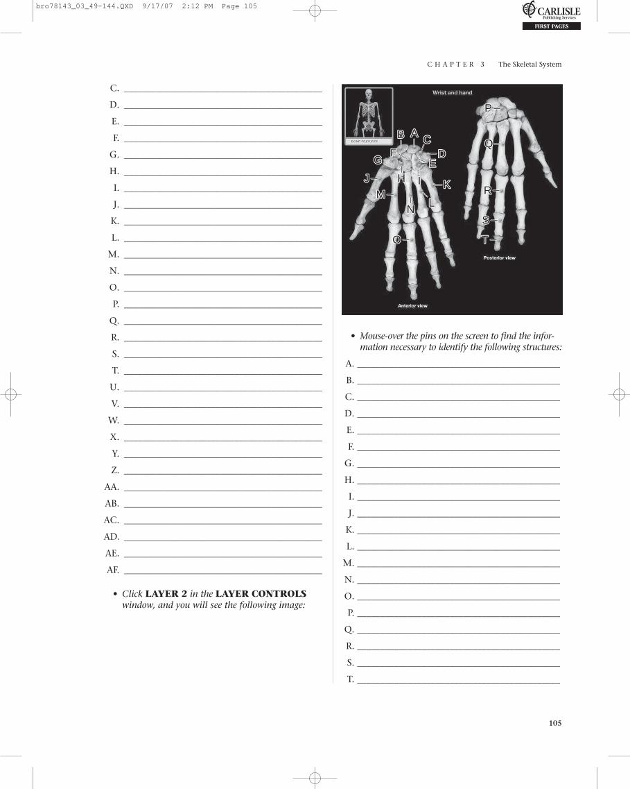
C H A P T E R 3 The Skeletal System
105
C. ___________________________________________
D. ___________________________________________
E. ___________________________________________
F. ___________________________________________
G. ___________________________________________
H. ___________________________________________
I. ___________________________________________
J. ___________________________________________
K. ___________________________________________
L. ___________________________________________
M. ___________________________________________
N. ___________________________________________
O. ___________________________________________
P. ___________________________________________
Q. ___________________________________________
R. ___________________________________________
S. ___________________________________________
T. ___________________________________________
U. ___________________________________________
V. ___________________________________________
W. ___________________________________________
X. ___________________________________________
Y. ___________________________________________
Z. ___________________________________________
AA. ___________________________________________
AB. ___________________________________________
AC. ___________________________________________
AD. ___________________________________________
AE. ___________________________________________
AF. ___________________________________________
• Click LAYER 2 in the LAYER CONTROLSwindow, and you will see the following image:
AABB CCDD
EEFF
GG
HH IIJJ KK
LLMM
NN
PP
OO
RR
SS
TT
• Mouse-over the pins on the screen to find the infor-mation necessary to identify the following structures:
A. ____________________________________________
B. ____________________________________________
C. ____________________________________________
D. ____________________________________________
E. ____________________________________________
F. ____________________________________________
G. ____________________________________________
H. ____________________________________________
I. ____________________________________________
J. ____________________________________________
K. ____________________________________________
L. ____________________________________________
M. ____________________________________________
N. ____________________________________________
O. ____________________________________________
P. ____________________________________________
Q. ____________________________________________
R. ____________________________________________
S. ____________________________________________
T. ____________________________________________
bro78143_03_49-144.QXD 9/17/07 2:12 PM Page 105
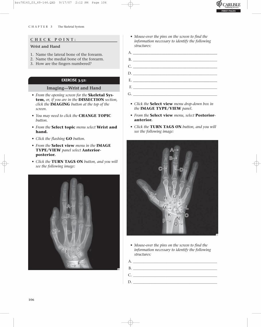
C H A P T E R 3 The Skeletal System
106
C H E C K P O I N T :
Wrist and Hand
1. Name the lateral bone of the forearm.2. Name the medial bone of the forearm.3. How are the fingers numbered?
• Mouse-over the pins on the screen to find theinformation necessary to identify the followingstructures:
A. ____________________________________________
B. ____________________________________________
C. ____________________________________________
D. ____________________________________________
E. ____________________________________________
F. ____________________________________________
G. ____________________________________________
• Click the Select view menu drop-down box inthe IMAGE TYPE/VIEW panel.
• From the Select view menu, select Posterior-anterior.
• Click the TURN TAGS ON button, and you willsee the following image:
EXERCISE 3.52:
Imaging—Wrist and Hand
• From the opening screen for the Skeletal Sys-tem, or, if you are in the DISSECTION section,click the IMAGING button at the top of thescreen.
• You may need to click the CHANGE TOPICbutton.
• From the Select topic menu select Wrist andhand.
• Click the flashing GO button.
• From the Select view menu in the IMAGETYPE/VIEW panel select Anterior-posterior.
• Click the TURN TAGS ON button, and you willsee the following image:
AA
BB
CC DD
EE
FFGG
AA
BB
CC DD
EEFF GGHH II
JJ
KK
LLMM
NNPPOO
• Mouse-over the pins on the screen to find the information necessary to identify the followingstructures:
A. ____________________________________________
B. ____________________________________________
C. ____________________________________________
D. ____________________________________________
bro78143_03_49-144.QXD 9/17/07 2:12 PM Page 106
C H A P T E R 3 The Skeletal System
107
E. ____________________________________________
F. ____________________________________________
G. ____________________________________________
H. ____________________________________________
I. ____________________________________________
J. ____________________________________________
K. ____________________________________________
L. ____________________________________________
M. ____________________________________________
N. ____________________________________________
O. ____________________________________________
P. ____________________________________________
Q. ____________________________________________
Self TestTake this opportunity to quiz yourself by taking theSELF TEST.
• From the opening screen for the Skeletal Sys-tem, or, if you are in the DISSECTION section,
click the SELF TEST button at the top of thescreen.
• From the Select test topic menu, selectSkeleton.
• From the Select region menu, select Upperlimb.
• From the Select test type menu, select Clickto identify.
• From the Select number of questions menu,select ALL.
• After completing the SELF TEST and saving yourresults, click the Select test type menu, selectMultiple choice.
• From the Select number of questions menu,select ALL.
• Complete the SELF TEST, and save your results.
What Have I Learned?
The following questions cover the material thatyou just learned: the pectoral girdle and upperlimb. Apply what you have learned in answering
these questions:
1. Which bony structures form the glenohumeraljoint?
2. Name the characteristic features of the scapula.
3. The lateral end of the clavicle articulates ______.
4. Name a landmark of the scapula for intramuscu-lar injections.
5. Name another visible subcutaneous landmark ofthe scapula.
6. How many necks are on the proximal end of thehumerus? Where are they located?
7. Which neck is a common site for fractures?
8. Why is your elbow referred to as your “funnybone”?
I N R E V I E W
Continued
bro78143_03_49-144.QXD 9/17/07 2:12 PM Page 107
C H A P T E R 3 The Skeletal System
108
9. List the proximal carpal bones from lateral tomedial.
10. List the distal carpal bones from lateral tomedial.
11. List the phalanges of fingers II through V fromproximal to distal.
12. List the phalanges of finger I from proximal todistal.
13. Describe the interosseous membrane of theforearm.
14. What structure holds the radio-ulnar joint inplace?
15. Name the distal pointed projection of the radius.Of the ulna?
16. Name the lateral bone of the forearm.
17. Name the medial bone of the forearm.
18. Name the bones and the bony structures thatform the shoulder joint.
19. Name the wrist bone with a prominent hook(hamulus).
20. What term describes a bone embedded in a ten-don? Which wrist bone is embedded in a ten-don? Which tendon?
21. Name the prominent ridge on the posteriorscapula.
22. Name the eight wrist bones.
Pelvic Girdle and Lower Limb
EXERCISE 3.53:
Hip and Thigh, Anterior View
• From the opening screen for the SkeletonSystem, click the DISSECTION button on themenu at the top of the screen. Or, if you are in theIMAGING section, click the DISSECTIONbutton at the top of the screen.
• You may need to click the CHANGE TOPIC/VIEW button to continue.
• From the Select topic menu, select Hip andthigh under the Pelvic girdle and lowerlimb heading.
• From the Select view menu, select Anterior.
• Click the flashing GO button.
Continued
bro78143_03_49-144.QXD 9/17/07 2:12 PM Page 108

C H A P T E R 3 The Skeletal System
109
• Click LAYER 1 in the LAYER CONTROLSwindow, and you will see the following image:
S.____________________________________________
T.____________________________________________
U.____________________________________________
V.____________________________________________
W.____________________________________________
X.____________________________________________
Y.____________________________________________
Z.____________________________________________
AA.____________________________________________
AA BB
AA AA
CCDD EEFF
GGHH
IIJJ
KK LL
MM
NNPPOO QQ
R
U WX
R
SS
TT
UVV
WX
ZZYY
• Mouse-over the pins on the screen to find theinformation necessary to identify the followingstructures:
A. ____________________________________________
B. ____________________________________________
C. ____________________________________________
D. ____________________________________________
E. ____________________________________________
F. ____________________________________________
G. ____________________________________________
H. ____________________________________________
I. ____________________________________________
J. ____________________________________________
K. ____________________________________________
L. ____________________________________________
M. ____________________________________________
N. ____________________________________________
O. ____________________________________________
P. ____________________________________________
Q. ____________________________________________
R. ____________________________________________
EXERCISE 3.54:
Hip and Thigh, Posterior View
• From the opening screen for the SkeletonSystem, click the DISSECTION button on themenu at the top of the screen. Or, if you are in theDISSECTION section, click the CHANGETOPIC/VIEW button at the top of the screen.
• From the Select topic menu, select Hip andthigh, if it is not already selected.
• From the Select view menu, select Posterior.
• Click the flashing GO button.
• Click LAYER 1 in the LAYER CONTROLSwindow, and you will see the following image:
AA BBCCDD
EEFF
GG HHII
JJKK
LLMM
NNPP
OO
R
U
R
SS TTUVV
bro78143_03_49-144.QXD 9/17/07 2:12 PM Page 109

C H A P T E R 3 The Skeletal System
110
• Mouse-over the pins on the screen to find theinformation necessary to identify the followingstructures:
A. ____________________________________________
B. ____________________________________________
C. ____________________________________________
D. ____________________________________________
E. ____________________________________________
F. ____________________________________________
G. ____________________________________________
H. ____________________________________________
I. ____________________________________________
J. ____________________________________________
K. ____________________________________________
L. ____________________________________________
M. ____________________________________________
N. ____________________________________________
O. ____________________________________________
P. ____________________________________________
Q. ____________________________________________
R. ____________________________________________
S. ____________________________________________
T. ____________________________________________
U. ____________________________________________
V. ____________________________________________
C H E C K P O I N T :
Hip and Thigh
1. Describe the patella.2. Describe the sacrum.3. Name the enlarged distal ends of the femur.
• From the Select topic menu, select Pelvis.
• Superior will be visible in the Select viewmenu.
• Click the flashing GO button.
• Click LAYER 1 in the LAYER CONTROLSwindow, and you will see the following image:
EXERCISE 3.55:
Pelvis
• From the opening screen for the Skeleton Sys-tem, click the DISSECTION button on themenu at the top of the screen. Or, if you are in theDISSECTION section, click the CHANGETOPIC/VIEW button at the top of the screen.
AABB
CCDDEE
FF GG HH
IIJJ KK
LLMM NN
PP OO
QQRR
• Mouse-over the pins on the screen to find the infor-mation necessary to identify the following structures:
A. ____________________________________________
B. ____________________________________________
C. ____________________________________________
D. ____________________________________________
E. ____________________________________________
F. ____________________________________________
G. ____________________________________________
H. ____________________________________________
I. ____________________________________________
J. ____________________________________________
K. ____________________________________________
L. ____________________________________________
M. ____________________________________________
N. ____________________________________________
O. ____________________________________________
bro78143_03_49-144.QXD 9/17/07 2:12 PM Page 110
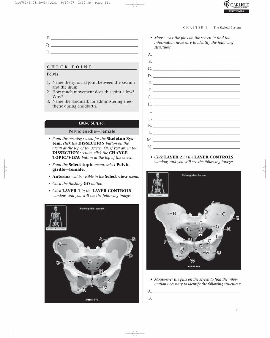
C H A P T E R 3 The Skeletal System
111
P. ____________________________________________
Q. ____________________________________________
R. ____________________________________________
C H E C K P O I N T :
Pelvis
1. Name the synovial joint between the sacrumand the ilium.
2. How much movement does this joint allow?Why?
3. Name the landmark for administering anes-thetic during childbirth.
• Mouse-over the pins on the screen to find theinformation necessary to identify the followingstructures:
A. ____________________________________________
B. ____________________________________________
C. ____________________________________________
D. ____________________________________________
E. ____________________________________________
F. ____________________________________________
G. ____________________________________________
H. ____________________________________________
I. ____________________________________________
J. ____________________________________________
K. ____________________________________________
L. ____________________________________________
M. ____________________________________________
N. ____________________________________________
• Click LAYER 2 in the LAYER CONTROLSwindow, and you will see the following image:
EXERCISE 3.56:
Pelvic Girdle—Female
• From the opening screen for the Skeleton Sys-tem, click the DISSECTION button on themenu at the top of the screen. Or, if you are in theDISSECTION section, click the CHANGETOPIC/VIEW button at the top of the screen.
• From the Select topic menu, select Pelvicgirdle—female.
• Anterior will be visible in the Select view menu.
• Click the flashing GO button.
• Click LAYER 1 in the LAYER CONTROLSwindow, and you will see the following image:
AA BBCC
DDEEFFGG
HHII
JJ KKLL MMNN
AA
BB CCDDEE
FFGGHHII JJ KK
LLMMNNPP OO
QQR
U
WX
RSS
TTUVV
WX
• Mouse-over the pins on the screen to find the infor-mation necessary to identify the following structures:
A. ____________________________________________
B. ____________________________________________
bro78143_03_49-144.QXD 9/17/07 2:12 PM Page 111
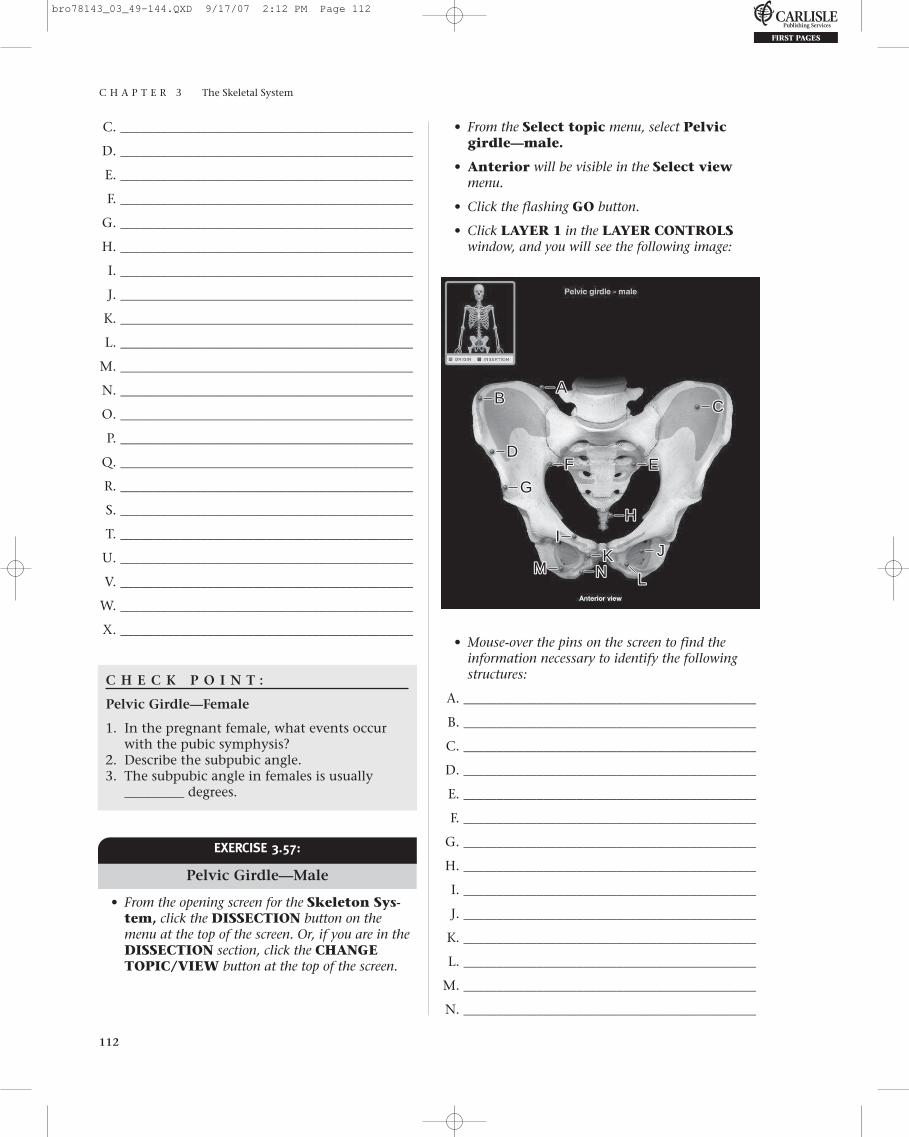
C H A P T E R 3 The Skeletal System
112
C. ____________________________________________
D. ____________________________________________
E. ____________________________________________
F. ____________________________________________
G. ____________________________________________
H. ____________________________________________
I. ____________________________________________
J. ____________________________________________
K. ____________________________________________
L. ____________________________________________
M. ____________________________________________
N. ____________________________________________
O. ____________________________________________
P. ____________________________________________
Q. ____________________________________________
R. ____________________________________________
S. ____________________________________________
T. ____________________________________________
U. ____________________________________________
V. ____________________________________________
W. ____________________________________________
X. ____________________________________________
C H E C K P O I N T :
Pelvic Girdle—Female
1. In the pregnant female, what events occurwith the pubic symphysis?
2. Describe the subpubic angle.3. The subpubic angle in females is usually
_________ degrees.
• From the Select topic menu, select Pelvicgirdle—male.
• Anterior will be visible in the Select viewmenu.
• Click the flashing GO button.
• Click LAYER 1 in the LAYER CONTROLSwindow, and you will see the following image:
EXERCISE 3.57:
Pelvic Girdle—Male
• From the opening screen for the Skeleton Sys-tem, click the DISSECTION button on themenu at the top of the screen. Or, if you are in theDISSECTION section, click the CHANGETOPIC/VIEW button at the top of the screen.
AABB CC
DDEEFF
GG
HHII
JJKK
LLMM NN
• Mouse-over the pins on the screen to find theinformation necessary to identify the followingstructures:
A. ____________________________________________
B. ____________________________________________
C. ____________________________________________
D. ____________________________________________
E. ____________________________________________
F. ____________________________________________
G. ____________________________________________
H. ____________________________________________
I. ____________________________________________
J. ____________________________________________
K. ____________________________________________
L. ____________________________________________
M. ____________________________________________
N. ____________________________________________
bro78143_03_49-144.QXD 9/17/07 2:12 PM Page 112

C H A P T E R 3 The Skeletal System
113
• Click LAYER 2 in the LAYER CONTROLSwindow, and you will see the following image:
O. ____________________________________________
P. ____________________________________________
Q. ____________________________________________
R. ____________________________________________
S. ____________________________________________
T. ____________________________________________
U. ____________________________________________
V. ____________________________________________
W. ____________________________________________
X. ____________________________________________
C H E C K P O I N T :
Pelvic Girdle—Male
1. Describe the difference between the male andfemale obturator foramena.
2. What structures are found in the right iliacfossa? The left iliac fossa?
3. The subpubic angle in males is usually_________ degrees.
AA
BB CCDDEEFF GG
HH IIJJ KK LL
MMNNPP
OO
QQ RU
WX
RSSTTU
VV WX
• Mouse-over the pins on the screen to find theinformation necessary to identify the followingstructures:
A. ____________________________________________
B. ____________________________________________
C. ____________________________________________
D. ____________________________________________
E. ____________________________________________
F. ____________________________________________
G. ____________________________________________
H. ____________________________________________
I. ____________________________________________
J. ____________________________________________
K. ____________________________________________
L. ____________________________________________
M. ____________________________________________
N. ____________________________________________
EXERCISE 3.58:
Hip Bone
• From the opening screen for the SkeletonSystem, click the DISSECTION button on themenu at the top of the screen. Or, if you are inthe DISSECTION section, click theCHANGE TOPIC/VIEW button at the top of the screen.
• From the Select topic menu, select Hip bone.
• Medial and lateral will be visible in theSelect view menu.
• Click the flashing GO button.
bro78143_03_49-144.QXD 9/17/07 2:12 PM Page 113

C H A P T E R 3 The Skeletal System
114
• Mouse-over the pins on the screen to find theinformation necessary to identify the followingstructures:
A. ____________________________________________
B. ____________________________________________
C. ____________________________________________
D. ____________________________________________
E. ____________________________________________
F. ____________________________________________
G. ____________________________________________
H. ____________________________________________
I. ____________________________________________
J. ____________________________________________
K. ____________________________________________
L. ____________________________________________
M. ____________________________________________
N. ____________________________________________
O. ____________________________________________
P. ____________________________________________
Q. ____________________________________________
R. ____________________________________________
S. ____________________________________________
T. ____________________________________________
U. ____________________________________________
V. ____________________________________________
• Click LAYER 2 in the LAYER CONTROLSwindow, and you will see the following image:
AA
BB CCDD
EE
FFGG HH
II JJKKLLMM
NN
PPOO
QQR
U
W X
RSSTT
UVV
W X
ZZ YY
AA BBCC
DDEE
FFGGHH
IIJJKK
LL MMNNPPOO
QQR
U
RSS
TT
U
VV
• Mouse-over the pins on the screen to find theinformation necessary to identify the followingstructures:
A. ____________________________________________
B. ____________________________________________
C. ____________________________________________
D. ____________________________________________
E. ____________________________________________
F. ____________________________________________
G. ____________________________________________
H. ____________________________________________
I. ____________________________________________
J. ____________________________________________
K. ____________________________________________
• Click LAYER 1 in the LAYER CONTROLSwindow, and you will see the following image:
bro78143_03_49-144.QXD 9/17/07 2:12 PM Page 114

C H A P T E R 3 The Skeletal System
115
L. ____________________________________________
M. ____________________________________________
N. ____________________________________________
O. ____________________________________________
P. ____________________________________________
Q. ____________________________________________
R. ____________________________________________
S. ____________________________________________
T. ____________________________________________
U. ____________________________________________
V. ____________________________________________
W. ____________________________________________
X. ____________________________________________
Y. ____________________________________________
Z. ____________________________________________
C H E C K P O I N T :
Hip Bone
1. Describe the acetabulum.2. Name a hip landmark for intramuscular
injections.3. Name a hip landmark for administering anes-
thetic during childbirth.
EXERCISE 3.59:
Femur
• From the opening screen for the SkeletonSystem, click the DISSECTION button on the menu at the top of the screen. Or, if you arein the DISSECTION section, click theCHANGE TOPIC/VIEW button at the top ofthe screen.
• From the Select topic menu, select Femur.
• Anterior and posterior will be visible in theSelect view menu.
• Click the flashing GO button.
• Click LAYER 1 in the LAYER CONTROLSwindow, and you will see the following image:
AABBCCDD
EE FF
GG
HH
II
JJKKLLMM
NN
PPOO
RR
SSTT
• Mouse-over the pins on the screen to find the infor-mation necessary to identify the following structures:
A. ____________________________________________
B. ____________________________________________
C. ____________________________________________
D. ____________________________________________
E. ____________________________________________
F. ____________________________________________
G. ____________________________________________
H. ____________________________________________
I. ____________________________________________
J. ____________________________________________
K. ____________________________________________
L. ____________________________________________
M. ____________________________________________
N. ____________________________________________
O. ____________________________________________
P. ____________________________________________
Q. ____________________________________________
R. ____________________________________________
S. ____________________________________________
T. ____________________________________________
bro78143_03_49-144.QXD 9/17/07 2:12 PM Page 115
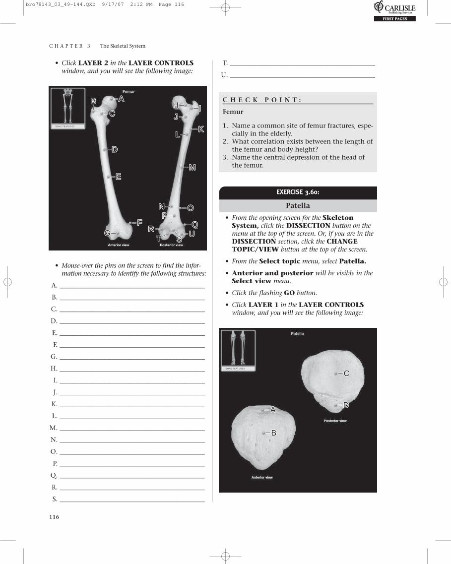
C H A P T E R 3 The Skeletal System
116
• Click LAYER 2 in the LAYER CONTROLSwindow, and you will see the following image:
T. ____________________________________________
U. ____________________________________________
C H E C K P O I N T :
Femur
1. Name a common site of femur fractures, espe-cially in the elderly.
2. What correlation exists between the length ofthe femur and body height?
3. Name the central depression of the head ofthe femur.
AABB
CC
DD
EE
FFGG
HH IIJJ
KKLL
MM
NNPP
OO
QQRU
RSSTT
U
• Mouse-over the pins on the screen to find the infor-mation necessary to identify the following structures:
A. ____________________________________________
B. ____________________________________________
C. ____________________________________________
D. ____________________________________________
E. ____________________________________________
F. ____________________________________________
G. ____________________________________________
H. ____________________________________________
I. ____________________________________________
J. ____________________________________________
K. ____________________________________________
L. ____________________________________________
M. ____________________________________________
N. ____________________________________________
O. ____________________________________________
P. ____________________________________________
Q. ____________________________________________
R. ____________________________________________
S. ____________________________________________
EXERCISE 3.60:
Patella
• From the opening screen for the SkeletonSystem, click the DISSECTION button on themenu at the top of the screen. Or, if you are in theDISSECTION section, click the CHANGETOPIC/VIEW button at the top of the screen.
• From the Select topic menu, select Patella.
• Anterior and posterior will be visible in theSelect view menu.
• Click the flashing GO button.
• Click LAYER 1 in the LAYER CONTROLSwindow, and you will see the following image:
AA
BB
CC
DD
bro78143_03_49-144.QXD 9/17/07 2:12 PM Page 116

C H A P T E R 3 The Skeletal System
117
• Mouse-over the pins on the screen to find the infor-mation necessary to identify the following structures:
A. ____________________________________________
B. ____________________________________________
C. ____________________________________________
D. ____________________________________________
C H E C K P O I N T :
Patella
1. Describe the location of the patella.2. What structures form the knee joint?3. What affect does the patella have on the ten-
don of the quadriceps femoris muscle?
• Mouse-over the pins on the screen to find theinformation necessary to identify the followingstructures:
A. ___________________________________________
B. ___________________________________________
C. ___________________________________________
D. ___________________________________________
E. ___________________________________________
F. ___________________________________________
G. ___________________________________________
H. ___________________________________________
I. ___________________________________________
J. ___________________________________________
K. ___________________________________________
L. ___________________________________________
M. ___________________________________________
N. ___________________________________________
O. ___________________________________________
P. ___________________________________________
Q. ___________________________________________
R. ___________________________________________
S. ___________________________________________
T. ___________________________________________
U. ___________________________________________
V. ___________________________________________
W. ___________________________________________
X. ___________________________________________
Y. ___________________________________________
Z. ___________________________________________
AA. ___________________________________________
AB. ___________________________________________
AC. ___________________________________________
EXERCISE 3.61:
Leg and Foot, Anterior View
• From the opening screen for the SkeletonSystem, click the DISSECTION button on themenu at the top of the screen. Or, if you are in theDISSECTION section, click the CHANGETOPIC/VIEW button at the top of the screen.
• From the Select topic menu, select Leg and foot.
• From the Select view menu, select Anterior.
• Click the flashing GO button.
• Click LAYER 1 in the LAYER CONTROLSwindow, and you will see the following image:
AABB
AA AAAABB
AA
CC
CC
DDEEFF
GGHH
IIJJ
KKLL MMNNPPOO
QQR
UW
X
RSS TT
UVV
WX ZZYY
bro78143_03_49-144.QXD 9/17/07 2:12 PM Page 117

C H A P T E R 3 The Skeletal System
118
H. ____________________________________________
I. ____________________________________________
J. ____________________________________________
K. ____________________________________________
L. ____________________________________________
M. ____________________________________________
N. ____________________________________________
O. ____________________________________________
P. ____________________________________________
Q. ____________________________________________
R. ____________________________________________
S. ____________________________________________
T. ____________________________________________
U. ____________________________________________
C H E C K P O I N T :
Leg and Foot
1. What bone forms the connecting linkbetween the foot and the leg?
2. What bone contributes to the knee and anklejoints?
3. Name the largest tarsal bone. Where is itlocated?
AABB
CCDD
EE FF
GGHH
II
JJ KK
LL MMNN
PPOO
QQR UR SS TT U
• Mouse-over the pins on the screen to find theinformation necessary to identify the followingstructures:
A. ____________________________________________
B. ____________________________________________
C. ____________________________________________
D. ____________________________________________
E. ____________________________________________
F. ____________________________________________
G. ____________________________________________
EXERCISE 3.63:
Tibia and Fibula
• From the opening screen for the SkeletonSystem, click the DISSECTION button on the menu at the top of the screen. Or, if you arein the DISSECTION section, click theCHANGE TOPIC/VIEW button at the top ofthe screen.
• From the Select topic menu, select Tibia andfibula.
• Anterior and posterior will be visible in theSelect view menu.
• Click the flashing GO button.
EXERCISE 3.62:
Leg and Foot, Posterior View
• From the opening screen for the SkeletonSystem, click the DISSECTION button on themenu at the top of the screen. Or, if you are in theDISSECTION section, click the CHANGETOPIC/VIEW button at the top of the screen.
• From the Select topic menu, select Leg andfoot, if it is not already selected.
• From the Select view menu, select Posterior.
• Click the flashing GO button.
• Click LAYER 1 in the LAYER CONTROLSwindow, and you will see the following image:
bro78143_03_49-144.QXD 9/17/07 2:12 PM Page 118
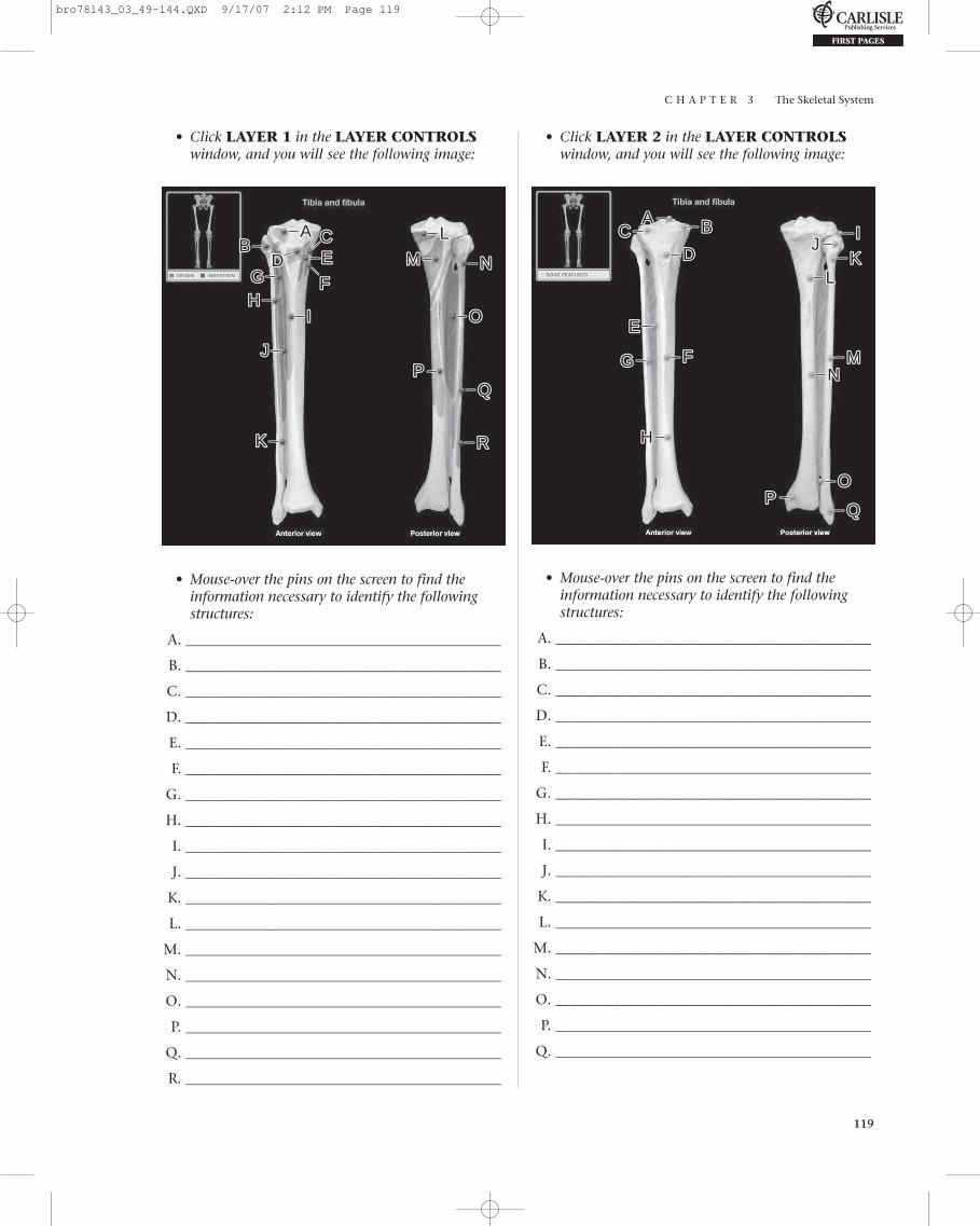
C H A P T E R 3 The Skeletal System
119
• Click LAYER 1 in the LAYER CONTROLSwindow, and you will see the following image:
• Click LAYER 2 in the LAYER CONTROLSwindow, and you will see the following image:
AABB CC
DD EE
FFGGHH
II
JJ
KK
LL
MM NN
PP
OO
RR
• Mouse-over the pins on the screen to find theinformation necessary to identify the followingstructures:
A. ____________________________________________
B. ____________________________________________
C. ____________________________________________
D. ____________________________________________
E. ____________________________________________
F. ____________________________________________
G. ____________________________________________
H. ____________________________________________
I. ____________________________________________
J. ____________________________________________
K. ____________________________________________
L. ____________________________________________
M. ____________________________________________
N. ____________________________________________
O. ____________________________________________
P. ____________________________________________
Q. ____________________________________________
R. ____________________________________________
AA BB
DDCC II
JJKK
LL
EE
FF MMGGNN
HH
PPOO
• Mouse-over the pins on the screen to find theinformation necessary to identify the followingstructures:
A. ____________________________________________
B. ____________________________________________
C. ____________________________________________
D. ____________________________________________
E. ____________________________________________
F. ____________________________________________
G. ____________________________________________
H. ____________________________________________
I. ____________________________________________
J. ____________________________________________
K. ____________________________________________
L. ____________________________________________
M. ____________________________________________
N. ____________________________________________
O. ____________________________________________
P. ____________________________________________
Q. ____________________________________________
bro78143_03_49-144.QXD 9/17/07 2:12 PM Page 119

C H A P T E R 3 The Skeletal System
120
C H E C K P O I N T :
Tibia and Fibula
1. What structure on the tibia contributes to theankle joint?
2. What structure on the fibula contributes tothe ankle joint?
3. What is unique about the anterior shaft ofthe tibia?
• Mouse-over the pins on the screen to find theinformation necessary to identify the followingstructures:
A. ___________________________________________
B. ___________________________________________
C. ___________________________________________
D. ___________________________________________
E. ___________________________________________
F. ___________________________________________
G. ___________________________________________
H. ___________________________________________
I. ___________________________________________
J. ___________________________________________
K. ___________________________________________
L. ___________________________________________
M. ___________________________________________
N. ___________________________________________
O. ___________________________________________
P. ___________________________________________
Q. ___________________________________________
R. ___________________________________________
S. ___________________________________________
T. ___________________________________________
U. ___________________________________________
V. ___________________________________________
W. ___________________________________________
X. ___________________________________________
Y. ___________________________________________
Z. ___________________________________________
AA. ___________________________________________
AB. ___________________________________________
AC. ___________________________________________
AD. ___________________________________________
AE. ___________________________________________
EXERCISE 3.64:
Ankle and Foot
• From the opening screen for the SkeletonSystem, click the DISSECTION button on themenu at the top of the screen. Or, if you are in theDISSECTION section, click the CHANGETOPIC/VIEW button at the top of the screen.
• From the Select topic menu, select Ankle andfoot.
• Superior and inferior will be visible in theSelect view menu.
• Click the flashing GO button.
• Click LAYER 1 in the LAYER CONTROLSwindow, and you will see the following image:
AA LL MMBB NNCCOO
PP
WW
DD EE
GG
KK
ACAC
RR
SSTT
FFUU
ADAD
AAAA
XXYY
HH
II
AEAE
ZZ
ABABJJ
VV
bro78143_03_49-144.QXD 9/17/07 2:12 PM Page 120
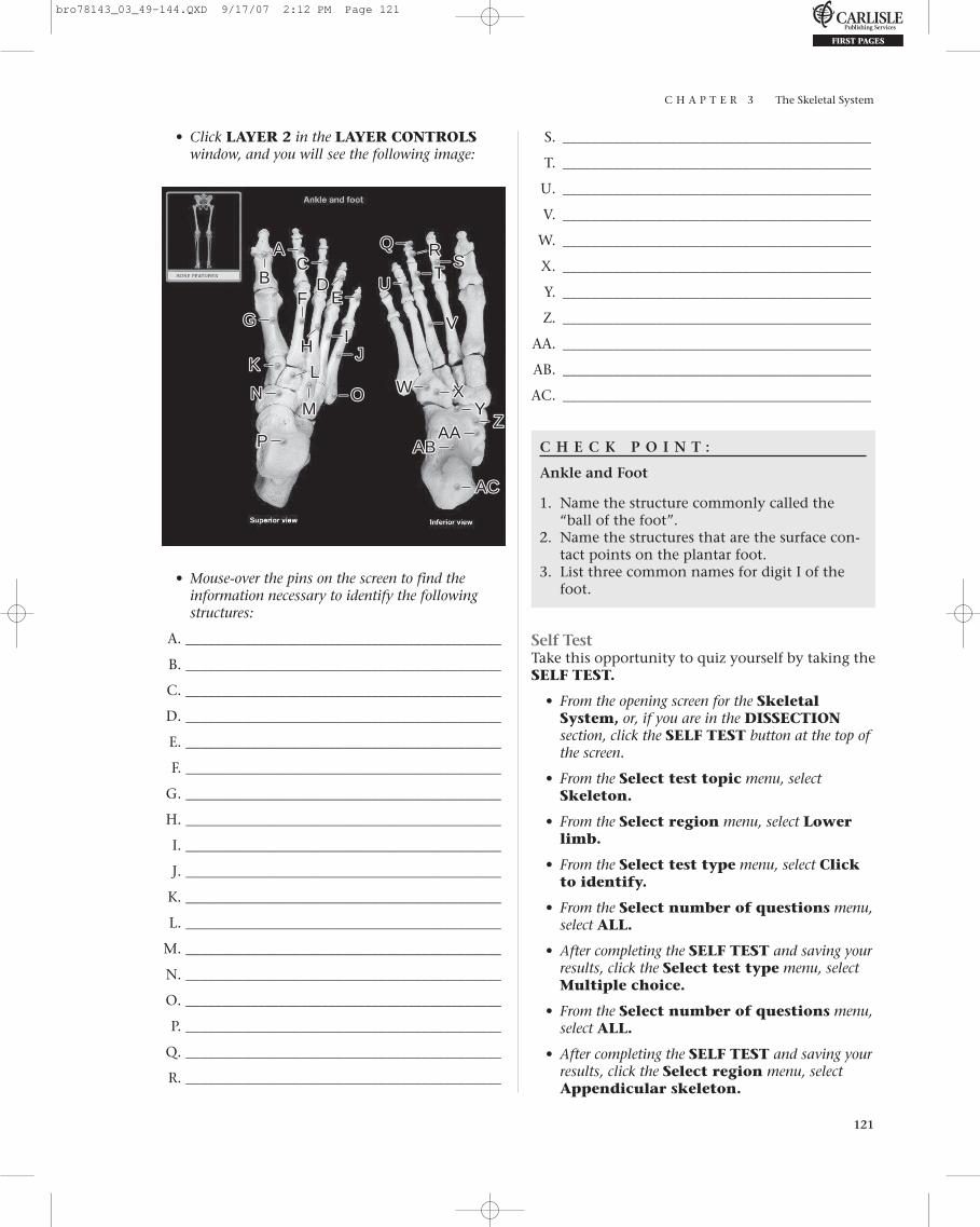
C H A P T E R 3 The Skeletal System
121
• Click LAYER 2 in the LAYER CONTROLSwindow, and you will see the following image:
S. ___________________________________________
T. ___________________________________________
U. ___________________________________________
V. ___________________________________________
W. ___________________________________________
X. ___________________________________________
Y. ___________________________________________
Z. ___________________________________________
AA. ___________________________________________
AB. ___________________________________________
AC. ___________________________________________
C H E C K P O I N T :
Ankle and Foot
1. Name the structure commonly called the“ball of the foot”.
2. Name the structures that are the surface con-tact points on the plantar foot.
3. List three common names for digit I of thefoot.
Self TestTake this opportunity to quiz yourself by taking theSELF TEST.
• From the opening screen for the SkeletalSystem, or, if you are in the DISSECTIONsection, click the SELF TEST button at the top ofthe screen.
• From the Select test topic menu, select Skeleton.
• From the Select region menu, select Lowerlimb.
• From the Select test type menu, select Clickto identify.
• From the Select number of questions menu,select ALL.
• After completing the SELF TEST and saving yourresults, click the Select test type menu, selectMultiple choice.
• From the Select number of questions menu,select ALL.
• After completing the SELF TEST and saving yourresults, click the Select region menu, selectAppendicular skeleton.
QQAA RR
BBCC SS
TTDD UU
EEGG
FF
XX
VV
MM
II
WW
HH JJ
OO
KK
ZZ
ACAC
YY
LLNN
AAAAPP ABAB
• Mouse-over the pins on the screen to find theinformation necessary to identify the followingstructures:
A. ____________________________________________
B. ____________________________________________
C. ____________________________________________
D. ____________________________________________
E. ____________________________________________
F. ____________________________________________
G. ____________________________________________
H. ____________________________________________
I. ____________________________________________
J. ____________________________________________
K. ____________________________________________
L. ____________________________________________
M. ____________________________________________
N. ____________________________________________
O. ____________________________________________
P. ____________________________________________
Q. ____________________________________________
R. ____________________________________________
bro78143_03_49-144.QXD 9/17/07 2:12 PM Page 121

C H A P T E R 3 The Skeletal System
122
• From the Select test type menu, select Clickto identify.
• From the Select number of questions menu,select ALL.
• After completing the SELF TEST and saving yourresults, click the Select test type menu, selectMultiple choice.
• From the Select number of questions menu,select ALL.
• Complete the SELF TEST, and save your results.
What Have I Learned?
The following questions cover the material thatyou have just learned: the pelvic girdle andlower limb. Apply what you have learned in
answering these questions:
1. Where on the femur does the lateral collateralligament attach?
2. How many tarsal bones are there on each foot?Name them.
3. The head of which bone is commonly referred toas the “ball of the foot”?
4. Name the small bones of the toes. How many ofthese bones make up each toe?
5. Name the two enlargement of the distal femur.What structures do they articulate with?
6. Describe the patella.
7. Describe the sacrum.
8. Name the synovial joint between the sacrum andthe ilium.
9. How much movement does this joint allow?Why?
10. Name the landmark for administering anestheticduring childbirth.
11. In the pregnant female, what events occur withthe pubic symphysis?
12. The subpubic angle in females is usually_________________ degrees.
13. The subpubic angle in males is usually_________________ degrees.
14. Describe the difference between the male andfemale obturator foramena.
15. Name a hip landmark for intramuscular injections.
16. Name a hip landmark for administering anes-thetic during childbirth.
17. Name a common site of femur fractures, espe-cially in the elderly.
I N R E V I E W
bro78143_03_49-144.QXD 9/17/07 2:12 PM Page 122

C H A P T E R 3 The Skeletal System
123
Animation: Synovial Joint• From the opening screen for the Skeletal
System, or, if you are in the DISSECTIONsection, click the ANIMATIONS button at thetop of the screen.
• From the Select animation menu, selectSynovial joint.
• Click the Play button, and view the animation.
• After viewing the animation, answer thesequestions:
1. A joint is . . . .
2. What is the most common type of joint?
3. How many types of synovial joints are foundin the body? What distinguishes each onefrom the others?
4. Which type of synovial joint allows the mostmobility? Give an example.
5. Which type of synovial joint allows the leastmobility? Give an example.
6. A typical synovial joint is characterized by . . . .
7. Describe the joint capsule.
18. What correlation exists between the length ofthe femur and body height?
19. Name the central depression of the head of thefemur.
20. What structures form the knee joint?
21. What bone forms the connecting link betweenthe foot and the leg?
22. What bone contributes to the knee and anklejoints?
23. Name the largest tarsal bone. Where is it located?
24. What structure on the tibia contributes to theankle joint?
25. What structure on the fibula contributes to theankle joint?
H E A D S U P !
With the study of the joints, or articulations, we
can tie together the concepts that you have
learned in the Skeletal System and integrate
them with those you will learn in the next
chapter – the Muscular System. With this in
mind, you will want to refer back to this section
of the Skeletal System while you are working on
the Muscular System.
bro78143_03_49-144.QXD 9/17/07 2:12 PM Page 123

C H A P T E R 3 The Skeletal System
124
8. What is synovial fluid? Where is it produced?
9. What tissue covers the surface of adjoiningbones? What is its function?
10. What is the function of the meniscus of theknee joint?
11. What is the function of bursae?
• Mouse-over the pins on the screen to find theinformation necessary to identify the followingstructures:
A. ____________________________________________
B. ____________________________________________
• Click LAYER 2 in the LAYER CONTROLSwindow, and you will see the following image:
EXERCISE 3.65:
Temporomandibular Joint
• From the opening screen for the SkeletalSystem, or from the Skeletal SystemAnimations Screen, click the DISSECTIONbutton on the menu at the top of the screen.
• From the Select topic menu, select Temporo-mandibular joint under the Joints heading.
• Lateral will be visible in the Select viewmenu.
• Click the flashing GO button.
• Click LAYER 1 in the LAYER CONTROLSwindow, and you will see the following image:
AABB
AA BB
CC DDEE
FFGG
bro78143_03_49-144.QXD 9/17/07 2:12 PM Page 124

C H A P T E R 3 The Skeletal System
125
• Mouse-over the pins on the screen to find theinformation necessary to identify the followingstructures:
A. ____________________________________________
B. ____________________________________________
C. ____________________________________________
D. ____________________________________________
E. ____________________________________________
F. ____________________________________________
G. ____________________________________________
• Click LAYER 3 in the LAYER CONTROLSwindow, and you will see the following image:
I. ____________________________________________
J. ____________________________________________
• Click LAYER 4 in the LAYER CONTROLSwindow, and you will see the following image:
AABB
CC
DD
GGHH
II
JJ
EE
FF
• Mouse-over the green pins on the screen to find theinformation necessary to identify the followingstructures:
A. ____________________________________________
B. ____________________________________________
C. ____________________________________________
Non-Skeletal System Structures (blue pins)
D. ____________________________________________
E. ____________________________________________
F. ____________________________________________
G. ____________________________________________
H. ____________________________________________
AABBCC
LL
NN
DDEE
FF
HH
GG
JJ
II
KKMM OO
PP
• Mouse-over the green pins on the screen to find theinformation necessary to identify the followingstructures:
A. ____________________________________________
B. ____________________________________________
C. ____________________________________________
D. ____________________________________________
E. ____________________________________________
F. ____________________________________________
G. ____________________________________________
Non-Skeletal System Structures (blue pins)
H. ____________________________________________
I. ____________________________________________
J. ____________________________________________
K. ____________________________________________
L. ____________________________________________
M. ____________________________________________
N. ____________________________________________
O. ____________________________________________
P. ____________________________________________
bro78143_03_49-144.QXD 9/17/07 2:12 PM Page 125

C H A P T E R 3 The Skeletal System
126
• Click LAYER 5 in the LAYER CONTROLSwindow, and you will see the following image: C H E C K P O I N T :
Temporomandibular Joint
1. Name the structure that encloses the temporomandibular joint.
2. What is the function of the synovial membrane?
3. What is the function of the lateral ligamentof the temporomandibular joint?
4. What is the cause of “jaw clicking”?5. What is the location of the articular disk of
the temporomandibular joint?
AAJJ
BB CCII
DD
EE
FFGG
HH
KKLL
MMPP
NN
OO
• Mouse-over the green pins on the screen to find theinformation necessary to identify the followingstructures:
A. ____________________________________________
B. ____________________________________________
C. ____________________________________________
D. ____________________________________________
E. ____________________________________________
Non-Skeletal System Structures (blue pins)
F. ____________________________________________
G. ____________________________________________
H. ____________________________________________
I. ____________________________________________
J. ____________________________________________
K. ____________________________________________
L. ____________________________________________
M. ____________________________________________
N. ____________________________________________
O. ____________________________________________
P. ____________________________________________
Q. ____________________________________________
EXERCISE 3.66:
Glenohumeral (Shoulder) Joint
• From the opening screen for the SkeletonSystem, click the DISSECTION button on themenu at the top of the screen. Or, if you are in theDISSECTION section, click the CHANGETOPIC/VIEW button at the top of the screen.
• From the Select topic menu, select Gleno-humeral (shoulder) joint.
• Anterior will be visible in the Select viewmenu.
• Click the flashing GO button.
• Click LAYER 1 in the LAYER CONTROLSwindow, and you will see the following image:
AABB
CC
bro78143_03_49-144.QXD 9/17/07 2:12 PM Page 126

C H A P T E R 3 The Skeletal System
127
• Mouse-over the pins on the screen to find theinformation necessary to identify the followingstructures:
A. ____________________________________________
B. ____________________________________________
C. ____________________________________________
• Click LAYER 2 in the LAYER CONTROLSwindow, and you will see the following image:
• Click LAYER 3 in the LAYER CONTROLSwindow, and you will see the following image:
AABB
EE
FF
CC
DD
GG
• Mouse-over the green pins on the screen to find theinformation necessary to identify the followingstructures:
A. ____________________________________________
B. ____________________________________________
Non-Skeletal System Structures (blue pins)
C. ____________________________________________
D. ____________________________________________
E. ____________________________________________
F. ____________________________________________
G. ____________________________________________
AABBCC DD
EEFF
GG
HH
IIJJ
KKLLMM
• Mouse-over the green pins on the screen to find theinformation necessary to identify the followingstructures:
A. ____________________________________________
B. ____________________________________________
C. ____________________________________________
D. ____________________________________________
E. ____________________________________________
F. ____________________________________________
G. ____________________________________________
Non-Skeletal System Structures (blue pins)
H. ____________________________________________
I. ____________________________________________
J. ____________________________________________
K. ____________________________________________
L. ____________________________________________
M. ____________________________________________
bro78143_03_49-144.QXD 9/17/07 2:12 PM Page 127
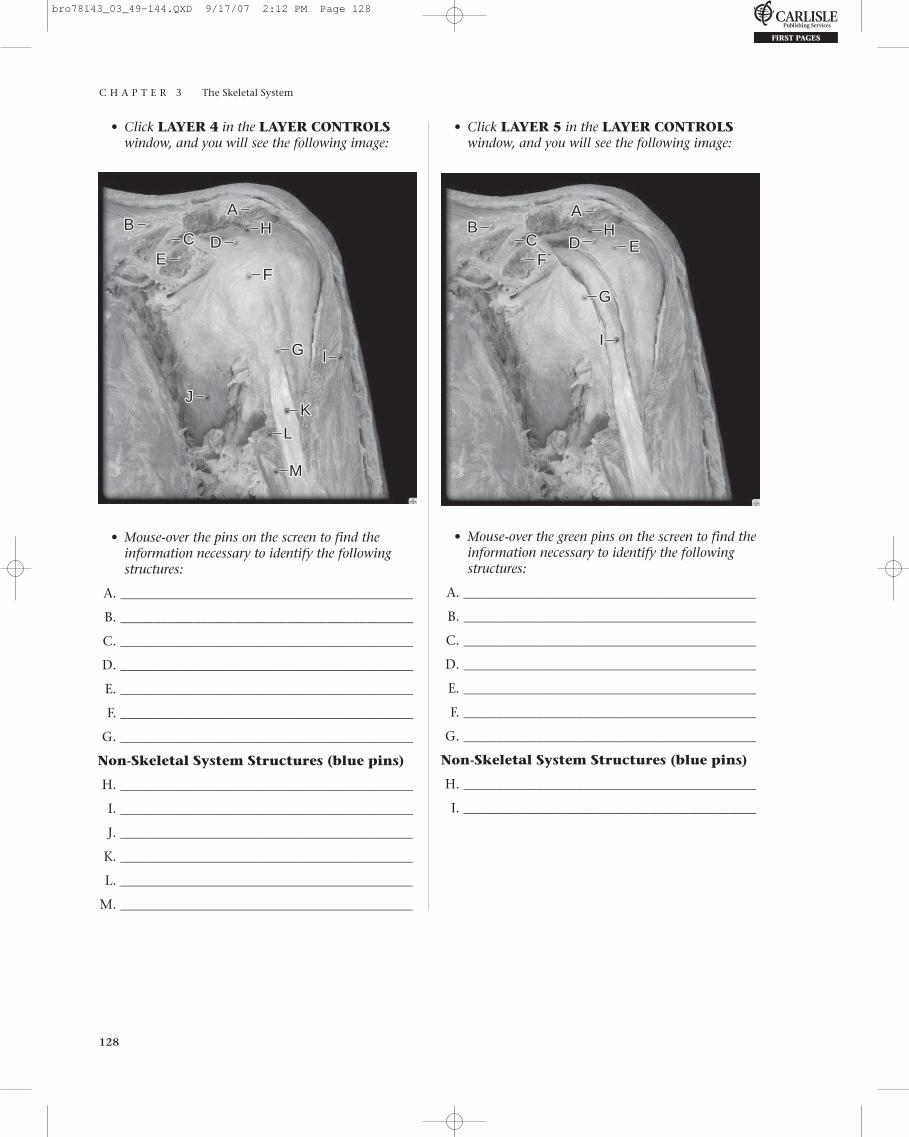
C H A P T E R 3 The Skeletal System
128
• Click LAYER 4 in the LAYER CONTROLSwindow, and you will see the following image:
• Click LAYER 5 in the LAYER CONTROLSwindow, and you will see the following image:
AABB
EECC DD
FF
GG
HH
II
JJKK
LL
MM
• Mouse-over the pins on the screen to find theinformation necessary to identify the followingstructures:
A. ____________________________________________
B. ____________________________________________
C. ____________________________________________
D. ____________________________________________
E. ____________________________________________
F. ____________________________________________
G. ____________________________________________
Non-Skeletal System Structures (blue pins)
H. ____________________________________________
I. ____________________________________________
J. ____________________________________________
K. ____________________________________________
L. ____________________________________________
M. ____________________________________________
AABB
CC EEFF
DD
GG
HH
II
• Mouse-over the green pins on the screen to find theinformation necessary to identify the followingstructures:
A. ____________________________________________
B. ____________________________________________
C. ____________________________________________
D. ____________________________________________
E. ____________________________________________
F. ____________________________________________
G. ____________________________________________
Non-Skeletal System Structures (blue pins)
H. ____________________________________________
I. ____________________________________________
bro78143_03_49-144.QXD 9/17/07 2:12 PM Page 128

C H A P T E R 3 The Skeletal System
129
• Click LAYER 6 in the LAYER CONTROLSwindow, and you will see the following image: C H E C K P O I N T :
Glenohumeral (Shoulder) Joint
1. Name the structure enclosing the acromio-clavicular joint.
2. What is the function of the coracohumeralligament?
3. What is the function of the coracoclavicularligament?
4. What is the result of a torn coracoclavicularligament?
5. Name the tough, fibrous envelope of theglenohumeral joint.
AA BB
AABB
CCDD
EE FFGG
HH IIJJ
KK
MMLL
• Mouse-over the pins on the screen to find theinformation necessary to identify the followingstructures:
A. ____________________________________________
B. ____________________________________________
C. ____________________________________________
D. ____________________________________________
E. ____________________________________________
F. ____________________________________________
G. ____________________________________________
H. ____________________________________________
I. ____________________________________________
J. ____________________________________________
K. ____________________________________________
L. ____________________________________________
M. ____________________________________________
EXERCISE 3.67:
Elbow Joint
• From the opening screen for the SkeletonSystem, click the DISSECTION button on themenu at the top of the screen. Or, if you are in theDISSECTION section, click the CHANGETOPIC/VIEW button at the top of the screen.
• From the Select topic menu, select Elbowjoint.
• Anterior will be visible in the Select view menu.
• Click the flashing GO button.
• Click LAYER 1 in the LAYER CONTROLSwindow, and you will see the following image:
bro78143_03_49-144.QXD 9/17/07 2:12 PM Page 129

C H A P T E R 3 The Skeletal System
130
• Mouse-over the pins on the screen to find theinformation necessary to identify the followingstructures:
A. ____________________________________________
B. ____________________________________________
• Click LAYER 2 in the LAYER CONTROLSwindow, and you will see the following image:
• Click LAYER 3 in the LAYER CONTROLSwindow, and you will see the following image:
AA
BB
DD
FF
II
KKJJ
GG
CC
EE
HH
• Mouse-over the pins on the screen to find theinformation necessary to identify the followingstructures:
A. ____________________________________________
B. ____________________________________________
C. ____________________________________________
D. ____________________________________________
E. ____________________________________________
F. ____________________________________________
G. ____________________________________________
H. ____________________________________________
I. ____________________________________________
J. ____________________________________________
K. ____________________________________________
AA
BB CCDD
FFEEGG
HHIIJJ KK LL
MMNN
PP
OO
• Mouse-over the pins on the screen to find theinformation necessary to identify the followingstructures:
A. ____________________________________________
B. ____________________________________________
C. ____________________________________________
D. ____________________________________________
E. ____________________________________________
F. ____________________________________________
G. ____________________________________________
H. ____________________________________________
I. ____________________________________________
J. ____________________________________________
K. ____________________________________________
L. ____________________________________________
M. ____________________________________________
N. ____________________________________________
O. ____________________________________________
P. ____________________________________________
bro78143_03_49-144.QXD 9/17/07 2:12 PM Page 130

C H A P T E R 3 The Skeletal System
131
• Click LAYER 4 in the LAYER CONTROLSwindow, and you will see the following image:
• Click LAYER 5 in the LAYER CONTROLSwindow, and you will see the following image:
AA
BB CCDD
EEFFGG
HH
IIJJ
• Mouse-over the green pins on the screen to find theinformation necessary to identify the followingstructures:
A. ____________________________________________
B. ____________________________________________
C. ____________________________________________
D. ____________________________________________
Non-Skeletal System Structures (blue pins)
E. ____________________________________________
F. ____________________________________________
G. ____________________________________________
H. ____________________________________________
I. ____________________________________________
J. ____________________________________________
AA
BBCC DDEE
FF GGHH IIJJ
KK
LL
• Mouse-over the pins on the screen to find the infor-mation necessary to identify the following structures:
A. ____________________________________________
B. ____________________________________________
C. ____________________________________________
D. ____________________________________________
E. ____________________________________________
F. ____________________________________________
G. ____________________________________________
H. ____________________________________________
I. ____________________________________________
J. ____________________________________________
K. ____________________________________________
L. ____________________________________________
C H E C K P O I N T :
Elbow Joint
1. Name the structure that encloses the elbow joint.2. What is the function of the radial collateral ligament?3. What is the function of the ulnar collateral ligament?4. What is the function of the anular ligament of the radius?5. Name the bones associated with the elbow joint.
bro78143_03_49-144.QXD 9/17/07 2:12 PM Page 131

C H A P T E R 3 The Skeletal System
132
• Mouse-over the pins on the screen to find theinformation necessary to identify the followingstructures:
A. ____________________________________________
B. ____________________________________________
C. ____________________________________________
• Click LAYER 2 in the LAYER CONTROLSwindow, and you will see the image at the top of the next column:
EXERCISE 3.68:
Knee Joint, Anterior View
• From the opening screen for the SkeletonSystem, click the DISSECTION button on themenu at the top of the screen. Or, if you are in theDISSECTION section, click the CHANGETOPIC/VIEW button at the top of the screen.
• From the Select topic menu, select Kneejoint.
• From the Select view menu, select Anterior.
• Click the flashing GO button.
• Click LAYER 1 in the LAYER CONTROLSwindow, and you will see the following image:
AA
BB
CC
AA
BB
• Mouse-over the green pin on the screen to find theinformation necessary to identify the followingstructures:
A. ____________________________________________
Non-Skeletal System Structures (blue pin)
B. ____________________________________________
• Click LAYER 3 in the LAYER CONTROLSwindow, and you will see the following image:
AA
BB CC
DD
EEFF
GG
bro78143_03_49-144.QXD 9/17/07 2:12 PM Page 132

C H A P T E R 3 The Skeletal System
133
• Mouse-over the pins on the screen to find the infor-mation necessary to identify the following structures:
A. ____________________________________________
B. ____________________________________________
C. ____________________________________________
D. ____________________________________________
E. ____________________________________________
F. ____________________________________________
G. ____________________________________________
• Click LAYER 4 in the LAYER CONTROLSwindow, and you will see the following image:
• Click LAYER 5 in the LAYER CONTROLSwindow, and you will see the following image:
AA
BB
CCEEDD
FFGG HH
II
JJ
• Mouse-over the pins on the screen to find the infor-mation necessary to identify the following structures:
A. ____________________________________________
B. ____________________________________________
C. ____________________________________________
D. ____________________________________________
E. ____________________________________________
F. ____________________________________________
G. ____________________________________________
H. ____________________________________________
I. ____________________________________________
J. ____________________________________________
AA
BBCCDD
GGHH
EE
FF
II JJKK LL
MM
NN
• Mouse-over the pins on the screen to find theinformation necessary to identify the followingstructures:
A. ____________________________________________
B. ____________________________________________
C. ____________________________________________
D. ____________________________________________
E. ____________________________________________
F. ____________________________________________
G. ____________________________________________
H. ____________________________________________
I. ____________________________________________
J. ____________________________________________
K. ____________________________________________
L. ____________________________________________
M. ____________________________________________
N. ____________________________________________
bro78143_03_49-144.QXD 9/17/07 2:12 PM Page 133

C H A P T E R 3 The Skeletal System
134
• Mouse-over the green pins on the screen to find theinformation necessary to identify the followingstructures:
A. ____________________________________________
B. ____________________________________________
C. ____________________________________________
D. ____________________________________________
Non-Skeletal System Structures (blue pins)
E. ____________________________________________
F. ____________________________________________
G. ____________________________________________
H. ____________________________________________
I. ____________________________________________
J. ____________________________________________
K. ____________________________________________
AA
• Mouse-over the pin on the screen to find theinformation necessary to identify the followingstructure:
A. ____________________________________________
EXERCISE 3.69:
Knee Joint, Posterior View
• From the opening screen for the Skeleton Sys-tem, click the DISSECTION button on themenu at the top of the screen. Or, if you are in theDISSECTION section, click the CHANGETOPIC/VIEW button at the top of the screen.
• From the Select topic menu, select Kneejoint, if it is not already selected.
• From the Select view menu, select Posterior.
• Click the flashing GO button.
• Click LAYER 1 in the LAYER CONTROLSwindow, and you will see the following image:
AA
BB CC
DD
JJ
EE
FF
HHII
KK
GG
• Click LAYER 2 in the LAYER CONTROLSwindow, and you will see the following image:
bro78143_03_49-144.QXD 9/17/07 2:12 PM Page 134
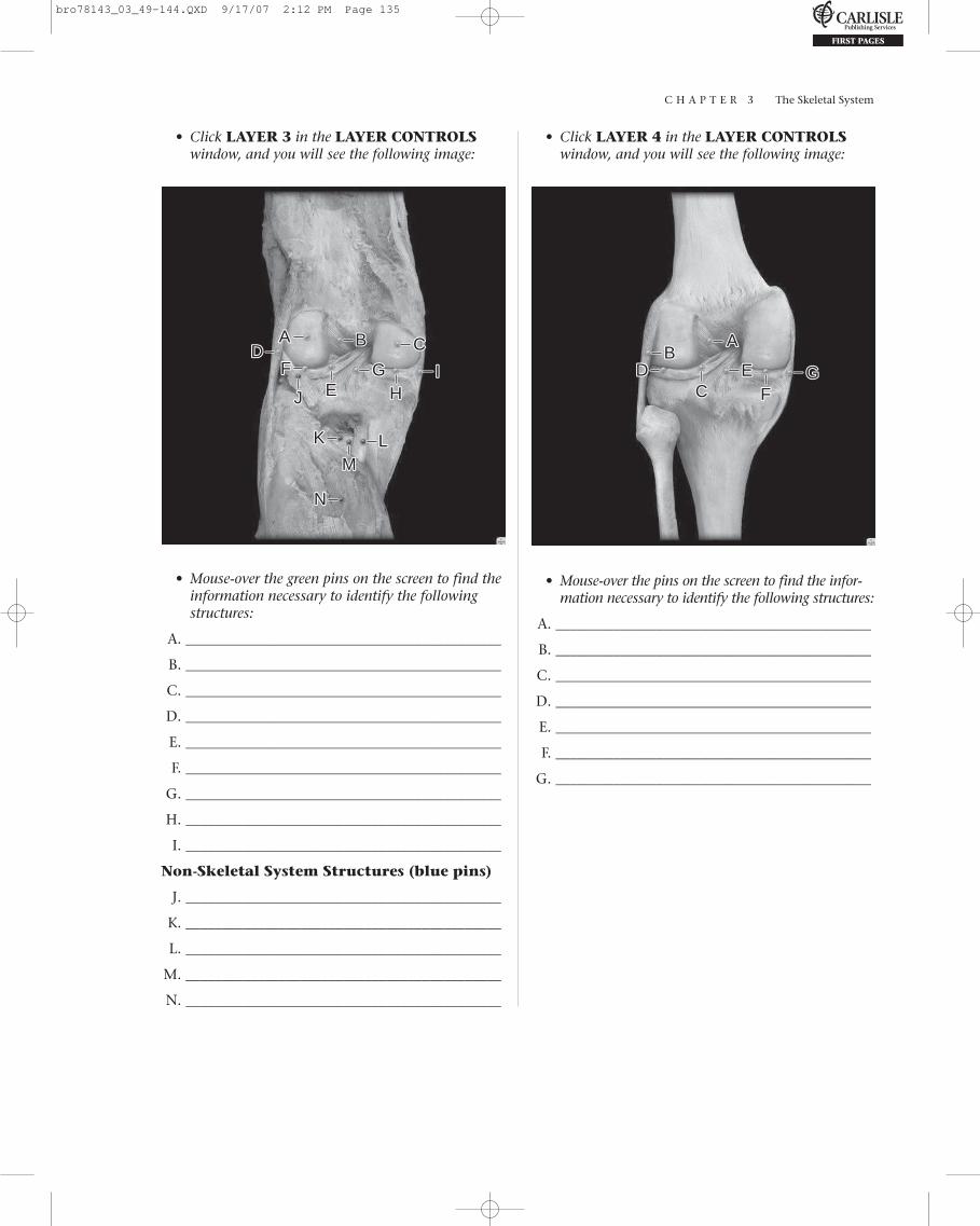
C H A P T E R 3 The Skeletal System
• Click LAYER 3 in the LAYER CONTROLSwindow, and you will see the following image:
AA BB CCDD
EE HH
FF II
KK LLMM
NN
JJ
GG
• Mouse-over the green pins on the screen to find theinformation necessary to identify the followingstructures:
A. ____________________________________________
B. ____________________________________________
C. ____________________________________________
D. ____________________________________________
E. ____________________________________________
F. ____________________________________________
G. ____________________________________________
H. ____________________________________________
I. ____________________________________________
Non-Skeletal System Structures (blue pins)
J. ____________________________________________
K. ____________________________________________
L. ____________________________________________
M. ____________________________________________
N. ____________________________________________
AABB
CCDD EE
FFGG
• Mouse-over the pins on the screen to find the infor-mation necessary to identify the following structures:
A. ____________________________________________
B. ____________________________________________
C. ____________________________________________
D. ____________________________________________
E. ____________________________________________
F. ____________________________________________
G. ____________________________________________
• Click LAYER 4 in the LAYER CONTROLSwindow, and you will see the following image:
bro78143_03_49-144.QXD 9/17/07 2:12 PM Page 135

C H A P T E R 3 The Skeletal System
136
• Click LAYER 5 in the LAYER CONTROLSwindow and you will see the following image: C H E C K P O I N T :
Knee Joint
1. Name the structures that make up the“unhappy triad.”
2. Why are these structures referred to as the“unhappy triad”?
3. Name a ligament absent in approximately 40 percent of knees.
4. What is the function of the anterior cruciateligament?
5. What is the function of the posterior cruciateligament?
AA
BB
CC DDEE
FFHHII
KK
LLMM
JJGG
• Mouse-over the pins on the screen to find theinformation necessary to identify the followingstructures:
A. ____________________________________________
B. ____________________________________________
C. ____________________________________________
D. ____________________________________________
E. ____________________________________________
F. ____________________________________________
G. ____________________________________________
H. ____________________________________________
I. ____________________________________________
J. ____________________________________________
K. ____________________________________________
L. ____________________________________________
M. ____________________________________________
EXERCISE 3.70:
Ankle Joint, Lateral View
• From the opening screen for the SkeletonSystem, click the DISSECTION button on themenu at the top of the screen. Or, if you are in theDISSECTION section, click the CHANGETOPIC/VIEW button at the top of the screen.
• From the Select topic menu, select Anklejoint.
• From the Select view menu, select Lateral.
• Click the flashing GO button.
• Click LAYER 1 in the LAYER CONTROLSwindow, and you will see the following image:
AA BB
bro78143_03_49-144.QXD 9/17/07 2:12 PM Page 136
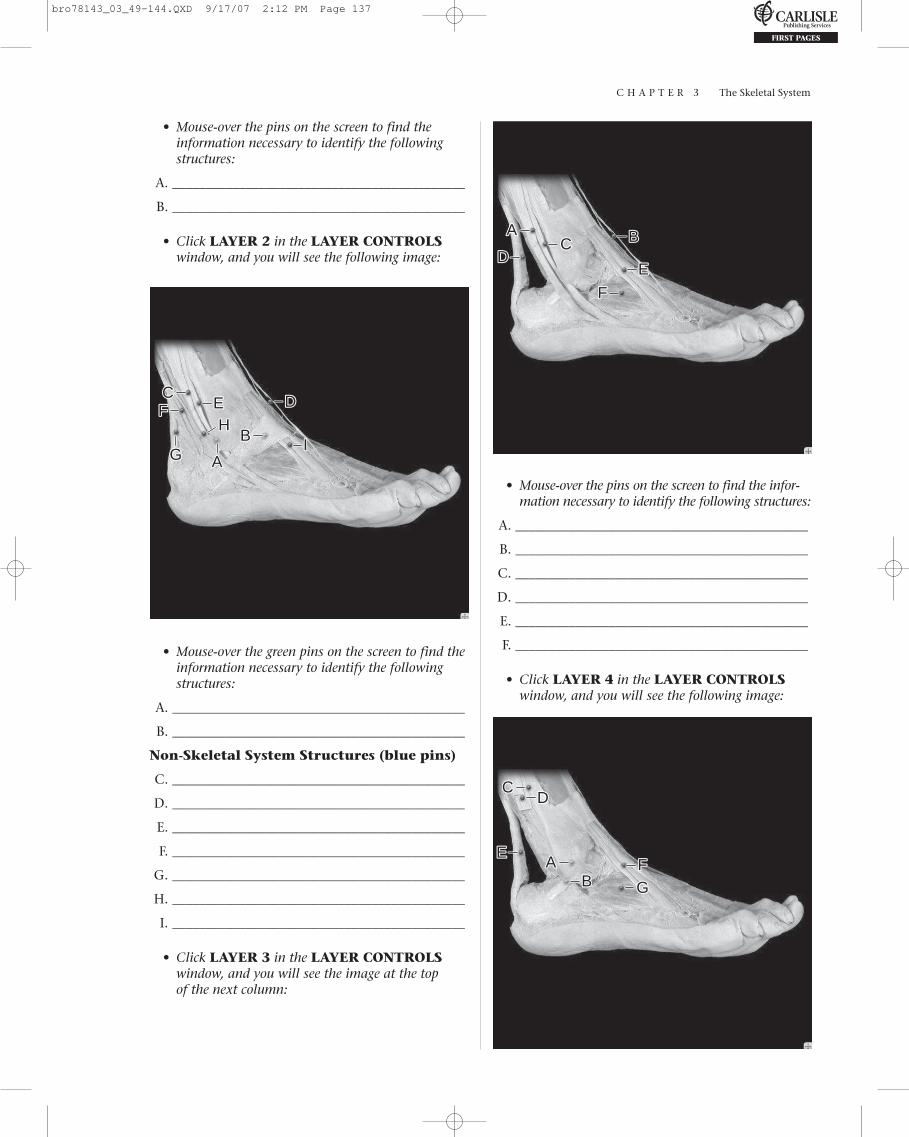
C H A P T E R 3 The Skeletal System
• Mouse-over the pins on the screen to find theinformation necessary to identify the followingstructures:
A. ____________________________________________
B. ____________________________________________
• Click LAYER 2 in the LAYER CONTROLSwindow, and you will see the following image:
AAGGBB
CC DDEEFF
IIHH
• Mouse-over the green pins on the screen to find theinformation necessary to identify the followingstructures:
A. ____________________________________________
B. ____________________________________________
Non-Skeletal System Structures (blue pins)
C. ____________________________________________
D. ____________________________________________
E. ____________________________________________
F. ____________________________________________
G. ____________________________________________
H. ____________________________________________
I. ____________________________________________
• Click LAYER 3 in the LAYER CONTROLSwindow, and you will see the image at the top of the next column:
AA BBCCDD
EE
FF
• Mouse-over the pins on the screen to find the infor-mation necessary to identify the following structures:
A. ____________________________________________
B. ____________________________________________
C. ____________________________________________
D. ____________________________________________
E. ____________________________________________
F. ____________________________________________
• Click LAYER 4 in the LAYER CONTROLSwindow, and you will see the following image:
AABB
CCDD
EEFFGG
bro78143_03_49-144.QXD 9/17/07 2:12 PM Page 137

C H A P T E R 3 The Skeletal System
138
• Mouse-over the green pins on the screen to find theinformation necessary to identify the followingstructures:
A. ____________________________________________
B. ____________________________________________
Non-Skeletal System Structures (blue pins)
C. ____________________________________________
D. ____________________________________________
E. ____________________________________________
F. ____________________________________________
G. ____________________________________________
• Click LAYER 5 in the LAYER CONTROLSwindow, and you will see the following image:
H. ____________________________________________
I. ____________________________________________
J. ____________________________________________
K. ____________________________________________
L. ____________________________________________
M. ____________________________________________
N. ____________________________________________
AABB
CC
DDGG
EEFF
HHII KK LL
MMNNJJ
• Mouse-over the pins on the screen to find theinformation necessary to identify the followingstructures:
A. ____________________________________________
B. ____________________________________________
C. ____________________________________________
D. ____________________________________________
E. ____________________________________________
F. ____________________________________________
G. ____________________________________________
EXERCISE 3.71:
Ankle Joint, Medial View
• From the opening screen for the SkeletonSystem, click the DISSECTION button on themenu at the top of the screen. Or, if you are in theDISSECTION section, click the CHANGETOPIC/VIEW button at the top of the screen.
• From the Select topic menu, select Anklejoint, if it is not already selected.
• From the Select view menu, select Medial.
• Click the flashing GO button.
• Click LAYER 1 in the LAYER CONTROLSwindow, and you will see the following image:
AA
• Mouse-over the pin on the screen to find the infor-mation necessary to identify the following structure:
A. ____________________________________________
bro78143_03_49-144.QXD 9/17/07 2:12 PM Page 138
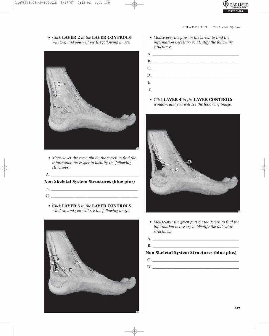
C H A P T E R 3 The Skeletal System
139
• Click LAYER 2 in the LAYER CONTROLSwindow, and you will see the following image:
• Mouse-over the pins on the screen to find theinformation necessary to identify the followingstructures:
A. ____________________________________________
B. ____________________________________________
C. ____________________________________________
D. ____________________________________________
E. ____________________________________________
F. ____________________________________________
• Click LAYER 4 in the LAYER CONTROLSwindow, and you will see the following image:
AA
BB
CC
• Mouse-over the green pin on the screen to find theinformation necessary to identify the followingstructures:
A. ____________________________________________
Non-Skeletal System Structures (blue pins)
B. ____________________________________________
C. ____________________________________________
• Click LAYER 3 in the LAYER CONTROLSwindow, and you will see the following image:
AABB
CCDDEE FF
AABB
CCDD
• Mouse-over the green pins on the screen to find theinformation necessary to identify the followingstructures:
A. ____________________________________________
B. ____________________________________________
Non-Skeletal System Structures (blue pins)
C. ____________________________________________
D. ____________________________________________
bro78143_03_49-144.QXD 9/17/07 2:12 PM Page 139

C H A P T E R 3 The Skeletal System
140
• Click LAYER 5 in the LAYER CONTROLSwindow, and you will see the following image:
AA
BBCC
DD EE FFHHIIKK
LLMM
JJGG
• Mouse-over the pins on the screen to find theinformation necessary to identify the followingstructures:
A. ____________________________________________
B. ____________________________________________
C. ____________________________________________
D. ____________________________________________
E. ____________________________________________
F. ____________________________________________
G. ____________________________________________
H. ____________________________________________
I. ____________________________________________
J. ____________________________________________
K. ____________________________________________
L. ____________________________________________
M. ____________________________________________
C H E C K P O I N T :
Ankle Joint
1. Name the strongest tendon of the body. Whyis it considered the strongest?
2. What is the extensor retinaculum of the foot?What is its function?
3. What is the function of the lateral ligamentof the ankle?
4. What is the flexor retinaculum of the foot?What is its function?
5. What is the function of the medial (deltoid)ligament of the ankle?
Self TestTake this opportunity to quiz yourself by taking theSELF TEST.
• From the opening screen for the Skeletal System,or, if you are in the DISSECTION section, click theSELF TEST button at the top of the screen.
• From the Select test topic menu, selectSkeleton.
• From the Select region menu, select Joints.
• From the Select test type menu, select Clickto identify.
• From the Select number of questions menu,select ALL.
• After completing the SELF TEST and saving yourresults, click the Select test type menu, selectMultiple choice.
• From the Select number of questions menu,select ALL.
• After completing the SELF TEST and saving yourresults, click the Select test topic menu, selectAnimation.
• From the Select animation menu, selectSynovial joint.
• Multiple choice is visible in the Select testtype menu.
• ALL is visible in the Select number of ques-tions menu.
• Complete the SELF TEST and save your results.
bro78143_03_49-144.QXD 9/17/07 2:12 PM Page 140

C H A P T E R 3 The Skeletal System
141
What Have I Learned?
The following questions cover the material thatyou have just learned: joints. Apply what youhave learned in answering these questions:
1. Name the structure that encloses the temporo-mandibular joint.
2. What is the function of the synovial membrane?
3. What is the cause of “jaw clicking”?
4. Name the structure enclosing the acromioclavic-ular joint.
5. What is the result of a torn coracoclavicularligament?
6. Name the tough, fibrous envelope of the gleno-humeral joint.
7. Name the structure that encloses the elbow joint.
8. Name the bones associated with the elbow joint.
9. Name the structures that make up the “unhappytriad.”
10. Name a ligament absent in approximately 40 percent of knees.
11. What is the function of the anterior cruciateligament?
12. What is the function of the posterior cruciateligament?
13. Name the strongest tendon of the body. Why isit considered the strongest?
14. What is the extensor retinaculum of the foot?What is its function?
I N R E V I E W
AABB
CC
DD
EE
EXERCISE 3.72:
Histology—Elastic Cartilage
• From the opening screen for the SkeletalSystem or, if you are in the DISSECTIONsection, click the HISTOLOGY button at the topof the screen.
• From the Select topic menu select Elasticcartilage.
• Click the flashing GO button.
• Click the TURN TAGS ON button, and you willsee the image to the right:
bro78143_03_49-144.QXD 9/17/07 2:12 PM Page 141

C H A P T E R 3 The Skeletal System
142
• Mouse-over the pins on the screen to find theinformation necessary to identify the followingstructures:
A. ____________________________________________
B. ____________________________________________
C. ____________________________________________
D. ____________________________________________
E. ____________________________________________
EXERCISE 3.73:
Histology—Fibrous Cartilage
• Click the CHANGE TOPIC button in theCURRENT TOPIC window.
• From the Select topic menu select Fibrouscartilage.
• Click the flashing GO button.
• Click the TURN TAGS ON button, and you willsee the following image:
AA
BB
• Mouse-over the pins on the screen to find theinformation necessary to identify the followingstructures:
A. ____________________________________________
B. ____________________________________________
EXERCISE 3.74:
Histology—Hyaline Cartilage
• Click the CHANGE TOPIC button in theCURRENT TOPIC window.
• From the Select topic menu select Hyalinecartilage.
• Click the flashing GO button.
• Click the TURN TAGS ON button, and you willsee the following image:
AABB CC
DD
EE
• Mouse-over the pins on the screen to find theinformation necessary to identify the followingstructures:
A. ____________________________________________
B. ____________________________________________
C. ____________________________________________
D. ____________________________________________
E. ____________________________________________
bro78143_03_49-144.QXD 9/17/07 2:12 PM Page 142

C H A P T E R 3 The Skeletal System
143
• Mouse-over the pins on the screen to find theinformation necessary to identify the followingstructures:
A. ____________________________________________
B. ____________________________________________
C. ____________________________________________
D. ____________________________________________
E. ____________________________________________
F. ____________________________________________
EXERCISE 3.75:
Histology—Compact Bone
• Click the CHANGE TOPIC button in theCURRENT TOPIC window.
• From the Select topic menu select Compactbone.
• Click the flashing GO button.
• From the Select magnification menu selectLow magnification.
• Click the TURN TAGS ON button, and you willsee the following image:
AABBCC DD
• Mouse-over the pins on the screen to find theinformation necessary to identify the followingstructures:
A. ____________________________________________
B. ____________________________________________
C. ____________________________________________
D. ____________________________________________
• From the Select magnification menu selectHigh magnification.
AABB
CCDD
EE
FF
EXERCISE 3.76:
Histology—Cancellous Bone
• Click the CHANGE TOPIC button in theCURRENT TOPIC window.
• From the Select topic menu select Cancellousbone.
• Click the flashing GO button.
• Click the TURN TAGS ON button, and you willsee the following image:
bro78143_03_49-144.QXD 9/17/07 2:12 PM Page 143

C H A P T E R 3 The Skeletal System
144
• Click the TURN TAGS ON button, and you willsee the following image:
• Mouse-over the pins on the screen to find theinformation necessary to identify the followingstructures:
A. ____________________________________________
B. ____________________________________________
C. ____________________________________________
Self TestTake this opportunity to quiz yourself by taking theSELF TEST.
• From the opening screen for the SkeletalSystem, or, if you are in the DISSECTIONsection, click the SELF TEST button at the top ofthe screen.
• From the Select test topic menu, selectHistology.
• From the Select number of questions menu,select ALL.
• Complete the SELF TEST and save your results.
AA
BB
CC
What Have I Learned?
The following questions cover the material thatyou have just learned: histology. Apply what youhave learned in answering these questions:
1. Name the outer layer of most cartilage.
2. Give four examples of elastic cartilage.
3. What are lacunae? What are contained withinthe lacunae?
4. Give four examples of fibrous cartilage.
5. What is the function of the chondrogenic layerof the perichondrium?
6. Describe an osteon.
7. Describe a perforating canal.
8. Interstitial lamellae result from_________________.
9. What component of bone tissue is essential tobone nutrition, growth, and repair?
10. What are trabeculae?
I N R E V I E W
bro78143_03_49-144.QXD 9/17/07 2:12 PM Page 144