Embed Size (px)
Citation preview

KEY KNOWLEDGE
The contents of this chapter are designed to enable students to: ■ develop understanding of the function of stem cells in antenatal (pre-birth) human development
■ gain knowledge of the different types of stem cells ■ become aware of the potential use of stem cells in medical treatment of certain disorders
■ recognise how disruption of the cell cycle can result in developmental abnormalities and cancers.
FIGURE 12.1 This baby was once a single fertilised egg, or zygote, that underwent a remarkable developmental journey involving growth in cell numbers, cell migration and cell differentiation. By the time of its birth, a baby is a complex organism composed of billions of cells, organised into tissues, organs and systems. In this chapter we will explore some of the processes involved in this transformation, including the role of stem cells.
CHAPTER
12 Cell growth and differentiation
CHAPTER12Cell growth and di� erentiationAntenatal human developmentKey events: embryonic development Abnormal embryonic developmentCancer and the cell cycleBiochallengeChapter review
ONLINE P
AGE PROOFS
PROOFSThe contents of this chapter are designed to enable students to:
PROOFSThe contents of this chapter are designed to enable students to:develop understanding of the function of stem cells in antenatal (pre-birth)
PROOFSdevelop understanding of the function of stem cells in antenatal (pre-birth)
gain knowledge of the different types of stem cells
PROOFSgain knowledge of the different types of stem cellsbecome aware of the potential use of stem cells in medical treatment of
PROOFSbecome aware of the potential use of stem cells in medical treatment of
PROOFS
PROOFSrecognise how disruption of the cell cycle can result in developmental
PROOFSrecognise how disruption of the cell cycle can result in developmental
PROOFS

NATURE OF BIOLOGY 1464
Antenatal human developmentIn the transition from a single-celled zygote to a newborn baby, remarkable changes will take place:• Many mitotic cell divisions occur that, by the time of birth, will increase the
total number of cells to many billions. Estimates of the number of cells in a newborn vary; however, a reliable indication that this � gure must be in the billions comes from one study that identi� ed, at birth, the number of cells in just the forebrain as 38 billion. (Source: GB Samuelsen et al., ‘� e changing number of cells in the human fetal forebrain and its subdivisions: A stereological analysis’, Cereb. Cortex, vol. 13, pp. 115–122, 2003.)
• A process of cell di� erentiation occurs, which will produce an estimated 200-plus di� erent cell types.
• A process of organisation of these di� erentiated cells of various types into tissue organs and systems occurs.Antenatal or pre-birth development in humans involves a number of stages.
� e starting point is a single-celled zygote formed by fertilisation of an egg by a sperm; then follows the development of an embryo and � nally a fetus.
Figure 12.2 shows a typical timeline of antenatal development. By con-vention, the standard historical method that is commonly used by doctors and hospitals to identify the duration of a pregnancy starts from the time of a woman’s last menstrual period. Why? � is is a known event, in contrast to the time of fertilisation, which is usually less well-de� ned (except of course in cases of in-vitro fertilisation (IVF)). Week and month numbers in this � gure, such as the sixth week of pregnancy, give the so-called gestational or menstrual age of a pregnancy. However, when talking about events in embryonic or fetal development in the sections below, the times given will refer to the days or weeks since fertilisation, and are based on direct observ-ations. So, 5 days after fertilisation corresponds to the third week of a preg-nancy as measured in gestational age.
11 2 3 4 5 6 7 8 9
Last menstruation
Fertilisation
Periods
Week no.Month no.
Fetal developmentEmbryogenesis
Antenatal (pre-birth) development
2 3 4 5 6 7 8 9 10 11 12 13 14 15 16 17 18 19 20 21 22 23 24 25 26 27 28 29 30 31 32 33 34 35 36 37 38 39 40
Full term
FIGURE 12.2 Diagram showing the typical course of a normal human pregnancy. The weeks of a pregnancy are, by convention, counted from the time of the last period because this is a known point in time.
Egg to zygoteAfter a female reaches puberty, as part of each menstrual cycle about 20 of the immature eggs in her ovary begin their development into a mature haploid egg cell or oocyte. � e immature eggs that start this development are each enclosed in a � uid-� lled sac called a follicle (see � gure 12.3). � is process begins in response to a hormonal signal from the pituitary gland that secretes follicle-stimulating hormone (FSH). Normally, only one of these eggs will complete the developmental process and will be released from the follicle and leave the ovary. Can you suggest what might be a possible outcome if two eggs simultaneously complete development and are released?
� e mature egg is released from a follicle and out of the ovary, then normally passes into the fallopian tube. Figure 12.4 shows a striking image of a human egg being released from a follicle and from the ovary.
Unit 2 Human prenatal developmentConcept summary and practice questions
AOS 1
Topic 4
Concept 3
ODD FACT
The use of the date of a woman’s last menstrual period as a starting point to measure the duration of a pregnancy means that this start date is actually 2 weeks before ovulation — the release from the woman’s ovary of the egg that was fertilised, thus producing the pregnancy.
ONLINE 3
ONLINE 3
ONLINE
ONLINE
ONLINE
ONLINE
ONLINE
ONLINE
ONLINE 9
ONLINE 9
ONLINE
ONLINE
ONLINE
ONLINE
ONLINE 10
ONLINE 10
ONLINE
ONLINE
ONLINE
ONLINE
ONLINE
ONLINE
ONLINE
ONLINE
ONLINE 11
ONLINE 11
ONLINE
ONLINE
ONLINE
ONLINE
ONLINE
ONLINE 12
ONLINE 12
ONLINE
ONLINE
ONLINE
ONLINE
ONLINE
ONLINE 13
ONLINE 13
ONLINE
ONLINE
ONLINE
ONLINE
ONLINE
ONLINE
ONLINE
ONLINE
ONLINE
ONLINE
ONLINE
Diagram showing the typical course of a normal human pregnancy. The weeks of a pregnancy are, by
ONLINE
Diagram showing the typical course of a normal human pregnancy. The weeks of a pregnancy are, by convention, counted from the time of the last period because this is a known point in time.
ONLINE
convention, counted from the time of the last period because this is a known point in time.
ONLINE
ONLINE
ONLINE
ONLINE
ODD FACT
ONLINE
ODD FACT
The use of the date of a ONLINE
The use of the date of a ONLINE
woman’s last menstrual period ONLINE
woman’s last menstrual period as a starting point to measure ONLIN
E
as a starting point to measure the duration of a pregnancy ONLIN
E
the duration of a pregnancy
PAGE the time of fertilisation, which is usually less well-de� ned (except of course
PAGE the time of fertilisation, which is usually less well-de� ned (except of course fertilisation (IVF)). Week and month numbers in this
PAGE fertilisation (IVF)). Week and month numbers in this � gure, such as the sixth week of pregnancy, give the so-called
PAGE � gure, such as the sixth week of pregnancy, give the so-called of a pregnancy. However, when talking about events in
PAGE of a pregnancy. However, when talking about events in
fetal development
PAGE fetal development
refer to the days or weeks
PAGE refer to the days or weeks since
PAGE since
ations. So, 5 days after fertilisation corresponds to the third week of a preg-
PAGE ations. So, 5 days after fertilisation corresponds to the third week of a preg-nancy as measured in gestational age.
PAGE nancy as measured in gestational age.
PAGE
PAGE
PAGE
Antenatal (pre-birth) developmentPAGE
Antenatal (pre-birth) development
PROOFSchanging number of cells in the human fetal forebrain and its subdivisions:
PROOFSchanging number of cells in the human fetal forebrain and its subdivisions:
vol. 13, pp. 115–122, 2003.)
PROOFSvol. 13, pp. 115–122, 2003.)A process of cell di� erentiation occurs, which will produce an estimated
PROOFSA process of cell di� erentiation occurs, which will produce an estimated
A process of organisation of these di� erentiated cells of various types into
PROOFSA process of organisation of these di� erentiated cells of various types into
or pre-birth development in humans involves a number of stages.
PROOFS or pre-birth development in humans involves a number of stages.
� e starting point is a single-celled zygote formed by
PROOFS� e starting point is a single-celled zygote formed by fertilisation
PROOFSfertilisation
a sperm; then follows the development of an embryo and � nally a fetus.
PROOFSa sperm; then follows the development of an embryo and � nally a fetus.
Figure 12.2 shows a typical timeline of antenatal development. By con-
PROOFSFigure 12.2 shows a typical timeline of antenatal development. By con-
vention, the standard historical method that is commonly used by doctors
PROOFSvention, the standard historical method that is commonly used by doctors and hospitals to identify the duration of a pregnancy starts from the time of
PROOFS
and hospitals to identify the duration of a pregnancy starts from the time of a woman’s last menstrual period. Why? � is is a known event, in contrast to PROOFS
a woman’s last menstrual period. Why? � is is a known event, in contrast to the time of fertilisation, which is usually less well-de� ned (except of course PROOFS
the time of fertilisation, which is usually less well-de� ned (except of course fertilisation (IVF)). Week and month numbers in this PROOFS
fertilisation (IVF)). Week and month numbers in this

465CHAPTER 12 Cell growth and differentiation
FIGURE 12.3 Photomicrograph (50X magni� cation) of a cross- section of a mammalian ovary showing egg cells (oocytes) at various stages of development within the ovarian follicles.
After releasing an egg cell, the follicle that remains develops into a structure known as the corpus luteum. � e corpus luteum releases the hormone estrogen. Release of estrogen causes the lining of the uterus to thicken. (Refer to � gure 11.4, which shows a corpus luteum developing alongside a follicle that contains a maturing egg cell.)
After release from a follicle, the oocyte moves into the fallopian tube where it remains capable of being fertilised for a period of up to 12 hours. For ferti-lisation to occur, one sperm must � rst penetrate the various layers that sur-round the egg (see Odd fact) and then enter the cytoplasm of the egg cell. When a sperm penetrates the egg, the egg rapidly completes the second div-ision of meiosis, forming a second polar body and a mature oocyte (refer to � gure 11.15). � e haploid sperm nucleus then fuses with the haploid nucleus of the egg to create a single diploid cell known as a zygote (see � gure 12.5). � e DNA of the chromosomes of the zygote, half from the mother and half from the father, creates a new genome that contains all the genetic information needed to form a unique human being.
(a)
(b)
(c)
Nucleus of matureegg cell
Fertilisingsperm
First and secondpolar bodies
Corona radiata
Zona pellucida
FIGURE 12.5 Process of fertilisation (a) A single sperm moves into the cytoplasm of the egg cell that then quickly completes the second division of meiosis, forming a second polar body and a mature oocyte. (b) Combination of the paternal chromosomes of the sperm (n = 23) and the maternal chromosomes of the egg (n = 23) (c) Formation of a diploid single-celled zygote (2n = 46) marks the completion of fertilisation.
FIGURE 12.4 Moment of ovulation in a human ovary. The egg within a jelly-like substance (yellow and arrowed) emerges from a follicle (red) that protrudes from the surface of the ovary. The silver object is a surgical instrument.
ODD FACT
As well as its plasma membrane, a mature egg (oocyte) is coated in a thick layer of carbohydrate, known as the zona pellucida (= transparent girdle). Outside this are several layers of cells that form a ring called the corona radiata (= radiating crown).
ONLINE DNA of the chromosomes of the zygote, half from the mother and half from the
ONLINE DNA of the chromosomes of the zygote, half from the mother and half from the
father, creates a new genome that contains all the genetic information needed
ONLINE father, creates a new genome that contains all the genetic information needed
to form a unique human being.
ONLINE to form a unique human being.
ONLINE
ONLINE
ONLINE
FIGURE 12.5
ONLINE
FIGURE 12.5 Process of
ONLINE
Process of fertilisation
ONLINE
fertilisation (a)
ONLINE
(a) A single sperm
ONLINE
A single sperm moves into the cytoplasm of ONLIN
E
moves into the cytoplasm of the egg cell that then quickly ONLIN
E
the egg cell that then quickly completes the second division ONLIN
E
completes the second division of meiosis, forming a second ONLIN
E
of meiosis, forming a second ONLINE
ONLINE
ONLINE
this are several layers of cells
ONLINE
this are several layers of cells that form a ring called the
ONLINE
that form a ring called the radiating
ONLINE
radiating
PAGE to � gure 11.4, which shows a corpus luteum developing alongside a follicle
PAGE to � gure 11.4, which shows a corpus luteum developing alongside a follicle that contains a maturing egg cell.)
PAGE that contains a maturing egg cell.)After release from a follicle, the oocyte moves into the fallopian tube where
PAGE After release from a follicle, the oocyte moves into the fallopian tube where it remains capable of being fertilised for a period of up to 12 hours. For ferti-
PAGE it remains capable of being fertilised for a period of up to 12 hours. For ferti-lisation to occur, one sperm must � rst penetrate the various layers that sur-
PAGE lisation to occur, one sperm must � rst penetrate the various layers that sur-round the egg (see Odd fact) and then enter the cytoplasm of the egg cell.
PAGE round the egg (see Odd fact) and then enter the cytoplasm of the egg cell. When a sperm penetrates the egg, the egg rapidly completes the second div-
PAGE When a sperm penetrates the egg, the egg rapidly completes the second div-ision of meiosis, forming a second polar body and a mature oocyte (refer to
PAGE ision of meiosis, forming a second polar body and a mature oocyte (refer to � gure 11.15). � e haploid sperm nucleus then fuses with the haploid nucleus PAGE � gure 11.15). � e haploid sperm nucleus then fuses with the haploid nucleus of the egg to create a single diploid cell known as a PAGE of the egg to create a single diploid cell known as a DNA of the chromosomes of the zygote, half from the mother and half from the PAGE
DNA of the chromosomes of the zygote, half from the mother and half from the father, creates a new genome that contains all the genetic information needed PAGE
father, creates a new genome that contains all the genetic information needed
PROOFS
PROOFSAfter releasing an egg cell, the follicle that remains develops into a structure
PROOFSAfter releasing an egg cell, the follicle that remains develops into a structure
� e corpus luteum releases the hormone
PROOFS
� e corpus luteum releases the hormone . Release of estrogen causes the lining of the uterus to thicken. (Refer PROOFS
. Release of estrogen causes the lining of the uterus to thicken. (Refer to � gure 11.4, which shows a corpus luteum developing alongside a follicle PROOFS
to � gure 11.4, which shows a corpus luteum developing alongside a follicle
NATURE OF BIOLOGY 1466
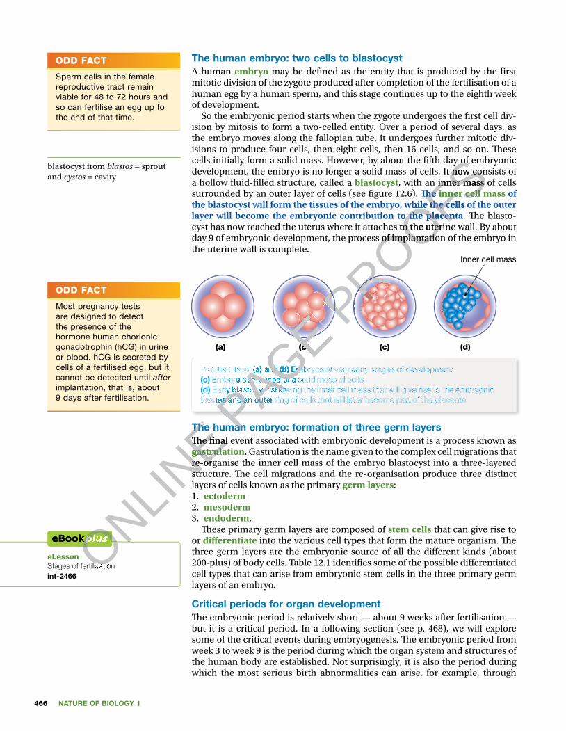
The human embryo: two cells to blastocystA human embryo may be de� ned as the entity that is produced by the � rst mitotic division of the zygote produced after completion of the fertilisation of a human egg by a human sperm, and this stage continues up to the eighth week of development.
So the embryonic period starts when the zygote undergoes the � rst cell div-ision by mitosis to form a two-celled entity. Over a period of several days, as the embryo moves along the fallopian tube, it undergoes further mitotic div-isions to produce four cells, then eight cells, then 16 cells, and so on. � ese cells initially form a solid mass. However, by about the � fth day of embryonic development, the embryo is no longer a solid mass of cells. It now consists of a hollow � uid-� lled structure, called a blastocyst, with an inner mass of cells surrounded by an outer layer of cells (see � gure 12.6). � e inner cell mass of the blastocyst will form the tissues of the embryo, while the cells of the outer layer will become the embryonic contribution to the placenta. � e blasto-cyst has now reached the uterus where it attaches to the uterine wall. By about day 9 of embryonic development, the process of implantation of the embryo in the uterine wall is complete.
(a) (b) (c) (d)
Inner cell mass
FIGURE 12.6 (a) and (b) Embryos at very early stages of development (c) Embryo composed of a solid mass of cells (d) Early blastocyst showing the inner cell mass that will give rise to the embryonic tissues and an outer ring of cells that will later become part of the placenta
The human embryo: formation of three germ layers � e � nal event associated with embryonic development is a process known as gastrulation. Gastrulation is the name given to the complex cell migrations that re-organise the inner cell mass of the embryo blastocyst into a three-layered structure. � e cell migrations and the re-organisation produce three distinct layers of cells known as the primary germ layers:1. ectoderm2. mesoderm3. endoderm.
� ese primary germ layers are composed of stem cells that can give rise to or di� erentiate into the various cell types that form the mature organism. � e three germ layers are the embryonic source of all the di� erent kinds (about 200-plus) of body cells. Table 12.1 identi� es some of the possible di� erentiated cell types that can arise from embryonic stem cells in the three primary germ layers of an embryo.
Critical periods for organ development� e embryonic period is relatively short — about 9 weeks after fertilisation — but it is a critical period. In a following section (see p. 468), we will explore some of the critical events during embryogenesis. � e embryonic period from week 3 to week 9 is the period during which the organ system and structures of the human body are established. Not surprisingly, it is also the period during which the most serious birth abnormalities can arise, for example, through
ODD FACT
Sperm cells in the female reproductive tract remain viable for 48 to 72 hours and so can fertilise an egg up to the end of that time.
blastocyst from blastos = sprout and cystos = cavity
ODD FACT
Most pregnancy tests are designed to detect the presence of the hormone human chorionic gonadotrophin (hCG) in urine or blood. hCG is secreted by cells of a fertilised egg, but it cannot be detected until after implantation, that is, about 9 days after fertilisation.
eLessonStages of fertilisationint-2466
ONLINE The human embryo: formation of three germ layers
ONLINE The human embryo: formation of three germ layers
� e � nal event associated with embryonic development is a process known as
ONLINE � e � nal event associated with embryonic development is a process known as
gastrulation
ONLINE
gastrulationre-organise the inner cell mass of the embryo blastocyst into a three-layered
ONLINE
re-organise the inner cell mass of the embryo blastocyst into a three-layered structure. � e cell migrations and the re-organisation produce three distinct
ONLINE
structure. � e cell migrations and the re-organisation produce three distinct
ONLINE
ONLINE
Stages of fertilisationONLINE
Stages of fertilisationONLINE
ONLINE
ONLINE
ONLINE
ONLINE
ONLINE P
AGE
PAGE
PAGE
PAGE
PAGE
PAGE
PAGE
PAGE
PAGE
PAGE
PAGE (b)
PAGE (b)
PAGE
PAGE FIGURE 12.6
PAGE FIGURE 12.6 (a)
PAGE (a) and
PAGE and (b)
PAGE (b) Embryos at very early stages of development
PAGE Embryos at very early stages of development
Embryo composed of a solid mass of cells
PAGE Embryo composed of a solid mass of cells Early blastocyst showing the inner cell mass that will give rise to the embryonic
PAGE Early blastocyst showing the inner cell mass that will give rise to the embryonic
tissues and an outer ring of cells that will later become part of the placentaPAGE tissues and an outer ring of cells that will later become part of the placentaPAGE
The human embryo: formation of three germ layers PAGE
The human embryo: formation of three germ layers
PROOFScells initially form a solid mass. However, by about the � fth day of embryonic
PROOFScells initially form a solid mass. However, by about the � fth day of embryonic development, the embryo is no longer a solid mass of cells. It now consists of
PROOFSdevelopment, the embryo is no longer a solid mass of cells. It now consists of , with an inner mass of cells
PROOFS, with an inner mass of cells � e
PROOFS� e inner cell mass
PROOFSinner cell massthe blastocyst will form the tissues of the embryo, while the cells of the outer
PROOFSthe blastocyst will form the tissues of the embryo, while the cells of the outer layer will become the embryonic contribution to the placenta
PROOFSlayer will become the embryonic contribution to the placentacyst has now reached the uterus where it attaches to the uterine wall. By about
PROOFScyst has now reached the uterus where it attaches to the uterine wall. By about day 9 of embryonic development, the process of implantation of the embryo in
PROOFSday 9 of embryonic development, the process of implantation of the embryo in
PROOFS
PROOFS
PROOFS
PROOFS
PROOFS
PROOFS
PROOFS
PROOFS
PROOFS
PROOFS
PROOFS
PROOFS
PROOFS
PROOFS
PROOFS
PROOFS
PROOFS
PROOFS
PROOFS
PROOFS
PROOFS
PROOFS

467CHAPTER 12 Cell growth and differentiation
exposure to damaging chemicals, known as teratogens (teratos = monster) or teratogenic agents. A well-known teratogen is thalidomide, which in the past was taken by women to treat morning sickness during pregnancy. Ingestion of thalidomide by women during the � rst trimester (3 months) of their preg-nancies resulted in the birth of babies with severe physical abnormalities. � alidomide was available from the late 1950s but was banned in most coun-tries by 1962, when the link was established between the use of the drug and serious congenital malformations, particularly of the limbs, which were either completely missing or severely shortened (see � gure 12.7).
TABLE 12.1 Some of the differentiated cell types that can arise from embryonic stem cells in the three primary germ layers
Primary germ layer Differentiated cells/tissues/organs
ectoderm skinmelanocytesbrain, spinal cord and nervespituitary glandadrenal medullasense organs: eyessense organs: inner ears
mesoderm heart and blood vesselsadrenal cortexsmooth, skeletal and cardiac musclepart of urogenital systembone marrowblood cellsbone and cartilagelymphatic tissue
endoderm larynx, trachea and lungslining of respiratory tractlining of gastrointestinal tractliverpancreasthymusthyroid glandurinary bladder and urethra
During very early embryonic development, exposure to a teratogen can cause the death of an embryo. Exposure later in the embryonic period can result in major malformations that may either lead to a spontaneous loss of a
pregnancy or to the appearance at birth of severe congenital malformations or birth defects (such as those experienced through exposure to thalidomide, discussed above). Exposure to teratogens during fetal development may be expected to give rise to less serious congenital malformations.
� e critical periods for organ development are shown in � gure 12.8. From these data, you can see that the embryonic period is the most critical time for organ development.
ODD FACT
By 1960, thalidomide was marketed as a completely safe drug in 46 countries, including Australia, and its level of sales were close to those of aspirin.
FIGURE 12.7 Thalidomide is a teratogenic agent that can result in the birth of babies with severe malformations of the limbs.
ONLINE
ONLINE
ONLINE
ONLINE During very early embryonic development, exposure to a teratogen can cause
ONLINE During very early embryonic development, exposure to a teratogen can cause
ONLINE P
AGE
PAGE
PAGE part of urogenital system
PAGE part of urogenital systembone marrow
PAGE bone marrowblood cells
PAGE blood cellsbone and cartilage
PAGE bone and cartilagelymphatic tissue
PAGE lymphatic tissue
PROOFS
PROOFS
PROOFS
PROOFS Some of the differentiated cell types that can arise from
PROOFS Some of the differentiated cell types that can arise from
Differentiated cells/tissues/organs
PROOFSDifferentiated cells/tissues/organs
brain, spinal cord and nerves
PROOFSbrain, spinal cord and nerves
adrenal medulla
PROOFSadrenal medullasense organs: eyes
PROOFSsense organs: eyessense organs: inner ears
PROOFSsense organs: inner ears
heart and blood vessels
PROOFS
heart and blood vesselsadrenal cortexPROOFS
adrenal cortexsmooth, skeletal and cardiac musclePROOFS
smooth, skeletal and cardiac musclepart of urogenital systemPROOFS
part of urogenital systembone marrowPROOFS
bone marrow

NATURE OF BIOLOGY 1468
Implant-ationphase
Weeks1–2
Week3
CNSEye
Eye Ear Palate EarBrain
External genitaliaTeeth
Arm
Leg
Heart Heart
Week4
Week5
Week6
Most common site of birth defect
Week7
Neural-tube defects, mental retardation
Cardiac defects
Absent/shortenedlimb
Absent/shortenedlimb
Low-set malformed ears, deafness Ears
CNS
Eyes
Teeth
Very small eyes, cataracts, glaucoma
Enamel hypoplasia,staining
Cleft palate Palate
Masculinisation offemale genitalia
Major malformationsSpont-aneousabortion
External genitalia
Minor and functional defects
Heart
Upper limb
Lower limb
Week8
Week12
Week15
Weeks20–36
Week38
Embryonic phase Fetal (growth) phase
FIGURE 12.8 The critical periods for organ development. Note that the major defects occur in the embryonic phase.
The human fetus Nine weeks after fertilisation is regarded as the point at which the fetal stage of development is reached. In general, a fetus is characterised by the presence of all the major body organs, although they are not fully developed. Organ systems that were formed during embryonic development will develop further in the fetus, and, in some cases, even after birth, for example, the circulatory system, the respiratory system and the nervous system.
Fetal development is the period during which the major growth in size of the fetus and the mass of its organs occurs (see � gure 12.9). Table 12.2 shows the change in average fetal length and weight at various times from the start of the fetal period (nine weeks after fertilisation) to full term (birth). Over a period of about 30 weeks, the mass of the fetus increases more than 400-fold (see � gure 12.9).
ONLINE
ONLINE
ONLINE
ONLINE
ONLINE
ONLINE
ONLINE
ONLINE
Low-set malformed ears, deafness
ONLINE
Low-set malformed ears, deafness
Major malformations
ONLINE
Major malformations
ONLINE
ONLINE P
AGE
PAGE
PAGE
PAGE
PAGE
PAGE
PAGE
PAGE
PAGE
PAGE
PAGE
PAGE
PAGE
PAGE
PAGE Very small eyes, cataracts, glaucoma
PAGE Very small eyes, cataracts, glaucoma
Enamel hypoplasia,
PAGE Enamel hypoplasia,
staining
PAGE staining
Lower limb
PAGE Lower limb
PROOFS
PROOFS
PROOFS
PROOFS
PROOFS
PROOFS
PROOFS
PROOFS
PROOFS
PROOFS
PROOFS
PROOFS
PROOFS
PROOFS
PROOFS
PROOFS
PROOFS
PROOFS
PROOFS
PROOFS
PROOFS
PROOFS
PROOFS
PROOFS
PROOFS
PROOFS
PROOFS
PROOFS
PROOFS
PROOFS
PROOFS
PROOFS
PROOFS
PROOFS
PROOFS
PROOFS
PROOFS
PROOFS
PROOFS
PROOFS
PROOFS
PROOFS
PROOFS
PROOFS
PROOFS
PROOFS
PROOFS
PROOFS
PROOFS
PROOFS
PROOFS
PROOFS
PROOFS
PROOFS
PROOFS

469CHAPTER 12 Cell growth and differentiation
TABLE 12.2 Approximate fetal lengths and weights over the period of fetal development. Up to 16 weeks, lengths are given as crown–rump lengths. From and including 22 weeks, lengths are given as crown–heel lengths.
Weeks after fertilisation Length Mass 9 3 cm 8 g10 5 cm 14 g16 14 cm 190 g22 30 cm 600 g28 40 cm 1320 g34 47 cm 2600 g
40 51−52 cm 3685 g
KEY IDEAS
■ Antenatal development involves the formation of a zygote, followed by periods of embryonic and fetal development.
■ The increase in cell numbers during the periods of both embryonic and fetal development is a result of cell division by mitosis.
■ Key events in embryonic development include blastocyst formation, which produces the inner cell mass that gives rise to all the tissues of the embryo, and gastrulation, which organises the migration of embryonic cells into the three primary germ layers.
■ Cells from the primary germ layers give rise to or differentiate into a variety of different cell types.
■ Fetal development commences at about week 9 after fertilisation and is a period of major growth as well as continued development of organ systems.
■ Severe congenital malformations can result from exposure to certain chemicals (teratogens) during critical periods of organ development, in particular during the embryonic stage of development.
FIGURE 12.9 Relative change in size from late embryonic stage, through fetal development to full term
ONLINE
ONLINE
ONLINE
ONLINE
ONLINE P
AGE
PAGE
PAGE
PAGE
PAGE
PAGE
PAGE
PAGE
PAGE
PAGE
PAGE Approximate fetal lengths and weights over the period of fetal
PAGE Approximate fetal lengths and weights over the period of fetal
development. Up to 16 weeks, lengths are given as crown–rump lengths. From
PAGE development. Up to 16 weeks, lengths are given as crown–rump lengths. From and including 22 weeks, lengths are given as crown–heel lengths.
PAGE and including 22 weeks, lengths are given as crown–heel lengths.
PAGE
PAGE
PAGE
PAGE Weeks after fertilisation
PAGE Weeks after fertilisation
9
PAGE 9 10 PAGE 10
PROOFS
PROOFS
PROOFS
PROOFS
PROOFS
PROOFS
PROOFS
PROOFS
PROOFS
PROOFS
PROOFS
PROOFS
PROOFS
PROOFS
PROOFS
PROOFS
PROOFS
PROOFS
PROOFS
PROOFS
PROOFS
PROOFS
PROOFS
PROOFS
PROOFS
PROOFS
PROOFS
PROOFS
PROOFS
PROOFS
PROOFS
PROOFS
PROOFS
PROOFS
PROOFS
PROOFS
PROOFS
PROOFS
PROOFS
PROOFS
PROOFS
PROOFS
PROOFS
PROOFS
PROOFS
PROOFS
PROOFS
PROOFS
PROOFS
PROOFS
PROOFS
PROOFS
PROOFS
PROOFS

NATURE OF BIOLOGY 1470
QUICK CHECK
1 What is the length of the period of embryonic development in humans?2 What event initiates the completion of the second meiotic division in a
human egg?3 Which is longer: the period of embryonic development or the period of fetal
development?4 What is a teratogen?5 Identify whether each of the following statements is either true or false.
a Fertilisation occurs in a woman’s ovary.b A zygote is the single diploid cell resulting from the fertilisation of an
egg by a sperm.c A zygote divides by meiosis to produce an increase in cells.d Gastrulation involves cell migrations to form a three-layered embryo.e The three primary germ layers are ectoderm, mesoderm and endoderm.
Key events: embryonic development � e embryonic period, from zygote formation to the end of about the eighth week of development, is the time during which key developmental events occur, as follows:• organisation of cells into the three primary germ layers from which all the
structures and organs of the body will develop • formation of a head–tail axis and a front–to-back (ventral-to-dorsal) axis of
the embryo• cell di� erentiation and beginning of formation of the brain, spinal cord and
nerve cells, heart, sense organs such as eyes, lungs, kidneys, digestive tract, and arms and legs. Key cells involved in the establishment of the organ systems are the embry-
onic stem cells.
Stem cells in actionStem cells are undi� erentiated or unspecialised cells that have the ability to di� erentiate into organ- or tissue-speci� c cells with specialised functions, such as nerve cells, blood cells, bone cells, heart cells, skin cells and so on.
� ese terminal cells with specialised functions, such as a liver cell or a muscle cell, are di� erentiated and, once di� erentiated, cannot normally revert to an undi� erentiated state. A second feature of stem cells is that they are capable of dividing and renewing themselves over long periods. Figure 12.10 shows mouse stem cells that have been stained to show the presence of one of the proteins (Oct4) that are essential to keep the stem cells in an undi� erentiated state.
Some stem cells in your body are constantly dividing to replace tissues. Examples of these are the stem cells in the basal layer of your skin (refer to � gure 9.17), and stem cells in the crypts of your intestine (refer to � gure 9.18). Each of these stem cells divides to produce a spe-cialised di� erentiated cell and a replacement stem cell (see � gure 12.11). � is is how stem cells self-renew.
Di� erent kinds of stem cell occur and they can be distinguished in terms of their potency to produce di� erent cell types. Descriptions of stem cell potencies include:• totipotent• pluripotent• multipotent• oligopotent• unipotent.
FIGURE 12.10 Mouse stem cells. The yellow colouring shows the presence of a protein, known as Oct4, which is essential to keep these stem cells in an undifferentiated state.
ONLINE Stem cells are undi� erentiated or unspecialised cells that have the ability to
ONLINE Stem cells are undi� erentiated or unspecialised cells that have the ability to
di� erentiate into organ- or tissue-speci� c cells with specialised functions
ONLINE di� erentiate into organ- or tissue-speci� c cells with specialised functions
such as nerve cells, blood cells, bone cells, heart cells, skin cells and so on.
ONLINE such as nerve cells, blood cells, bone cells, heart cells, skin cells and so on.
� ese terminal cells with specialised functions, such as a liver cell or a
ONLINE
� ese terminal cells with specialised functions, such as a liver cell or a
ONLINE P
AGE structures and organs of the body will develop
PAGE structures and organs of the body will develop formation of a head–tail axis and a front–to-back (ventral-to-dorsal) axis of
PAGE formation of a head–tail axis and a front–to-back (ventral-to-dorsal) axis of
cell di� erentiation and beginning of formation of the brain, spinal cord and
PAGE cell di� erentiation and beginning of formation of the brain, spinal cord and nerve cells, heart, sense organs such as eyes, lungs, kidneys, digestive tract,
PAGE nerve cells, heart, sense organs such as eyes, lungs, kidneys, digestive tract,
PAGE and arms and legs.
PAGE and arms and legs. Key cells involved in the establishment of the organ systems are the embry-
PAGE Key cells involved in the establishment of the organ systems are the embry-
onic stem cells.
PAGE onic stem cells.
Stem cells in actionPAGE Stem cells in actionStem cells are undi� erentiated or unspecialised cells that have the ability to PAGE
Stem cells are undi� erentiated or unspecialised cells that have the ability to di� erentiate into organ- or tissue-speci� c cells with specialised functionsPAGE
di� erentiate into organ- or tissue-speci� c cells with specialised functions
PROOFS
PROOFS
PROOFSA zygote is the single diploid cell resulting from the fertilisation of an
PROOFSA zygote is the single diploid cell resulting from the fertilisation of an
A zygote divides by meiosis to produce an increase in cells.
PROOFSA zygote divides by meiosis to produce an increase in cells.Gastrulation involves cell migrations to form a three-layered embryo.
PROOFSGastrulation involves cell migrations to form a three-layered embryo.The three primary germ layers are ectoderm, mesoderm and endoderm.
PROOFSThe three primary germ layers are ectoderm, mesoderm and endoderm.
Key events: embryonic development
PROOFSKey events: embryonic development � e embryonic period, from zygote formation to the end of about the eighth
PROOFS� e embryonic period, from zygote formation to the end of about the eighth week of development, is the time during which key developmental events
PROOFSweek of development, is the time during which key developmental events
organisation of cells into the three primary germ layers from which all the PROOFS
organisation of cells into the three primary germ layers from which all the structures and organs of the body will develop PROOFS
structures and organs of the body will develop formation of a head–tail axis and a front–to-back (ventral-to-dorsal) axis of PROOFS
formation of a head–tail axis and a front–to-back (ventral-to-dorsal) axis of

471CHAPTER 12 Cell growth and differentiation
FIGURE 12.11 The division of a stem cell by mitosis gives rise to two daughter cells, one of which differentiates to become a speci� c cell type and the other that replaces or renews the original stem cell. Why is this self-renewal important?
Stem cell
Self-renewal
Differentiated cellDifferentiation
� e fertilised egg is said to be totipotent (totus = entire) because such a cell has the potential to give rise to all cell types. Other totipotent cells include embryonic cells of a two-, four- or eight-cell embryo.
� e embryonic stem cells from the inner cell mass of the embryonic blastocyst are pluripotent (plures = several, many), as are the cells of the pri-mary germ layers — ectoderm, mesoderm and endoderm. � ese cells can dif-ferentiate into many cell types.
Multipotent cells have the ability to di� erentiate into a closely related family of cells; for example, a multipotent blood stem cell can develop into a red blood cell or a white blood cell or platelets (all specialised cells).
Oligopotent cells have the ability to di� erentiate into a few cells, for example, adult (somatic) lymphoid or myeloid stem cells.
Unipotent stem cells have the ability to produce only cells of their own type, but because they can self-renew they are termed stem cells. Examples include adult (somatic) muscle stem cells.
Sources of stem cellsStem cells can be obtained from the following sources:• Embryonic stem cells (ESCs) may be obtained from the inner cell mass of
an early embryo at a stage known as a blastocyst (see � gure 12.12), that is, the clump of cells adhered to the inside surface of a blastocyst (see � gure 12.13a). A single cell is isolated from the inner cell mass of a blastocyst and is grown in culture, dividing by mitosis to produce a culture of stem cells. � ese ESCs are obtained from extra embryos created as part of IVF pro-cedures and they are in excess of requirements. Taking these cells from the inner mass of a blastocyst destroys an embryo and this procedure has raised ethical issues (see below).
Fertilisedegg
Two-cellstage
Morula(10–30 cells)
(day 4)
Blastocyst(day 5)
Outer cells(form placenta)
Inner cell mass(forms embryo)Egg
nucleusPolarbody
Eggcytoplasm
FIGURE 12.12 The development of a fertilised cell to a blastocyst stage. To see some real cells, use the Embryos weblink for this chapter in your eBookPLUS.
Unit 2 Stem cell differentiationConcept summary and practice questions
AOS 1
Topic 4
Concept 1
ODD FACT
Scientists have discovered that proteins produced by three key genes maintain pluripotent stem cells in their undifferentiated state. These proteins, known as Oct4, Sox2 and Klf4, repress or silence the genes needed for embryonic development. When production of these proteins stops, the stem cells start to differentiate and are no longer stem cells.
ONLINE Embryonic stem cells
ONLINE Embryonic stem cells an early embryo at a stage known as a blastocyst (see � gure 12.12), that is,
ONLINE an early embryo at a stage known as a blastocyst (see � gure 12.12), that is, the clump of cells adhered to the inside surface of a blastocyst (see � gure
ONLINE the clump of cells adhered to the inside surface of a blastocyst (see � gure 12.13a). A single cell is isolated from the inner cell mass of a blastocyst and
ONLINE 12.13a). A single cell is isolated from the inner cell mass of a blastocyst and is grown in culture, dividing by mitosis to produce a culture of stem cells.
ONLINE is grown in culture, dividing by mitosis to produce a culture of stem cells. � ese ESCs are obtained from extra embryos created as part of IVF pro-
ONLINE � ese ESCs are obtained from extra embryos created as part of IVF pro-
ONLINE
ONLINE P
AGE blood cell or a white blood cell or platelets (all specialised cells).
PAGE blood cell or a white blood cell or platelets (all specialised cells). cells have the ability to di� erentiate into a few cells, for example,
PAGE cells have the ability to di� erentiate into a few cells, for example, adult (somatic) lymphoid or myeloid stem cells.
PAGE adult (somatic) lymphoid or myeloid stem cells. stem cells have the ability to produce only cells of their own type,
PAGE stem cells have the ability to produce only cells of their own type,
but because they can self-renew they are termed stem cells. Examples include
PAGE but because they can self-renew they are termed stem cells. Examples include adult (somatic) muscle stem cells.
PAGE adult (somatic) muscle stem cells.
Sources of stem cells
PAGE Sources of stem cellsStem cells can be obtained from the following sources:PAGE Stem cells can be obtained from the following sources:
Embryonic stem cells PAGE Embryonic stem cells an early embryo at a stage known as a blastocyst (see � gure 12.12), that is, PAGE
an early embryo at a stage known as a blastocyst (see � gure 12.12), that is,
PROOFS
PROOFS
PROOFSThe division of a stem cell by mitosis gives rise to two daughter
PROOFSThe division of a stem cell by mitosis gives rise to two daughter
cells, one of which differentiates to become a speci� c cell type and the other that
PROOFScells, one of which differentiates to become a speci� c cell type and the other that replaces or renews the original stem cell. Why is this self-renewal important?
PROOFSreplaces or renews the original stem cell. Why is this self-renewal important?
PROOFS=
PROOFS= entire) because such a cell
PROOFS entire) because such a cell
has the potential to give rise to all cell types. Other totipotent cells include
PROOFShas the potential to give rise to all cell types. Other totipotent cells include embryonic cells of a two-, four- or eight-cell embryo.
PROOFSembryonic cells of a two-, four- or eight-cell embryo.
� e embryonic stem cells from the inner cell mass of the embryonic
PROOFS� e embryonic stem cells from the inner cell mass of the embryonic
several, many), as are the cells of the pri-
PROOFS several, many), as are the cells of the pri-
mary germ layers — ectoderm, mesoderm and endoderm. � ese cells can dif-
PROOFSmary germ layers — ectoderm, mesoderm and endoderm. � ese cells can dif-
cells have the ability to di� erentiate into a closely related family PROOFS
cells have the ability to di� erentiate into a closely related family of cells; for example, a multipotent blood stem cell can develop into a red PROOFS
of cells; for example, a multipotent blood stem cell can develop into a red blood cell or a white blood cell or platelets (all specialised cells). PROOFS
blood cell or a white blood cell or platelets (all specialised cells). cells have the ability to di� erentiate into a few cells, for example, PROOFS
cells have the ability to di� erentiate into a few cells, for example,

NATURE OF BIOLOGY 1472
• Parthenotes are another potential source of embryonic stem cells. � ese are derived from unfertilised human eggs that are arti� cially stimulated to begin development. Such an egg, of course, may start development but it is not capable of developing into a human being.
• Adult stem cells (more accurately called somatic stem cells) can be obtained from various sources throughout the body such as bone marrow, skin, the liver, the brain, adipose tissue and blood. In addition, another source of stem cells is cord blood that can be harvested from the umbilical cord of a baby after birth (see � gure 12.13b). Samples of some of these tissues are more accessible than others, such as blood, bone marrow that can be harvested by drilling into bones — typically the iliac crest or the femur, and adipose tissue, which can be obtained by liposuction. Somatic stem cells are multipotent. � is means that they can give rise to particular cell types such as di� erent kinds of blood cells or skin cells. Cord blood, for example, contains mainly stem cells that give rise to various blood cells.
Embryonic stem cells
Bone cells
Nerve cells
Skin cells
(a)
(b)
Blood cells
Adult (somatic) stem cells
Stem cells removedfrom inner cell massof blastocyst Stem cells
cultured inlaboratory
Stem cells removed from umbilical-cordblood and bone marrow
FIGURE 12.13 Stem cell lines can be created from various sources. (a) One source of stem cells is embryonic stem cells from the inner cell mass of a blastocyst. (b) Somatic stem cells can be extracted from bone marrow and from umbilical cord blood. Somatic stem cells is the preferred term for adult stem cells. Can you suggest why?
• Induced pluripotent stem cells (iPSCs) — research by Shinya Yamamaka in Japan in 2006 led to the discovery that some specialised adult somatic (skin) cells could be genetically reprogrammed to return to an undi� erentiated embryonic state. � is reprogramming was achieved by the addition to these cells of four speci� c embryonic genes, which encode proteins that are known to keep stem cells in an undi� erentiated state. One of these genes is the OCT4 gene that encodes the Oct4 protein (refer to � gure 12.10). � e creation of iPSCs does not involve the ethical issues related to the embryo deaths that necessarily accompany embryonic cell stem cells derived from blastocysts. � e ability to produce iPSCs is supporting new lines of research into disease and drug development. For example, iPSCs can be made from skin samples of patients with Parkinson’s disease, and these cells show signs of that disease. � is means that aspects of the disease can be studied in detail in cell cultures in the laboratory, allowing the e� ectiveness of new drugs to be explored using these iPSCs. Cell-based therapies using iPSCs are not practical at present. � e current procedure for reprogramming of somatic cells involves genetic modi� cation, which can sometimes cause cells to produce tumours.
ODD FACT
One advantage of somatic stem cells is that, if used for the person from whom they were taken, there is no risk of rejection.
ODD FACT
Shinya Yamamaka was the co-recipient of the Nobel Prize for Physiology or Medicine in 2012 for ‘the discovery that mature cells can be reprogrammed to become pluripotent’.
ONLINE
ONLINE
ONLINE FIGURE 12.13
ONLINE FIGURE 12.13
source of stem cells is embryonic stem cells from the inner cell mass of a
ONLINE source of stem cells is embryonic stem cells from the inner cell mass of a
blastocyst.
ONLINE blastocyst.
umbilical cord blood.
ONLINE
umbilical cord blood. cells
ONLINE
cells. Can you suggest why?
ONLINE
. Can you suggest why?
ONLINE
ONLINE
ONLINE P
AGE
PAGE
PAGE
PAGE Adult (somatic) stem cells
PAGE Adult (somatic) stem cells
Stem cells removed from umbilical-cord
PAGE Stem cells removed from umbilical-cordblood and bone marrow
PAGE blood and bone marrow
PAGE
PAGE
FIGURE 12.13 PAGE
FIGURE 12.13 Stem cell lines can be created from various sources. PAGE
Stem cell lines can be created from various sources. source of stem cells is embryonic stem cells from the inner cell mass of a PAGE
source of stem cells is embryonic stem cells from the inner cell mass of a PAGE PROOFS
are more accessible than others, such as blood, bone marrow that can be
PROOFSare more accessible than others, such as blood, bone marrow that can be harvested by drilling into bones — typically the iliac crest or the femur, and
PROOFSharvested by drilling into bones — typically the iliac crest or the femur, and
Somatic stem cells are multipotent. � is means that they can give rise to
PROOFS Somatic stem cells are multipotent. � is means that they can give rise to particular cell types such as di� erent kinds of blood cells or skin cells. Cord
PROOFSparticular cell types such as di� erent kinds of blood cells or skin cells. Cord blood, for example, contains mainly stem cells that give rise to various blood
PROOFSblood, for example, contains mainly stem cells that give rise to various blood
PROOFS
PROOFS
Stem cellsPROOFS
Stem cellscultured inPROOFS
cultured inlaboratoryPROOFS
laboratory

473CHAPTER 12 Cell growth and differentiation
Stem cells in regenerative medicineAs people age, a number of degenerative disorders appear more commonly, such as Parkinsonism or Parkinson’s disease. � is particular disorder results from the death of certain brain cells that normally produce a chemical (dopa-mine) that controls muscle movements. People with Parkinsonism show impairment of their motor movements, balance and speech. Early treatment for Parkinsonism involved administering dopamine to a� ected persons. � is treatment gave only short-term improvement.
Is there a way in which the lost dopamine-producing cells can be replaced? Experimental work is now proceeding on the potential use of stem cells to replace the lost cells in the brain.
Because stem cells have the unique ability to regenerate damaged tissue, research is being carried out on the potential use of stem cells in the treatment of Parkinson’s disease and on some other human disorders or conditions. Potential uses of stem cells for these purposes are called cell-based therapies, and the � eld of research is termed regenerative medicine.
In addition to Parkinson’s disease, other conditions that are potential targets for cell-based therapies include:• type 1 diabetes, a condition in which the insulin-producing cells of the pan-
creas are destroyed by people’s own immune system so that they cannot control their blood glucose levels and require the administration of insulin
• heart disease where sectors of heart muscle have died as a result of a heart attack (myocardial infarction) (refer to chapter 6)
• spinal cord injuries as a result of accident or trauma where the interrup-tions to the passage of nerve signals along the spinal cord have resulted in quadriplegia.Cell-based therapies also have the potential to restore normal function in
conditions such as macular degeneration of the retina, burns, and various forms of arthritis. However, the � eld of regenerative medicine is still experi-mental. � e present challenges are to improve the culturing of stem cells under laboratory conditions to increase their numbers, and to increase the under-standing of how stem cells di� erentiate into speci� c cell types. Once these challenges are overcome and the use of stem cells can be shown to be predict-able, reliable and to do no harm, the use of speci� c di� erentiated cell types
from stem cells in the treatment of certain diseases and injuries would be expected to become routine. However, at present, for most diseases, conditions and injuries, safe and e� ective treatments using cell-based therapies are yet to be realised.
Research in the � eld of regenerative medicine is ongoing. Scientists at the University of California reported that, following the injection of human stem cells from nerve tissue into the spinal cords of par-alysed mice, the test group of mice displayed better mobility than the non-injected controls after just nine days and, after four months, the test group of mice could walk. � e stem cells migrated up the spinal cord and developed into di� erent kinds of cells including those cells that form insulating layers of myelin around nerve cells. Figure 12.14 shows the growth of myelin around nerve cells in the damaged region of a mouse spinal cord following injection of stem cells.
Scientists at the Walter and Eliza Hall Institute of Medical Research in Melbourne identi� ed a cell line within mouse breast tissue that included multipotent
FIGURE 12.14 Injured spinal cord of mouse following injection of human stem cells. These stem cells developed into myelin-producing cells that form a wrapping (green) around nerve cells (red) (see the areas marked by arrowheads). Other nerve cells remained without a myelin wrapping (see the areas indicated with arrows).
ONLINE standing of how stem cells di� erentiate into speci� c cell types. Once these
ONLINE standing of how stem cells di� erentiate into speci� c cell types. Once these
challenges are overcome and the use of stem cells can be shown to be predict-
ONLINE challenges are overcome and the use of stem cells can be shown to be predict-
able, reliable and to do no harm, the use of speci� c di� erentiated cell types
ONLINE able, reliable and to do no harm, the use of speci� c di� erentiated cell types
ONLINE
ONLINE
ONLINE
ONLINE
ONLINE P
AGE attack (myocardial infarction) (refer to chapter 6)
PAGE attack (myocardial infarction) (refer to chapter 6)spinal cord injuries as a result of accident or trauma where the interrup-
PAGE spinal cord injuries as a result of accident or trauma where the interrup-tions to the passage of nerve signals along the spinal cord have resulted in
PAGE tions to the passage of nerve signals along the spinal cord have resulted in
Cell-based therapies also have the potential to restore normal function in
PAGE Cell-based therapies also have the potential to restore normal function in
conditions such as macular degeneration of the retina, burns, and various
PAGE conditions such as macular degeneration of the retina, burns, and various forms of arthritis. However, the � eld of regenerative medicine is still experi-
PAGE forms of arthritis. However, the � eld of regenerative medicine is still experi-mental. � e present challenges are to improve the culturing of stem cells under
PAGE mental. � e present challenges are to improve the culturing of stem cells under laboratory conditions to increase their numbers, and to increase the under-PAGE laboratory conditions to increase their numbers, and to increase the under-standing of how stem cells di� erentiate into speci� c cell types. Once these PAGE standing of how stem cells di� erentiate into speci� c cell types. Once these challenges are overcome and the use of stem cells can be shown to be predict-PAGE
challenges are overcome and the use of stem cells can be shown to be predict-
PROOFSIs there a way in which the lost dopamine-producing cells can be replaced?
PROOFSIs there a way in which the lost dopamine-producing cells can be replaced?
Experimental work is now proceeding on the potential use of stem cells to
PROOFSExperimental work is now proceeding on the potential use of stem cells to
Because stem cells have the unique ability to regenerate damaged tissue,
PROOFSBecause stem cells have the unique ability to regenerate damaged tissue, research is being carried out on the potential use of stem cells in the treatment
PROOFSresearch is being carried out on the potential use of stem cells in the treatment of Parkinson’s disease and on some other human disorders or conditions.
PROOFSof Parkinson’s disease and on some other human disorders or conditions. Potential uses of stem cells for these purposes are called
PROOFSPotential uses of stem cells for these purposes are called cell-based therapies
PROOFScell-based therapies
regenerative medicine
PROOFSregenerative medicine
In addition to Parkinson’s disease, other conditions that are potential targets
PROOFSIn addition to Parkinson’s disease, other conditions that are potential targets
type 1 diabetes, a condition in which the insulin-producing cells of the pan-
PROOFStype 1 diabetes, a condition in which the insulin-producing cells of the pan-creas are destroyed by people’s own immune system so that they cannot
PROOFScreas are destroyed by people’s own immune system so that they cannot control their blood glucose levels and require the administration of insulin
PROOFS
control their blood glucose levels and require the administration of insulinheart disease where sectors of heart muscle have died as a result of a heart PROOFS
heart disease where sectors of heart muscle have died as a result of a heart attack (myocardial infarction) (refer to chapter 6)PROOFS
attack (myocardial infarction) (refer to chapter 6)spinal cord injuries as a result of accident or trauma where the interrup-PROOFS
spinal cord injuries as a result of accident or trauma where the interrup-

NATURE OF BIOLOGY 1474
stem cells. Remarkably, they found that just one of these cells introduced to fat tissue in the laboratory could produce a branching mammary gland (see � gure 12.15a). � ese stem cells were able to give rise to the various cell types present in mammary glands (see � gure 12.15b).
FIGURE 12.15 (a) Branching mammary gland produced from a single mouse breast stem cell (b) Section through mammary tissue produced from stem cell with different cell types shown by arrows (L = lumen)
(a) (b)
Therapeutic cloning� e purpose of therapeutic cloning is to produce stem cells for use in treatment.
� erapeutic cloning involves the creation of an embryo, through the tech-nique of somatic nuclear transfer, for the purpose of obtaining stem cells from that embryo. � ese stem cells are intended for use in treating a patient who has a degenerative disease. � e cell that provides the nucleus in therapeutic cloning is a healthy cell from the patient who is to receive treatment. As a result, the embryo that is created is a genetic match to the patient and these cells do not cause an immune response. Figure 12.16 shows the process of therapeutic cloning.
Disease-freecells taken frompatient
Enucleationof egg cell
Fusion ofcell andegg
Embryocultured and stem cellsremoved
Embryonic stem cells cultured and speci�c celltypes obtained
Required celltypes introducedinto patient
FIGURE 12.16 Therapeutic cloning involves the creation of an embryo that is genetically identical to a patient. The patient’s cell is fused with an enucleated egg cell and develops into an early embryo. Stem cells are then taken from the inner cell mass of the early embryo (blastocyst) and grown in culture as pluripotent stem cells. Would these cultured cells be expected to cause an immune response if injected into the patient? Why?
ONLINE that is created is a genetic match to the patient and these cells do not cause an
ONLINE that is created is a genetic match to the patient and these cells do not cause an
immune response. Figure 12.16 shows the process of therapeutic cloning.
ONLINE immune response. Figure 12.16 shows the process of therapeutic cloning.
ONLINE P
AGE Therapeutic cloning
PAGE Therapeutic cloningtherapeutic cloning
PAGE therapeutic cloning
� erapeutic cloning involves the creation of an embryo, through the tech-
PAGE � erapeutic cloning involves the creation of an embryo, through the tech-
nique of somatic nuclear transfer, for the purpose of obtaining stem cells from
PAGE nique of somatic nuclear transfer, for the purpose of obtaining stem cells from that embryo.
PAGE that embryo. � ese stem cells are intended for use in treating a patient who has a
PAGE � ese stem cells are intended for use in treating a patient who has a
degenerative disease. � e cell that provides the nucleus in therapeutic cloning is PAGE degenerative disease. � e cell that provides the nucleus in therapeutic cloning is a healthy cell from the patient who is to receive treatment. As a result, the embryo PAGE a healthy cell from the patient who is to receive treatment. As a result, the embryo that is created is a genetic match to the patient and these cells do not cause an PAGE
that is created is a genetic match to the patient and these cells do not cause an immune response. Figure 12.16 shows the process of therapeutic cloning.PAGE
immune response. Figure 12.16 shows the process of therapeutic cloning.
PROOFS
PROOFS

475CHAPTER 12 Cell growth and differentiation
�e use of early embryos as a source of stem cells raises ethical issues because establishing an embryonic stem cell line destroys an embryo. Like-wise, ethical issues arise for therapeutic cloning because this procedure involves the arti�cial creation of an embryo solely for the purpose of obtaining stem cells, a process that then destroys the embryo.
In December 2002, the Research Involving Human Embryos Act 2002 was passed in the Australian Parliament. �is Act established a framework that regulated the use of ‘excess’ embryos. Provisions of this Act included the state-ment that ‘embryos cannot be created solely for research purposes’. Under the provisions of this Act, therapeutic cloning was not permitted in Australia. How-ever, in December 2006, the legislation was amended with Parliament lifting the ban of the cloning of human embryos for stem cell research and allowing therapeutic cloning to be undertaken.
KEY IDEAS
■ Stem cells are undifferentiated or unspecialised cells that have the ability to differentiate into organ- or tissue-speci�c cells with specialised functions and to self renew.
■ Stem cells include embryonic stem cells and somatic (adult) stem cells. ■ Stem cells from different sources differ in their potency or ability to produce differentiated cells of various types.
■ Therapeutic cloning is the creation, through the technique of somatic nuclear transfer, of an embryo for the purpose of obtaining stem cells from that embryo.
■ Regenerative medicine is still at an experimental stage but it raises promises for the treatment of degenerative conditions and severe trauma injuries.
QUICK CHECK
6 What is the difference between the members of the following pairs?a Totipotent and pluripotentb Undifferentiated and differentiatedc Embryonic stem cell and somatic (adult) stem cell
7 Identify one source of embryonic stem cells.8 List two sources that could be used to obtain somatic (adult) stem cells.9 What is a parthenote?
Abnormal embryonic developmentAbnormalities arising during antenatal development are described as con-genital malformations or birth defects. One estimate is that about three per-cent of babies are born with a congenital abnormality. Some of these cause the death of a baby soon after birth, while others leave the child with a life-long defective function, either physical or physiological. Some congenital mal-formations can be treated by surgery, such as cleft lip and palate, or by other forms of treatment.
Congenital defects arise in several ways and may be due to: 1. genetic factors. Genetic factors include single gene defects and chromosomal
abnormalities• Single-gene defects are usually inherited, for example, phenylketonuria
and cystic �brosis, which most commonly appear in newborns of unaf-fected parents who are heterozygous carriers of the allele involved.
In other cases, the single gene defect may be the result of a mutation in one of the gametes involved in fertilisation. For example, a form of
Unit 2 Factors affecting cell growthConcept summary and practice questions
AOS 1
Topic 4
Concept 4ONLIN
E
ONLINE
ONLINE c
ONLINE c7
ONLINE 7 Identify one sour
ONLINE Identify one sour8
ONLINE 8 List two sour
ONLINE
List two sour9
ONLINE 9
ONLINE
ONLINE
ONLINE
ONLINE
ONLINE
ONLINE
ONLINE
ONLINE
ONLINE
ONLINE
ONLINE
ONLINE
ONLINE
ONLINE
ONLINE
ONLINE
nit
ONLINE
nit 2
ONLINE
2
ONLINE
Factors
ONLINE
Factors affecting ONLIN
E
affecting cell growthONLIN
E
cell growthConcept summary ONLIN
E
Concept summary A ONLIN
E
AO ONLINE
OS ONLINE
S 1ONLINE
1ONLINE
Topic ONLINE
Topic 4ONLINE
4ONLINE
ONLINE
ONLINE P
AGE
PAGE
PAGE nuclear transfer, of an embryo for the purpose of obtaining stem cells from
PAGE nuclear transfer, of an embryo for the purpose of obtaining stem cells from
Regenerative medicine is still at an experimental stage but it raises promises
PAGE Regenerative medicine is still at an experimental stage but it raises promises for the treatment of degenerative conditions and severe trauma injuries.
PAGE for the treatment of degenerative conditions and severe trauma injuries.
PAGE
PAGE
PAGE QUICK CHECK
PAGE QUICK CHECK
What is the difPAGE What is the difference between the members of the following pairs?PAGE
ference between the members of the following pairs?What is the difference between the members of the following pairs?What is the difPAGE What is the difference between the members of the following pairs?What is the dif
T PAGE Totipotent and pluripotentPAGE otipotent and pluripotentTotipotent and pluripotentT PAGE
Totipotent and pluripotentTUndifPAGE
Undifferentiated and differentiatedPAGE
ferentiated and differentiatedUndifferentiated and differentiatedUndifPAGE
Undifferentiated and differentiatedUndifEmbryonic stem cell and somatic (adult) stem cellPAGE
Embryonic stem cell and somatic (adult) stem cell
PROOFSprovisions of this Act, therapeutic cloning was not permitted in Australia. How
PROOFSprovisions of this Act, therapeutic cloning was not permitted in Australia. However, in December 2006, the legislation was amended with Parliament lifting
PROOFSever, in December 2006, the legislation was amended with Parliament lifting the ban of the cloning of human embryos for stem cell research and allowing
PROOFSthe ban of the cloning of human embryos for stem cell research and allowing
PROOFS
PROOFS
PROOFSStem cells are undifferentiated or unspecialised cells that have the
PROOFSStem cells are undifferentiated or unspecialised cells that have the ability to differentiate into organ- or tissue-speci�c cells with specialised
PROOFSability to differentiate into organ- or tissue-speci�c cells with specialised
Stem cells include embryonic stem cells and somatic (adult) stem cells.
PROOFSStem cells include embryonic stem cells and somatic (adult) stem cells.Stem cells from different sources differ in their potency or ability to
PROOFSStem cells from different sources differ in their potency or ability to produce differentiated cells of various types.
PROOFS
produce differentiated cells of various types.Therapeutic cloning is the creation, through the technique of somatic PROOFS
Therapeutic cloning is the creation, through the technique of somatic nuclear transfer, of an embryo for the purpose of obtaining stem cells from PROOFS
nuclear transfer, of an embryo for the purpose of obtaining stem cells from

NATURE OF BIOLOGY 1476
dwar� sm, known as achondroplasia, is a dominant trait, with about 80 per cent of cases being due to such a mutation.
• Chromosomal abnormalities, such as the presence of an extra copy of a chromosome so that instead of the normal two copies of an autosome, three copies are present — a condition termed trisomy. Examples of these chromosomal abnormalities surviving to birth are found in Down syndrome (2n = 47, +21), Edward syndrome (2n = 47, +18), and Patau syndrome (2n = 47, +13). � ese chromosomal abnormalities arise as a result of the disruption of the normal cell cycle, either during gamete formation by meiosis, or during the early cell divisions of the embryo by mitosis. If the DNA is damaged in cells undergoing mitosis, cell division is held up at the G1 checkpoint to prevent replication of damaged. DNA cell division is normally held up at another checkpoint to ensure that homologous chromosomes are attached to the correct spindle � bres. � is means that, normally, cell division continues only when DNA is not faulty and when homologous chromosomes are attached to the correct spindle � bres so that at anaphase they disjoin to opposite poles of the spindle. In chro-mosomal abnormalities, the normal operation of this second checkpoint breaks down so that two copies of a chromosome move to the same pole of the spindle. Research has shown that the risk of an egg with a chromosomal abnor-mality is much higher in older women than in younger women (see � gure 12.17). � is suggests that the operation of checkpoints is more at risk of malfunctioning in older women that in younger women.
In some cases, interactions may occur between genetic factors and environmental factors.
100%
90%
80%
70%
60%
50%
Per
cen
t ch
rom
oso
mal
ly a
bno
rmal
40%
30%
20%
10%
0%28 30 32 34 36
Female age
38 40 42 44
FIGURE 12.17 Graph showing the increase in production of chromosomally abnormal eggs with increasing maternal age
2. environmental factors. Environmental factors that can result in a birth defect are known as teratogens, for example, chemicals in the pregnant woman’s bloodstream that reach the developing embryo, such as thalidomide (refer to p. 467).
Other known teratogens include physical agents such as radiation, hyper-thermia (resulting from use of hot tubs, saunas); viral infections, such as rubella (German measles); drugs and chemicals such as alcohol and
ONLINE 60%
ONLINE 60%
50%
ONLINE 50%
Per
cen
t ch
rom
oso
mal
ly a
bno
rmal
ONLINE
Per
cen
t ch
rom
oso
mal
ly a
bno
rmal
40%
ONLINE
40%
PAGE � gure 12.17). � is suggests that the operation of checkpoints is more at
PAGE � gure 12.17). � is suggests that the operation of checkpoints is more at risk of malfunctioning in older women that in younger women.
PAGE risk of malfunctioning in older women that in younger women.In some cases, interactions may occur between genetic factors and
PAGE In some cases, interactions may occur between genetic factors and environmental factors.
PAGE environmental factors.
80% PAGE 80%
70% PAGE 70% PAGE
PAGE
PAGE
PAGE
PAGE
PAGE
PAGE
PAGE
PAGE PROOFS
of the normal cell cycle, either during gamete formation by meiosis,
PROOFSof the normal cell cycle, either during gamete formation by meiosis, or during the early cell divisions of the embryo by mitosis. If the DNA
PROOFSor during the early cell divisions of the embryo by mitosis. If the DNA is damaged in cells undergoing mitosis, cell division is held up at the
PROOFSis damaged in cells undergoing mitosis, cell division is held up at the G1 checkpoint to prevent replication of damaged. DNA cell division is
PROOFSG1 checkpoint to prevent replication of damaged. DNA cell division is normally held up at another checkpoint to ensure that homologous
PROOFSnormally held up at another checkpoint to ensure that homologous chromosomes are attached to the correct spindle � bres. � is means that,
PROOFSchromosomes are attached to the correct spindle � bres. � is means that, normally, cell division continues only when DNA is not faulty and when
PROOFSnormally, cell division continues only when DNA is not faulty and when homologous chromosomes are attached to the correct spindle � bres so
PROOFShomologous chromosomes are attached to the correct spindle � bres so that at anaphase they disjoin to opposite poles of the spindle. In chro-
PROOFSthat at anaphase they disjoin to opposite poles of the spindle. In chro-mosomal abnormalities, the normal operation of this second checkpoint
PROOFSmosomal abnormalities, the normal operation of this second checkpoint breaks down so that two copies of a chromosome move to the same pole
PROOFSbreaks down so that two copies of a chromosome move to the same pole
Research has shown that the risk of an egg with a chromosomal abnor-PROOFS
Research has shown that the risk of an egg with a chromosomal abnor-mality is much higher in older women than in younger women (see PROOFS
mality is much higher in older women than in younger women (see � gure 12.17). � is suggests that the operation of checkpoints is more at PROOFS
� gure 12.17). � is suggests that the operation of checkpoints is more at risk of malfunctioning in older women that in younger women.PROOFS
risk of malfunctioning in older women that in younger women.

477CHAPTER 12 Cell growth and differentiation
cocaine; and seafood with a high mercury content. Factors such as maternal diabetes, smoking or malnutrition can also contribute to the development of birth abnormalities. A dietary de� ciency may result in a birth defect, for example, a de� ciency of vitamin A; in experiments using pigs exposed to a vitamin A-de� cient diet, o� spring were born with the absence of one or both eyes, and with cleft palates.
One tragic example of the action of a teratogen is the methylmercury poi-soning that occurred in Minamata Bay, Japan for several decades as a result of unchecked industrial pollution. As well as a� ecting adults who ate � sh with high levels of methyl mercury, pregnant women gave birth to babies with severe congenital abnormalities (see � gure 12.18).
� e earlier in the stages of development that exposure to a teratogen occurs, the more serious its e� ects will be. In some cases, spontaneous mis-carriage may occur even before a woman is aware that she is pregnant.
3. unknown factors. In some cases, the cause of a birth abnormality may be unknown.
Cancer and the cell cycleIn the healthy body, some cells can divide to produce new cells and this pro-cess usually occurs in an orderly and carefully controlled manner. � is normal process enables the body to grow during childhood and adolescence, and to replace damaged, dead or lost cells during adulthood.
In contrast, cancer is a disease in which cells divide in an uncontrolled manner, forming an abnormal mass of cells (see � gure 12.19). Cancer cells are out of control because of mutations in their genes that result in a breakdown of the normal regulation of the cell cycle. So cancer is a disease that results from the loss of control of the cell cycle.
FIGURE 12.19 Section through human kidney: (a) normal kidney and (b) kidney with cancer growing
(a) (b)
FIGURE 12.18 Kazumitsu Hannaga, a congenital Minamata disease patient, at Meisui-en Hospital, Minamata, 1991. The hospital opened in 1972 to care for Minamata victims.
ODD FACT
The � rst teratogen that was not a chemical was identi� ed in 1941 by an Australian physician, Norman Gregg. He recognised the link between the rubella virus infection of mothers during the � rst two months of their pregnancies and the occurrence of eye defects in their newborn babies. ONLIN
E
ONLINE
ONLINE
ONLINE
ONLINE
The � rst teratogen that was not
ONLINE
The � rst teratogen that was not a chemical was identi� ed in
ONLINE
a chemical was identi� ed in 1941 by an Australian physician,
ONLINE
1941 by an Australian physician, Norman Gregg. He recognised
ONLINE
Norman Gregg. He recognised the link between the rubella
ONLINE
the link between the rubella virus infection of mothers
ONLINE
virus infection of mothers during the � rst two months
ONLINE
during the � rst two months of their pregnancies and the
ONLINE
of their pregnancies and the
ONLINE
ONLINE
ONLINE
ONLINE
occurrence of eye defects in ONLINE
occurrence of eye defects in their newborn babies. ONLIN
E
their newborn babies.
PAGE is a disease in which cells divide in an uncontrolled
PAGE is a disease in which cells divide in an uncontrolled manner, forming an abnormal mass of cells (see � gure 12.19). Cancer cells are
PAGE manner, forming an abnormal mass of cells (see � gure 12.19). Cancer cells are out of control because of mutations in their genes that result in a breakdown of
PAGE out of control because of mutations in their genes that result in a breakdown of the normal regulation of the cell cycle. So cancer is a disease that results from
PAGE the normal regulation of the cell cycle. So cancer is a disease that results from the loss of control of the cell cycle.
PAGE the loss of control of the cell cycle.
PAGE PROOFS
with high levels of methyl mercury, pregnant women gave birth to babies
PROOFSwith high levels of methyl mercury, pregnant women gave birth to babies
� e earlier in the stages of development that exposure to a teratogen
PROOFS� e earlier in the stages of development that exposure to a teratogen occurs, the more serious its e� ects will be. In some cases, spontaneous mis-
PROOFSoccurs, the more serious its e� ects will be. In some cases, spontaneous mis-carriage may occur even before a woman is aware that she is pregnant.
PROOFScarriage may occur even before a woman is aware that she is pregnant. In some cases, the cause of a birth abnormality may be
PROOFS In some cases, the cause of a birth abnormality may be
Cancer and the cell cycle
PROOFSCancer and the cell cycleIn the healthy body, some cells can divide to produce new cells and this pro-
PROOFSIn the healthy body, some cells can divide to produce new cells and this pro-cess usually occurs in an orderly and carefully controlled manner. � is normal
PROOFScess usually occurs in an orderly and carefully controlled manner. � is normal process enables the body to grow during childhood and adolescence, and to PROOFS
process enables the body to grow during childhood and adolescence, and to replace damaged, dead or lost cells during adulthood.PROOFS
replace damaged, dead or lost cells during adulthood. is a disease in which cells divide in an uncontrolled PROOFS
is a disease in which cells divide in an uncontrolled manner, forming an abnormal mass of cells (see � gure 12.19). Cancer cells are PROOFS
manner, forming an abnormal mass of cells (see � gure 12.19). Cancer cells are

NATURE OF BIOLOGY 1478
Normal control or regulation of the cell cycle involves several mechanisms including:• Checkpoints, such as the G1 checkpoint, ensure that the replication of chro-
mosomal DNA is error-free. If errors such as lesions, breaks or other damage are found, the progress of the cell cycle is delayed to allow the DNA to be repaired.
• Proteins, known as cyclins, ensure that chromosomes are attached to the correct spindle �bres so that an equal distribution of chromosomes to daughter cells occurs.
• Normal cells display so-called ‘contact inhibition’, which means they will stop dividing if they begin to overgrow adjacent cells.
• Chemical signals convey information to cells about when to divide faster and when to slow down or stop dividing. Two kinds of genes are involved in this signalling: (1) oncogenes that signal cells to continue dividing and (2) tumour suppressor genes that signal cells to stop dividing. Mutations of these genes would be expected to disrupt the control of the cell cycle.
• If cells are chromosomally abnormal or are damaged, they receive a signal to self-destruct in a process known as programmed cell death or apoptosis.In cancer cells, however, these various controls of the cell division cycle
are lost and the cells do not respond to signals to stop dividing. Instead, cancer cells divide in an uncontrolled manner, forming tumours that damage the surrounding healthy tissues. Cancer cells do not show ‘contact inhibition’ but will overgrow existing cells. While some tumours are benign, others are malignant because cells from these tumours can enter the blood-stream or lymphatics and spread to other regions of the body — a process known as metastasis (see �gure 12.20).
Many cancers have a genetic component. Some cancers are inherited, for example, retinoblastoma, a cancer of the eye. In other cases, the pres-ence of a particular gene can increase the risk of occurrence of a cancer (see table 12.3). �e BRCA1 and BRCA2 genes are rare, but between 45 and 90 per cent of women with one of those genes develop breast cancer. �ese genes also increase the risk of ovarian cancer. �e BRCA2 gene in men increases their risk of developing breast or prostate cancer. However, it should be noted that these genes are involved in only a small percentage of cancers of a particular organ; for example, less than 3 per cent of breast can-cers are caused by a faulty gene.
TABLE 12.3 Some genes that are associated with an increased risk of developing cancer in a speci�c tissue
Genes that increase risk Associated cancer
APC, MYH bowel cancer
BRCA1, BRCA2 breast and ovarian cancer
VHL, TCS1, TCS2 kidney cancer
FAMMM, CDKN2A melanoma
�e majority of cancers result from the action of environmental factors, such as exposure to ultraviolet (UV) radiation, carcinogenic chemicals in cigarette smoke, viruses and diet. Tobacco use in combination with excessive alcohol consumption is a risk factor in the development of oral cancer (cancer of the mouth). �ese environmental factors cause gene mutations and so can be called mutagens.
ODD FACT
Cancer Research UK suggests that around 4 out of 100 cancers (4%) are linked to alcohol. It increases the risk of mouth cancer, liver cancer, breast cancer, bowel cancer and throat cancer.
Unit 2 Uncontrolled cell growthConcept summary and practice questions
AOS 1
Topic 4
Concept 5
ONLINE and 90 per cent of women with one of those genes develop breast cancer.
ONLINE and 90 per cent of women with one of those genes develop breast cancer.
�ese genes also increase the risk of ovarian cancer. �e
ONLINE �ese genes also increase the risk of ovarian cancer. �e
men increases their risk of developing breast or prostate cancer. However, it
ONLINE men increases their risk of developing breast or prostate cancer. However, it
should be noted that these genes are involved in only a small percentage of
ONLINE
should be noted that these genes are involved in only a small percentage of cancers of a particular organ; for example, less than 3 per cent of breast can
ONLINE
cancers of a particular organ; for example, less than 3 per cent of breast cancers are caused by a faulty gene.
ONLINE
cers are caused by a faulty gene.
PAGE damage the surrounding healthy tissues. Cancer cells do not show ‘contact
PAGE damage the surrounding healthy tissues. Cancer cells do not show ‘contact inhibition’ but will overgrow existing cells. While some tumours are benign,
PAGE inhibition’ but will overgrow existing cells. While some tumours are benign, others are malignant because cells from these tumours can enter the blood
PAGE others are malignant because cells from these tumours can enter the bloodstream or lymphatics and spread to other regions of the body — a process
PAGE stream or lymphatics and spread to other regions of the body — a process
PAGE metastasis
PAGE metastasis (see �gure 12.20).
PAGE (see �gure 12.20).
Many cancers have a genetic component. Some cancers are inherited,
PAGE Many cancers have a genetic component. Some cancers are inherited,
for example, retinoblastoma, a cancer of the eye. In other cases, the pres
PAGE for example, retinoblastoma, a cancer of the eye. In other cases, the presence of a particular gene can increase the risk of occurrence of a cancer
PAGE ence of a particular gene can increase the risk of occurrence of a cancer (see table 12.3). �e PAGE (see table 12.3). �e and 90 per cent of women with one of those genes develop breast cancer. PAGE and 90 per cent of women with one of those genes develop breast cancer. �ese genes also increase the risk of ovarian cancer. �e PAGE
�ese genes also increase the risk of ovarian cancer. �e
PROOFS’, which means they will
PROOFS’, which means they will
Chemical signals convey information to cells about when to divide
PROOFSChemical signals convey information to cells about when to divide faster and when to slow down or stop dividing. Two kinds of genes are
PROOFSfaster and when to slow down or stop dividing. Two kinds of genes are that signal cells to continue
PROOFS that signal cells to continue that signal cells to stop dividing.
PROOFS that signal cells to stop dividing.
Mutations of these genes would be expected to disrupt the control of the
PROOFSMutations of these genes would be expected to disrupt the control of the
If cells are chromosomally abnormal or are damaged, they receive a
PROOFSIf cells are chromosomally abnormal or are damaged, they receive a signal to self-destruct in a process known as
PROOFSsignal to self-destruct in a process known as programmed cell death
PROOFSprogrammed cell death
In cancer cells, however, these various controls of the cell division cycle
PROOFSIn cancer cells, however, these various controls of the cell division cycle
are lost and the cells do not respond to signals to stop dividing. Instead, PROOFS
are lost and the cells do not respond to signals to stop dividing. Instead, cancer cells divide in an uncontrolled manner, forming tumours that PROOFS
cancer cells divide in an uncontrolled manner, forming tumours that damage the surrounding healthy tissues. Cancer cells do not show ‘contact PROOFS
damage the surrounding healthy tissues. Cancer cells do not show ‘contact inhibition’ but will overgrow existing cells. While some tumours are benign, PROOFS
inhibition’ but will overgrow existing cells. While some tumours are benign,

479CHAPTER 12 Cell growth and differentiation
KEY IDEAS
■ Abnormalities arising during antenatal development are described as congenital defects.
■ Congenital defects may be due to genetic or environmental factors or be of unknown origin.
■ Teratogens are physical, chemical or biological agents that can cause birth defects.
■ Cancer is a disease that results from the loss of normal control of the cell cycle, resulting in an uncontrolled growth of cells.
QUICK CHECK
10 Identify two of the control mechanisms that are in place in normal cells to prevent uncontrolled growth by cell division.
11 What was the teratogen that caused Minamata disease?12 What is a mutagen?13 Give two examples of environmental agents that are believed to act as
mutagens.
(b) (i)
(ii)
(iii)
Tumour
Lymphvessel
Secretory lobule
Rib
(a)
Milk duct
FIGURE 12.20 (a) Longitudinal section of a breast showing normal tissues (b) (i) Development of a discrete tumour or ‘lump in the breast’ (ii) Development of tumour into cancer. Cancer comes from the Latin for crab because cancers typically have an irregular shape. (iii) S pread of cancer within breast and migration of cancer cells from the breast through lymph vessels and blood vessel to new sites where secondary cancers develop, the process of metastasis. (c) Spread of breast cancer cell in vitro, that is, in a cell culture in the laboratory. Migrating cancer cells show the presence of a speci� c protein, vimentin, that stains green and is not present in normal cells. Note how the cancer cells (green) migrate and multiply � lling the space and crowding out the normal cells (red). (Image courtesy of Professor Leigh Ackland)
(c) (i)
(iii)
(ii)
ONLINE
ONLINE
ONLINE
ONLINE KEY IDEAS
ONLINE KEY IDEAS
ONLINE
ONLINE
secondary cancers develop,
ONLINE
secondary cancers develop,
ONLINE
the process of metastasis.
ONLINE
the process of metastasis. Spread of breast cancer
ONLINE
Spread of breast cancer , that is, in a cell
ONLINE
, that is, in a cell culture in the laboratory.
ONLINE
culture in the laboratory. Migrating cancer cells show
ONLINE
Migrating cancer cells show the presence of a speci� c
ONLINE
the presence of a speci� c protein, vimentin, that stains ONLIN
E
protein, vimentin, that stains green and is not present in ONLIN
E
green and is not present in ONLINE
normal cells. Note how the ONLINE
normal cells. Note how the cancer cells (green) migrate ONLIN
E
cancer cells (green) migrate ONLINE P
AGE
PAGE PROOFS
PROOFS
PROOFS
PROOFS(ii)
PROOFS(ii)

480 NATURE OF BIOLOGY 1
BIOCHALLENGE
Cell growth and cell differentiation
1 The following statement appeared in an article by IR Fox et al., ‘Use of differentiated pluripotent stem cells in replacement therapy for treating disease’, which was published in the journal Science on 22 August 2014.
Unlimited populations of differentiated PSCs [pluripotent stem cells] should facilitate blood therapies and hematopoietic stem cell transplantation, as well as the treatment of heart, pancreas, liver, muscle, and neurologic disorders. However, successful cell transplantation will require optimizing the best cell type and site for engraftment, overcoming limitations to cell migration and tissue integration, and possibly needing to control immunologic reactivity.
Consider this statement and answer the following questions.a What challenges must be overcome before the use of
PSCs can become a routine clinical practice?b What is meant by the phrase ‘overcoming
immunologic reactivity’?c If this article was about the use of induced pluripotent
stem cells (iPSCs), would immunologic reactivity be a challenge? Explain your response.
2 Fetal stem cells were used to treat a boy with the genetic disorder, Ataxia Telangiectasia (A-T), a rare neurodegenerative disease that produces signi� cant disability including poor coordination, a weakened immune system and a breakdown in the DNA repair mechanism so that an affected person’s risk of cancer is increased.
Four years after the treatment with these stem cells, the boy developed abnormal growths in his brain and spinal cord. This rare event was believed to have been a result of the boy’s weakened immune system.Fortunately these tumours were benign and were able to be surgically removed. a When the tumours were removed, it was found that
they were a result of the treatment, not an independent event that occurred in the boy. How might this have been determined?
b Person A commented: ‘The use of stem cell therapy should be stopped. No further research should be done’.
Person B commented: ‘More research is needed with a focus on the safety of stem cell therapy, � nding out what can potentially go wrong and developing safeguards to reduce any risks’.
With which person do you agree, and why?
3 One form of stem cell therapy that has been in use for decades is the use of stem cells (see � gure 12.21) from bone marrow transplants for treatment of disorders of the blood and the immune system, as well as the acquired loss of bone marrow function. Advanced techniques are in practice for collecting blood stem cells and their use is well established clinically. In contrast, other stem cell therapies are still experimental.
FIGURE 12.21
a What is the difference between a clinical procedure and an experimental procedure?
b Suggest why stem cells from bone marrow have been used for treatment of blood and immune disorders for decades while stem-cell therapies for other conditions are still experimental.
ONLINE mechanism so that an affected person’s risk of cancer is
ONLINE mechanism so that an affected person’s risk of cancer is
Four years after the treatment with these stem cells,
ONLINE Four years after the treatment with these stem cells,
the boy developed abnormal growths in his brain and
ONLINE
the boy developed abnormal growths in his brain and spinal cord. This rare event was believed to have
ONLINE
spinal cord. This rare event was believed to have been a result of the boy’s weakened immune system.
ONLINE
been a result of the boy’s weakened immune system.Fortunately these tumours were benign and were able
ONLINE
Fortunately these tumours were benign and were able to be surgically removed.
ONLINE
to be surgically removed. When the tumours were removed, it was found that
ONLINE
When the tumours were removed, it was found that they were a result of the treatment, not an independent
ONLINE
they were a result of the treatment, not an independent event that occurred in the boy. How might this have
ONLINE
event that occurred in the boy. How might this have been determined?
ONLINE
been determined? Person A commented: ‘The use of stem cell therapy ONLIN
E
Person A commented: ‘The use of stem cell therapy should be stopped. No further research should be ONLIN
E
should be stopped. No further research should be done’. ONLIN
E
done’.
PAGE genetic disorder, Ataxia Telangiectasia (A-T), a rare
PAGE genetic disorder, Ataxia Telangiectasia (A-T), a rare neurodegenerative disease that produces signi� cant
PAGE neurodegenerative disease that produces signi� cant disability including poor coordination, a weakened
PAGE disability including poor coordination, a weakened immune system and a breakdown in the DNA repair PAGE immune system and a breakdown in the DNA repair mechanism so that an affected person’s risk of cancer is PAGE mechanism so that an affected person’s risk of cancer is PAGE
PAGE PROOFS
With which person do you agree, and why?
PROOFSWith which person do you agree, and why?
One form of stem cell therapy that has been in use for
PROOFSOne form of stem cell therapy that has been in use for decades is the use of stem cells (see � gure 12.21) from
PROOFSdecades is the use of stem cells (see � gure 12.21) from bone marrow transplants for treatment of disorders of the
PROOFSbone marrow transplants for treatment of disorders of the blood and the immune system, as well as the acquired
PROOFSblood and the immune system, as well as the acquired loss of bone marrow function. Advanced techniques are
PROOFSloss of bone marrow function. Advanced techniques are in practice for collecting blood stem cells and their use
PROOFSin practice for collecting blood stem cells and their use is well established clinically. In contrast, other stem cell
PROOFSis well established clinically. In contrast, other stem cell
experimental.
PROOFS experimental.
PROOFS
PROOFS

481CHAPTER 12 Cell growth and differentiation
Unit 2Cell growth and cell differentiation
Sit topic test
AOS 1
Topic 4Chapter review
Key wordsadult stem cellantenatal birth defectblastocyst cancercell-based therapy congenital
malformation contact inhibition corpus luteum di�erentiation ectoderm embryo
embryonic development
embryonic stem cell endoderm estrogen fertilisation fetal development fetus follicle follicle-stimulating
hormone gastrulation germ layer
gestational age induced pluripotent
stem cell inner cell mass menstrual age mesoderm metastasis multipotent mutagenoligipotent oncogene ovulation parthenote
pluripotent programmed cell death regenerative medicine somatic (adult) stem cell stem cell teratogen thalidomide therapeutic cloning totipotent tumour suppressor gene unipotent zygote
Questions1 Making connections ➜ Use at least eight chapter
key words to draw a concept map. You may use other words to draw your map.
2 Demonstrating knowledge ➜ Stem cell therapy is a treatment that uses stem cells, or cells that come from stem cells, to replace or to repair damage to a patient’s cells or tissues. �e stem cells might be put into the blood, or transplanted into the damaged tissue directly, or even recruited from the patient’s own tissues for self-repair.a What di�erentiated cells might come from stem
cells in the case of pluripotent stem cells?b What di�erentiated cells might come from
unipotent stem cells?c Outline one procedure by which a patient’s own
cells might be recruited for self-repair.d Which, if any of these procedures, would
not entail the problem of immunological reactivity?
3 Interpreting graphical data ➜ Refer to �gure 12.8 on page 468.a Which organ is most susceptible to damage over
the most extended period of antenatal life?b Which organs would be susceptible to
damage by teratogens in the embryonic period only?
c What level of damage (major, minor or nil) would you predict from exposure to a teratogen as follows?
i To ears in weeks 10 to 12 ii To limbs in weeks 4 and 5 iii To palate in weeks 5 and 6 iv To an embryo in week 2
d A newborn has a cleft lip and a cleft palate. Over what period might exposure to a teratogen have occurred to produce this birth defect?
4 Making comparisons ➜ What is the di�erence between the members of the following pairs?a Embryonic stem cells and somatic stem cellsb Parthenote and fertilised eggc Totipotent and multipotentd Induced pluripotent stem cell and pluripotent
stem cells 5 Applying your understanding ➜ In 2012,
researchers at Harvard University’s School of Public Health and Boston University’s Slone School of Epidemiology compared the birth outcomes of 4524 pregnant women who had been prescribed anti-nausea medication ondansetron during the �rst trimester to those of 5859 women who had not sought medical treatment for morning sickness. �ey found that women who had taken ondansetron as a morning sickness treatment were 2.37 times more likely to deliver babies with a cleft palate. (Source: http://montco.legalexaminer.com/ 2015/04/09/causes-cleft-lip-palate)a What tentative conclusion might be drawn from
this observation?b What further research, if any, might be done
to provide further evidence in support of your conclusion?
ONLINE hat di�erentiated cells might come from stem
ONLINE hat di�erentiated cells might come from stem
cells in the case of pluripotent stem cells?
ONLINE cells in the case of pluripotent stem cells?
hat di�erentiated cells might come from
ONLINE
hat di�erentiated cells might come from unipotent stem cells?
ONLINE
unipotent stem cells?utline one procedure by which a patient’s own
ONLINE
utline one procedure by which a patient’s own cells might be recruited for self-repair.
ONLINE
cells might be recruited for self-repair.hich, if any of these procedures, would
ONLINE
hich, if any of these procedures, would not entail the problem of immunological
ONLINE
not entail the problem of immunological reactivity?
ONLINE
reactivity? nterpreting graphical data
ONLINE
nterpreting graphical data on page 468.ONLIN
E
on page 468.W ONLIN
E
Which organ is most susceptible to damage over ONLINE
hich organ is most susceptible to damage over Which organ is most susceptible to damage over W ONLINE
Which organ is most susceptible to damage over Wthe most extended period of antenatal life?ONLIN
E
the most extended period of antenatal life?hich organs would be susceptible to ONLIN
E
hich organs would be susceptible to
PAGE Stem cell therapy is
PAGE Stem cell therapy is
a treatment that uses stem cells, or cells that come
PAGE a treatment that uses stem cells, or cells that come from stem cells, to replace or to repair damage to a
PAGE from stem cells, to replace or to repair damage to a patient’s cells or tissues. �e stem cells might be put
PAGE patient’s cells or tissues. �e stem cells might be put into the blood, or transplanted into the damaged
PAGE into the blood, or transplanted into the damaged tissue directly, or even recruited from the patient’s PAGE tissue directly, or even recruited from the patient’s
hat di�erentiated cells might come from stem PAGE
hat di�erentiated cells might come from stem
d
PAGE d A new
PAGE A newwhat period might exposure to a teratogen have
PAGE what period might exposure to a teratogen have occurred to produce this birth defect?
PAGE occurred to produce this birth defect?4
PAGE 4 M
PAGE Making comparisons
PAGE aking comparisons
between the members of the following pairs?
PAGE between the members of the following pairs?a
PAGE a
PROOFSregenerative medicine
PROOFSregenerative medicine somatic (adult) stem cell
PROOFSsomatic (adult) stem cell stem cell
PROOFSstem cell teratogen
PROOFSteratogen thalidomide
PROOFSthalidomide therapeutic cloning
PROOFStherapeutic cloning totipotent
PROOFStotipotent tumour suppressor gene
PROOFStumour suppressor gene unipotent
PROOFSunipotent zygote
PROOFSzygote
PROOFS
A new PROOFS
A newborn has a cleft lip and a cleft palate. Over PROOFS
born has a cleft lip and a cleft palate. Over what period might exposure to a teratogen have PROOFS
what period might exposure to a teratogen have

482 NATURE OF BIOLOGY 1
6 Interpreting data ➜ Figure 12.22 shows the number of scienti� c publications in the domain of stem cell research in the period from 1996 to 2012. (ESC: embryonic stem cells; hESCs: human embryonic stem cells; iPSCs: induced pluripotent stem cells)a Explain why global research publications
on iPSCs appear much later than research publication on hECSs.
b What area of stem cell research dominates the publications?
c By what approximate factor has the number of research publication on iPSCs increased in the period from 2008 to 2012?
d In what year did the total number of publications on stem cells of all types � rst exceed 10 000?
Number of publications3000
ESCshESCsiPSCs
Stem cells
2500
2000
1500
1000
500
0
30 000
25 000
20 000
15 000
10 000
5000
01996 1997 1998 1999 2000 2001 2002 2003 2004 2005 2006 2007 2008 2009 2010 2011 2012
# of
pub
licat
ions
: ste
m c
ells
, all
typ
es (l
ine)
# of
pub
licat
ions
: ES
Cs,
hE
SC
s, iP
SC
s (b
ars)
FIGURE 12.22
7 Analysing information ➜ Figure 12.23 shows the distribution of causes of congenital malformations.a Give one example of each of the environmental
factors listed in this chapter.b Two types of genetic causes exist. What are
they?
Genetic
Multifactorialof unknown
EnvironmentalIntrauterine infections: 3%Maternal metabolic disorders: 4%Environmental chemicals: 4%Drugs and medications: < 1%Ionising radiation: 1%–2%
65%–75%
20%–25%
FIGURE 12.23
8 Demonstrating understanding ➜ Outline the events that can initiate the conversion of a normal cell to a cancer cell.
ONLINE P
AGE
PAGE 15 000
PAGE 15 000
10 000
PAGE 10 000
5000
PAGE 5000
1996 1997 1998 1999 2000 2001 2002 2003 2004 2005 2006 2007 2008 2009 2010 2011 2012PAGE 1996 1997 1998 1999 2000 2001 2002 2003 2004 2005 2006 2007 2008 2009 2010 2011 2012
# of
pub
licat
ions
: ste
m c
ells
, all
typ
es (l
ine)
PAGE #
of p
ublic
atio
ns: s
tem
cel
ls, a
ll ty
pes
(lin
e)
PROOFS
EnvironmentalPROOFS
EnvironmentalPROOFS
PROOFS
PROOFS
PROOFS
PROOFS
PROOFS65%–75%
PROOFS65%–75%
20%–25%
PROOFS20%–25%
PROOFS
PROOFS
PROOFS