Embed Size (px)
Citation preview
doi:10.1016/j.jmb.2007.09.042 J. Mol. Biol. (2008) 375, 837–854
Available online at www.sciencedirect.com
Characterization and Further Stabilization of DesignedAnkyrin Repeat Proteins by Combining MolecularDynamics Simulations and Experiments
Gianluca Interlandi†, Svava K. Wetzel†, Giovanni Settanni,Andreas Plückthun⁎ and Amedeo Caflisch⁎
Department of Biochemistry,University of Zürich, CH-8057Zürich, Switzerland
Received 15 January 2007;received in revised form11 August 2007;accepted 6 September 2007Available online21 September 2007
*Corresponding authors. E-mail [email protected]; caflisch@b†G.I. and S.K.W. contributed equaPresent addresses: G. Interlandi, D
Bioengineering, University of WashiUSA; G. Settanni, MRC Center for PUniversity of Cambridge, CambridgAbbreviations used: AR, ankyrin
designed AR proteins; MD, moleculProtein Data Bank; GdnHCl, guanidCD, circular dichroism; MALS, multi
0022-2836/$ - see front matter © 2007 E
Multiple molecular dynamics simulations with explicit solvent at roomtemperature and at 400 K were carried out to characterize designed ankyrinrepeat (AR) proteins with full-consensus repeats. Using proteins with one tofive repeats, the stability of the native structure was found to increase withthe number of repeats. The C-terminal capping repeat, originating fromthe natural guanine-adenine-binding protein, was observed to denaturefirst in almost all high-temperature simulations. Notably, a stableintermediate is found in experimental equilibrium unfolding studies ofone of the simulated consensus proteins. On the basis of simulation results,this intermediate is interpreted to represent a conformation with adenatured C-terminal repeat. To validate this interpretation, constructswithout C-terminal capping repeat were prepared and did not show thisintermediate in equilibrium unfolding experiments. Conversely, thecapping repeats were found to be essential for efficient folding in the celland for avoiding aggregation, presumably because of their highly chargedsurface. To design a capping repeat conferring similar solubility propertiesyet even higher stability, eight point mutations adapting the C-cap to theconsensus AR and adding a three-residue extension at the C-terminus werepredicted in silico and validated experimentally. The in vitro full-consensusproteins were also compared with a previously published designed ARprotein, E3_5, whose internal repeats show 80% identity in primary se-quence. A detailed analysis of the simulations suggests that networks of saltbridges between β-hairpins, as well as additional interrepeat hydrogenbonds, contribute to the extraordinary stability of the full consensus.
© 2007 Elsevier Ltd. All rights reserved.
Keywords: protein denaturation; protein engineering; network of saltbridges; folding pathways; ankyrin repeat proteins
Edited by F. Schmidresses:ioc.uzh.ch.lly to this work.epartment ofngton, Seattle, WA,rotein Engineering,e, UK.repeat; DARPins,ar dynamics; PDB,ine hydrochloride;angle light scattering.
lsevier Ltd. All rights reserve
Introduction
The hallmark of repeat proteins is their modularnative-state architecture, which has been discov-ered in a variety of polypeptide families in the lastdecade.1–3 The ankyrin repeat (AR) consists of 33amino acids forming a loop, a β-turn and twoantiparallel α-helices connected by a tight turn.1
Multiple ARs are stacked in a linear array to forma rigid solenoidal native structure, which is sta-bilized predominantly by interactions betweenresidues that are close in sequence (Fig. 1). Fur-thermore, the hydrophobic core of repeat proteinshas a toroidal shape, unlike globular proteins. AR-
d.
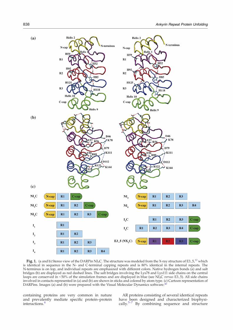
Fig. 1. (a and b) Stereo view of the DARPin NI3C. The structure wasmodeled from the X-ray structure of E3_5,12 whichis identical in sequence in the N- and C-terminal capping repeats and is 80% identical in the internal repeats. TheN-terminus is on top, and individual repeats are emphasized with different colors. Native hydrogen bonds (a) and saltbridges (b) are displayed as red dashed lines. The salt bridges involving the Lys78 and Lys111 side chains on the centralloops are conserved in b50% of the simulation frames and are displayed in blue (see NI3C versus E3_5). All side chainsinvolved in contacts represented in (a) and (b) are shown in sticks and colored by atom type. (c) Cartoon representation ofDARPins. Images (a) and (b) were prepared with the Visual Molecular Dynamics software.40
838 Ankyrin Repeat Protein Unfolding
containing proteins are very common in natureand prevalently mediate specific protein–proteininteractions.4
AR proteins consisting of several identical repeatshave been designed and characterized biophysi-cally.5–7 By combining sequence and structure
839Ankyrin Repeat Protein Unfolding
consensus analyses, an AR module was designedwith seven randomized positions in the loop and inthe first helix.7,8 Different numbers of this modulecould be joined to generate combinatorial librariesof AR proteins. Because of the self-complementarityof consensus repeats, the size of the binding site canbe altered simply by adding or removing repeats.To reduce the solvent exposure of hydrophobicsurfaces, internal modules were flanked by N- andC-terminal “capping” repeats, which were bor-rowed from the guanine-adenine (GA)-bindingprotein9 and slightly modified to approach theconsensus and for cloning purposes.7 Librarymembers were then selected to function as specificbinders or even enzyme inhibitors.7,10,11 DesignedAR proteins (DARPins) were shown experimentallyto be thermodynamically stable, soluble and highlyexpressed in native form in bacteria.7,12
Up to now, only a few studies have been carriedout to gain insight into the folding or unfoldingpathways of AR proteins. The unfolding of the four-repeat tumor suppressor p16INK4a, an inhibitor of acyclin-dependent kinase, starts at the twoN-terminalrepeats, as suggested by mutagenesis experiments13
and as verified by molecular dynamics (MD) simu-lations.14 These repeats deviate more from theconsensus sequence and may thus be intrinsicallyless stable. The role of topology in the energylandscape of AR proteins has recently been investi-gated by a simplified structure-based model.15 Theequilibrium folding behavior16,17 and a kinetic on-pathway intermediate18 have been experimentallycharacterized for the Drosophila Notch receptor andvariants with different numbers of repeats. How-ever, a much more detailed understanding of thefolding andunfoldingmechanism is essential to shedlight on the stabilizing factors of AR proteins,especially in view of the increasing interest in theirapplication in biotechnology.
Table 1. Simulation systems
Proteinstructure
Number ofrepeatsa
Number ofamino acids
Ne(elec
NI1C 3 (1) 90NI2C 4 (2) 123 −NI3C 5 (3) 156 −E3_5 5 (3) 156 −I1 1 (1) 22f
I2 2 (2) 55f
I3 3 (3) 88f
a Total number of repeats and, in parentheses, the number of nonrepeats flank the indicated number of identical consensus repeats. Iindicated number of identical repeats is present. E3_5 is a member ofrepeat, but has identical capping sequences.
b Total net charge of the protein at pH 7.c Initial side length of the cubic box. The box adjusts its volume acco
(see Materials and Methods). A larger water box is used to simulate Nd The total simulation time of the 300-K runs includes the initial 1
Methods).e Two 300-K trajectories of NI1C and two 400-K trajectories of NI1C a
velocities.f The 11 residues preceding the first helix of the first repeat were d
simulations of these proteins.
Here, stabilizing interactions at room temperatureand the unfolding pathways of several AR proteinsand mutants thereof have been investigated by acombination of equilibrium unfolding experimentsand multiple MD runs in explicit water for a totalsimulation time of N2 μs (Table 1). We first addressedthe question of how the number of repeats affectsstability. For this purpose, a full-consensus sequencewith identical repeats was chosen.19 We denote theseproteins as NIxC, where N and C refer to the N- andC-terminal capping repeats, respectively, I refers tothe internal “full”-consensus repeat and the subscriptx gives the number of identical internal-consensusrepeats. Furthermore, the protein E3_5, a member ofthe NX3C library12 (where X denotes a library repeatmodule) with an 80% sequence identity to the full-consensus repeats, was chosen for MD analysis.As mentioned above, the primary structure of
flanking repeats differs from the consensus design.Hence, interrepeat interfaces involving the terminalrepeats are different from interfaces between inter-nal repeats.To understand the role of the capping repeats
in favoring solubility but potentially limiting stability,they were removed both experimentally and in MDsimulations at room temperature and at high tem-perature. Moreover, mutations to further improve thestability of the C-terminal cap were suggested, andthe MD simulations and equilibrium unfoldingexperiments of six NI1C mutants and two NI3Cmutants were performed at room temperature.The aim of the present study, combining simula-
tions and experimental work, is to dissect thearchitecture of ARs to identify mutations that arecritical for stability and to shed light on the role ofthe capping repeats and the relationship betweenstability and number of repeats. Furthermore, by ananalysis of the high-temperature unfolding mecha-nism at the atomic level of detail, weak links may
t chargeb
tron units)Box sizec
(Å)
Simulation timed (ns)
300 K 400 K
−8 65.1 50, 40e 150, 200e
10 80.6 50 100, 150e
12 80.6/99.2 50 10016 80.6/99.2 50 100−1 49.6 40 40−3 55.8 50 150−5 62.0 50 150
capping repeats. In NI1C, NI2C and NI3C, the N- and C-cappingn I1, I2 and I3, these capping repeats are missing, and only thethe NX3C library and differs at about seven positions per internal
rding to the given temperature and pressure during the simulationI3C and E3_5 at 400 K.0 ns that were discarded during the analysis (see Materials and
ndNI2Cwere run with different initial random assignments of the
eleted because large displacements were observed in preliminary
840 Ankyrin Repeat Protein Unfolding
become apparent, in turn helping to understand theexperimental unfolding data and the further designof ankyrins.
Results
Fluctuations and stabilizing interactions at roomtemperature
Comparison with crystallographic B-factors
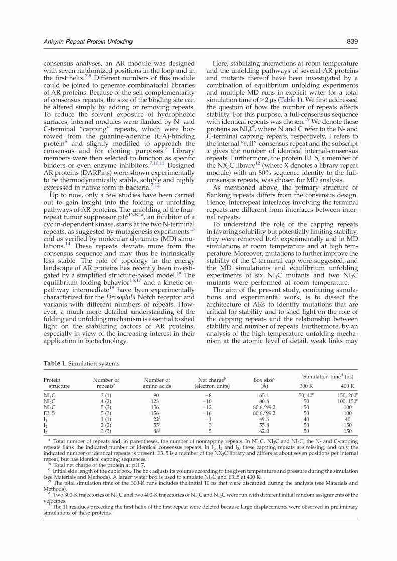
In the loop regions of E3_5, larger fluctuationvalues of Cα atoms were observed along the MDtrajectory at 300 K than those derived from B-factors(Fig. 2a). This discrepancy is probably due to inter-molecular contacts between Loops 1 and 2 and twoneighboring protein molecules in the crystal [ProteinData Bank (PDB) code 1MJ012]. On the other hand,very low B-factors and fluctuations during the MDsimulations characterize the helical and tight-turnregions.
Stabilizing interactions
DARPins are stable at 300 K. The values of theCα root mean square deviation (RMSD) averagedover the interval 10–50 ns are 1.55±0.32 Å, 1.94±0.24 Å, 1.66±0.30 Å and 1.87±0.30 Å for NI1C,NI2C, NI3C and E3_5, respectively. The corre-sponding all-atom RMSDs are 2.43±0.20 Å, 2.70±0.21 Å, 2.38±0.24 Å and 2.59±0.24 Å. Several polarinteractions observed during 300-K runs contributeto the observed stability (see Materials andMethods for the definition of native contacts from
the simulations). Interesting examples are theHis59, His92 and His125 side chains (numberingaccording to PDB file 1MJ0) at the tight turnbetween helices 1 and 2 of each repeat (Fig. 1a),which are involved as donors in hydrogen bondswith carbonyl groups in the C-terminal turn of thefirst helix of the respective preceding repeat. Thesehistidine side chains not only provide an inter-repeat interaction but also contribute to theshielding of the interrepeat interface from thesolvent. Additional hydrogen bonds involve theside chains of His52, His85 and His118, which arepart of a conserved TPLH motif at the beginning ofthe first helix of each internal repeat, and thebackbone carbonyl group of Ala75, Tyr81, Ala108,Tyr114 and Ala141 in the loop of the followingrepeat (Fig. 1a). Furthermore, each of these his-tidines accepts a hydrogen bond from the main-chain NH of the residue n−3 and thereby links twoadjacent repeats. Shielding of these hydrogenbonds from the solvent is provided mainly by theside chain of the tyrosine in the loops of NI3C(positions 48, 81 and 114).
NI3C versus E3_5
In the 300-K simulations, there is a slightly highernumber of hydrogen bonds in the full-consensusprotein NI3C than in the library member E3_5, inparticular in the internal repeats (Table 2), which isconsistent with the smaller fluctuations in NI3C thanin E3_5 (Fig. 2a). Furthermore, the larger number ofcharged residues in NI3C compared to E3_5 (48versus 36) leads to an increased number of saltbridges in NI3C (Table 2 and Fig. 1b). In each
Fig. 2. Cα root mean squarefluctuations (RMSFs) at 300 K. (a)NI3C versus E3_5. Values of RMSFderived from crystallographic B-factors of the E3_5 Cα atoms(PDB file 1MJ0) were calculatedusing the formula RMSFi;exp ¼ffiffiffiffiffiffiffiffiffiffi
38p2 Bi
q, where Bi is the B- factor
of Cα residue i. The interval 10–50 ns of each trajectory was used tocalculate RMSF values. For eachtrajectory, a very similar behavioris observed for the interval 10–50 nsand for the eight 5-ns intervals (notshown). Blue triangles indicate theresidues where E3_5 differs fromNI3C in primary sequence. (b) NI1Cversus I3 (50-ns run). In the 40-nsrun, NI1C has a Cα RMSF sequenceprofile similar to that in the 50-nstrajectory. The amino acids locatedin the helices are represented asfilled circles. Helical segments areemphasized by curled braces belowthe x-axis. h1 to h10 denote helices1–10.
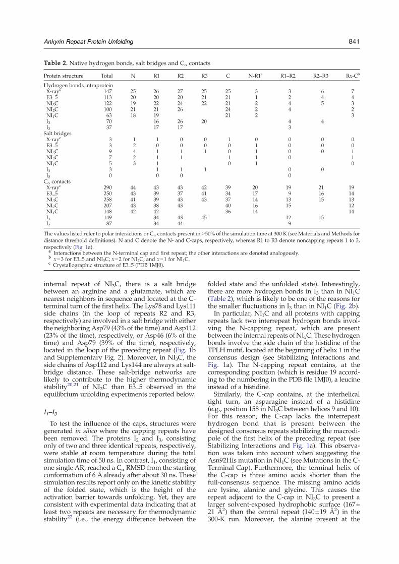
Table 2. Native hydrogen bonds, salt bridges and Cα contacts
Protein structure Total N R1 R2 R3 C N-R1a R1–R2 R2–R3 Rx-Cb
Hydrogen bonds intraproteinX-rayc 147 25 26 27 25 25 3 3 6 7E3_5 113 20 20 20 21 21 1 2 4 4NI3C 122 19 22 24 22 21 2 4 5 3NI2C 100 21 21 26 24 2 4 2NI1C 63 18 19 21 2 3I3 70 16 26 20 4 4I2 37 17 17 3Salt bridgesX-rayc 3 1 1 0 0 1 0 0 0 0E3_5 3 2 0 0 0 0 1 0 0 0NI3C 9 4 1 1 1 0 1 0 0 1NI2C 7 2 1 1 1 1 0 1NI1C 5 3 1 0 1 0I3 3 1 1 1 0 0I2 0 0 0 0Cα contactsX-rayc 290 44 43 43 42 39 20 19 21 19E3_5 250 43 39 37 41 34 17 9 16 14NI3C 258 41 39 43 43 37 14 13 15 13NI2C 207 43 38 43 40 16 15 12NI1C 148 42 42 36 14 14I3 149 34 43 45 12 15I2 87 34 44 9
The values listed refer to polar interactions or Cα contacts present in N50% of the simulation time at 300 K (see Materials andMethods fordistance threshold definitions). N and C denote the N- and C-caps, respectively, whereas R1 to R3 denote noncapping repeats 1 to 3,respectively (Fig. 1a).
a Interactions between the N-terminal cap and first repeat; the other interactions are denoted analogously.b x=3 for E3_5 and NI3C; x=2 for NI2C; and x=1 for NI1C.c Crystallographic structure of E3_5 (PDB 1MJ0).
841Ankyrin Repeat Protein Unfolding
internal repeat of NI3C, there is a salt bridgebetween an arginine and a glutamate, which arenearest neighbors in sequence and located at the C-terminal turn of the first helix. The Lys78 and Lys111side chains (in the loop of repeats R2 and R3,respectively) are involved in a salt bridge with eitherthe neighboring Asp79 (43% of the time) and Asp112(23% of the time), respectively, or Asp46 (6% of thetime) and Asp79 (39% of the time), respectively,located in the loop of the preceding repeat (Fig. 1band Supplementary Fig. 2). Moreover, in NI3C, theside chains of Asp112 and Lys144 are always at salt-bridge distance. These salt-bridge networks arelikely to contribute to the higher thermodynamicstability20,21 of NI3C than E3_5 observed in theequilibrium unfolding experiments reported below.
I1–I3To test the influence of the caps, structures were
generated in silico where the capping repeats havebeen removed. The proteins I2 and I3, consistingonly of two and three identical repeats, respectively,were stable at room temperature during the totalsimulation time of 50 ns. In contrast, I1, consisting ofone single AR, reached a Cα RMSD from the startingconformation of 6 Å already after about 30 ns. Thesesimulation results report only on the kinetic stabilityof the folded state, which is the height of theactivation barrier towards unfolding. Yet, they areconsistent with experimental data indicating that atleast two repeats are necessary for thermodynamicstability22 (i.e., the energy difference between the
folded state and the unfolded state). Interestingly,there are more hydrogen bonds in I3 than in NI1C(Table 2), which is likely to be one of the reasons forthe smaller fluctuations in I3 than in NI1C (Fig. 2b).In particular, NI1C and all proteins with capping
repeats lack two interrepeat hydrogen bonds invol-ving the N-capping repeat, which are presentbetween the internal repeats of NIxC. These hydrogenbonds involve the side chain of the histidine of theTPLH motif, located at the beginning of helix 1 in theconsensus design (see Stabilizing Interactions andFig. 1a). The N-capping repeat contains, at thecorresponding position (which is residue 19 accord-ing to the numbering in the PDB file 1MJ0), a leucineinstead of a histidine.Similarly, the C-cap contains, at the interhelical
tight turn, an asparagine instead of a histidine(e.g., position 158 in NI3C between helices 9 and 10).For this reason, the C-cap lacks the interrepeathydrogen bond that is present between thedesigned consensus repeats stabilizing the macrodi-pole of the first helix of the preceding repeat (seeStabilizing Interactions and Fig. 1a). This observa-tion was taken into account when suggesting theAsn92His mutation in NI1C (see Mutations in the C-Terminal Cap). Furthermore, the terminal helix ofthe C-cap is three amino acids shorter than thefull-consensus sequence. The missing amino acidsare lysine, alanine and glycine. This causes therepeat adjacent to the C-cap in NI3C to present alarger solvent-exposed hydrophobic surface (167±21 Å2) than the central repeat (140±19 Å2) in the300-K run. Moreover, the alanine present at the
842 Ankyrin Repeat Protein Unfolding
C-terminal end of the full-consensus repeatsincreases the helical propensity of the C-terminalhelix. This evidence was considered when suggest-ing a Lys-Ala-Ala extension of the C-cap terminalhelix in NI1C (see below).
Structural stability and high-temperatureunfolding mechanism
Correlation between structural stability and numberof repeats
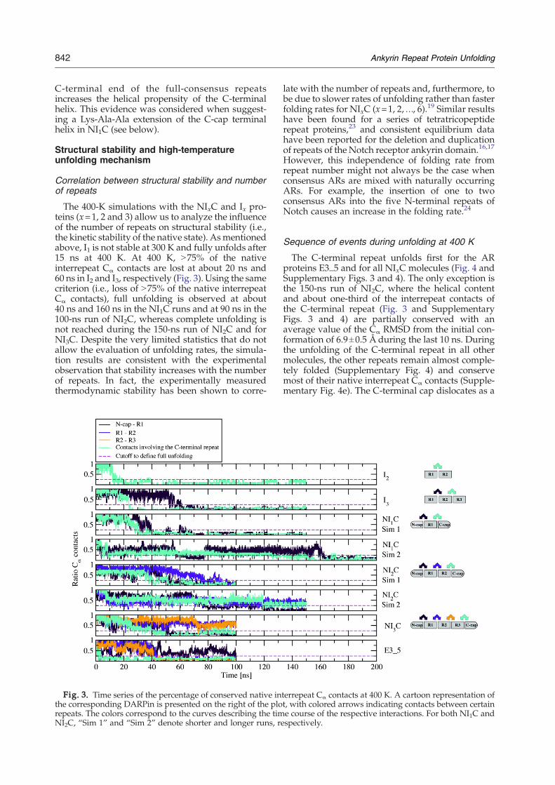
The 400-K simulations with the NIxC and Ix pro-teins (x=1, 2 and 3) allow us to analyze the influenceof the number of repeats on structural stability (i.e.,the kinetic stability of the native state). Asmentionedabove, I1 is not stable at 300 K and fully unfolds after15 ns at 400 K. At 400 K, N75% of the nativeinterrepeat Cα contacts are lost at about 20 ns and60 ns in I2 and I3, respectively (Fig. 3). Using the samecriterion (i.e., loss of N75% of the native interrepeatCα contacts), full unfolding is observed at about40 ns and 160 ns in the NI1C runs and at 90 ns in the100-ns run of NI2C, whereas complete unfolding isnot reached during the 150-ns run of NI2C and forNI3C. Despite the very limited statistics that do notallow the evaluation of unfolding rates, the simula-tion results are consistent with the experimentalobservation that stability increases with the numberof repeats. In fact, the experimentally measuredthermodynamic stability has been shown to corre-
Fig. 3. Time series of the percentage of conserved native inthe corresponding DARPin is presented on the right of the plorepeats. The colors correspond to the curves describing the timNI2C, “Sim 1” and “Sim 2” denote shorter and longer runs, re
late with the number of repeats and, furthermore, tobe due to slower rates of unfolding rather than fasterfolding rates for NIxC (x=1, 2,…, 6).19 Similar resultshave been found for a series of tetratricopeptiderepeat proteins,23 and consistent equilibrium datahave been reported for the deletion and duplicationof repeats of the Notch receptor ankyrin domain.16,17
However, this independence of folding rate fromrepeat number might not always be the case whenconsensus ARs are mixed with naturally occurringARs. For example, the insertion of one to twoconsensus ARs into the five N-terminal repeats ofNotch causes an increase in the folding rate.24
Sequence of events during unfolding at 400 K
The C-terminal repeat unfolds first for the ARproteins E3_5 and for all NIxC molecules (Fig. 4 andSupplementary Figs. 3 and 4). The only exception isthe 150-ns run of NI2C, where the helical contentand about one-third of the interrepeat contacts ofthe C-terminal repeat (Fig. 3 and SupplementaryFigs. 3 and 4) are partially conserved with anaverage value of the Cα RMSD from the initial con-formation of 6.9±0.5 Å during the last 10 ns. Duringthe unfolding of the C-terminal repeat in all othermolecules, the other repeats remain almost comple-tely folded (Supplementary Fig. 4) and conservemost of their native interrepeat Cα contacts (Supple-mentary Fig. 4e). The C-terminal cap dislocates as a
terrepeat Cα contacts at 400 K. A cartoon representation oft, with colored arrows indicating contacts between certaine course of the respective interactions. For both NI1C andspectively.
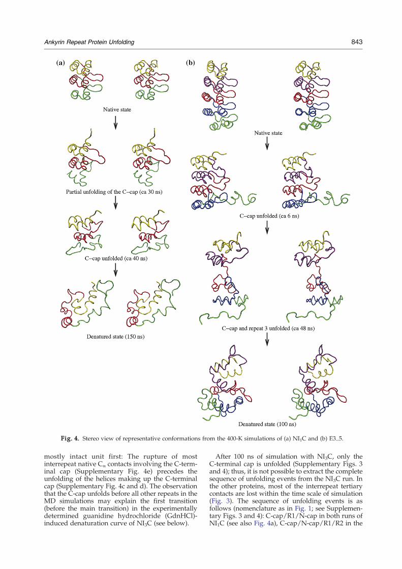
Fig. 4. Stereo view of representative conformations from the 400-K simulations of (a) NI1C and (b) E3_5.
843Ankyrin Repeat Protein Unfolding
mostly intact unit first: The rupture of mostinterrepeat native Cα contacts involving the C-term-inal cap (Supplementary Fig. 4e) precedes theunfolding of the helices making up the C-terminalcap (Supplementary Fig. 4c and d). The observationthat the C-cap unfolds before all other repeats in theMD simulations may explain the first transition(before the main transition) in the experimentallydetermined guanidine hydrochloride (GdnHCl)-induced denaturation curve of NI3C (see below).
After 100 ns of simulation with NI3C, only theC-terminal cap is unfolded (Supplementary Figs. 3and 4); thus, it is not possible to extract the completesequence of unfolding events from the NI3C run. Inthe other proteins, most of the interrepeat tertiarycontacts are lost within the time scale of simulation(Fig. 3). The sequence of unfolding events is asfollows (nomenclature as in Fig. 1; see Supplemen-tary Figs. 3 and 4): C-cap/R1/N-cap in both runs ofNI1C (see also Fig. 4a), C-cap/N-cap/R1/R2 in the
844 Ankyrin Repeat Protein Unfolding
100-ns run of NI2C (as mentioned above, in the 150-ns run of NI2C, the helical content was partiallyconserved) and C-cap/R2/R3/R1/N-cap in E3_5(see also Fig. 4b). The unfolding of the central repeatR2 of E3_5 is preceded by the rupture of thehydrophobic interface between repeats R1 and R2,as indicated by the decrease of their native inter-repeat Cα contacts (turquoise line in Fig. 3). The factthat the unfolding of repeat R2 of E3_5 directlyfollows the denaturation of the C-cap is consistentwith the smaller number of native interrepeat hy-drogen bonds between repeats R1 and R2 of E3_5,compared to NI3C (Table 2), and to the relativelysmall number of native Cα contacts between repeatsR1 and R2 of E3_5 (Table 2). Moreover, the fact thatsome internal repeats (e.g., repeat R2 of E3_5 andrepeat R1 of NI1C) unfold before the N-terminal capprovides evidence that the very high temperature(400 K) does not lead to artificial deformations atthe protein surface in the simulations. Thus, it canbe excluded that the observed early unfolding of theC-cap in the simulations is an artifact caused by thehigh temperature. Interestingly, the N-terminal capseems to be more stable than the C-terminal cap,which is consistent with the fact that the N-terminalcap is more similar to the consensus repeat.
Denatured state
During the interval 155–200 ns in one of the twounfolding runs ofNI1C, helix 2 in theN-capping repeatelongates up to residue 47,withπ-helical turns at its C-terminal region (Supplementary Fig. 3). The sameelongation of helix 2 in the N-capping repeat takesplace for NI3C, where otherwise only the C-terminalcapping repeat unfolded during a total simulationtime of 100 ns (Supplementary Fig. 3). Nonnative π-helical structure is also present at residues 126–146 ofE3_5 (Supplementary Fig. 3). Similarly, at the end ofone of the two unfolding runs of NI2C, residues 107–121 form a nonnative α-helical structure.
Experimental studies on the equilibriumunfolding of NI3C and variants without cappingrepeats
Previously, the stability of several DARPins hasbeen measured by both GdnHCl and thermal dena-turation.7,12 These proteins were unselected mem-bers of the DARPin library. They were all highlystable, even though some differences betweenindividual library members can be noted. Therewas a general trend towards higher stability with anincreasing number of repeats. However, since theindividual library members of the same lengthcovered a range of stabilities,25 a quantitative rela-tionship could not be established. Most of theproteins tested have previously shown highlycooperative reversible transitions that were consis-tent with a two-state equilibrium system.The full-consensus proteins are even more stable,
and the details of the dependence of folding andunfolding rates on the number of repeats in the
protein are reported in the accompanying ma-nuscript.19 By the design of the consensus sequence,the previously variable library positions were nowchosen according to the most frequent residues,which turn out to be charged or polar; a possiblereason for the stability is that additional favorableelectrostatic interactions are formed (see NI3C versusE3_5).When the full-consensus protein NI3C is com-
pared to E3_5, a member of the NX3C library,two differences in GdnHCl equilibrium denatura-tion experiments are apparent (Fig. 5). First, themain transition is shifted to higher GdnHCl (by0.8 M). Second, the denaturation is no more fullycooperative and is not consistent with a two-stateequilibrium system. Instead, there is a small ”pre-transition” visible at 3.7 M GdnHCl, before the maintransition, which occurs at about 5.6 M GdnHCl.The two transitions can be well described by a se-quential three-state model, and the calculated ΔG(19.7 kcal/mol) is about double that obtained forE3_5 (11.2 kcal/mol; Fig. 5a).A possible explanation is that the higher stability
of the central domains in NI3C uncouples theunfolding of one or both of the capping repeats,which may therefore unfold already at a lowerdenaturant concentration and give rise to an equi-librium intermediate with one or both of the capsdetached from the central repeats. At this inter-mediate state, a portion of the circular dichroism(CD) signal is lost. This explanation is alsosupported by the unfolding behavior of all NIxCproteins and E3_5 in the MD simulations where theC-cap unfolds prior to the other repeats.To test this hypothesis, additional proteins, which
were devoid of one or both of the capping repeats,were constructed. We denote them NI3 or NI4, toindicate that they have only the N-cap and three orfour full-consensus repeats, respectively, and I3Cand I4C, to indicate that they carry only the C-capand the number of consensus repeats indicated bythe number. Finally, we also created the moleculeswithout any caps, which consist only of theconsensus repeats and are named I3 and I4 (seeMaterials and Methods and Supplementary Fig. 1for the definition of the respective sequences).It is immediately apparent that the molecules
lacking both N- and C-terminal caps show signifi-cant amounts of insoluble protein upon expressionin Escherichia coli (Fig. 6), in contrast to E3_5 andNI3C, which are completely soluble. The moleculeslacking only one of the capping repeats could bepurified from the soluble fraction and were furtheranalyzed using multiangle light scattering (MALS).A portion of NI3 and NI4 precipitated after elutionfrom the Ni–NTA column, while I3C and I4Cremained soluble. MALS analysis showed that theproteins I3C and I4C form soluble aggregates,however (data not shown). In contrast, the solubleportion of the proteins NI3 and NI4, lacking theC-terminal cap, remains mainly monomeric. How-ever, these proteins do remain aggregation-prone, asthey aggregate at intermediate concentrations of
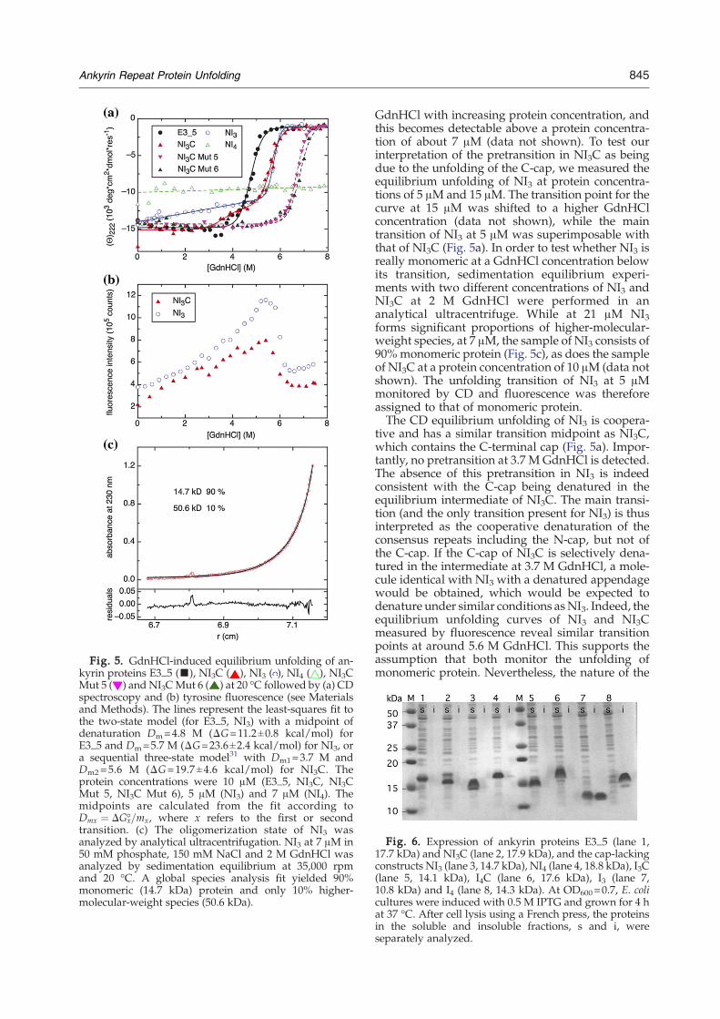
Fig. 5. GdnHCl-induced equilibrium unfolding of an-kyrin proteins E3_5 (▪), NI3C ( ), NI3 ( ), NI4 ( ), NI3CMut 5 ( ) and NI3CMut 6 ( ) at 20 °C followed by (a) CDspectroscopy and (b) tyrosine fluorescence (see Materialsand Methods). The lines represent the least-squares fit tothe two-state model (for E3_5, NI3) with a midpoint ofdenaturation Dm=4.8 M (ΔG=11.2±0.8 kcal/mol) forE3_5 and Dm=5.7 M (ΔG=23.6±2.4 kcal/mol) for NI3, ora sequential three-state model31 with Dm1=3.7 M andDm2=5.6 M (ΔG=19.7±4.6 kcal/mol) for NI3C. Theprotein concentrations were 10 μM (E3_5, NI3C, NI3CMut 5, NI3C Mut 6), 5 μM (NI3) and 7 μM (NI4). Themidpoints are calculated from the fit according toDmx ¼ DGx-=mx, where x refers to the first or secondtransition. (c) The oligomerization state of NI3 wasanalyzed by analytical ultracentrifugation. NI3 at 7 μM in50 mM phosphate, 150 mM NaCl and 2 M GdnHCl wasanalyzed by sedimentation equilibrium at 35,000 rpmand 20 °C. A global species analysis fit yielded 90%monomeric (14.7 kDa) protein and only 10% higher-molecular-weight species (50.6 kDa).
845Ankyrin Repeat Protein Unfolding
GdnHCl with increasing protein concentration, andthis becomes detectable above a protein concentra-tion of about 7 μM (data not shown). To test ourinterpretation of the pretransition in NI3C as beingdue to the unfolding of the C-cap, we measured theequilibrium unfolding of NI3 at protein concentra-tions of 5 μM and 15 μM. The transition point for thecurve at 15 μM was shifted to a higher GdnHClconcentration (data not shown), while the maintransition of NI3 at 5 μM was superimposable withthat of NI3C (Fig. 5a). In order to test whether NI3 isreally monomeric at a GdnHCl concentration belowits transition, sedimentation equilibrium experi-ments with two different concentrations of NI3 andNI3C at 2 M GdnHCl were performed in ananalytical ultracentrifuge. While at 21 μM NI3forms significant proportions of higher-molecular-weight species, at 7 μM, the sample of NI3 consists of90% monomeric protein (Fig. 5c), as does the sampleof NI3C at a protein concentration of 10 μM (data notshown). The unfolding transition of NI3 at 5 μMmonitored by CD and fluorescence was thereforeassigned to that of monomeric protein.The CD equilibrium unfolding of NI3 is coopera-
tive and has a similar transition midpoint as NI3C,which contains the C-terminal cap (Fig. 5a). Impor-tantly, no pretransition at 3.7 M GdnHCl is detected.The absence of this pretransition in NI3 is indeedconsistent with the C-cap being denatured in theequilibrium intermediate of NI3C. The main transi-tion (and the only transition present for NI3) is thusinterpreted as the cooperative denaturation of theconsensus repeats including the N-cap, but not ofthe C-cap. If the C-cap of NI3C is selectively dena-tured in the intermediate at 3.7 M GdnHCl, a mole-cule identical with NI3 with a denatured appendagewould be obtained, which would be expected todenature under similar conditions asNI3. Indeed, theequilibrium unfolding curves of NI3 and NI3Cmeasured by fluorescence reveal similar transitionpoints at around 5.6 M GdnHCl. This supports theassumption that both monitor the unfolding ofmonomeric protein. Nevertheless, the nature of the
Fig. 6. Expression of ankyrin proteins E3_5 (lane 1,17.7 kDa) and NI3C (lane 2, 17.9 kDa), and the cap-lackingconstructs NI3 (lane 3, 14.7 kDa), NI4 (lane 4, 18.8 kDa), I3C(lane 5, 14.1 kDa), I4C (lane 6, 17.6 kDa), I3 (lane 7,10.8 kDa) and I4 (lane 8, 14.3 kDa). At OD600=0.7, E. colicultures were induced with 0.5 M IPTG and grown for 4 hat 37 °C. After cell lysis using a French press, the proteinsin the soluble and insoluble fractions, s and i, wereseparately analyzed.
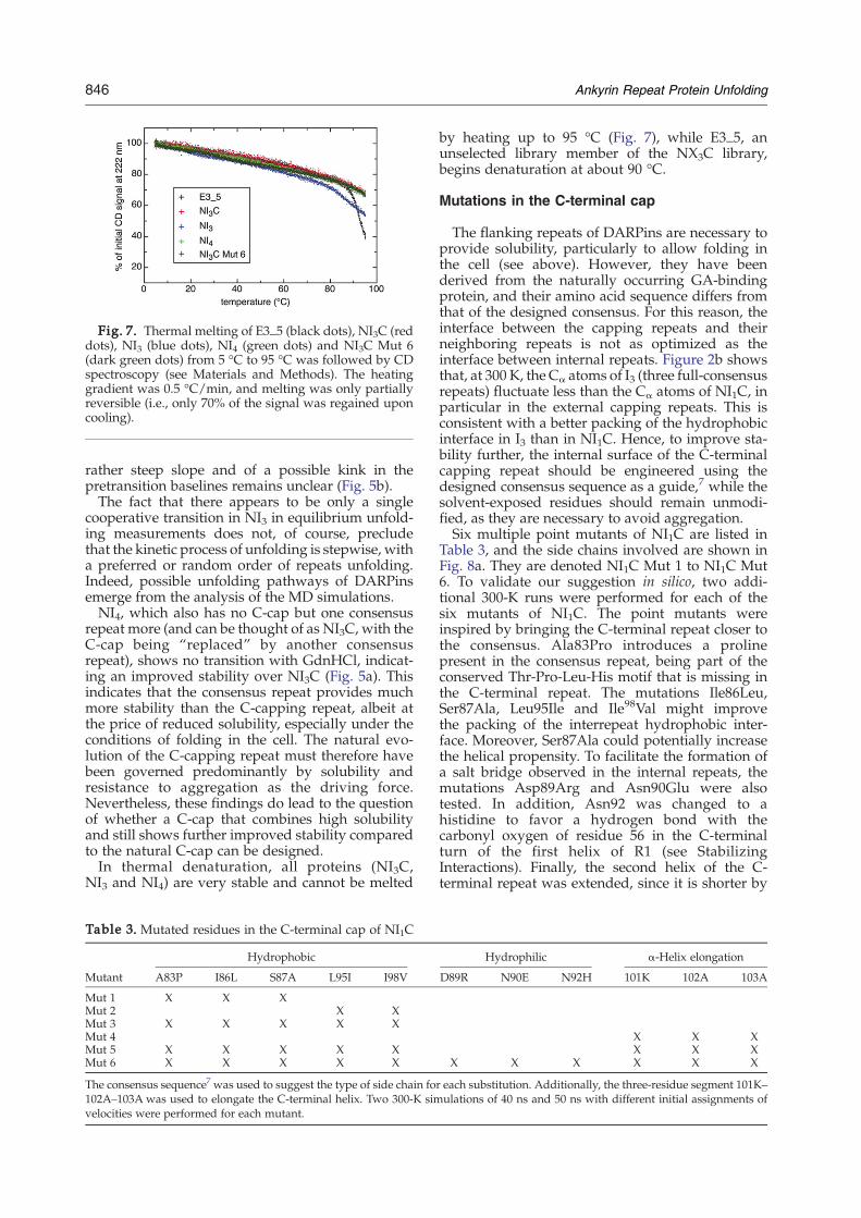
Fig. 7. Thermal melting of E3_5 (black dots), NI3C (reddots), NI3 (blue dots), NI4 (green dots) and NI3C Mut 6(dark green dots) from 5 °C to 95 °C was followed by CDspectroscopy (see Materials and Methods). The heatinggradient was 0.5 °C/min, and melting was only partiallyreversible (i.e., only 70% of the signal was regained uponcooling).
846 Ankyrin Repeat Protein Unfolding
rather steep slope and of a possible kink in thepretransition baselines remains unclear (Fig. 5b).The fact that there appears to be only a single
cooperative transition in NI3 in equilibrium unfold-ing measurements does not, of course, precludethat the kinetic process of unfolding is stepwise, witha preferred or random order of repeats unfolding.Indeed, possible unfolding pathways of DARPinsemerge from the analysis of the MD simulations.NI4, which also has no C-cap but one consensus
repeat more (and can be thought of as NI3C, with theC-cap being “replaced” by another consensusrepeat), shows no transition with GdnHCl, indicat-ing an improved stability over NI3C (Fig. 5a). Thisindicates that the consensus repeat provides muchmore stability than the C-capping repeat, albeit atthe price of reduced solubility, especially under theconditions of folding in the cell. The natural evo-lution of the C-capping repeat must therefore havebeen governed predominantly by solubility andresistance to aggregation as the driving force.Nevertheless, these findings do lead to the questionof whether a C-cap that combines high solubilityand still shows further improved stability comparedto the natural C-cap can be designed.In thermal denaturation, all proteins (NI3C,
NI3 and NI4) are very stable and cannot be melted
Table 3. Mutated residues in the C-terminal cap of NI1C
Mutant
Hydrophobic
A83P I86L S87A L95I I98V
Mut 1 X X XMut 2 X XMut 3 X X X X XMut 4Mut 5 X X X X XMut 6 X X X X X
The consensus sequence7 was used to suggest the type of side chain fo102A–103A was used to elongate the C-terminal helix. Two 300-K simvelocities were performed for each mutant.
by heating up to 95 °C (Fig. 7), while E3_5, anunselected library member of the NX3C library,begins denaturation at about 90 °C.
Mutations in the C-terminal cap
The flanking repeats of DARPins are necessary toprovide solubility, particularly to allow folding inthe cell (see above). However, they have beenderived from the naturally occurring GA-bindingprotein, and their amino acid sequence differs fromthat of the designed consensus. For this reason, theinterface between the capping repeats and theirneighboring repeats is not as optimized as theinterface between internal repeats. Figure 2b showsthat, at 300 K, the Cα atoms of I3 (three full-consensusrepeats) fluctuate less than the Cα atoms of NI1C, inparticular in the external capping repeats. This isconsistent with a better packing of the hydrophobicinterface in I3 than in NI1C. Hence, to improve sta-bility further, the internal surface of the C-terminalcapping repeat should be engineered using thedesigned consensus sequence as a guide,7 while thesolvent-exposed residues should remain unmodi-fied, as they are necessary to avoid aggregation.Six multiple point mutants of NI1C are listed in
Table 3, and the side chains involved are shown inFig. 8a. They are denoted NI1C Mut 1 to NI1C Mut6. To validate our suggestion in silico, two addi-tional 300-K runs were performed for each of thesix mutants of NI1C. The point mutants wereinspired by bringing the C-terminal repeat closer tothe consensus. Ala83Pro introduces a prolinepresent in the consensus repeat, being part of theconserved Thr-Pro-Leu-His motif that is missing inthe C-terminal repeat. The mutations Ile86Leu,Ser87Ala, Leu95Ile and Ile98Val might improvethe packing of the interrepeat hydrophobic inter-face. Moreover, Ser87Ala could potentially increasethe helical propensity. To facilitate the formation ofa salt bridge observed in the internal repeats, themutations Asp89Arg and Asn90Glu were alsotested. In addition, Asn92 was changed to ahistidine to favor a hydrogen bond with thecarbonyl oxygen of residue 56 in the C-terminalturn of the first helix of R1 (see StabilizingInteractions). Finally, the second helix of the C-terminal repeat was extended, since it is shorter by
Hydrophilic α-Helix elongation
D89R N90E N92H 101K 102A 103A
X X XX X X
X X X X X X
r each substitution. Additionally, the three-residue segment 101K–ulations of 40 ns and 50 ns with different initial assignments of
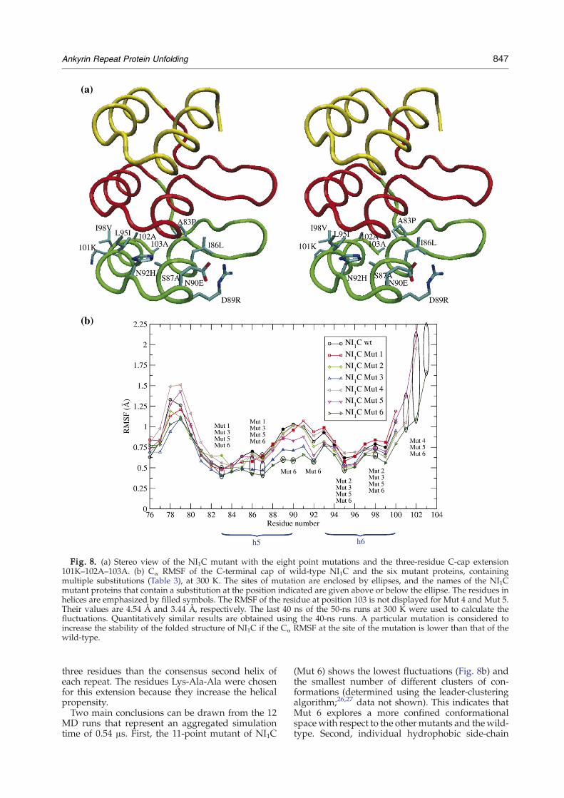
Fig. 8. (a) Stereo view of the NI1C mutant with the eight point mutations and the three-residue C-cap extension101K–102A–103A. (b) Cα RMSF of the C-terminal cap of wild-type NI1C and the six mutant proteins, containingmultiple substitutions (Table 3), at 300 K. The sites of mutation are enclosed by ellipses, and the names of the NI1Cmutant proteins that contain a substitution at the position indicated are given above or below the ellipse. The residues inhelices are emphasized by filled symbols. The RMSF of the residue at position 103 is not displayed for Mut 4 and Mut 5.Their values are 4.54 Å and 3.44 Å, respectively. The last 40 ns of the 50-ns runs at 300 K were used to calculate thefluctuations. Quantitatively similar results are obtained using the 40-ns runs. A particular mutation is considered toincrease the stability of the folded structure of NI1C if the Cα RMSF at the site of the mutation is lower than that of thewild-type.
847Ankyrin Repeat Protein Unfolding
three residues than the consensus second helix ofeach repeat. The residues Lys-Ala-Ala were chosenfor this extension because they increase the helicalpropensity.Two main conclusions can be drawn from the 12
MD runs that represent an aggregated simulationtime of 0.54 μs. First, the 11-point mutant of NI1C
(Mut 6) shows the lowest fluctuations (Fig. 8b) andthe smallest number of different clusters of con-formations (determined using the leader-clusteringalgorithm;26,27 data not shown). This indicates thatMut 6 explores a more confined conformationalspace with respect to the othermutants and thewild-type. Second, individual hydrophobic side-chain
848 Ankyrin Repeat Protein Unfolding
replacements do not contribute significantly to thestructural stability of the folded structure (Table 4).On the other hand, the mutations Asp89Arg andAsn90Glu together seem to be favorable, as theyallow the Arg89–Glu90 salt bridge to form. In fact,the Arg89–Glu90 salt bridge is present in 84% of allframes sampled during the intervals 10–40 ns and10–50 ns of the two 300-K simulations with Mut 6.To test these suggestions, six mutants of NI1C (see
SupplementaryData), which contain the sixmultiplepoint mutations, were constructed (Table 3) in theC-terminal capping repeat. Expression inE. coli led tocompletely soluble proteins for all six mutants, andMALS analysis showed that all of the purifiedproteins are monomeric (data not shown). The sta-bilities of the six mutants were compared to thewild-type protein NI1C using thermal denaturationexperiments (Fig. 9a). While Mut 2 shows a Tm=57 °C that is slightly below the transition midpointof NI1C wild-type (i.e., Tm=60 °C), the othermutants show increased Tm values in the followingorder: Mut 4 with Tm=64 °C, Mut 1 and Mut 3 withTm=68 °C, and Mut 5 and Mut 6 with 77 °C.These results validate the hypotheses that repla-
cing hydrophobic residues in the interrepeat inter-face with those present in the consensus sequenceand elongation of the C-terminal helix can furtherimprove the stability of DARPins. Indeed, Mut 1and Mut 3, which present mutations of hydro-phobic residues but no helix elongation, have anincreased melting point of 8 degrees. On the otherhand, Mut 4, which differs from the wild-type onlyby the elongation of the helix, shows a 4 degreesincrease in stability. When mutations of hydro-phobic residues and elongation of the C-terminalhelix are combined, as in Mut 5 and Mut 6, an evenlarger increase in the melting point (17 degrees intotal) is observed. However, mutations of hydro-phobic residues in the C-terminal helix (i.e., L95Iand I98V) cause a slight destabilization (the Tm of
Table 4. Side-chain contributions to the energy difference bet
Mutation
Side chain/systema
Total van der Waa
HydrophobicA83P −3±2 (−5±1) −4±1 (−4±1I86L −1±1 (−13±1) −1±1 (−12±S87A 11±1 (−16±3) 1±1 (−6±1L95I −1±1 (−13±1) −1±1 (−12±I98V 2±1 (−11±2) −1±1 (−11±PolarD89R 58±40 (−156±46) −11±2 (2±3)N90E −147±37 (−30±6) 6±3 (−7±2N92H −6±7 (−28±6) −3±2 (−8±2
All values are expressed in kilocalories per mole. A negative value indicare the energy difference between Mut 6 and wild-type, while wild-tyOnly values of Mut 6 are shown (averages over the intervals 10–40 ns aall substitutions. The results obtained with the other mutants for the sastandard deviation of those reported here for Mut 6.
a Total and van der Waals energies are calculated for a mutated sidexcluding the backbone of the mutated side chain.
b Total and van der Waals energies are calculated for a mutated sidmutated side chain.
Mut 2 is 3 degrees less than for the wild-type),while these mutations have essentially no effect ifthey are combined with mutations in the first helix(Mut 3 and Mut 1 behave almost identically).Furthermore, the hypothesis of increased stabilityby additional electrostatic interactions (i.e., a saltbridge between the side chains at positions 89 and90, and an interrepeat hydrogen bond involving ahistidine at position 92) could not be confirmed. Infact, Mut 5 shows the same transition midpoint asMut 6, although Mut 6 differs from Mut 5 by theadditional mutations at positions 89, 90 and 92.In summary, the stabilizing effect of the 11
mutations present in Mut 6 seems to be largelycaused by six of them: the three present in Mut 1,which all bring the C-cap closer to the consensus(A83P, introducing a conserved Pro; I86L and S87A,increasing the helical propensity), as well as thosepresent in Mut 4 (the extension of the second helixby Lys-Ala-Ala).Mut 5 andMut 6 were chosen for GdnHCl equilib-
rium unfolding measured by CD (Fig. 9b). The tran-sitions of both mutants have the samemidpoint, andthey are cooperative, consistent with a two-statemodel. The calculated ΔG value (7.8 kcal/mol) ismore than double that obtained for NI1C wild-type(3.7 kcal/mol) and is 85% of the value for the four-repeat molecule NI2C containing the wild-typeC-cap.19Because of the cooperative nature of NI1C folding,
this small protein serves well to quantify the effectsof the stabilizing cap mutations. However, we alsowished to test their contributions in the context of thelarger NI3C, to test their effect on the pretransitionand the main transition. Therefore, in addition tointroducing the C-cap mutations in NI1C, they werealso introduced in NI3C. NI3CMut 5 andNI3CMut 6are fully soluble, as are all the other mutants. How-ever, NI3C Mut 5 and NI3C Mut 6 are a mixture ofmonomer and dimer (with about 15% dimer) at
ween Mut 6 and wild-type
Side chain/proteinb
ls Total van der Waals
) −6±2 (−4±1) −8±1 (−4±1)1) −3±1 (−10±1) −2±1 (−10±1)) 11±1 (−16±2) 0±1 (−5±1)1) −1±1 (−12±1) −1±1 (−11±1)1) −0±1 (−7±2) 1±1 (−8±1)
−34±20 (−17±23) −2±2 (−3±1)) −52±24 (−9±4) 1±2 (−6±1)) −8±3 (−10±3) −2±1 (−7±1)
ates that the mutation is favorable. Values outside the parenthesespe energy is given inside the parentheses.nd 10–50 ns of the two 300-K simulations), as this mutant containsme substitutions are not shown because their values fall within the
e chain and the rest of the system (i.e., protein, ions and water),
e chain and the rest of the protein, excluding the backbone of the
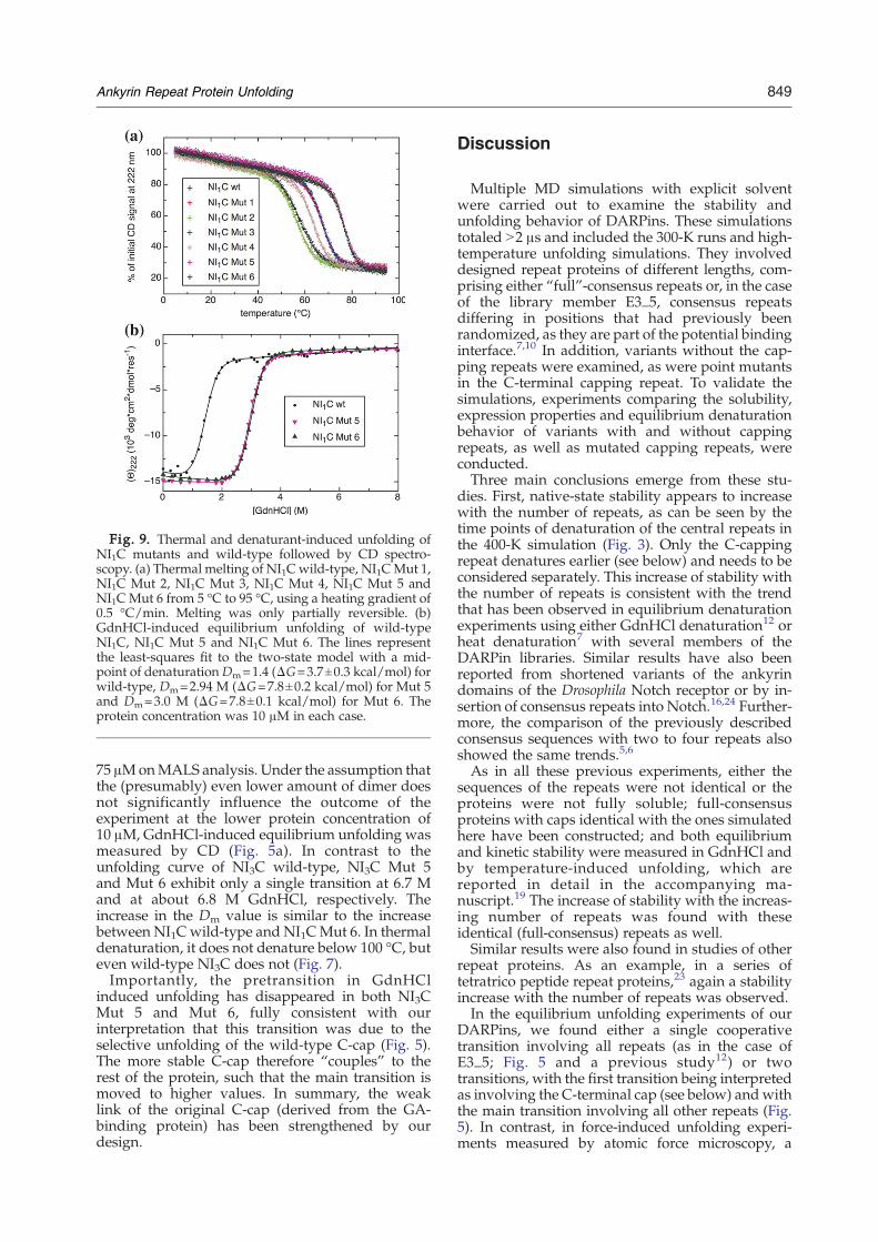
Fig. 9. Thermal and denaturant-induced unfolding ofNI1C mutants and wild-type followed by CD spectro-scopy. (a) Thermal melting of NI1Cwild-type, NI1CMut 1,NI1C Mut 2, NI1C Mut 3, NI1C Mut 4, NI1C Mut 5 andNI1C Mut 6 from 5 °C to 95 °C, using a heating gradient of0.5 °C/min. Melting was only partially reversible. (b)GdnHCl-induced equilibrium unfolding of wild-typeNI1C, NI1C Mut 5 and NI1C Mut 6. The lines representthe least-squares fit to the two-state model with a mid-point of denaturationDm=1.4 (ΔG=3.7±0.3 kcal/mol) forwild-type, Dm=2.94 M (ΔG=7.8±0.2 kcal/mol) for Mut 5and Dm=3.0 M (ΔG=7.8±0.1 kcal/mol) for Mut 6. Theprotein concentration was 10 μM in each case.
849Ankyrin Repeat Protein Unfolding
75 μMonMALS analysis. Under the assumption thatthe (presumably) even lower amount of dimer doesnot significantly influence the outcome of theexperiment at the lower protein concentration of10 μM, GdnHCl-induced equilibrium unfolding wasmeasured by CD (Fig. 5a). In contrast to theunfolding curve of NI3C wild-type, NI3C Mut 5and Mut 6 exhibit only a single transition at 6.7 Mand at about 6.8 M GdnHCl, respectively. Theincrease in the Dm value is similar to the increasebetween NI1C wild-type and NI1CMut 6. In thermaldenaturation, it does not denature below 100 °C, buteven wild-type NI3C does not (Fig. 7).Importantly, the pretransition in GdnHCl
induced unfolding has disappeared in both NI3CMut 5 and Mut 6, fully consistent with ourinterpretation that this transition was due to theselective unfolding of the wild-type C-cap (Fig. 5).The more stable C-cap therefore “couples” to therest of the protein, such that the main transition ismoved to higher values. In summary, the weaklink of the original C-cap (derived from the GA-binding protein) has been strengthened by ourdesign.
Discussion
Multiple MD simulations with explicit solventwere carried out to examine the stability andunfolding behavior of DARPins. These simulationstotaled N2 μs and included the 300-K runs and high-temperature unfolding simulations. They involveddesigned repeat proteins of different lengths, com-prising either “full”-consensus repeats or, in the caseof the library member E3_5, consensus repeatsdiffering in positions that had previously beenrandomized, as they are part of the potential bindinginterface.7,10 In addition, variants without the cap-ping repeats were examined, as were point mutantsin the C-terminal capping repeat. To validate thesimulations, experiments comparing the solubility,expression properties and equilibrium denaturationbehavior of variants with and without cappingrepeats, as well as mutated capping repeats, wereconducted.Three main conclusions emerge from these stu-
dies. First, native-state stability appears to increasewith the number of repeats, as can be seen by thetime points of denaturation of the central repeats inthe 400-K simulation (Fig. 3). Only the C-cappingrepeat denatures earlier (see below) and needs to beconsidered separately. This increase of stability withthe number of repeats is consistent with the trendthat has been observed in equilibrium denaturationexperiments using either GdnHCl denaturation12 orheat denaturation7 with several members of theDARPin libraries. Similar results have also beenreported from shortened variants of the ankyrindomains of the Drosophila Notch receptor or by in-sertion of consensus repeats into Notch.16,24 Further-more, the comparison of the previously describedconsensus sequences with two to four repeats alsoshowed the same trends.5,6
As in all these previous experiments, either thesequences of the repeats were not identical or theproteins were not fully soluble; full-consensusproteins with caps identical with the ones simulatedhere have been constructed; and both equilibriumand kinetic stability were measured in GdnHCl andby temperature-induced unfolding, which arereported in detail in the accompanying ma-nuscript.19 The increase of stability with the increas-ing number of repeats was found with theseidentical (full-consensus) repeats as well.Similar results were also found in studies of other
repeat proteins. As an example, in a series oftetratrico peptide repeat proteins,23 again a stabilityincrease with the number of repeats was observed.In the equilibrium unfolding experiments of our
DARPins, we found either a single cooperativetransition involving all repeats (as in the case ofE3_5; Fig. 5 and a previous study12) or twotransitions, with the first transition being interpretedas involving the C-terminal cap (see below) andwiththe main transition involving all other repeats (Fig.5). In contrast, in force-induced unfolding experi-ments measured by atomic force microscopy, a
850 Ankyrin Repeat Protein Unfolding
sequential unfolding was observed.28 This differ-ence in unfolding behavior is not surprising, asforce-induced unfolding imposes a particular direc-tion on the unfolding trajectory. Furthermore, theforce-induced unfolding is a kinetic experiment,while the solution experiments described here havebeen equilibrium experiments. A stepwise unfold-ing is observed in the high-temperature unfoldingsimulations described here. This is not at variancewith experimental results. In the kinetic unfolding ofa NX1C library member protein, no intermediatewas detected at 5 °C, but differential scanning calo-rimetry experiments revealed a deviation from atwo-state model at higher temperatures.29 Also, theinterpretation of the equilibrium and kinetic unfold-ing data of the full consensus proteins19 are con-sistent with folding models different from 2-state.The sequence of unfolding events observed in the
high-temperature simulations here is not at variancewith Go-type simulations of the DARPin E3_5.15
That study suggests that folding of E3_5 starts withthe formation of the N-cap and propagates sequen-tially through neighboring repeats to the C-cap.However, in the unfolding simulation presentedhere, R3 unfolds after R2. This discrepancy could bedue to the limited statistics (only one run with E3_5),the fact that unfolding might follow a slightlydifferent pathway than folding or the presence ofmultiple folding pathways that are not detected bythe simplified model used in that study.15 Interest-ingly, that same study suggests that folding of theNotch receptor starts at the second or the sixth AR.15
This disagrees with recent mutagenesis experimentsshowing that folding of the Notch receptor beginswith the formation of repeats 3–5.30 There, it ispointed out by the authors that the discrepancybetween simulations and experiments of the Notchreceptor could be due to the coarse-grained modelused in the former.30
The second main conclusion derived from thepresent studies is that the full-consensus proteinsshow higher stability than even the most favorablelibrary members. For example, E3_5, a member ofthe NX3C library, can be compared with the full-consensus protein NI3C. It should be noted that thestability of E3_5 is already very high, with a ΔGvalue of 11.2 ± 0.8 kcal/mol (determined byGdnHCl-induced equilibrium denaturation) and amelting temperature of N85 °C (Figs. 5 and 7).Nevertheless, further stability improvement can beobserved in the full-consensus structure. Interest-ingly, the NI3C molecule no longer shows a fullycooperative transition in GdnHCl-induced unfold-ing but a first transition where about 20% of thehelical CD signal is lost. On the basis of the sequenceof events observed in the MD simulations ofunfolding, the first transition was interpreted asthe loss of the C-cap structure (see below). Measure-ments with a protein lacking the C-cap, NI3,supported this interpretation. The unfolding tran-sition of NI3 is cooperative, with a ΔG value of23.6±2.4 kcal/mol, and occurs at the same GdnHClconcentration as the second transition (I⇌U) for
NI3C (Fig. 5a). Although NI3 aggregates in GdnHClat higher protein concentrations, it was mainlymonomeric at 7 μM, as shown by analyticalultracentrifugation (Fig. 5c). A ΔG value of 19.7±4.6 kcal/mol was calculated from the experimentalequilibrium unfolding data for NI3C using a se-quential three-state model.31 The presence of a thirdspecies in equilibrium unfolding has been observedas well for the natural ankyrin p19;32 however, inthis protein, several repeats strongly deviate fromthe consensus sequence and might constitute a“weak link.” Furthermore, this protein is substan-tially less stable (Dm,urea=2.9 M). GdnHCl-inducedequilibrium unfolding experiments with NI4showed no transition, indicating an even higher sta-bility for a protein with four full-consensus repeats.However, the unfolding study with this proteinis difficult, as it is very prone to aggregation inGdnHCl, as also observed with NI3 in GdnHCl.In designing this “full-consensus” sequence, those
residues that mediate binding to target proteinsin DARPins7,10 were replaced by the most frequentresidues,19 and thereby a number of charged residueswere newly introduced. These are involved in addi-tional salt bridges (e.g., betweenβ-hairpins), and theyare likely to contribute to the unusual stability.The third main conclusion is that the C-cap used
here is the limiting part for the stability of the whole-consensus repeat protein. Note, however, that thisbecomes only experimentally noticeable in the mostextremely stable molecules. In the majority of librarymembers, which still have ΔG values of 10–20 kcal/mol measured in equilibrium denaturation experi-ments and melting temperatures of between 70 and90 °C7,12 and are thus already at the upper edge ofnatural proteins, a single cooperative transitioncharacterizing obviously very stable molecules isfound. This indicates that the engineering of theC-cap will be of importance only for applicationsunder the most extreme conditions and might pushthe already highly stable DARPins even further.As mentioned above, the C-cap denatured first in
almost all MD runs of the designed consensus ARs. Apossible reason is that the C-cap originates from thenatural GA-binding protein and has not been underparticular evolutionary pressure. Thus, its amino acidsequence significantly differs from the full-consensusdesign and is characterized by a shorter helix. Thesedifferences possibly lead to a low structural stabilityof the shorter helix, to a poor packing of thehydrophobic core at the interface to the precedingrepeat and to the lack of one interrepeat hydrogenbond and one intrarepeat salt bridge. The last twoelectrostatic interactions are indeed present amongconsensus repeats. These observations were takeninto account to suggest the design of an even morestable C-terminal capping repeat (see below).The C-capping repeat plays a very important role:
In its absence, the expression of DARPins in E. colileads to a significant amount of insoluble aggre-gated protein. The C-capping repeat prevents for-mation of insoluble aggregates and the N-cappingrepeat prevents soluble aggregates, while the con-
851Ankyrin Repeat Protein Unfolding
structs I3 and I4 (missing both capping repeats) areexpressed almost entirely in inclusion bodies (Fig.6). These observations explain the evolution of thecapping repeats to secure the cellular folding andfunction of AR proteins and also demonstrate theimportance of the capping repeats for the practicalutilization of DARPins in biotechnology.These findings are also consistent with the report
on another design study of full-consensus ARproteins5 with a slightly different sequence andwithout any caps, which were only soluble at acidicpH. The introduction of positive charges in the C-cap then allowed the protein to be soluble at neutralpH, but it still had to be produced from inclusionbodies made in E. coli with subsequent refolding.6
However, the gain in solubility was accompanied bya significant loss in stability at pH 4. Furthermore,the stability could not be measured at pH 7 becausethe protein was not soluble.The C-cap has thus been identified as being
absolutely necessary to provide a highly chargedsurface to the protein to allow it to fold to the nativestate in a bacterial expression system, but at the sametime to become a liability if one wants to drive thestability of these proteins to evenmore extreme values.The combined simulation and experimental results ledto the question on whether it might be possible todesign an equally soluble C-cap that nevertheless wasof a similar stability as the internal consensus repeats.Eight different point mutations were considered, aswas an extension of three amino acids to the last helix.The variant containing all mutations, as well as the C-terminal extension (NI1CMut 6), showed significantlysmaller fluctuations than the wild-type in room-temperature MD simulations (Fig. 8b).Testing the mutations in equilibrium unfolding
experiments largely confirmed the suggested design.All the six mutants are equally soluble as the wild-type protein. Both NI1CMut 5 and NI1CMut 6 showa remarkable increase in stability, as indicated by amelting point that lies 17 degrees higher and by a N2-fold increase in ΔG value when compared to thewild-type NI1C. These results confirmed the impor-tance of a better packing of the hydrophobic core.However, the introduction of additional electrostaticinteractions does not further increase the stability, asindicated by the similar curves of Mut 5 and Mut 6(Figs. 5a and 9). When the eight point mutations andthe three-residue helical extension are introducedinto NI3C (NI3CMut 5 andMut 6), we also observe alarge increase in stability compared to NI3C wild-type, but more importantly, the pretransition at3.7 M GdnHCl is absent (Fig. 5a). This experiment isfurther proof that the equilibrium intermediate inNI3C wild-type corresponds to a state wherein theless stable wild-type C-terminal capping repeat isselectively denatured.The current study has a number of direct impli-
cations for the understanding of repeat proteins andthe design of further improved libraries. It helps torationalize the minimal number of ARs found innatural proteins, as the critical interactions betweenrepeats are important for stabilizing the repeat
domain. It also helps to understand the vitalimportance of the capping repeats and shows thatif extreme stabilities are needed, the C-cap ofGA-binding protein can become limiting. However,with the improved design of the C-cap, DARPins ofeven more extreme stability can be designed.
Materials and Methods
Sequences and initial conformations
Table 1 lists the systems that were simulated in thepresent study. Simulations of E3_5 were started from itsX-ray structure12 (PDB code 1MJ0). The initial conforma-tions of NI1C, NI2C and NI3C were modeled from thestructure of E3_5. The mutated side chains were con-structed with a library of rotamers using the programInsight II (Accelrys, Inc.). The experimental structure ofNI3C has meanwhile been determined and is described inthe accompanying paper.33
The first and last residues of each repeat are definedhere differently from Refs. 7, 12 and 34 for topologicalreasons. In the present work, each internal repeat includessix amino acids preceding the β-hairpin at the tip of theloops, while in Refs. 7, 12 and 34, that β-hairpin was usedas the start (Supplementary Fig. 1). The present positionAla1 of each repeat would correspond to Ala28 of theprevious repeat in Refs. 7, 12 and 34. In this way, intra-loop contacts (such as hydrogen bonds and salt bridges)being counted as interrepeat contacts is avoided. Thisdefinition is also used to calculate repeatwise RMSDsfrom the initial conformation.For the “full-consensus” AR proteins NIxC, the
variable positions in Ref. 7 were fixed.19 In the notationof this manuscript, the primary structure of the full-con-sensus internal repeats of NIxC is A1DVNAKD KDG10-YTPLHLAARE20 GHLEIVEVLL30KAG33, where thenewly defined residues that differ in E3_5 and othermembers of the library are in boldface (cf. Supplemen-tary Fig. 1). For the discussion of E3_5 and NI3C, theresidue number refers to the numbering scheme accord-ing to PDB file 1MJ0. The constructs missing the N-cap(I1, I2, I3, I4, I3C, and I4C) start in front of the first helix ofthe internal-consensus repeat (sequence TPLHL, position12 of the numbering scheme shown above, correspond-ing to position 49 in PDB file 1MJ0; see also Supple-mentary Fig. 1). The constructs missing the C-cap (I1, I2,I3, I4, NI3 and NI4) end with the second helix of theinternal repeat (sequence LLKAG, position 33 of thenumbering scheme shown above, corresponding toposition 136 in the PDB file of E3_5; PDB code 1MJ0).This molecule, E3_5, has the same length as an NI3Cmolecule (see also Supplementary Fig. 1).For the simulations, the C-terminal cap of the NI1C
mutants was modeled by homology by superimposing thecentral repeat of NI1C to the C-terminal cap to generate thecoordinates of the mutated side chains and the three-amino-acid extension as well.
Simulations
The MD simulations were performed with the programNAMD235 using the CHARMM all-hydrogen force field(PARAM22)36 and the TIP3Pmodel of water. To effectivelycompare simulations with experimental results (e.g., a pH
852 Ankyrin Repeat Protein Unfolding
of 7.4 in the CD experiments; see below), side chains ofaspartates and glutamates were negatively charged, thoseof lysines and arginines were positively charged andhistidines were considered neutral. Initial conformationswere minimized in vacuo by performing 100 steps ofsteepest descent and subsequently 500 steps of conjugategradient minimization with CHARMM.37 The proteinswere then inserted into a cubic water box of different sidelengths, depending on the number of amino acids. In thecase of NI3C and E3_5, a larger box was used for the 400-Kthan for the 300-K simulations. The minimal distancebetween the protein and the boundary of the boxwas 12 Å.The different box sizes and durations of the simulations aresummarized in Table 1. Furthermore, for each of the sixmutants of NI1C, two simulations at 300 K (50 ns and 40 ns)were performed using a box with the same dimensions asthe one used for NI1C. Chloride and sodium ions wereadded to neutralize the system and approximate a saltconcentration of 150 mM. The water molecules over-lapping with the protein or the ions were removed if thedistance between the water oxygen and any atom of theprotein or any ion was smaller than 3.1 Å. The number ofwater molecules ranged from 3906 to 31,443, and the totalnumber of atoms ranged between 12,087 and 96,878. Toavoid finite-size effects, periodic boundary conditionswere applied. Different initial random velocities wereassigned whenever more than one simulation was per-formed with the same protein. Electrostatic interactionswere calculated within a cutoff of 12 Å, while long-rangeelectrostatic effects were taken into account by the ParticleMesh Ewald summation method.38 Van der Waals inter-actions were treated with the use of a switch functionstarting at 10 Å and turning off at 12 Å. The temperaturewas kept constant by using the Berendsen thermostat39
with a relaxation time of 0.2 ps,while the pressurewas heldconstant at 1 atm by applying a pressure piston.Before production runs, harmonic constraints were
applied to the positions of all the atoms of the protein toequilibrate the system at 300 K or 400 K during a timelength of 0.2 ns. After this equilibration phase, the harmonicconstraints were released. For the runs at 300 K, the first10 ns of unconstrained simulation time were also con-sidered part of the equilibration and were thus not used forthe analysis. For the six mutants of NI1C, the equilibrationwas elongated by 2 ns without restraints on the mutatedamino acids and its two neighbors. The dynamics wasintegrated with a time step of 2 fs. The covalent bondsinvolving hydrogens were rigidly constrained by means ofthe SHAKE algorithm with a tolerance of 10−8. Snapshotswere saved every 2 ps for trajectory analysis.
‡http://www.analyticalultracentrifugation.com/sedphat/(by P. Schuck, National Institutes of Health, Bethesda,MD).
Determination of native contacts
The conformations sampled at room temperature wereused to determine native hydrogen bonds, salt bridgesand Cα contacts. To define a hydrogen bond, a H⋯Odistance cutoff of 2.7 Å and a D–H⋯O angle cutoff of 120°were used, where a donor D could either be an oxygen or anitrogen. An interaction was defined as a salt bridge if theNζ of Lys or the Cζ of Arg was closer than 4 Å or 5 Å,respectively, from either the Cγ of Asp or the Cδ of Glu. Allhistidines were assumed to be neutral. A Cα contactinvolves two Cα atoms with a distance smaller than 6.5 Åand not adjacent in sequence (i.e., residue pairs i,j, withjN i+2). Only those hydrogen bonds, salt bridges and Cαcontacts present in at least half of the simulation frames at300 K were selected as native contacts (Table 2). They wereused to compare the conformational flexibility of different
proteins at room temperature and to monitor the changesin secondary and tertiary structures during unfolding.
Design and synthesis of DNA-encoding AR proteins,protein expression and purification
The process of the sequence design of the full-consensusARs is described in the accompanying manuscript.19 Thecloning, expression and purification of DARPins havebeen performed as described elsewhere.7 The constructionof the C-cap mutants is described in the SupplementaryData.
CD spectroscopy
All CD experiments were performed in 50 mM sodiumphosphate buffer (pH 7.4) and 150 mM NaCl, using 5–10 μM protein purified by immobilized metal ion affinitychromatography as described.7,10 To measure the dena-turant-induced equilibrium unfolding curves, the sampleswere equilibrated at 20 °C overnight at the correspondingGdnHCl concentrations. The CD signal at 222 nm wasrecorded on a Jasco J-715 instrument (Jasco, Japan)equipped with a computer-controlled water bath, usinga cylindrical quartz cell of 1 mm path length. CD datawere collected at 222 nm and 20 °C every 5 s with abandwidth of 2 nm and a response time of 4 s, averagedover 2 min. A baseline correction was made with thebuffer. The CD signal was converted to mean residueellipticity (ΘMRE) using the concentration of the sampledetermined spectrophotometrically at 280 nm. Thermalunfolding was recorded by continuous heating from 5 °Cto 95 °C with a temperature gradient of 0.5 °C/min. CDdata were collected at 222 nm every 5 s with a bandwidthof 2 nm and a response time of 4 s. Reversibility wasdetermined from the recovery of ellipticity after cooling.
Fluorescence spectroscopy
Tyrosine fluorescence was excited at 274 nm, and emis-sion spectra were recorded from 290 to 350 nm using a PTIAlpha Scan spectrofluorimeter (Photon Technologies, Inc.).Slid widths of 5 nm were used for both excitation andemission. Samples were prepared as for the CD measure-ments. After buffer correction, the intensity of the emissionmaximum at 304 nm or 303 nm, respectively, was plottedagainst the denaturant concentration.
Analytical ultracentrifugation
Sedimentation equilibrium experiments were per-formed with a Beckman XL-A centrifuge with a NA-50Ti rotor at 20 °C using optical absorbance detection. NI3protein at two concentrations (0.1 mg/ml and 0.3 mg/ml)was measured in 2 M GdnHCl, 50 mM phosphate bufferand 150 mM NaCl (pH 7.4). Protein and buffer sampleswere placed in cells fitted with double-sector centerpiecesand quartz windows. Sedimentation equilibrium wasapproached at a rotor speed of 35,000 rpm. The cellswere scanned at 230 nm, and 40 scans were collected. Thescans were analyzed using the software SEDPHAT‡.
853Ankyrin Repeat Protein Unfolding
Solvent density was calculated from the weight of thesalts, and the partial specific volume of the protein wasdetermined from the amino acid sequence of the proteinusing the software UltraScan§, not taking into account theinfluence of dissolved GdnHCl on the partial specificvolume.
Acknowledgements
We thank Dr. H.K. Binz for preparing thehomology model of NI3C, and Dr. C. Bodenreider(Universität Basel) for help with data fitting. We aregrateful to Dr. C. Briand for valuable help withanalytical ultracentrifugation, and Dr. P. Forrer forhelpful discussions about library design and bio-physical properties of AR proteins. We thank A.Widmer (Novartis Pharma, Basel, Switzerland) forproviding the molecular modeling program WIT-NOTP, which was used for visual analysis of thetrajectories. The simulations were performed on theMatterhorn Beowulf cluster at the ComputingCenter of the University of Zürich. We thank C.Bolliger and Dr. A. Godknecht for setting up thecluster, and the Canton of Zürich for generoushardware support. This work was supported by aSwiss National Science Foundation grant to A.C.and by a National Center of Competence in Re-search in Structural Biology grant to A.P.
Supplementary Data
Supplementary data associated with this articlecan be found, in the online version, at doi:10.1016/j.jmb.2007.09.042
References1. Andrade, M. A., Perez-Iratxeta, C. & Ponting, C. P.
(2001). Protein repeats: structures, functions, andevolution. J. Struct. Biol. 134, 117–131.
2. Kobe, B. & Kajava, A. V. (2000). When protein foldingis simplified to protein coiling: the continuum ofsolenoid protein structures. Trends Biochem. Sci. 25,509–515.
3. Groves, M. R. & Barford, D. (1999). Topologicalcharacteristics of helical repeat proteins. Curr. Opin.Struct. Biol. 9, 383–389.
4. Bork, P. (1993). Hundreds of ankyrin-like repeats infunctionally diverse proteins: mobile modules thatcross phyla horizontally? Proteins: Struct. Funct. Bioinf.17, 363–374.
5. Mosavi, L. K., Minor, D. L. & Peng, Z. Y. (2002).Consensus-derived structural determinants of theankyrin repeat motif. Proc. Natl Acad. Sci. USA, 99,16029–16034.
§http://www.ultrascan.uthscsa.edu/ (by B. Demeler,University of Texas Health Science Center, San Antonio,TX).
6. Mosavi, L. K. & Peng, Z. Y. (2003). Structure-basedsubstitutions for increased solubility of a designedprotein. Protein Eng. 10, 739–745.
7. Binz, H. K., Stumpp, M. T., Forrer, P., Amstutz, P. &Plückthun, A. (2003). Designing repeat proteins: well-expressed, soluble and stable proteins from combina-torial libraries of consensus ankyrin repeat proteins.J. Mol. Biol. 332, 489–503.
8. Forrer, P., Stumpp, M. T., Binz, H. K. & Plückthun, A.(2003). A novel strategy to design binding moleculesharnessing the modular nature of repeat proteins.FEBS Lett. 539, 2–6.
9. Batchelor, A. H., Piper, D. E., de la Brousse, F. C. &McKnight, S. L. (1998). The structure of GABP alpha/beta: an ETS domain ankyrin repeat heterodimerbound to DNA. Science, 279, 1037–1041.
10. Binz, H. K., Amstutz, P., Kohl, A., Stumpp, M. T.,Briand, C., Forrer, P. et al. (2004). High-affinity bindersselected from designed ankyrin repeat proteinlibraries. Nat. Biotechnol. 22, 575–582.
11. Amstutz, P., Binz, H. K., Parizek, P., Stumpp, M. T.,Kohl, A., Grütter, M. G. et al. (2005). Intracellularkinase inhibitors selected from combinatorial librariesof designed ankyrin repeat proteins. J. Biol. Chem. 280,24715–24722.
12. Kohl, A., Binz, H. K., Forrer, P., Stumpp, M. T., Grütter,M. G. & Plückthun, A. (2003). Designed to be stable:crystal structure of a consensus ankyrin repeat protein.Proc. Natl Acad. Sci. USA, 100, 1700–1705.
13. Tang, K. S., Fersht, A. R. & Itzhaki, L. S. (2003).Sequential unfolding of ankyrin repeats in tumorsuppressor p16. Structure, 11, 67–73.
14. Interlandi, G., Settanni, G. & Caflisch, A. (2006).Unfolding transition state and intermediates of thetumor suppressor p16ink4a investigated by moleculardynamics simulations. Proteins: Struct. Funct. Bioinf.64, 178–192.
15. Ferreiro, D. U., Cho, S. S., Komives, E. A. &Wolynes, P. G. (2005). The energy landscape ofmodular repeat proteins: topology determines fold-ing mechanism in the ankyrin family. J. Mol. Biol.354, 679–692.
16. Mello, C. C. & Barrick, D. (2004). An experimentallydetermined protein folding energy landscape. Proc.Natl Acad. Sci. USA, 101, 14102–14107.
17. Tripp, K. W. & Barrick, D. (2004). The tolerance of amodular protein to duplication and deletion of inter-nal repeats. J. Mol. Biol. 344, 169–178.
18. Mello, C. C., Bradley, C. M., Tripp, K. W. & Barrick, D.(2005). Experimental characterization of the foldingkinetics of the notch ankyrin domain. J. Mol. Biol. 352,266–281.
19. Wetzel, S. K., Settanni, G., Kenig, M., Binz, H. K.& Plückthun, A. (2007). Folding and unfoldingmechanism of highly stable full consensus ankyrinrepeat proteins. J. Mol. Biol. In press. doi:10.1016/j.jmb.11.046.
20. Karshikoff, A. & Ladenstein, R. (2001). Ion pairs andthe thermotolerance of proteins from hyperthermo-philes: a ‘traffic rule’ for hot roads. Trends Biochem. Sci.26, 550–556.
21. Berezovsky, I. N. & Shakhnovich, E. I. (2005). Physicsand evolution of thermophilic adaptation. Proc. NatlAcad. Sci. USA, 102, 12742–12747.
22. Zhang, B. & Peng, Z.-Y. (2000). A minimum foldingunit in the ankyrin repeat protein p16(ink4). J. Mol.Biol. 299, 1121–1132.
23. Main, E. R. G., Stott, K., Jackson, S. E. & Regan, L.(2005). Local and long-range stability in tandemly
854 Ankyrin Repeat Protein Unfolding
arrayed tetratricopeptide repeats. Proc. Natl Acad. Sci.USA, 102, 5721–5726.
24. Tripp, K.W.& Barrick, D. (2007). Enhancing the stabilityand folding rate of a repeat protein through the additionof consensus repeats. J. Mol. Biol. 26, 1187–1200.
25. Binz, H. K., Kohl, A., Plückthun, A. & Grütter, M. G.(2006). Crystal structure of a consensus-designedankyrin repeat protein: implications for stability.Proteins: Struct. Funct. Bioinf. 65, 280–284.
26. Hartigan, J. A. (1975). Clustering Algorithms. Wiley,New York.
27. Settanni, G., Rao, F. & Caflisch, A. (2005). Φ-Valueanalysis by molecular dynamics simulations of rever-sible folding. Proc. Natl Acad. Sci. USA, 102, 628–633.
28. Li, L. W., Wetzel, S., Plückthun, A. & Fernandez, J. M.(2006). Stepwise unfolding of ankyrin repeats in asingle protein revealed by atomic force microscopy.Biophys. J. 90, L30–L32.
29. Devi, V. S., Binz, H. K., Stumpp, M. T., Plückthun, A.,Bosshard, H. R. & Jelesarov, I. (2004). Folding of adesigned simple ankyrin repeat protein. Protein Sci.13, 2864–2870.
30. Bradley, C. M. & Barrick, D. (2006). The notch ankyrindomain folds via a discrete, centralized pathway.Structure, 14, 1303–1312.
31. Barrick, D. & Baldwin, R. L. (1993). Three-state analy-sis of sperm whale apomyoglobin folding. Biochem-istry, 32, 3790–3796.
32. Zeeb, M., Rosner, H., Zeslawski, W., Canet, D., Holak,T. A. & Balbach, J. (2002). Protein folding and stability
of human cdk inhibitor p19(INK4d). J. Mol. Biol. 315,447–457.
33. Merz, T., Wetzel, S. K., Firbank, S., Plückthun, A.,Grütter, M. G. & Mittl, P. R. E. (2007). Stabilizing ionicinteractions in a full consensus ankyrin repeat protein.J. Mol. Biol. In press. doi:10.1016/j.jmb.11.047.
34. Sedgwick, S. G. & Smerdon, S. J. (1999). The ankyrinrepeat: a diversity of interactions on a commonstructural framework. Trends Biochem. Sci. 24, 311–316.
35. Kalé, L., Skeel, R., Bhandarkar, M., Brunner, R.,Gursoy, A., Krawetz, N. et al. (1999). NAMD2: greaterscalability for parallel molecular dynamics. J. Comp.Phys. 151, 283–312.
36. MacKerell, A. D. E. A., Jr (1998). All-atom empiricalpotential for molecular modeling and dynamicsstudies of proteins. J. Phys. Chem. B, 102, 3586–3616.
37. Brooks, B. R., Bruccoleri, R. E., Olafson, B. D., States,D. J., Swaminathan, S. & Karplus, M. (1983).CHARMM: a program for macromolecular energy,minimization, and dynamics calculations. J. Comput.Chem. 4, 187–217.
38. Darden, T., York, D. & Pedersen, L. (1993). ParticleMesh Ewald—an N·log(N) method for Ewald sums inlarge systems. J. Chem. Phys. 98, 10089–10092.
39. Berendsen, H. J. C., Postma, J. P. M., Van Gunsteren,W. F., Dinola, A. & Haak, J. R. (1984). Molecular-dynamics with coupling to an external bath. J. Chem.Phys. 81, 3684–3690.
40. Humphrey, W., Dalke, A. & Schulten, K. (1996). VMD:visual molecular dynamics. J. Mol. Graphics, 14, 33–38.


![Formation characterization and rheological properties of ... · of rheology is of paramount importance for zirconia and partially stabilized zirconia [33-35], especially when stabilization](https://img.pdfslide.net/doc/110x75/5e513efd0343e320cf1e4764/formation-characterization-and-rheological-properties-of-of-rheology-is-of-paramount.jpg)